Results and Discussion
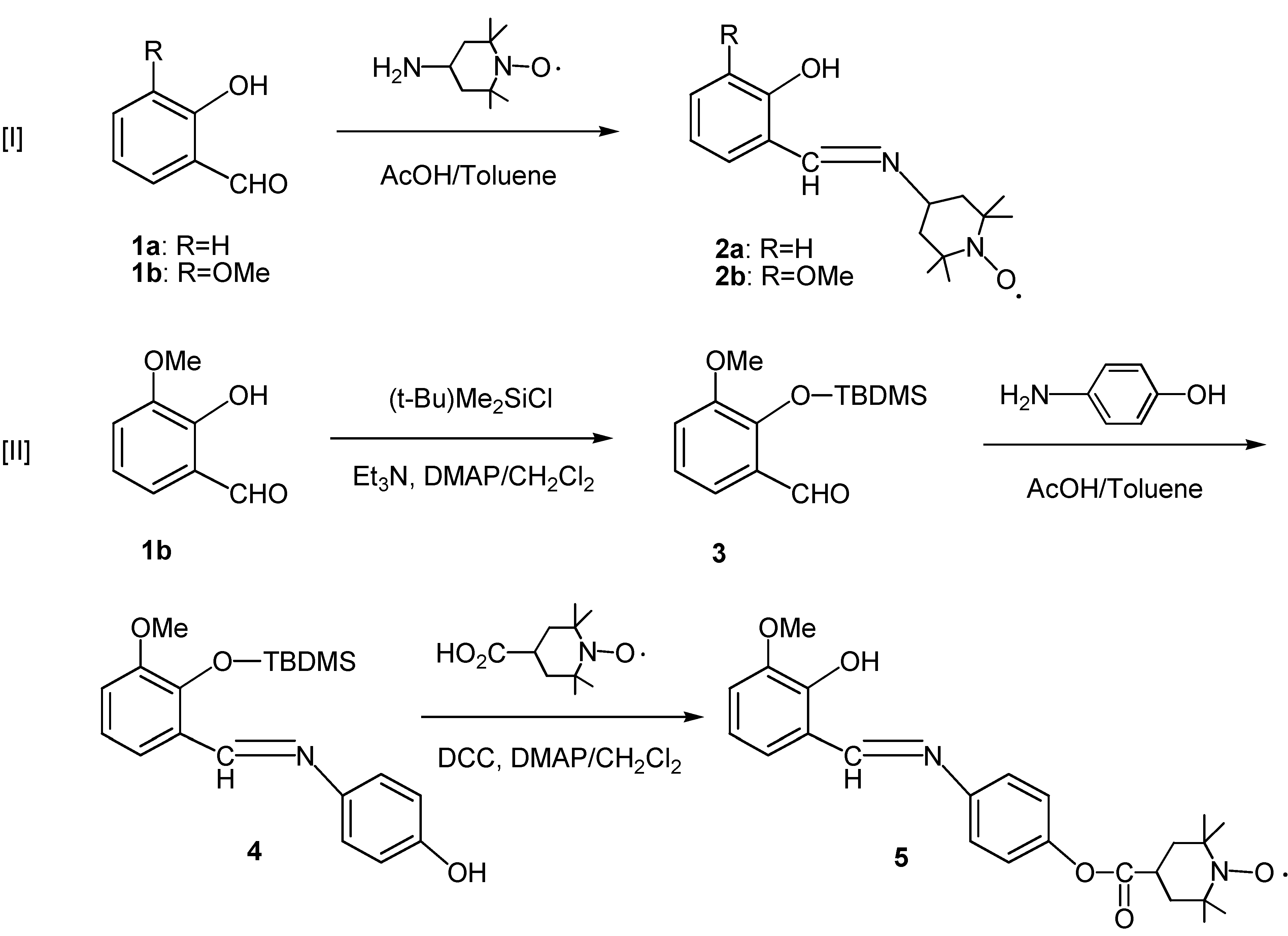
The preparation of the
N-salicylideneamine/aniline derivatives with TEMPO substituents was carried out as shown in
Scheme 1. Condensation of salicylaldehyde (
1a) or
o-methoxysalicylaldehyde (
1b) with 4-amino-TEMPO in toluene solution gave the corresponding Schiff bases
2a [
4] and
2b in moderate yields (reaction [I]). The salicylideneaniline derivative
5 was prepared from
o-methoxysalicylaldehyde (
1b) according to reaction [II]. The hydroxy group in
1b was at first protected with a TBDMS (
tert-butyldimethylsilyl) group to avoid the anticipated self-condensation reaction in the subsequent step to give the derivative
3 in 52% yield, which was then condensed with
p-aminophenol to afford the phenol derivative
4 in low yield (10%). The phenol
4 was finally condensed with 4-carboxy-TEMPO using DCC (1,3-dicyclohexylcarbodiimide) and DMAP [4-(dimethylamino)- pyridine] in dichloromethane solution, with concomitant deprotection of the TBDMS group, to give the desired salicylideneaniline derivative
5 in 38% yield. A similar approach to the unsubstituted derivative starting from salicylaldehyde (
1a) has so far been unsuccessful because of the failure of the reaction with
p-aminophenol in this case.
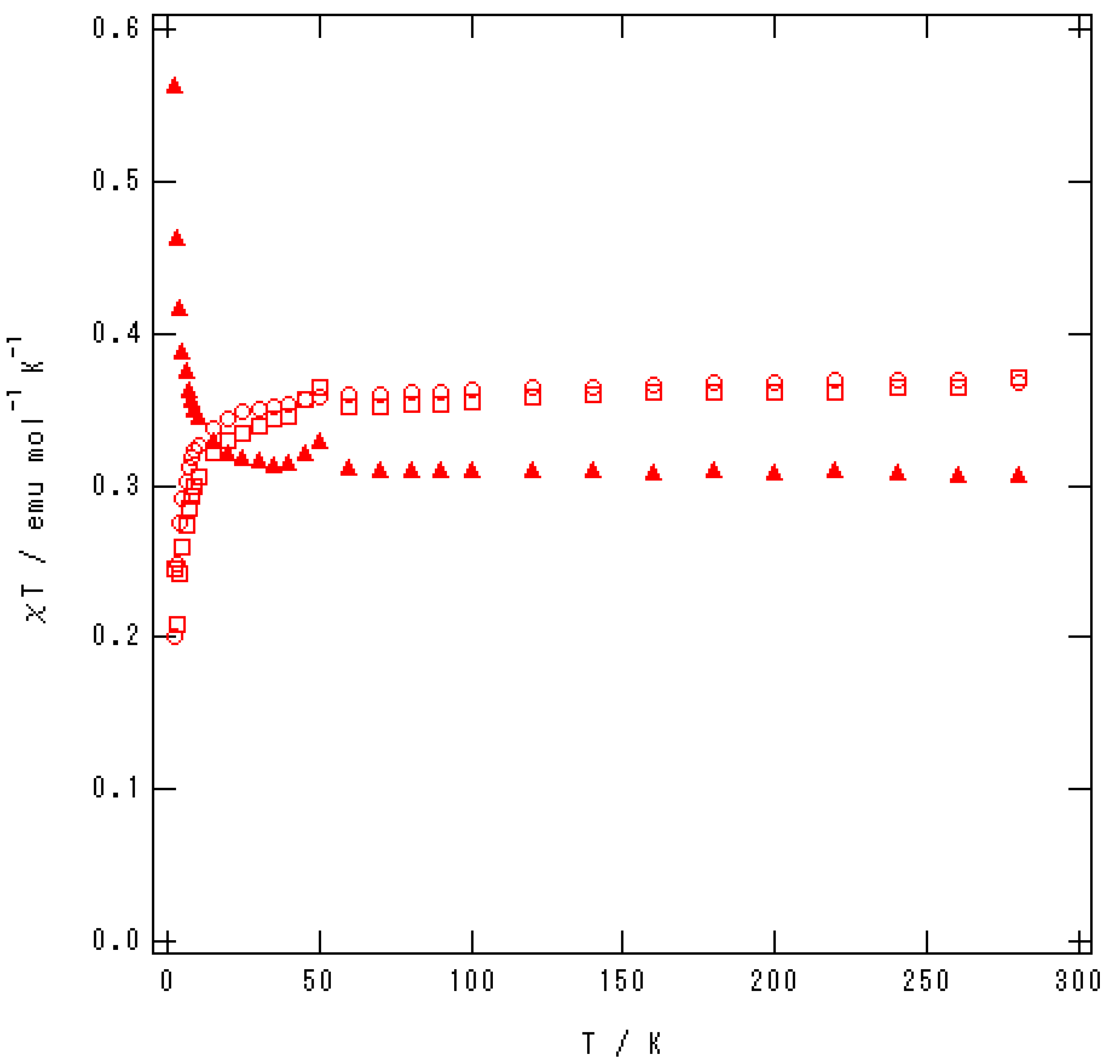
The temperature dependence data of their magnetic susceptibilities were obtained by the SQUID measurements from 2 to 300 K and the results are shown in
Figure 1. Curie-Weiss behaviour with weak intermolecular magnetic interactions are observed in all three radical compounds and ferromagnetic interactions with Weiss temperature of ca. 1 K are found, among them, only for
2b, while antiferromagnetic interactions are predominant in the other two radicals with apparently larger Weiss temperature for
5 (-2.06 K) than for
2a (-1.34 K, lit. –1.4 K [
4]). The Curie constants are reasonable values as S=+1/2 spin (0.37 emu·K·mol
–1) for
2a and
5 but it is apparently less than the anticipated value for
2b (0.31 emu·K·mol
–1), indicating the existence of some diamagnetic species in the radical.
Figure 1.
Temperature dependence data of χT for 2a (○), 2b (▲), and 5 (□).
Figure 1.
Temperature dependence data of χT for 2a (○), 2b (▲), and 5 (□).
Single crystals of the compounds could be obtained by recrystallization of the radicals and their X-ray diffraction data are summarized in
Table 1.
Table 1.
Summary of crystal data for 2a, 2b and 5
Table 1.
Summary of crystal data for 2a, 2b and 5
| Parameter | 2a | 2b | 5 |
|---|
| Formula | C16H23N2O2 | C17H25N2O3 | C24H29N2O5 |
| Formula weight | 275.37 | 305.40 | 425.50 |
| Crystal system | orthorhombic | monoclinic | monoclinic |
| Space group | Pca21 | P21/a | P21/c |
| a/Å | 15.6355(9) | 11.287(2) | 7.755(1) |
| b/Å | 8.2552(3) | 11.121(2) | 27.707(1) |
| c/Å | 24.276(2) | 13.823(2) | 11.038(1) |
| α/degrees | 90 | 90 | 90 |
| β/degrees | 90 | 103.17(1) | 107.66(1) |
| γ/degrees | 90 | 90 | 90 |
| V/Å3 | 3133.4(4) | 1690.1(5) | 2260.0(4) |
| Z | 8 | 4 | 4 |
| D (calc)/gcm-3 | 1.167 | 1.200 | 1.250 |
| No. of measured reflections | 13559 | 4292 | 4969 |
| No. of independent reflections | 6690 | 4094 | 4858 |
| No. of used reflection sin refinement F>2σ | 4138 | 1392 | 19571 |
| No. of parameters refined | 407 | 199 | 280 |
| R | 0.069 | 0.057 | 0.068 |
| RW | 0.070 | 0.031 | 0.046 |
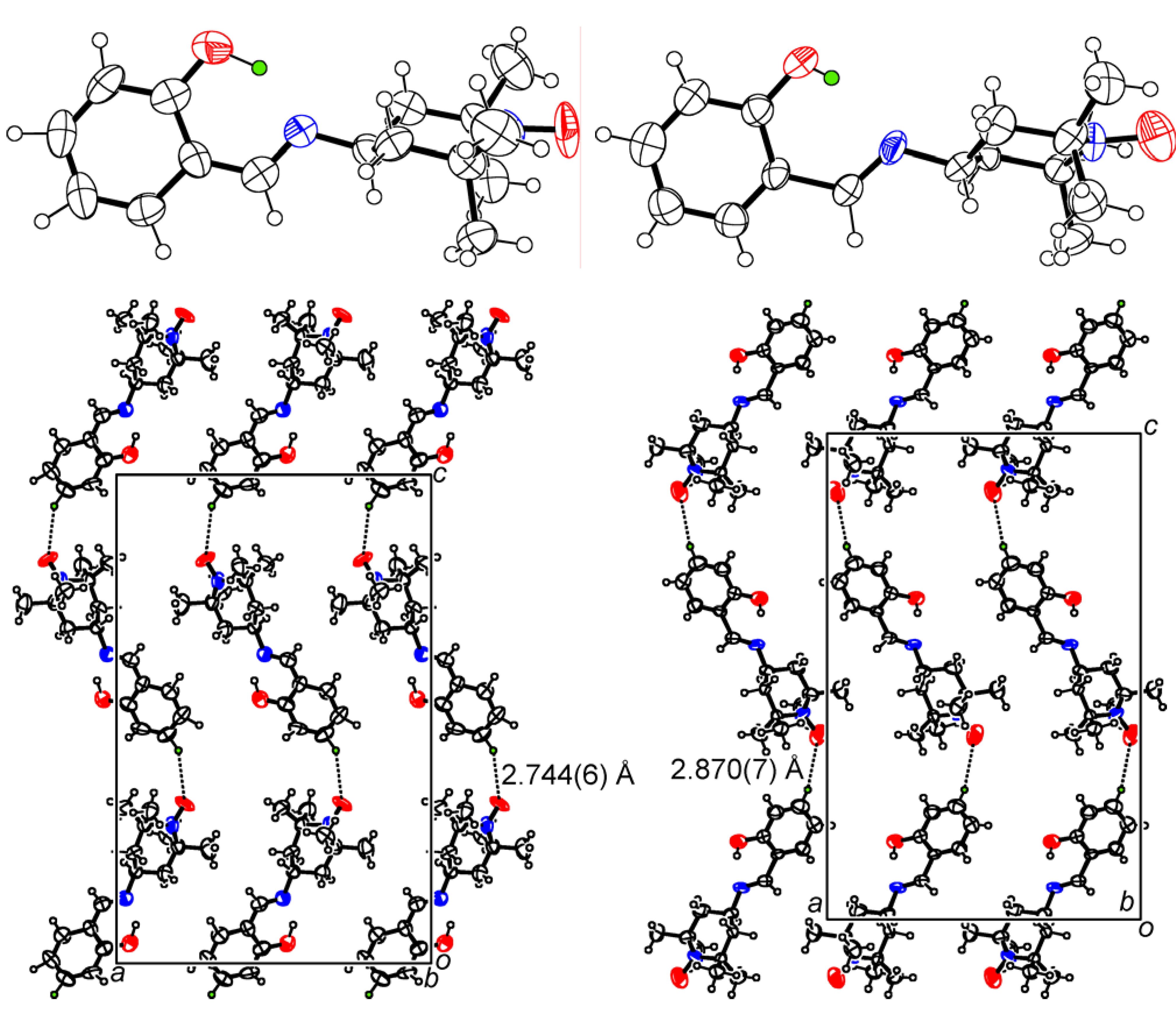
There are two crystallographically independent molecules in the crystal of
2a (molecule A and molecule B) as shown in
Figure 2 (upper). Inspection of the bond lengths in the molecules about the four bonds concerning the salicylideneamine moieties (see the chart above and
Table 2) indicates that the molecule A has some tendency towards the NH form whilst the predominance of the OH form is suggested in the molecule B, taking the difference of their bond alternations into consideration [
5]. The molecules of molecule A are found to form sheet-like structures on the
ac-plane and the similar sheet-like structures formed on the
ac-plane by the molecules of molecule B are stacked along the
b-axis to form an alternated columnar structure of sheets along the
b-axis (
Figure 2, lower).
Table 2.
Selected bond lengths in the molecules of 2a, 2b and 5
Table 2.
Selected bond lengths in the molecules of 2a, 2b and 5
| bond1 | 2a | 2b | 5 |
|---|
| a | 1.344(6) 2
1.359(5) 3 | 1.355(4) | 1.341(4) |
| b | 419(7) 2
1.391(6) 3 | 1.385(4) | 1.404(5) |
| c | 423(8) 2
1.488(7) 3 | 1.465(5) | 1.440(5) |
| d | 240(6) 2
1.293(6) 3 | 1.259(4) | 1.279(4) |
Figure 2.
Upper: molecular structures of 2a (left: molecule A, right: molecule B). Lower: sheet-like structures in the crystal structure of 2a (left: a sheet from molecules A, right: a sheet from molecules B).
Figure 2.
Upper: molecular structures of 2a (left: molecule A, right: molecule B). Lower: sheet-like structures in the crystal structure of 2a (left: a sheet from molecules A, right: a sheet from molecules B).
The nearest oxygen-oxygen distance of the spin centers amounts to 5.50 Å and is observed between a molecule of A and a molecule of B in the neighboring sheet along the
b-axis, which appears to be well separated to afford sufficient intermolecular magnetic interactions and, although it is difficult to assign the origin of the magnetic interactions exactly, the antiferromagnetic intermolecular interactions of this radical might be due to the spatial arrangements of the aryl groups within each sheet [
6].
The short contacts between the oxygen atoms of the spin centers and the
p-hydrogen atoms on the benzene rings of the neighboring molecules are shown in dotted lines in
Figure 2 (lower) and that are assumed to be one of the possible origins for the antiferromagnetic interactions observed in this radical. It is apparent from the bond lengths in
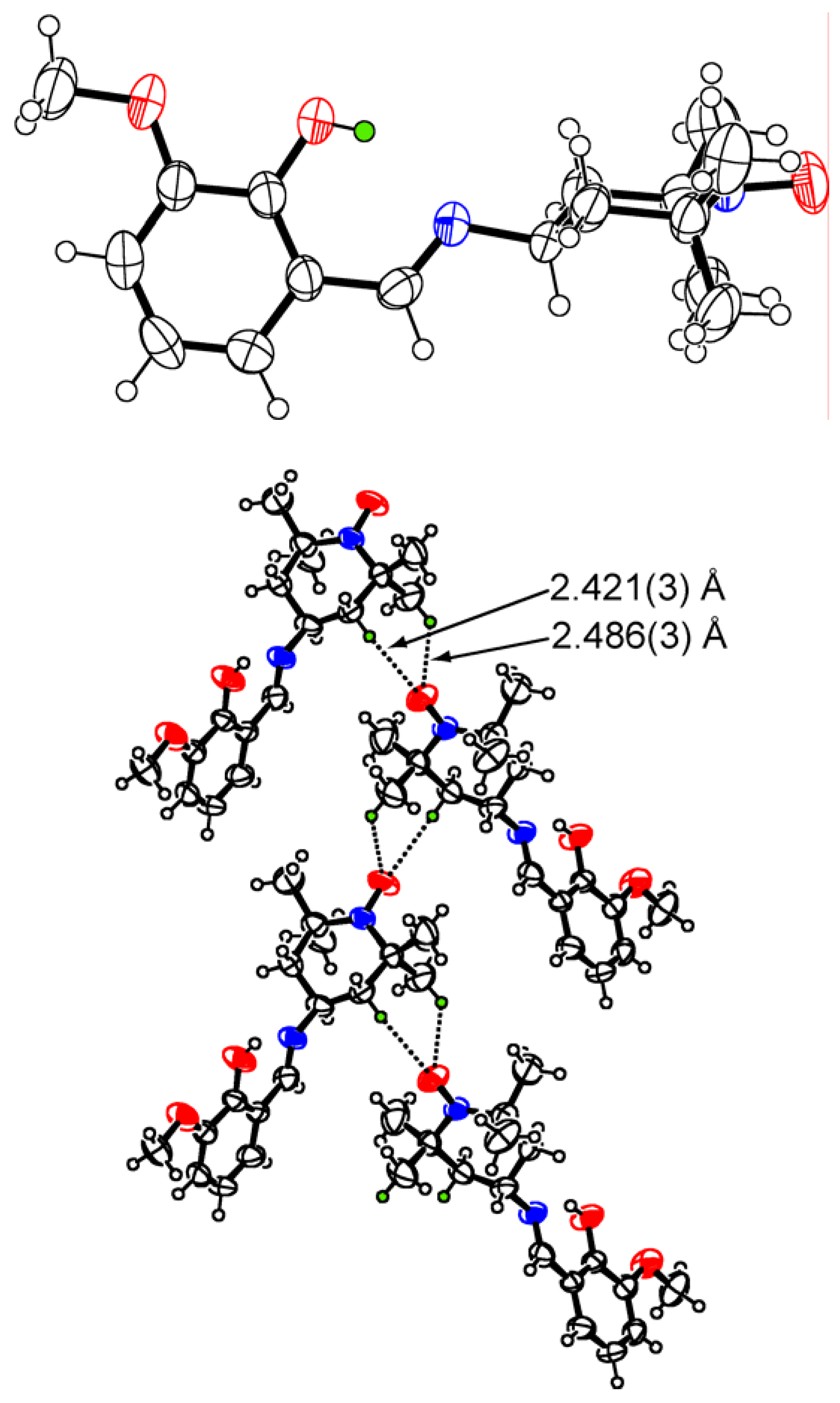
Table 2 that the molecular structure of the radical
2b has predominantly OH form and the mean plane of the six-membered ring of TEMPO moiety is oriented almost perpendicular from that of the benzene ring (
Figure 3, upper).
Figure 3.
Molecular (upper) and crystal structure (lower) of 2b.
Figure 3.
Molecular (upper) and crystal structure (lower) of 2b.
The crystal structure of the radical
2b is shown in the lower side of
Figure 3, in which ferromagnetic intermolecular interactions were uniquely observed within the radicals prepared in this work. There exist a couple of short contacts (3.29 Å and 3.31 Å) between the oxygen atom of the aminoxyl moiety in a molecule and one of the methyl as well as one of the methylene carbon atoms in the neighboring molecule and a closer look indicates the existence of the short contacts between the oxygen atom and the β−hydrogen atoms on the methyl as well as the methylene group of the neighboring molecule, amounting to 2.49 Å and 2.42 Å, respectively. The ferromagnetic interactions observed in this radical crystal may be understood on the basis of the spin polarization effect through the hydrogen bonds between the spin centers and the β−hydrogen atoms [
4].
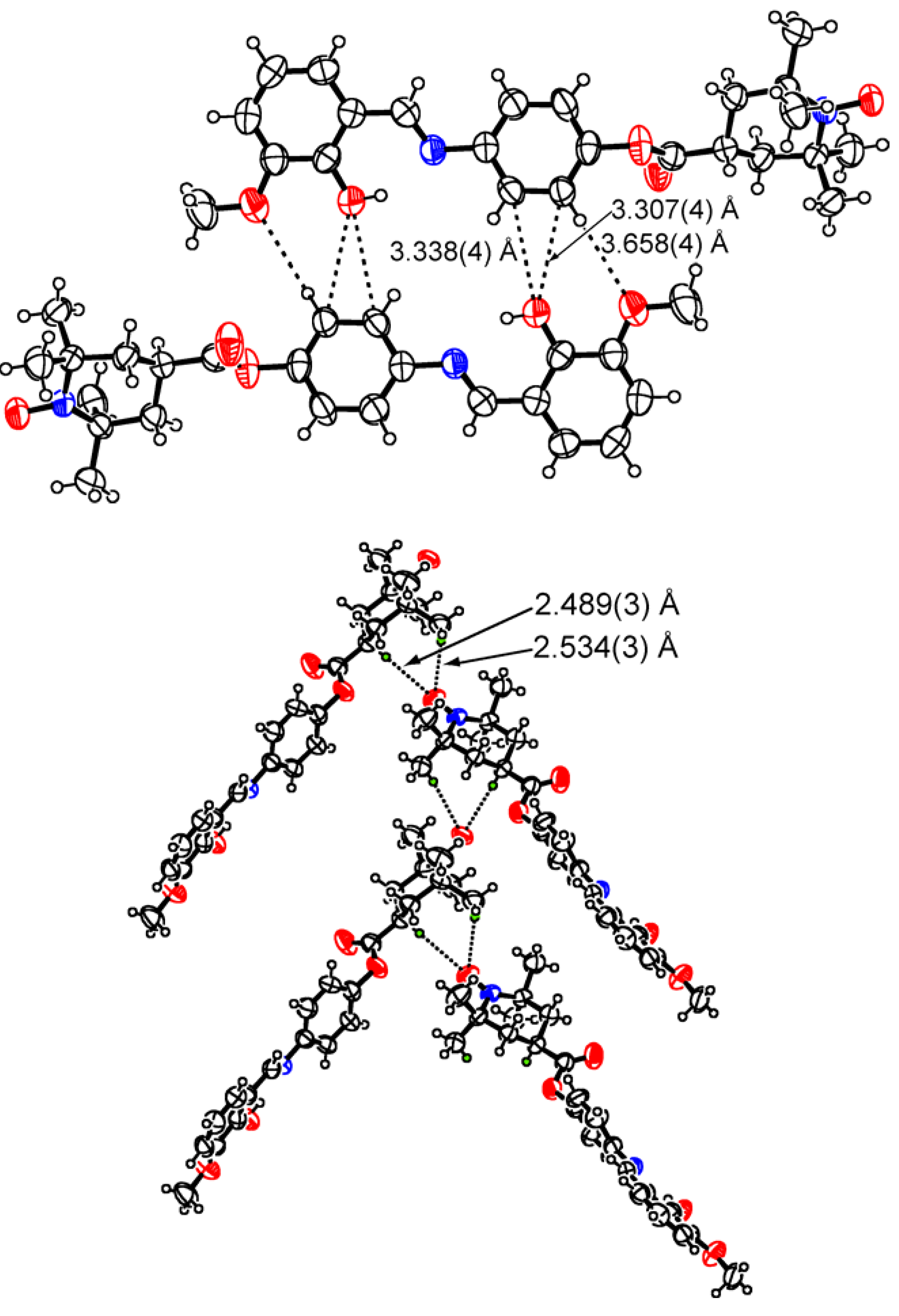
Figure 4.
Molecular structures of 5 (two molecules are depicted) with indication of the short contacts found between the molecules (upper) and the crystal structure of 5 (lower).
Figure 4.
Molecular structures of 5 (two molecules are depicted) with indication of the short contacts found between the molecules (upper) and the crystal structure of 5 (lower).
From the bond length data in
Table 2 the molecular structure of the radical
5 is also thought to correspond to that of the OH form and the corresponding structure is shown in
Figure 4 (upper). It is of some interest from the viewpoint of crystal structure formation that there are several side-by-side interactions between the molecules through the oxygen atoms of a molecule and the hydrogen atoms and/or the carbon atoms of the neighboring molecule to form such a side-by-side structure as depicted in
Figure 4 (upper). It is anticipated at the same time that the intramolecular hydrogen bonds are surpassing the intermolecular ones around the salicylidene aniline moieties. In the radical crystal, short contacts are found between the oxygen atoms of the spin centers and the β–hydrogen atoms of one of the methyl groups and the γ–hydrogen atoms on the methane carbon atoms in the neighboring molecules to form one-dimensional zigzag chains (
Figure 4, lower). The antiferromagnetic interactions observed in this radical suggest that the magnetic interactions are controlled mainly by the spin-spin interactions through the hydrogen bond found between the oxygen atom of a spin center and the γ–hydrogen atom on the methine carbon atom in the neighboring molecule.
Insofar as we have determined in our preliminary investigations, no distinct photochromic properties of these three radicals could be discerned upon irradiation with ultraviolet light, nor was any apparent thermochromic behaviour detected for the radicals 2a and 2b. On the other hand, an appreciable color change was observed for the crystals of 5, i.e., the original brownish yellow color of the crystals turned first to deep orange when they were heated over 160°C, then gradually to deep red up to 168°C, and finally they melted. The behavior suggests the existence of thermochromic properties in the radical compound and according a study on the magnetic properties of the radical with heating is now under way.


