Article

The Individual and Combined Effects of Deoxynivalenol and Aflatoxin B1 on Primary Hepatocytes of Cyprinus Carpio

1
College of Veterinary Medicine, Nanjing Agricultural University, Nanjing, 210095, China
2
Wujiang agricultural commission, Wujiang, 215200, China
*
Author to whom correspondence should be addressed.
Int. J. Mol. Sci. 2010, 11(10), 3760-3768; https://doi.org/10.3390/ijms11103760
Submission received: 26 August 2010 / Revised: 9 September 2010 / Accepted: 16 September 2010 / Published: 29 September 2010
(This article belongs to the Special Issue Advances in Molecular Toxicology)

Abstract

:
Aflatoxin B1 (AFB1) and deoxynivalenol (DON) are important food-borne mycotoxins that have been implicated in animal and human health. In this study, individual and combinative effects of AFB1 and DON were tested in primary hepatocytes of Cyprinus carpio. The results indicated that the combinative effects of AFB1 and DON (0.01 μg/mL AFB1 and 0.25 μg/mL DON; 0.02 μg/mL AFB1 and 0.25 μg/mL DON; 0.02 μg/mL AFB1 and 0.5 μg/mL DON) were higher than that of individual mycotoxin (P < 0.05). The activity of AST, ALT and LDH in cell supernatant was higher than that of control group (P < 0.05) when the mycotoxins were exposed to primary hepatocytes for 4 h. The decreased cell number was observed in tested group by inverted light microscopy. The mitochondrial swelling, endoplasmic reticulum dilation and a lot of lipid droplets were observed in primary hepatocytes by transmission electron microscope. Therefore, this combination was classified as an additive response of the two mycotoxins.

1. Introduction

Common carp are native to Europe but have been widely introduced and are now found worldwide except for the poles and northern Asia [1]. Carp are an important food fish throughout most of the world except for in Australia and North America where the fish is considered unpalatable. The world catch rate of carp per year exceeds 200,000 tons.
Aflatoxins are toxic chemicals produced by some species of naturally occurring moulds (Aspergillus flavus and Aspergillus parasiticus). Aflatoxins are common contaminants of oilseed crops such as cottonseed, peanut meal, and corn. Wheat, sunflower, soybean, fish meal, and nutritionally complete feeds can also be contaminated with aflatoxins, especially in warm and humid climates [25]. Four major aflatoxins (AFB1, AFB2, AFG1 and AFG2) are direct contaminants of grains and finished feeds. AFB1 is one of the most potent, naturally occurring, cancer-causing agents in animals. Aflatoxicosis is a disease that can affect many species of fish when feed contaminated with aflatoxins is eaten by the fish [6]. The first documented incidences of aflatoxicosis affecting fish health occurred in the 1960s in trout hatcheries [7]. The carcinogenic effect of AFB1 has been studied in fishes such as salmonid, rainbow trout, channel catfish, tilapia, guppy and Indian major carps [813].
Deoxynivalenol (DON) is prevalent worldwide in crops used for food and feed production, including in Canada and the United States [14]. Although DON is one of the least acutely toxic trichothecenes, it should be treated as an important food safety issue because it is a very common contaminant of grain. Toxic effects of DON in animals have been well documented and focused mainly on the immune system and the gastrointestinal tract. In particular, the symptoms of acute toxicosis at high dosage included diarrhea, vomiting, leukocytosis, hemorrhage, circulatory shock and ultimately death. Chronic toxicosis would lead to anorexia, reduced weight gain, nutrients malabsorption, neuroendocrine changes and immuno-suppress [15].
AFB1 and DON are important food-borne mycotoxins. They occur simultaneously in food including aquatic feed [16]. The use of more plant-based ingredients in aquafeeds enhances both the risk of introducing mycotoxins into the feed at the point of feed manufacturing, and mycotoxin production during storage of compounded feed [17]. After the fish feed were contaminated by fungi, more than one mycotoxin existed. In China, the AFB1 and DON were the most common mycotoxins in fish feed. In this research, the individual and combined effects of DON and AFB1 in primary hepatocytes of Cyprinus carpio were reported.

2. Results and Discussion

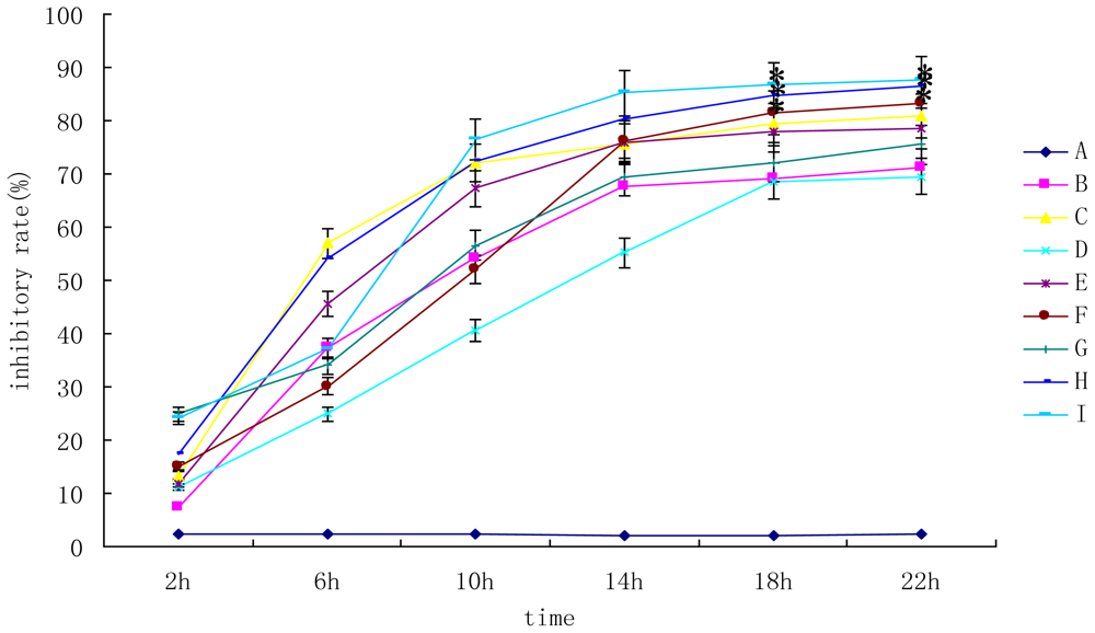
2.1. Individual and Combined Toxicity of DON and AFB1 on Primary Hepatocytes

To evaluate the influence of the mycotoxins on primary hepatocytes, the MTT assay was used. The results were shown in Figure 1. The results showed that the toxicity of mixture (group F, H and I) was higher than that of individual mycotoxin (P < 0.05). Group I was the most toxic (cell inhibitory rate was 87.67%). After the mycotoxins added, the inhibitory rate increased with the time lasted and increasing mycotoxin concentration. However, the concentration-response curves were not clear.

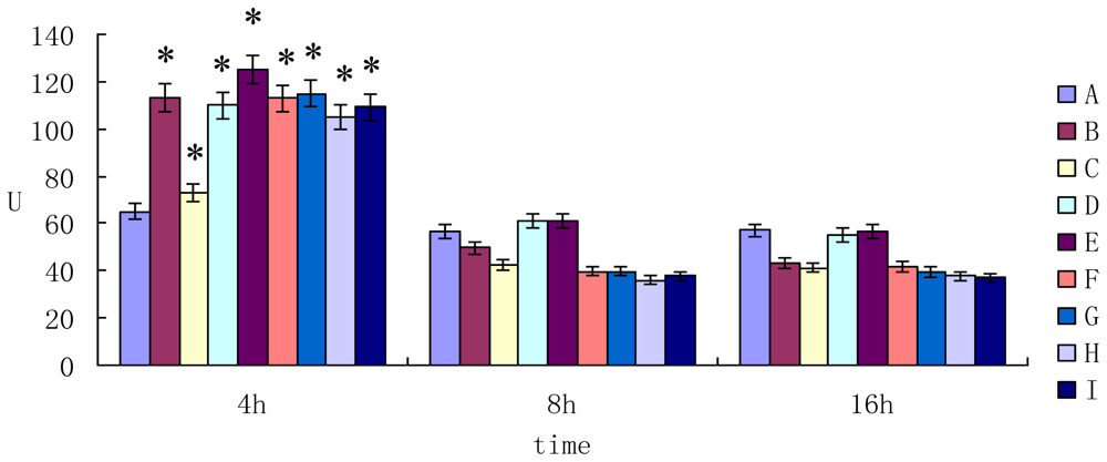
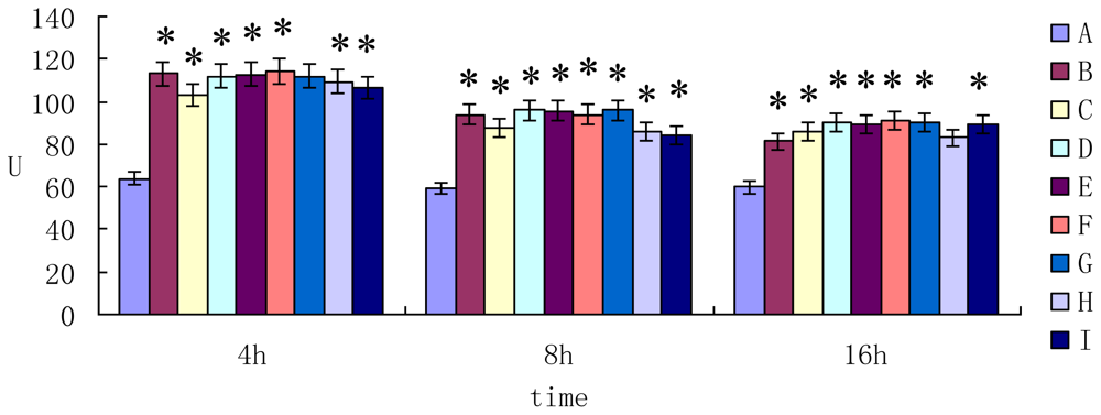
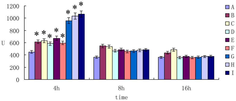
2.2. The Enzyme Activity of AST ALT and LDH in Cell Supernatant

The activity of aspartate aminotransferase (AST) in cell supernatant was shown in Figure 2. When the mycotoxin was exposed to primary hepatocytes for 4 h, the activity of AST was higher than that of control group (P < 0.05). However, the activity of AST decreased after 8h and 16h incubation (P > 0.05) compared to the control group.
The activity of alanine transarninase (ALT) in cell supernatant was shown in Figure 3. The results showed that the activity of ALT in mycotoxin group was higher than that of control group after 4 h, 8 h and 16 h incubation (P < 0.05).
The activity of lactate dehydrogenase (LDH) in cell supernatant was shown in Figure 4. The results showed that the activity of LDH in mycotoxin group was higher than that of control group after 4 h incubation (P < 0.05). However, prolonging the cultured time (for 8 h and 16 h incubation), the difference of the activity of LDH between the groups was not significant (P > 0.05).

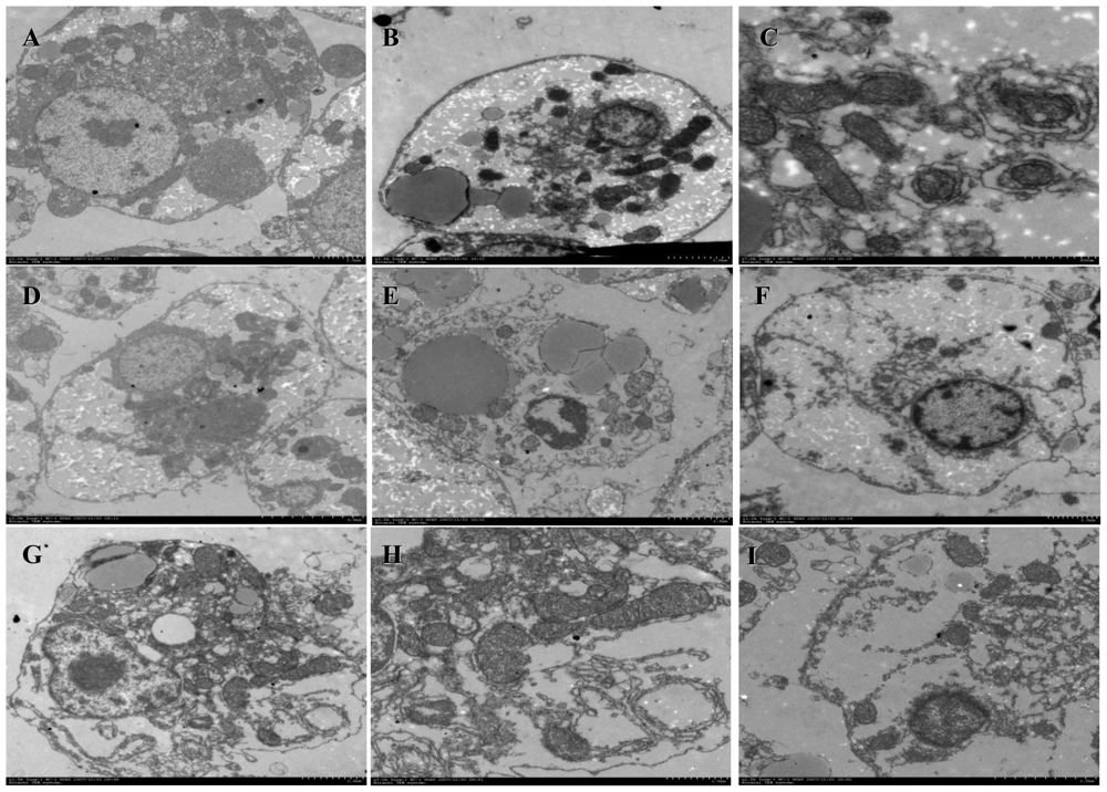
2.3. Morphological Observation on Cells

The ultrastructure of primary hepatocytes was shown in Figure 5. The cell membrane of normal hepatocytes was intact. The intranuclear chromatins were homogeneous granules. The organelles were clearness (Figure 5A). When the primary hepatocytes were cultured with the AFB1 (0.01 μg/mL), the lipid droplets, nucleus and chromatin condensation were observed clearly (Figure 5B). The saccular ectasia and vesicular structure were observed in rough endoplasmic reticulum and the smooth endoplasmic reticulum, respectively (Figure 5C). There were a lot lipid droplets and shrinkage nucleuses in hepatocytes (Figures 5D, 5E). The mitochondrial swelling was observed in hepatocytes (Figure 5F). The nuclear membrane dissolves and the endoplasmic reticulum dilation were observed in hepatocytes (Figures 5G, 5H, 5I).
AFB1 and DON always occur simultaneously in aquatic feed. They play an important role in the mycotoxicoses of Cyprinus carpio. In order to analyze the predictability of the combined effects of a mixture of AFB1 and DON, it is necessary to examine the individual effect of each mycotoxin on primary hepatocytes of Cyprinus carpio. The results showed that the combined toxicity of AFB1 and DON (Group F, H and I) was higher than that of individual mycotoxin (P < 0.05). The toxic effects included inhibition of cell growth, destruction of cell structure and increasing activity of AST, ALT and LDH. Up to now, many reports were focused on the single mycotoxin on cells [1823]. The principal target organ for aflatoxins was the liver. After the invasion of AFB1, it would lead to necrosis or death of liver cells. In the experiment, we found that AFB1 could lead to primary hepatocytes death. There were a few reports about the toxicology of combinative mycotoxin [2427]. The toxicity of AFB1 and DON on the injury and repair of DNA was studied [28]. The report only showed that AFB1 and DON produced marked effects on engendering tumor as genetoxic and nongenotoxic carcinogens complementarily.
The AST, ALT and LDH were secreted into cell supernatant from liver cells when they were destroyed. The activity of these enzymes only lasted for 4 h. So, these enzymes should be tested immediately. When primary hepatocytes were cultured with mycotoxins for 4 h, the activity of these enzymes increased significantly. However, the activity of these enzymes decreased at 8 h and 16 h, because almost primary hepatocytes were dead.
Many apoptotic cells were observed in individual mycotoxin group, however, a lot of necrotic cells in combinative group. The all results could indicate that a combined intake of mycotoxins would lead to a possible higher risk for adverse health effects than the intake of one of these mycotoxins alone. However, the data on combined toxic effects of mycotoxins were generally limited, particularly with respect to the AFB1 and DON. The further research would focus on the mechanism of apoptosis and necrosis induced by the AFB1 and DON.

3. Experimental Section

3.1. Mycotoxin

AFB1 and DON were purchased from Sigma–Aldrich Co. (mainland, China) and dissolved in dimethylsulfoxide (DMSO, Merck, Darmstadt, Germany).

3.2. Cell Cultures and Treatments

The method of isolation of primary hepatocytes was performed following the reference[29]. The primary hepatocytes were cultured in L-5 medium at 27 °C in CO2 incubator without CO2. Cells were seeded at a density of 2 × 10⁶ cells/mL in 24-well tissue culture plates. The experiment was designed with two factors and three levels. The final concentration of AFB1 and DON was 0 μg/mL, 0.01 μg/mL, 0.02 μg/mL and 0 μg/mL, 0.25 μg/mL, 0.5 μg/mL, respectively. The DMSO (2 μL/mL, final concentration) was the control group. Each group has 48 duplications.

3.3. Cytotoxicity Assay by MTT Test

The cytotoxicity were determined using the colorimetric method described by T.Mosmann [30]. Cells were seeded on 96-well culture plates (Polylabo, France) at 2 × 10⁶ cells/mL and treated with different concentrations of AFB1 or DON and/or AFB1 + DON combined for 2, 6, 10, 14, 18, 22 h at 27 °C, respectively. Then, the medium was moved carefully from the wells and the wells were washed once with PBS. MTT (concentration 5 mg/mL in PBS; 20 μL/well) was then added to the wells. The cultured cells were returned to the incubator for 4 h. The medium was discarded and the wells were dried. Formazan crystals produced were solubilized using 100 μL dimethyl sulfoxide (DMSO)/well and misce bene. Absorbance was measured in a Stat Fax 2000 ELIASA (Bio-Rad, USA) at 570 nm wavelength, to assess cell proliferation of the primary hepatocytes. The results were expressed as:
Inhibitory rate = ( 1 - mean OD mycotoxin in sample / mean OD solvent control ) × 100 %

3.4. The Enzyme Activity of AST, ALT and LDH in Cell Supernatant

After the mycotoxin added, the cell supernatant was collected at 4 h, 8 h and 16 h, respectively. The enzyme activity of AST, ALT and LDH was detected in cell supernatant by automated biochemical analyzer (HITACHI 7150, Japan).

3.5. Morphological Observation on Cells

The growth condition was observed by inverted light microscopy (Nikon, Japan). After the primary hepatocytes incubation with mycotoxins for 24 h, the cells were collected and fixed with 2.5% glutaraldehyde (pH 7.4). The ultrastructure of cell was observed by transmission electron microscope (HITACHI H-7650).

3.6. Data Analysis

Data were analyzed by SPSS 12.0 with One-Way ANOVA mode.

4. Conclusions

The experiments reported here were conducted to evaluate the effect of these toxins on primary hepatocytes. The results indicated that the combinative toxicity of AFB1 and DON (Groups F, H and I) was higher than that of individual mycotoxin (P < 0.05). However, what we research was the single mycotoxin on the cells. Therefore, we should more focus on the combinative mycotoxins on animal and human health.

Acknowledgements

This study was supported by the National Natural Science Foundation of China (Grant No. A2000748 and A200955), the Research Award Fund for Outstanding Young Teachers in Higher Education Institutions, China (Grant No. 20090097120024) and the National High Technology Research and Development Program of China (Grant No. 2007AA10Z429).

References

  1. Liu, ZJ; Zhu, ZY; Roberg, K; Faras, A; Guise, K; Kapuscinski, AR; Hackett, PB. Isolation and characterization of beta-actin gene of carp (Cyprinus carpio). DNA Seq 1990, 1, 125–136. [Google Scholar]
  2. Hutanasu, C; Sfarti, C; Trifan, A; Hutanasu, M; Stanciu, C. Aflatoxin contamination of food: additional risk factor for chronic liver diseases. Rev. Med. Chir. Soc. Med. Nat. Iasi 2009, 113, 1061–1065. [Google Scholar]
  3. Tchana, AN; Moundipa, PF; Tchouanguep, FM. Aflatoxin contamination in food and body fluids in relation to malnutrition and cancer status in Cameroon. Int. J. Environ. Res. Public Health 2010, 7, 178–188. [Google Scholar]
  4. Kitya, D; Bbosa, GS; Mulogo, E. Aflatoxin levels in common foods of South Western Uganda: a risk factor to hepatocellular carcinoma. Eur. J. Cancer Care (Engl.) 2010, 19, 516–521. [Google Scholar]
  5. Kumagai, S; Nakajima, M; Tabata, S; Ishikuro, E; Tanaka, T; Norizuki, H; Itoh, Y; Aoyama, K; Fujita, K; Kai, S; Sato, T; Saito, S; Yoshiike, N; Sugita-Konishi, Y. Aflatoxin and ochratoxin A contamination of retail foods and intake of these mycotoxins in Japan. Food Addit. Contam. Part A Chem. Anal. Control Expo. Risk Assess 2008, 25, 1101–1106. [Google Scholar]
  6. Hussain, M; Gabal, MA; Wilson, T; Summerfelt, RC. Effect of aflatoxin-contaminated feed on morbidity and residues in walleye fish. Vet. Hum. Toxicol 1993, 35, 396–398. [Google Scholar]
  7. Halver, JE; Ashley, LM; Smith, RR. Aflatoxicosis in coho salmon. Natl. Cancer Inst. Monogr 1969, 31, 141–155. [Google Scholar]
  8. Gallagher, EP. Using salmonid microarrays to understand the dietary modulation of carcinogenesis in rainbow trout. Toxicol. Sci 2006, 90, 1–4. [Google Scholar]
  9. Bailey, GS; Williams, DE; Wilcox, JS; Loveland, PM; Coulombe, RA; Hendricks, JD. Aflatoxin B1 carcinogenesis and its relation to DNA adduct formation and adduct persistence in sensitive and resistant salmonid fish. Carcinogenesis 1988, 9, 1919–1926. [Google Scholar]
  10. Gallagher, EP; Eaton, DL. In vitro biotransformation of aflatoxin B1 (AFB1) in channel catfish liver. Toxicol. Appl. Pharmacol 1995, 132, 82–90. [Google Scholar]
  11. Abdel-Wahhab, MA; Hasan, AM; Aly, SE; Mahrous, KF. Adsorption of sterigmatocystin by montmorillonite and inhibition of its genotoxicity in the Nile tilapia fish (Oreachromis nilaticus). Mutat. Res 2005, 582, 20–27. [Google Scholar]
  12. Sato, S; Matsushima, T; Tanaka, N; Sugimura, T; Takashima, F. Hepatic tumors in the guppy (Lebistes reticulatus) induced by aflatoxin B1, dimethylintrosamine, and 2-acetylaminofluorene. J. Natl. Cancer Inst 1973, 50, 767–778. [Google Scholar]
  13. Sahoo, PK; Mukherjee, SC. The effect of dietary immunomodulation upon Edwardsiella tarda vaccination in healthy and immunocompromised Indian major carp (Labeo rohita). Fish Shellfish Immunol 2002, 12, 1–16. [Google Scholar]
  14. Wood, GE; Carter, L, Jr. Limited survey of deoxynivalenol in wheat and corn in the United States. J. Assoc. Off. Anal. Chem 1989, 72, 38–40. [Google Scholar]
  15. Rotter, BA; Prelusky, DB; Pestka, JJ. Toxicology of deoxynivalenol (vomitoxin). J. Toxicol. Environ. Health 1996, 48, 1–34. [Google Scholar]
  16. Ali, N; Sardjono; Yamashita, A; Yoshizawa, T. Natural co-occurrence of aflatoxins and Fusarium mycotoxins (fumonisins, deoxynivalenol, nivalenol and zearalenone) in corn from Indonesia. Food Addit. Contam 1998, 15, 377–384. [Google Scholar]
  17. Tacon, AG; Metian, M. Aquaculture feed and food safety. Ann. N. Y. Acad. Sci 2008, 1140, 50–59. [Google Scholar]
  18. Billings, PC; Uwaifo, AO; Heidelberger, C. Influence of benzoflavone on aflatoxin B1-induced cytotoxicity, mutation, and transformation of C3H/10T1/2 cells. Cancer Res 1983, 43, 2659–2663. [Google Scholar]
  19. Loquet, C; Wiebel, FJ. Geno- and cytotoxicity of nitrosamines, aflatoxin B1, and benzo[a]-pyrene in continuous cultures of rat hepatoma cells. Carcinogenesis 1982, 3, 1213–1218. [Google Scholar]
  20. Palanee, T; Dutton, MF; Chuturgoon, AA. Cytotoxicity of aflatoxin B1 and its chemically synthesised epoxide derivative on the A549 human epithelioid lung cell line. Mycopathologia 2001, 151, 155–159. [Google Scholar]
  21. Zuckerman, AJ; Tsiquaye, KN; Fulton, F. Tissue culture of human embryo liver cells and the cytotoxicity of aflatoxin B1. Br. J. Exp. Pathol 1967, 48, 20–27. [Google Scholar]
  22. Iwaki, M; Kitagawa, T; Akamatsu, Y; Aibara, K. Cytotoxic effects of aflatoxin B1 and its association with cellular components in chicken embryo primary cultured cells. Biochim. Biophys. Acta 1990, 1035, 146–153. [Google Scholar]
  23. Gabliks, J; Barter, S. Comparative cytotoxicity of aflatoxin B1 and saxitoxin in cell cultures. Mol. Toxicol 1987, 1, 209–216. [Google Scholar]
  24. Golli-Bennour, EE; Kouidhi, B; Bouslimi, A; Abid-Essefi, S; Hassen, W; Bacha, H. Cytotoxicity and genotoxicity induced by aflatoxin B1, ochratoxin A, and their combination in cultured Vero cells. J. Biochem. Mol. Toxicol 2010, 24, 42–50. [Google Scholar]
  25. Pal, L; Dublecz, K; Weber, M; Balogh, K; Erdelyi, M; Szigeti, G; Mezes, M. Effect of combined treatment with aflatoxin B1 and T-2 toxin and metabolites on some production traits and lipid peroxide status parameters of broiler chickens. Acta Vet. Hung 2009, 57, 75–84. [Google Scholar]
  26. Orsi, RB; Oliveira, CA; Dilkin, P; Xavier, JG; Direito, GM; Correa, B. Effects of oral administration of aflatoxin B1 and fumonisin B1 in rabbits (Oryctolagus cuniculus). Chem. Biol. Interact 2007, 170, 201–208. [Google Scholar]
  27. McKean, C; Tang, L; Tang, M; Billam, M; Wang, Z; Theodorakis, CW; Kendall, RJ; Wang, JS. Comparative acute and combinative toxicity of aflatoxin B1 and fumonisin B1 in animals and human cells. Food Chem. Toxicol 2006, 44, 868–876. [Google Scholar]
  28. Li, B; Guo, H. Study on the combined toxicity of aflatoxin B1 and deoxynivalenol. Wei Sheng Yan Jiu 2000, 29, 393–395. [Google Scholar]
  29. Yanhong, F; Chenghua, H; Guofang, L; Haibin, Z. Optimization of the isolation and cultivation of Cyprinus carpio primary hepatocytes. Cytotechnology 2008, 58, 85–92. [Google Scholar]
  30. Mosmann, T. Rapid colorimetric assay for cellular growth and survival: application to proliferation and cytotoxicity assays. J. Immunol. Methods 1983, 65, 55–63. [Google Scholar]
Figure 1. The individual and combined inhibitory rate of DON and AFB1 on primary hepatocytes.A, control; B, 0.01 μg/mL AFB1; C, 0.02μg/mL AFB1; D, 0.25 μg/mL DON; E, 0.5 μg/mL DON; F, 0.01 μg/mL AFB1 and 0.25 μg/mL DON; G, 0.01 μg/mL AFB1 and 0.5 μg/mL DON; H, 0.02 μg/mL AFB1 and 0.25 μg/mL DON; I 0.02 μg/mL AFB1 and 0.5 μg/mL DON. * means significant difference at 0.05 level (P < 0.05). The same as follows.
Ijms 11 03760f1
Figure 2. The activity of AST in cell supernatant.
Ijms 11 03760f2
Figure 3. The activity of ALT in cell supernatant.
Ijms 11 03760f3
Figure 4. The activity of LDH in cell supernatant.
Ijms 11 03760f4
Figure 5. The ultrastructure of primary hepatocytes.
Ijms 11 03760f5

