Article

Sequence Analysis and Potentials of the Native RbcS Promoter in the Development of an Alternative Eukaryotic Expression System Using Green Microalga Ankistrodesmus convolutus

1
Department of Cell and Molecular Biology, Faculty of Biotechnology and Biomolecular Sciences, Universiti Putra Malaysia, 43400 UPM-Serdang, Selangor Darul Ehsan, Malaysia
2
Rubber Research Institute of Vietnam, 236Bis Nam Ky Khoi Nghia, District 3, Ho Chi Minh City, Vietnam
3
Department of Biology, Faculty of Science, Universiti Putra Malaysia, 43400 UPM-Serdang, Selangor Darul Ehsan, Malaysia
*
Author to whom correspondence should be addressed.
Int. J. Mol. Sci. 2012, 13(3), 2676-2691; https://doi.org/10.3390/ijms13032676
Submission received: 23 December 2011 / Revised: 2 February 2012 / Accepted: 5 February 2011 / Published: 28 February 2012
(This article belongs to the Section Biochemistry)

Abstract

:
The availability of highly active homologous promoters is critical in the development of a transformation system and improvement of the transformation efficiency. To facilitate transformation of green microalga Ankistrodesmus convolutus which is considered as a potential candidate for many biotechnological applications, a highly-expressed native promoter sequence of ribulose-1,5-bisphosphate carboxylase/oxygenase small subunit (AcRbcS) has been used to drive the expression of β-glucuronidase (gusA) gene in this microalga. Besides the determination of the transcription start site by 5′-RACE, sequence analysis revealed that AcRbcS promoter contained consensus TATA-box and several putative cis-acting elements, including some representative light-regulatory elements (e.g., G-box, Sp1 motif and SORLIP2), which confer light responsiveness in plants, and several potential conserved motifs (e.g., CAGAC-motif, YCCYTGG-motifs and CACCACA-motif), which may be involved in light responsiveness of RbcS gene in green microalgae. Using AcRbcS promoter::gusA translational fusion, it was demonstrated that this promoter could function as a light-regulated promoter in transgenic A. convolutus, which suggested that the isolated AcRbcS promoter was a full and active promoter sequence that contained all cis-elements required for developmental and light-mediated control of gene expression, and this promoter can be used to drive the expression of heterologous genes in A. convolutus. This achievement therefore advances the development of A. convolutus as an alternative expression system for the production of recombinant proteins. This is the first report on development of gene manipulation system for unicellular green alga A. convolutus.

1. Introduction

As photoautotrophs, green microalgae have many advantages as expression systems for the production of recombinant proteins: (i) they can be cultured simply, rapidly and economically although they are eukaryotic; (ii) they can produce complicated eukaryotic proteins which need post-translational modification [1]; and (iii) they are considered as safe food because they are free from human pathogens and endotoxins [2]. Although a number of expression systems have been developed and are available for the production of recombinant proteins, it is necessary to develop alternative expression systems which offer distinct advantages in terms of protein yield, ease of manipulation, cost of operation and that are generally regarded as safe. The heterologous protein expression systems using green algae meet these criteria. On using algae as the expression systems, it is necessary to use the strong constitutive and native promoters or the promoters from very close species to overcome the hurdles of the low-efficient expression of the transgenes [36].
Among the popular strong constitutive promoters, cauliflower mosaic virus 35S (CaMV35S) was used to drive the expression of foreign genes in several green microalgae species such as Chlamydomonas reinhardtii [7], Chlorella kessleri [8], Chlorella ellipsoidea [9] and Dunaliella salina [10,11]; however, all attempts using this promoter to express foreign genes in Chlamydomonas reinhardtii have failed [12]. The other constitutive promoters, Simian virus 40 (SV40) and ubiquitin (Ubi) promoters, were used in transformation systems of Chlamydomonas reinhardtii [13], Haematococcus pluvialis [14] and Chlorella ellipsoidea [15], respectively. The low-efficiency or the failure to express the foreign genes using these heterologous promoters reported in these algae species may be due to the lack of necessary regulatory elements or the poor codon usage of the transgenes, and these problems can be overcome by using the native promoters and codon optimization in the transformation constructs [16,17].
In order to improve the efficiency transformation of green microalgae as well as to establish the transformation procedures, several native promoters have been isolated and used. As such, the native heat shock protein 70 (HSP70) promoter has allowed the efficient transgene expression in green microalga Chlamydomonas reinhardtii [4,17]. Moreover, the promoter of abundant protein of photosystem I complex (PsaD) genes have proved to be of high value for efficient expression of chimeric constructs [18]. Similarly, the native promoter of ribulose-1,5-bisphosphate carboxylase/oxygenase small subunit (RbcS) gene has given high transformation efficiency in transgenic C. reinhardtii [7,1923] and D. salina [24]. Likewise, the RbcS promoter isolated from D. tertiolecta resulted in high expression level of the heterologous gene in transgenic C. reinhardtii [5]. Therefore, the highly active homologous promoter of RbcS genes is currently a priority choice to drive the expression of heterologous proteins in green microalgae.
The promising opportunity to use the fast growing green microalgae species as transgenic green cell factories has already resulted in several business start-ups during the last few years. As a fast growing alga producing appreciable amount of carotenoids and polyunsaturated fatty acids, especially lute [25], green microalga A. convolutus is being considered as an interesting candidate for many biotechnological applications. The simple, rapid and cheap culture of A. convolutus offers great advantages in bioengineering this species for producing valuable polypeptides and proteins safely and economically. Furthermore, the ability of A. convolutus to form floating aggregates during its normal growth besides other beneficiary attributes facilitates harvesting the biomass as well as the natural products and recombinant proteins [26]. Therefore, this species has high potential of being used as sources of lutein in poultry industry. Prospectively, A. convolutus is to be used as economic host to express the therapeutic proteins that can be served as an oral vaccine for poultry using the whole cell as the vector, as such no protein purification steps are needed meanwhile the animals are supplied with the amount of lutein in the cells. In order to establish an alternative expression system using A. convolutus, it is important to develop gene manipulation systems for this alga. Together with the knowledge of promoters and expression patterns of transgenes in other green microalgae species, exploiting the constitutive and native promoters is an important means for overcoming the hurdles of the low-efficient expression of transgenes in these algae. In the present study, the sequence of RbcS promoter from A. convolutus was analyzed and confirmed its ability to drive the expression of foreign genes in this alga. Also, the influence of light on the regulation of AcRbcS promoter activity was also reported. This is the first report on the development of gene manipulation system for green microalga A. convolutus.

2. Results and Discussion

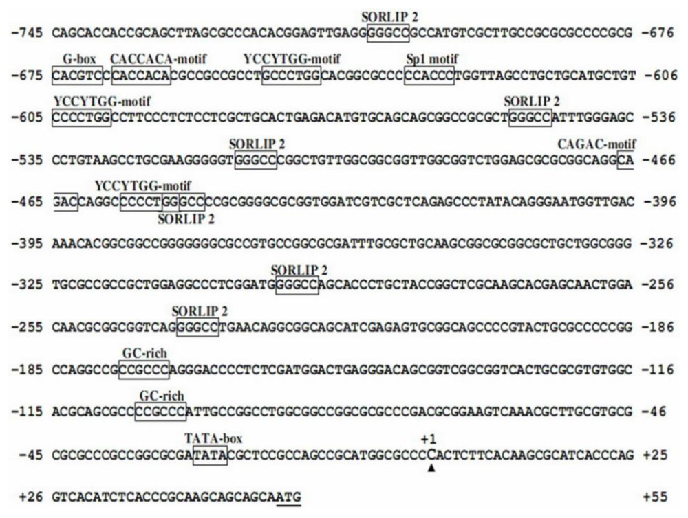
2.1. Sequence Analysis of AcRbcS Promoter

The AcRbcS promoter sequence was searched against PLACE and PlantCARE databases, and the results are shown in Figure 1. The eukaryotic consensus TATA-box was identified at −28 nucleotides upstream of the transcription start site (TSS). The AcRbcS promoter contained several putative cis-acting elements, which included some representative light-responsive elements such as G-box [2730] at position −675, Sp1 motif [31] at position −633, and six copies of SORLIP2 (sequences over-represented in light-induced promoter) [29] at position −706, −549, −512, −451, −297 and −239. Moreover, the AcRbcS promoter also contained putative GC-rich element (CCGCCC) at position −177 and −105. TATA box, the critical binding site, can be found in most eukaryotic gene promoters while other cis-acting elements frequently appeared in photosynthesis gene promoters of higher plants and microalgae; their specific roles were clearly described by a number of reports which concluded that these elements were essential for the light-responsive expression. It was interesting to note that G-box and Sp1 motif located in the upstream region, far from the transcription start site, but six copies of element SORLIP2 were found throughout the AcRbcS promoter region. The repetitive distribution of this element implied that AcRbcS promoter might be an active promoter. It was also noted that the GC-rich element was not found in the RbcS promoter of other green microalgae but it was found in RbcS promoter of A. convolutus, which suggested that the GC-rich element may not be essential for the light-responsive expression. Indeed, as described in a previous report, this element was found in maize RbcS promoter but not in rice RbcS promoter; the deletion of this element did not cause any strong effects on light-responsive expression of a reporter gene in maize mesophyll cells [32]. However, there was not enough proof to demonstrate the specific role of GC-rich elements in light-regulated gene promoters; therefore, the functional characterization of this element from light-regulated promoters should be carried out.
Sequence analysis of AcRbcS promoter also revealed the presence of other potential motifs such as the CAGAC-motif at position −467 and three copies of YCCYTGG-motifs at position −650, −605 and −457; both motifs were commonly found in the RbcS promoters of non-angiosperm plants. In addition, a potential CACCACA-motif which was apparent in the RbcS promoter of Dunaliella tertiolecta [5] was also recognized at position −668 bp of the AcRbcS promoter region. These findings suggested that these motifs were conserved and might play an important role in light-responsive gene expression in some of the green microalgae RbcS genes. However, deletion of these motifs in RbcS gene promoters of green microalgae species should be carried out to verify the specific functions of each motif.
Taken all together, the presence of several cis-acting elements known to be involved in photo-responsiveness of plant photosynthesis gene promoters and green microalgae RbcS gene in the upstream regions of the predicted transcription start site indicated that the isolated AcRbcS promoter reported in this study could be a full and active promoter.

2.2. Determination of the Transcription Start Site

The putative transcription start site (TSS, +1) of AcRbcS mRNA was determined using the SMARTer RACE PCR approach. A single band of 167 bp was obtained and cloned into pGEM-T Easy vector (Figure 2). Upon sequencing, this fragment exhibited complete homology to the 28 bases in the 5′ untranslated region of the previously cloned AcRbcS cDNA [26]. Nucleotide sequence analysis of three independent clones indicated that AcRbcS mRNA initiates 52 nucleotides upstream from the translation start codon (ATG). This result was also consistent with that predicted by using Promoter 2.0 and TSSP-TCM programs.

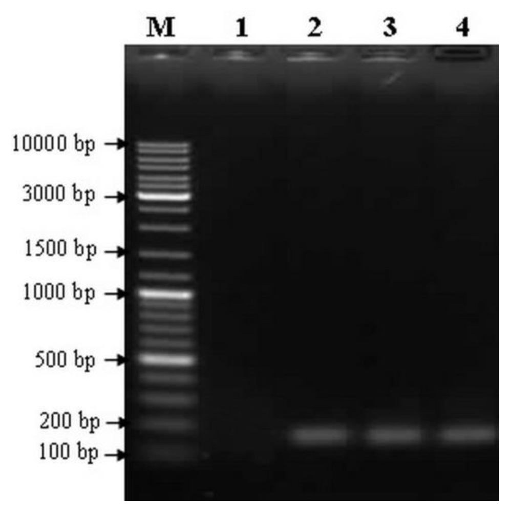
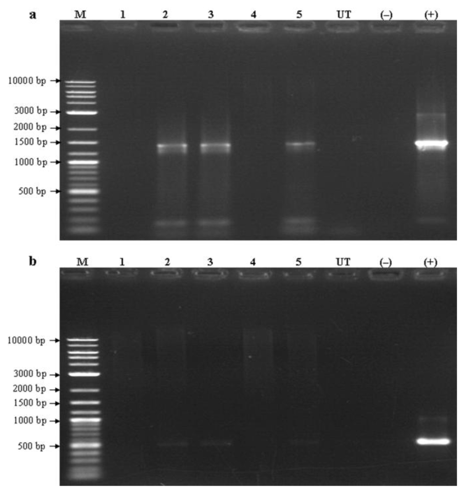
2.3. Detection of Transgenes Using PCR Analysis

Recently, many techniques have been developed to detect transgenes in transgenic organisms. Among these techniques, PCR-based techniques followed by gel electrophoresis and detection are routinely used to detect transgenes in plants as well as in microalgae [33,34]. Besides, other techniques such as the use of molecular beacon assay [35], fluorescence in situ hybridization [36] was also used. In the present study, the isolated AcRbcS promoter sequence was used to drive the expression of the transgenes in A. convolutus. The preliminary analysis of putative transformed A. convolutus lines was carried out using PCR. Two primer sets designed based on the coding sequence of β-glucuronidase (gusA) and hygromycin phosphotransferase (hpt) genes from destination vector pMDC163 were used in subsequent PCR analysis. Amplified with these transgene-specific primers, three out of five putative transformed lines (lines 2, 3 and 5) gave expected amplified products of 1433 bp for gusA (Figure 3a) and 509 bp for hpt gene (Figure 3b). This indicated the presence of gusA and hpt in the genome of these transformed lines. In contrast, no bands were detected in the untransformed cell and negative control (without DNA template) (Figure 3). The failure in detection of the transgenes in two putative transformed lines (Figure 3, lines 1 and 4) could have resulted from the rearrangement of pAcRbcS::gusA T-DNA or the transient expression of transgene in A. convolutus.

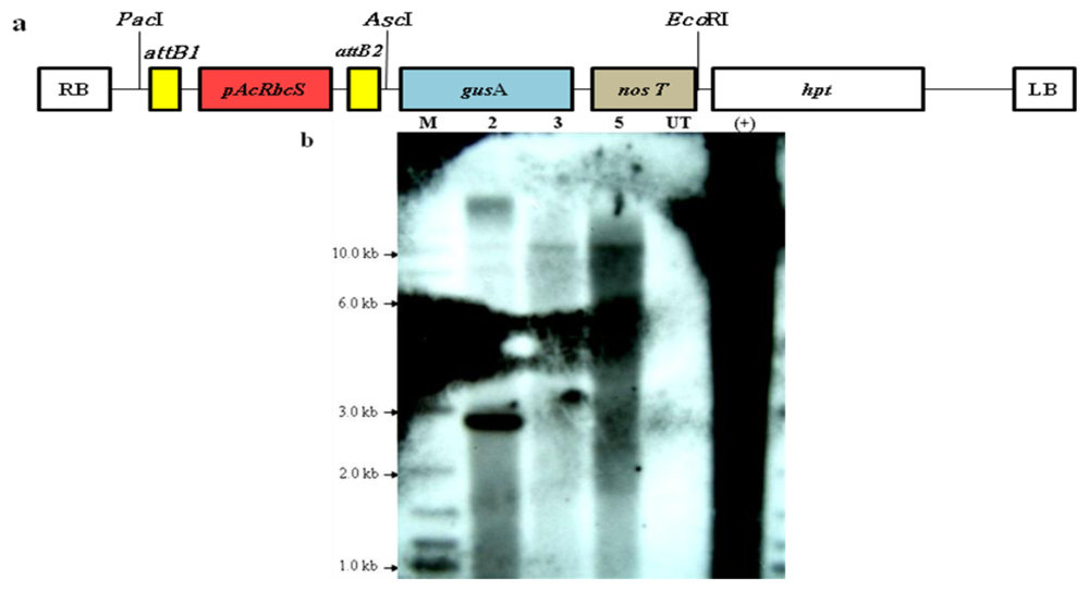
2.4. Integration of the Transgenes Driven by the AcRbcS Promoter

It is known that efficient promoters, including species-specific and universal promoters, are essential for foreign gene expression. For instance, the native ubiquitin promoter is more efficient than the heterologous CaMV35S promoter in driving the expression of foreign genes in monocot plant [37]. The regulatory region of the RbcS gene is an excellent candidate for the development of a promoter that can be widely used to achieve a high level of transgene expression in green algae as well as eukaryotic organisms [3,5,38,39].
In order to confirm the integration of the transgenes into the nuclear genome of A. convolutus, Southern hybridization analysis has been carried out. Approximately 15 μg genomic DNA isolated from the untransformed A. convolutus and three PCR-positive transformed lines was digested with EcoRI restriction enzyme and then hybridized with the gusA-gene-specific probe. EcoRI was chosen since it does not interfere with the gusA gene and has only one restriction site on the expression vector pAcRbcS::gusA (Figure 4a), which helped to linearize the expression vector when it was totally digested. In addition, after being digested with this enzyme, the genome of transgenic A. convolutus gave good fragmentation which was sufficient for Southern blot hybridization. Further, with the restriction site of EcoRI being located outside the gusA gene, this ensured that the copy number of the transgenes were determined precisely. The result showed that all of transformed lines yielded one or more bands, while no hybridization signal was detected from untransformed A. convolutus (Figure 4b). This was tangible evidence for the integration of transgenes into the nuclear genome of A. convolutus. The result also demonstrated that these transgenic lines may contain one to four copies in the nuclear genomes, which revealed that the transgenes could be integrated randomly into the genome, and gusA gene served as a strong selection for successful transformation of this alga. An identical hybridized banding pattern shown in two of the transformed lines (Figure 4, lines 3 and 5) suggested that these lines might be derived from a single transformed cell, which later divided into two cells during the overnight recovery step, before plated onto selection medium. This is the first report using the native promoter to drive the expression of the foreign genes in green microalga A. convolutus. The fact that the AcRbcS promoter functioned in A. convolutus suggested that this promoter could be used to establish a transformation system for A. convolutus to develop this microalga as an alternative host for the expression of heterologous proteins.

2.5. Influence of Light on the Regulation of AcRbcS Promoter Activity

It is known that the expression level of RbcS mRNA is regulated by light in plants [40] as well as green microalgae [26,41]. The fusion of RbcS promoters to reporter genes has been used to study light regulation in transgenic plants [38,39] but no such reports dealt with green algae.
In order to support the confirmation of integration of transgenes as well as to determine whether the AcRbcS promoter showed light regulation in transgenic A. convolutus, the levels of gusA transcript were preliminary examined in A. convolutus grown in continuous light for 3 days after 1-day subculture (L) and then transferred to dark condition for 24 h (D). This light regime has previously been used to examine the expression level of RbcS mRNA in A. convolutus with minor modification in this study, where 24 h was used instead of 12 h in dark conditions. Under such a regime, the expression level of A. convolutus RbcS gene was highest in continuous light for 3 days after 1-day subculture (L) and then reduced after 12 h in the dark [26]. In this study, the expression of gusA gene at RNA level was examined in two out of three transformed lines. As can be seen from the results, gusA mRNA was clearly present in all transgenic A. convolutus lines before being placed in dark conditions (Figure 5). After 24 h in total darkness, however, gusA transcript levels in these transgenic lines were drastically decreased (Figure 5), suggesting a marked down-regulation of the AcRbcS promoter in the absence of light. It implied that the gusA gene has been integrated into the genome of transgenic A. convolutus. It also revealed that the activity of AcRbcS promoter obviously depended on light. This suggested that the AcRbcS promoter seems to be light-regulated in transgenic A. convolutus as demonstrated by the large decrease of corresponding gusA transcripts. This pattern was also reported in the other studies conducted on higher plants, of which the expression levels of gusA gene in leaves driven by the RbcS promoters also reduced after being transferred from the light to the dark [38,39].

3. Experimental Section

3.1. Culture Conditions

Green microalga Ankistrodesmus convolutus was collected from axenic freshwater and deposited in the Department of Cell and Molecular Biology, Faculty of Biotechnology and Biomolecular Sciences, Universiti Putra Malaysia. The culture is grown and maintained in Bold’s Basal Medium (BBM) [42]. Cultures were agitated on a gyratory shaker (150 rpm) at 25 °C without aeration under illumination at a light intensity of 55 μmol photons m−2 s−1 with a photocycle of 12 h light/12 h dark.

3.2. Isolation and Sequence Analysis of AcRbcS Promoter

The AcRbcS promoter region from A. convolutus was obtained by TAIL-PCR amplification and cloned into pGEM-T Easy vector (Promega, USA) as described in our previous report [43]. Plasmid DNA was isolated using PureYield Plasmid Midiprep System (Promega, USA) according to the manufacture’s instruction and sequenced commercially. The location and distribution of putative cis-acting elements were analyzed by using the Plant Cis-acting Regulatory DNA Elements (PLACE) [44] and Plant Cis-acting Regulatory Elements (PlantCARE) [45] databases.

3.3. Identification of the Transcription Start Site by 5′-Rapid Amplification of cDNA Ends (5′-RACE)

Total RNA was isolated from A. convolutus as described in our previous report [46]. The 5′ end of AcRbcS cDNA was determined using the SMARTer RACE cDNA Amplification Kit (Clontech, USA) according to the manufacturer’s instructions. Briefly, approximately 1 μg of total RNA was converted into first-strand cDNA using SMARTScribe RT at 42 °C for 1.5 h. The 5′-RACE-ready cDNA was obtained using the 5′-CDS Primer A and SMARTer IIA oligonucleotides supplied in the kit. Upon reaching the end of the mRNA template, the terminal transferase activity of SMARTScribe RT adds several dC residues to the 3′ end of the first-strand cDNA, and the SMARTer oligonucleotides contains a terminal stretch of modified bases that anneal to the extended cDNA tail, allowing the oligonucleotides to serve as a template for the reverse transcription. A touchdown PCR was performed with the Universal Primer A Mix (UPM) from the kit and the first gene-specific primer (RGSP1: 5′-CGCGATCTGCTCGTCGTTCA-3′) as the forward and reverse primers, respectively. The PCR program consisted of 5 cycles of 94 °C for 30 s and 72 °C for 3 min, followed by 5 cycles of 94 °C for 30 s, 70 °C for 30 s and 72 °C for 3 min, then ended with 25 cycles of 94 °C for 30 s, 68 °C for 30 s and 72 °C for 3 min. The nested PCR reaction was then conducted with the Nested Universal Primer A (NUP) provided in the kit and the second gene-specific primer (RGSP2: 5′-TTCACGGGCTGCCACACCAT-3′) as the forward and reverse primers, respectively. The amplification was performed through 30 cycles of 94 °C for 30 s, 62 °C for 45 s and 72 °C for 1 min, followed by a final elongation step of 72 °C for 5 min. After separation on 1.2% (w/v) agarose gel electrophoresis, the obtained single band of approximately 170 bp was excised from the gel, purified using QiaquickGel Extraction Kit (Qiagen, Germany), cloned into pGEM-T Easy vector (Promega, USA) and sequenced commercially. Identical nucleotide sequence corresponding to the 5′ end of AcRbcS cDNA was obtained by sequence analysis of three independent clones.

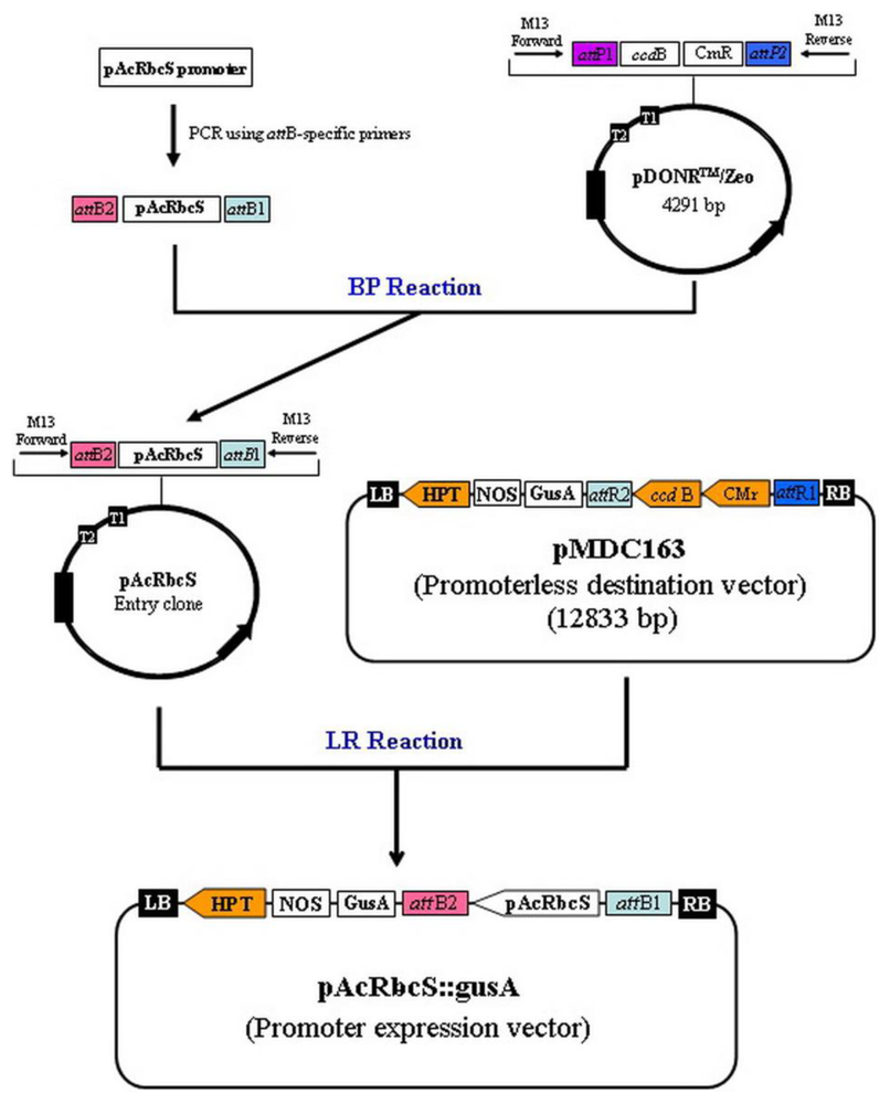
3.4. Construction of Promoter::gusA Fusing Vector

The construction of promoter::gusA fusing vector was made using the Gateway® cloning technology with Clonase II (Invitrogen, USA) according to the manufacturer’s instructions. A schematic diagram for the construction of binary vector was depicted in Figure 6. PCR amplification of AcRbcS promoter was performed using a promoter-specific primer set: pAcRbcS-F (5′-GGGGAC AAGTTTGTACAAAAAAGCAGGCTGCACCACCGCAGCTTAGCGCCCA-3′) and pAcRbcS-R (5′-G GGGACCACTTTGTACAAGAAAGCTGGGTCATTGCTGCTGCTTGCGGGTGA-3′), in which the attB1 and attB2 sites (underlined) were added to the 5′-ends of the forward and reverse primers, respectively. The PCR reaction consists of 1× Pfu buffer, 0.2 mM dNTPs mix, 0.2 μM forward primer, 0.2 μM reverse primer, 0.5 U Pfu DNA polymerase (Fermentas, USA) and 100 ng plasmid DNA. PCR conditions were initial denaturing at 95 °C for 5 min, followed by 35 cycles of 95 °C for 45 s, 68 °C for 45 s and 72 °C for 1 min, and a final elongation step of 72 °C for 7 min. The obtained PCR fragment was recombined into the entry vector pDONR/Zeo using a BP clonase enzyme mixture. The BP recombination reaction was then transformed into E. coli DH5α competent cells by electroporation and zeocin-resistant colonies were selected. Plasmid DNA was isolated and used for a second reaction with the LR clonase enzyme mixture, in this reaction the promoter fragment was recombined into the promoterless destination vector pMDC163 containing the β-glucuronidase (gusA) reporter gene to create the promoter expression vector, pAcRbcS::gusA. E. coli DH5α was transformed with pAcRbcS::gusA, the resulted recombinant plasmid was verified by sequencing and introduced into Ankistrodesmus convolutus using electroporation method.

3.5. Nuclear Transformation of A. convolutus

Cells in early stationary phase were collected by centrifugation at 6000 g for 5 min, treated with cellulase (2%) and pectinase (0.3%) to partially remove cell wall. Enzymes treated cells were electroporated using the Electroporator 2510 (Eppendorf, Germany) according to the method described by Chi et al. (unpublished). Briefly, the enzymes treated cells were resuspended in electroporation buffer (5 mM CaCl2, 20 mM HEPES, 200 mM sorbitol and 200 mM mannitol, pH 7.0) to the final cell density of 1–2 × 10⁶ cells/mL. A total of 2 μg pAcRbcS::gusA and 6 μg carrier DNA (salmon sperm DNA) were added to cell suspension. The resuspended cells were transferred into electroporation chamber and electroporated at a pulse voltage of 1.8 kV with the constant pulse duration of 5 ms using the Electroporator 2510 (Eppendorf, Germany). After electroporation, the cells were transferred into 5 mL of liquid BBM medium for recovery for 24 h in the dark and later they were spread onto solid BBM plates supplemented with hygromycin (40 μg/mL), and grown at 25 °C under 12:12 h light-dark cycle.
Transformed colonies appeared in a week, and independent colonies were maintained on selection medium. The hygromycin resistant colonies were then cultured in liquid BBM supplementing hygromycin (40 μg/mL) at 25 °C with agitation on a gyratory shaker (150 rpm) without aeration under 55–60 μmol photons m−2 s−1 illumination with a photocycle of 12 h light/12 h dark.

3.6. Analysis of T-DNA Integration and Transgenes Expression

Total RNA and genomic DNA of transgenic A. convolutus were extracted as described in our previous reports [46,47]. Putative transformed lines were initially screened by PCR, which was performed with two set of primers: gus-F (5′-ACCGAAGTTCATGCCAGTCCAGCG-3′) and gus-R (5′-ATGTCACGCCGTATGTTATTGCCG-3′) specific to β-glucuronidase (gusA) gene to amplify a 1433-bp fragment, and hpt-F (5′-AGCTGCGCCGATGGTTTCTACAA-3′) and hpt-R (5′-ATCGCCT CGCTCCAGTCAATG-3′) to amplify a 509-bp fragment of the hygromycin phosphotransferase (hpt) gene.
The transformed lines were also analyzed by Southern hybridization. Approximately 15 μg of genomic DNA was digested with EcoRI overnight, electrophoresed on 0.8% (w/v) agarose gel. The DNA was then overnight transferred by capillarity onto nylon membrane (Hybond N+, Amersham Bioscience) with 0.4 N NaOH, and autocrosslinked by UV-crosslinker (Ultra-Violet Products, UK) at the wavelength of 254 nm for 2 min. The membrane with cross-linked DNA was prehybridized at 50 °C for 2 h before hybridized at 55 °C overnight using the biotin-labelled gusA gene fragment as probe. This probe was prepared by PCR amplification of the gusA gene using gus-F and gus-R primers and incorporating biotin-14-dCTP. The hybridized membrane was washed twice with low stringency washing solution (2× SSC, 0.1% (w/v) SDS) for 15 min, and twice with medium stringency washing solution (0.5× SSC, 0.1% (w/v) SDS) for 30 min at 65 °C. The membrane was exposed to X-ray film (Kodak Medical X-ray Film, Kodak) in an X-ray cassette before developing the film for signal visualization.
For Northern hybridization analysis, approximately 15 μg of total RNA from selected transformed lines was denatured and separated in 1.2% (w/v) agarose gel containing 6% (v/v) formaldehyde followed by staining with ethidium bromide. The RNA samples was then transferred overnight by capillarity with 10× SSC buffer onto nylon membrane (Hybond N+, Amersham Bioscience) and autocrosslinked by UV-crosslinker (Ultra-Violet Products, UK) at the wavelength of 254 nm for 2 min. The blotted membrane was hybridized at 67 °C overnight with the labeled probe for gusA gene prepared by PCR incorporation of biotin-14-dCTP. The hybridized membrane was washed twice with 2× SSC, 0.1% (w/v) SDS for 15 min, and twice with 0.5× SSC, 0.1% (w/v) SDS for 30 min at 67 °C. The membrane was exposed to X-ray film (Kodak Medical X-ray Film, Kodak) in an X-ray cassette before developing the film for signal visualization.

4. Conclusions

Green algae have proven their utility and tractability as a system for the production of therapeutic or industrial proteins and peptides. They now seem poised to become the “green” alternative to the current mammalian, yeast, or bacterial recombinant protein production systems. Development of an expression system using green microalga Ankistrodesmus convolutus is worthwhile because of its importance as a potential model organism for genetic engineering. Like other unicellular green algae, A. convolutus meets the characteristics required for production of recombinant proteins, its cells grow fast in a simple medium and this species can be cultured simply, rapidly and economically.
In the present study, the AcRbcS promoter sequence which was isolated from green microalga A. convolutus in our previous work has been analyzed and then used to drive the expression of gusA in this microalga species. The presence of several cis-acting elements and motifs known to be involved in the light-dependent expression of photosynthesis genes of higher plants and other green microalgae species, and the retrieval of the transformed A. convolutus expressing transgenes driven by AcRbcS promoter suggested that the isolated promoter has contained the promoter elements necessary for the transcription and regulation of gene expression. The transformation construct may be functional in other closely related species. Indeed, describing the use of promoter-constructs from a closely related species to develop a nuclear transformation has been recently reported [5]; it is likely that the AcRbcS promoter and the construct developed in this study may be useful as a universal promoter and in the development of a transformation system, respectively, for other green microalgae species such as Chlamydomonas reinhardtii and Dunaliella spp.
In this study, it was found that the AcRbcS promoter is regulated by light as demonstrated by a reduction of corresponding gusA-specific transcripts when the transgenic A. convolutus was transferred from light to dark conditions; however, further experiments, such as determination of the presence of any negative regulatory elements within the AcRbcS promoter sequence, should be performed to increase the expression levels of the transgenes under the control of this promoter. This work demonstrates, for the first time, the use of the native promoter to drive the expression of foreign genes in nuclear genome of green microalga A. convolutus. The achievement of a transformation system and the practical application of the AcRbcS promoter have reliably contributed to the development of A. convolutus as a model organism for algae research, and it is possible to develop this microalga as an expression system for the production of recombinant proteins.

Acknowledgments

This work was supported by the MOSTI grant number 02-01-04-SF0041 from the Ministry of Science, Technology and Innovation of Malaysia. The first author is indebted to the Rubber Research Institute of Vietnam on the financial support for the postgraduate scholarship.

References

  1. Mayfield, S.P.; Franklin, S.E.; Lerner, R.A. Expression and assembly of a fully active antibody in algae. Proc. Natl. Acad. Sci. USA 2003, 100, 438–442. [Google Scholar]
  2. Griesbeck, C.; Kobl, I.; Heitzer, M. Chlamydomonas reinhardtii: A protein expression system for pharmaceutical and biotechnological proteins. Mol. Biotechnol 2006, 34, 213–223. [Google Scholar]
  3. Stevens, D.R.; Rochaix, J.D.; Purton, S. The bacterial phleomycin resistance gene ble as a dominant selectable marker in Chlamydomonas. Mol. Gen. Genet 1996, 251, 23–30. [Google Scholar]
  4. Schroda, M.; Blocker, D.; Beck, C.F. The HSP70A promoter as a tool for the improved expression of transgenes in Chlamydomonas. Plant J 2000, 21, 121–131. [Google Scholar]
  5. Walker, T.L.; Becker, D.K.; Collet, C. Characterisation of the Dunaliella tertiolecta RbcS genes and their promoter activity in Chlamydomonas reinhardtii. Plant Cell Rep 2005, 23, 727–735. [Google Scholar]
  6. Lu, Y.; Li, J.; Xue, L.; Yan, H.; Yuan, H.; Wang, C. A duplicated carbonic anhydrase 1 (DCA1) promoter mediates the nitrate reductase gene switch of Dunaliella salina. J. Appl. Phycol 2010. [Google Scholar] [CrossRef]
  7. Tang, D.K.H.; Qiao, S.Y.; Wu, M. Insertion mutagenesis of Chlamydomonas reinhardtii by electroporation and heterologous DNA. Biochem. Mol. Biol. Int 1995, 36, 1025–1035. [Google Scholar]
  8. El-Sheekh, M.M. Stable transformation of the intact cells of Chlorella kessleri with high velocity microprojectiles. Biol. Plant 1999, 42, 209–216. [Google Scholar]
  9. Kim, D.H.; Kim, Y.T.; Cho, J.J.; Bae, J.H.; Hur, S.B.; Hwang, I.; Choi, T.J. Stable integration and functional expression of flounder growth hormone gene in transformed microalga, Chlorella ellipsoidea. Mar. Biotechnol 2002, 4, 63–73. [Google Scholar]
  10. Tan, C.; Qin, S.; Zhang, Q.; Jiang, P.; Zhao, F. Establishment of a microparticle bombardment transformation system for Dunaliella salina. J. Microbiol 2005, 43, 361–365. [Google Scholar]
  11. Feng, S.; Xue, L.; Liu, H.; Lu, P. Improvement of efficiency of genetic transformation for Dunaliella salina by glass beads method. Mol. Biol. Rep 2009, 36, 1433–1439. [Google Scholar]
  12. Blankenship, E.; Kindle, K. Expression of chimeric genes by the light-regulated cabII-1 promoter in Chlamydomonas reinhardtii: A cabII-1/nit1 gene functions as a dominant selectable marker in a nit1 nit2 strain. Mol. Cell. Biol 1992, 12, 5268–5279. [Google Scholar]
  13. Butanaev, A.M. Hygromicin phosphotransferase gene as a dominant selective marker for transformation of Chlamydomonas reinhardtii. Mol. Biol 1994, 28, 682–686. [Google Scholar]
  14. Teng, C.; Qin, S.; Liu, J.; Yu, D.; Liang, C.; Tseng, C. Transient expression of lacZ in bombarded unicellular green alga Haematococcus pluvialis. J. Appl. Phycol 2002, 14, 495–500. [Google Scholar]
  15. Chen, Y.; Wang, Y.; Sun, Y.; Zhang, L.; Li, W. Highly efficient expression of rabbit neutrophil peptide-1 gene in Chlorella ellipsoidea cells. Curr. Genet 2001, 39, 365–370. [Google Scholar]
  16. Franklin, S.; Ngo, B.; Efuet, E.; Mayfield, S.P. Development of a GFP reporter gene for Chlamydomonas reinhardtii chloroplast. Plant J 2002, 30, 733–744. [Google Scholar]
  17. Shao, N.; Bock, R. A codon-optimized luciferase from Gaussia princeps facilitates the in vivo monitoring of gene expression in the model alga Chlamydomonas reinhardtii. Curr. Genet 2008, 53, 381–388. [Google Scholar]
  18. Fischer, N.; Rochaix, J.D. The flanking regions of PsaD drive efficient gene expression in the nucleus of the green alga Chlamydomonas reinhardtii. Mol. Genet. Genomics 2001, 265, 888–894. [Google Scholar]
  19. Kindle, K.L. High-frequency nuclear transformation of Chlamydomonas reinhardtii. Proc. Natl. Acad. Sci. USA 1990, 87, 1228–1232. [Google Scholar]
  20. Cerutti, H.; Johnson, A.M.; Gillham, N.W.; Boynton, J.E. A eubacterial gene conferring spectinomycin resistance on Chlamydomonas reinhardtii: integration into the nuclear genome and gene expression. Genetics 1997, 145, 97–110. [Google Scholar]
  21. Auchincloss, A.H.; Loroch, A.I.; Rochaix, J.D. The argininosuccinate lyase gene of Chlamydomonas reinhardtii: cloning of the cDNA and its characterization as a selectable shuttle marker. Mol. Gen. Genet 1999, 261, 21–30. [Google Scholar]
  22. Fuhrmann, M.; Oertel, W.; Hegemann, P. A synthetic gene coding for the green fluorescent protein (GFP) is a versatile reporter in Chlamydomonas reinhardtii. Plant J 1999, 19, 353–361. [Google Scholar]
  23. Kovar, J.L.; Zhang, J.; Funke, R.P.; Weeks, D.P. Molecular analysis of the acetolactate synthase gene of Chlamydomonas reinhardtii and development of a genetically engineered gene as a dominant selectable marker for genetic transformation. Plant J 2002, 29, 109–117. [Google Scholar]
  24. Sun, Y.; Yang, Z.Y.; Gao, X.S.; Li, Q.Y.; Zhang, Q.Q.; Xu, Z.K. Expression of foreign genes in Dunaliella by electroporation. Mol. Biotechnol 2005, 30, 185–192. [Google Scholar]
  25. Chu, W.L.; Phang, S.M.; Goh, S.H.; Blakebrough, N. Promising Microalgae for Production of Useful Chemicals. In Proceeding of the Conference on Medicinal Products from Tropical Rain Forests, Forest Research Institute of Malaysia, Kuala Lumpur, Malaysia, 13–15 May 1992; Shaari, K., Kadir, A.A., Ali, A.R.M., Eds.; pp. 338–345.
  26. Thanh, T.; Chi, V.T.Q.; Abdullah, M.P.; Omar, H.; Noroozi, M.; Ky, H.; Napis, S. Construction of cDNA library and preliminary analysis of expressed sequence tags from green microalga Ankistrodesmus convolutus Corda. Mol. Biol. Rep 2011, 38, 177–182. [Google Scholar]
  27. Giuliano, G.; Pechersky, E.; Malik, V.S.; Timko, M.P.; Scolnik, P.A.; Cashmore, A.R. An evolutionarily conserved protein binding sequence upstream of a plant light-regulated gene. Proc. Natl. Acad. Sci. USA 1988, 85, 7089–7093. [Google Scholar]
  28. Williams, M.E.; Foster, R.; Chua, N.H. Sequences flanking the hexameric G-box core CACGTG affect the specificity of protein binding. Plant Cell 1992, 4, 485–496. [Google Scholar]
  29. Hudson, M.E.; Quail, P.H. Identification of promoter motifs involved in the network of phytochrome A-regulated gene expression by combined analysis of genomic sequence and microarray data. Plant Physiol 2003, 133, 1605–1616. [Google Scholar]
  30. Li, T.; Gong, C.; Wang, T. The rice light-regulated gene RA68 encodes a novel protein interacting with oxygen-evolving complex PsbO mature protein. Plant Mol. Biol. Rep 2010, 28, 136–143. [Google Scholar]
  31. Ahrazem, O.; Rubio-Moraga, A.; López, R.C.; Gómez-Gómez, L. The expression of a chromoplast-specific lycopene beta cyclase gene is involved in the high production of saffron’s apocarotenoid precursors. J. Exp. Bot 2010, 61, 105–119. [Google Scholar]
  32. Nomura, M.; Katayama, K.; Nishimura, A.; Ishida, Y.; Ohta, S.; Komari, T.; Miyao-Tokutomi, M.; Tajima, S.; Matsuoka, M. The promoter of rbcS in a C3 plant (rice) directs organ-specific, light-dependent expression in a C4 plant (maize), but does not confer bundle sheath cell-specific expression. Plant Mol. Biol 2000, 44, 99–106. [Google Scholar]
  33. Higuchi, R.; Dollinger, G.; Walsh, P.S.; Griffith, R. Simultaneous amplification and detection of specific DNA sequences. Nat. Biotechnol 1992, 10, 413–417. [Google Scholar]
  34. Kumar, S.C.; Misqitta, R.W.; Reddy, V.S.; Rao, B.J.; Rajam, M.V. Genetic transformation of the green alga Chlamydomonas reinhardtii by Agrobacterium tumefaciens. Plant Sci 2004, 166, 731–738. [Google Scholar]
  35. Kota, R.; Holton, T.A.; Henry, R.J. Detection of transgenes in crop plants using molecular beacon assays. Plant Mol. Biol. Rep 1999, 17, 363–370. [Google Scholar]
  36. Jin, W.W.; Li, Z.Y.; Fang, Q.; Altosaar, I.; Liu, L.H.; Song, Y.C. Fluorescence in situ hybridization analysis of alien genes in Agrobacterium-mediated Cry1A(b)-transformed rice. Ann. Bot 2002, 90, 31–36. [Google Scholar]
  37. Wang, J.; Oard, J.H. Rice ubiquitin promoters: Deletion analysis and potential usefulness in plant transformation systems. Plant Cell Rep 2003, 22, 129–134. [Google Scholar]
  38. Gittins, J.R.; Pellny, T.K.; Hiles, E.R.; Rosa, C.; Biricolti, S.; James, D.J. Transgene expression driven by heterologous ribulose-1,5-bisphosphate carboxylase/oxygenase small-subunit gene promoters in the vegetative tissues of apple (Malus pumila Mill.). Planta 2000, 210, 232–240. [Google Scholar]
  39. Marraccini, P.; Courjault, C.; Caillet, V.; Lausanne, F.; Lepage, B.; Rogers, W.J.; Tessereau, S.; Deshayes, A. Rubisco small subunit of Coffea arabica: cDNA sequence, gene cloning and promoter analysis in transgenic tobacco plants. Plant Physiol. Biochem 2003, 41, 17–25. [Google Scholar]
  40. Terzaghi, W.B.; Cashmore, A.R. Light-regulated transcription. Annu. Rev. Plant Physiol. Plant Mol. Biol 1995, 46, 445–474. [Google Scholar]
  41. Yamazaki, T.; Yamamoto, M.; Sakamoto, W.; Kawano, S. Isolation and molecular characterization of rbcS in the unicellular green alga Nannochloris bacillaris (Chlorophyta, Trebouxiophyceae). Phycol. Res 2005, 53, 67–76. [Google Scholar]
  42. Nichols, H.W. Growth Media-Freshwater. In Handbook of Phycological Methods: Culture Methods and Growth Measurements; Stein, J.R., Ed.; Cambridge University Press: Cambridge, UK, 1973; pp. 7–24. [Google Scholar]
  43. Thanh, T.; Chi, V.T.Q.; Abdullah, M.P.; Omar, H.; Napis, S. Efficiency of the ligation-mediated PCR and TAIL-PCR methods on isolation of RbcS promoter sequences from green microalga Ankistrodesmus convolutus. Mol. Biol 2011. In Press. [Google Scholar]
  44. Higo, K.; Ugawa, Y.; Iwamoto, M.; Korenaga, T. Plant cis-acting regulatory DNA elements (PLACE) database. Nucleic Acids Res 1999, 27, 297–300. [Google Scholar]
  45. Lescot, M.; Déhais, P.; Thijs, G.; Marchal, K.; Moreau, Y.; van de Peer, Y.; Rouzé, P.; Rombauts, P. PlantCARE, a database of plant cis-acting regulatory elements and a portal to tools for in silico analysis of promoter sequences. Nucleic Acids Res 2002, 30, 325–327. [Google Scholar]
  46. Thanh, T.; Omar, H.; Abdullah, M.P.; Chi, V.T.Q.; Noroozi, M.; Ky, H.; Napis, S. Rapid and effective method of RNA isolation from green microalga Ankistrodesmus convolutus. Mol. Biotechnol 2009, 43, 148–153. [Google Scholar]
  47. Thanh, T.; Chi, V.T.Q.; Abdullah, M.P.; Omar, H.; Noroozi, M.; Napis, S. Cloning and characterization of ribulose-1,5-bisphosphate carboxylase/oxygenase small subunit (RbcS) cDNA from green microalga Ankistrodesmus convolutus. Mol. Biol. Rep 2011. [Google Scholar] [CrossRef]
Figure 1. Nucleotide sequence of the AcRbcS promoter. The start codon (ATG) was underlined and the putative transcriptional start site (TSS) was indicated by a vertical arrow and referred to as position +1. The putative TATA-box and other cis-acting elements were boxed and the names are given above the elements. The accession number of the AcRbcS promoter deposited in the GenBank database is JF802127.
Ijms 13 02676f1
Figure 2. Determination of the transcription start site using 5′-RACE. Lane M: 2-log DNA marker (New England Biolabs, UK); Lane 1: negative control using total RNA without reverse transcriptase; Lanes 2−4: SMARTer RACE PCR products from different cDNA as templates.
Ijms 13 02676f2
Figure 3. PCR analysis of putative transformed lines. (a) and (b) The presence of β-glucuronidase (gusA) and hygromycin phosphotransferase (hpt) genes were detected using gus- and hpt-specific primers, respectively. Three out of five putative lines tested (lines 2, 3 and 5) showed the presence of both gusA and htp genes. Lane M: 2-log DNA marker (New England Biolabs, UK); Lanes 1−5: putative transformed lines; Lane UT: untransformed control; Lane (−): negative control (without DNA template); Lane (+): positive control (pAcRbcS::gusA plasmid DNA).
Ijms 13 02676f3
Figure 4. Southern hybridization analysis of transformed A. convolutus. (a) The presentation of the T-DNA region with AcRbcS promoter fused with gusA and hpt genes. The position of EcoRI restriction site was indicated; (b) Genomic DNA (15 μg) from three PCR-positive transformed lines (lines 2, 3 and 5) and untransformed cell was digested with EcoRI restriction enzyme and probed with a biotin-labeled gusA gene-specific probe. Lane M: 2-log DNA marker (New England Biolabs, UK); Lane UT: untransformed (negative control); Lane (+): pAcRbcS::gusA plasmid DNA (positive control, 10 μg linearized using EcoRI restriction enzyme).
Ijms 13 02676f4
Figure 5. Light regulation of the AcRbcS promoter in transformed A. convolutus. Total RNA loaded on the gel was hybridized with a gusA-specific probe (upper panel). Total RNA was isolated from two transformed lines (lines 2 and 3) and an untransformed cell (UT) grown under continuous light for 3 days after 1-day subculture (L) and then transferred to dark for 24 h (D). Ethidium bromide staining of ribosomal RNA samples used to demonstrate equivalent loading (lower panel).
Ijms 13 02676f5
Figure 6. Schematic diagram showing the construction of pAcRbcS::gusA vector.
Ijms 13 02676f6

