Article

A Jacalin-Related Lectin Regulated the Formation of Aerial Mycelium and Fruiting Body in Flammulina velutipes

1
Mycological Research Center, College of Life Sciences, Fujian Agriculture and Forestry University, Fuzhou 350002, China
2
College of Food Sciences, Fujian Agriculture and Forestry University, Fuzhou 350002, China
*
Authors to whom correspondence should be addressed.
Int. J. Mol. Sci. 2016, 17(12), 1884; https://doi.org/10.3390/ijms17121884
Submission received: 27 September 2016 / Revised: 2 November 2016 / Accepted: 8 November 2016 / Published: 28 November 2016
(This article belongs to the Section Biochemistry)

Abstract

:
Flammulina velutipes, one of the most popular mushroom species in the world, has been recognized as a useful model system to study the biochemical and physiological aspects of the formation and elongation of fruit body. However, few reports have been published on the regulation of fruiting body formation in F. velutipes at the molecular level. In this study, a jacalin-related lectin gene from F. velutipes was characterized. The phylogenetic tree revealed that Fv-JRL1 clustered with other basidiomycete jacalin-like lectins. Moreover, the transcriptional pattern of the Fv-JRL1 gene in different developmental stages of F. velutipes implied that Fv-JRL1 could be important for formation of fruit body. Additionally, RNA interference (RNAi) and overexpression analyses provided powerful evidence that the lectin gene Fv-JRL1 from F. velutipes plays important roles in fruiting body formation.

Graphical Abstract

1. Introduction

Lectins are non-immunoglobulin proteins and that are widely distributed among organisms, including animals, plants, and microorganisms [1,2], in which they perform a variety of functions. In plants, most lectins act as a defensive system against fungi and insects [3,4,5,6], and some lectins mediate the symbiotic relationship of leguminous plants and nitrogen-fixing bacteria [7]. Animal lectins play a regulative role in organ differentiation and formation [8], and participate in metastasis of cancer cells [9,10] as well as the migration of lymphocytes from the bloodstream into the lymphoid organs [11]. In some viruses, lectins are primarily employed to attachment to host cells [2].
Mushrooms, defined as macrofungi with conspicuous fruiting structures, have been consumed by humans since earliest recorded history, due to their high nutritional properties, as well as their desirable taste and flavor [12,13]. Medicinal attributes, the second major property of mushrooms, have also been recognized and utilized by Oriental cultures, particularly in China and Japan, for a very long time [13,14,15].
Mushrooms contain large amounts of medicinal and pharmacological bioactive compounds [16,17,18,19]. Among them, mushroom lectins have captured increased attention because of their antitumor, antiproliferative, and immunomodulatory activities [20,21,22]. The edible basidiomycete Flammulina velutipes, one of the six most widely grown and consumed mushroom species in the world with an annual yield greater than 300,000 tons, expresses high level of lectins, approximately 77 mg of purified lectin (FVA) per 200 g of fruiting bodies [23]. However, the physiological roles of lectins from F. velutipes in nature are not well known. Previous studies have demonstrated that lectins from Pleurotus cornucopiae have been involved in the process of fruit body formation [24]. Thus, it was speculated that some lectins from F. velutipes may similarly be involved in formation of the fruiting body.
In this study, a jacalin-related lectin (JRL) gene from F. velutipes, designated as Fv-JRL1, was identified and characterized. The expression levels of the Fv-JRL1 gene were also characterized in different developmental stages of F. velutipes. The transcriptional pattern suggested that Fv-JRL1 could be important for formation of the fruiting body. Additionally, RNAi and overexpression were also conducted in order to study the function of this gene, and the results strongly suggest that Fv-JRL1 from F. velutipes does really play an important regulatory role in formation of aerial mycelium and fruiting body.

2. Results

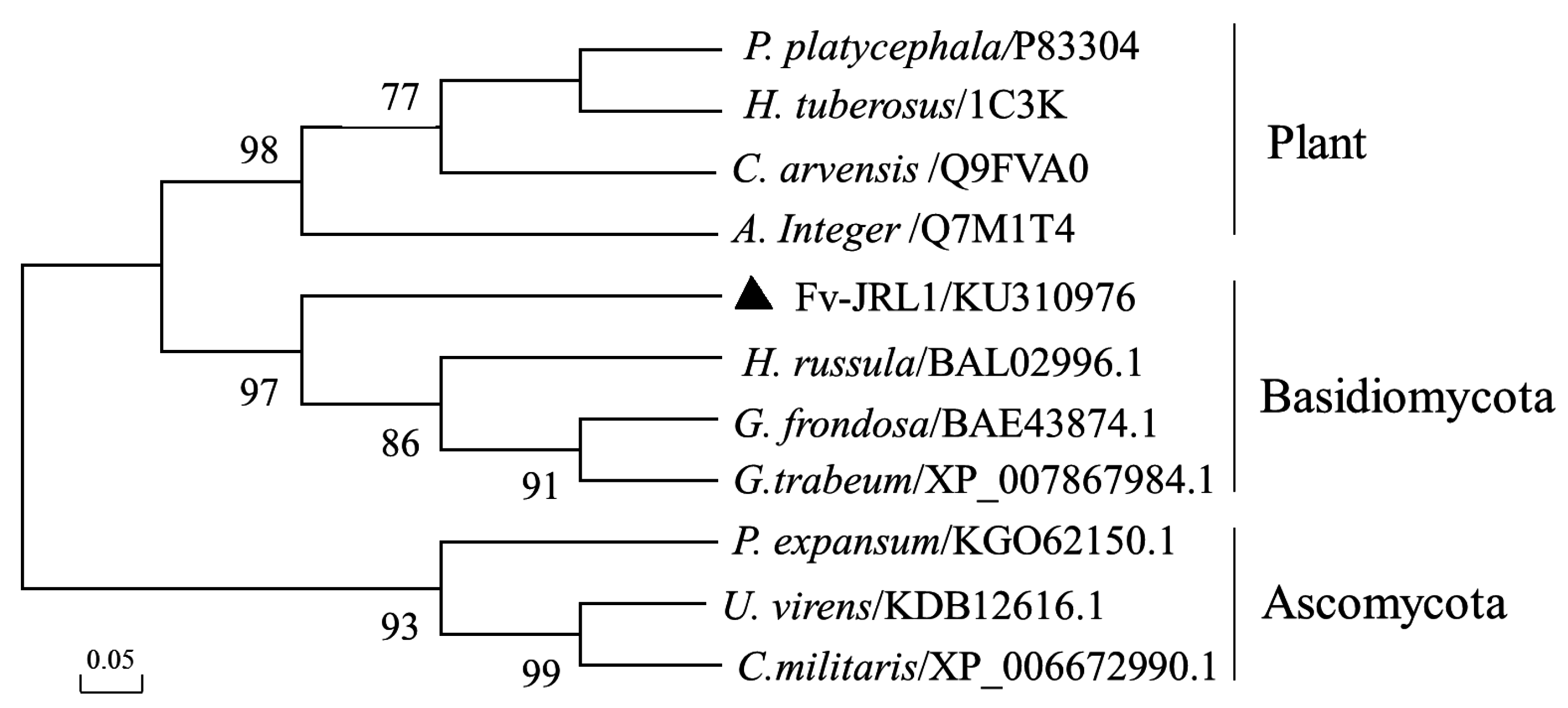
2.1. The Structure of the Fv-JRL1 Gene and Phylogenetic Analysis of the Fv-JRL1 Protein

The sequence of the Fv-JRL1 gene was retrieved from the genome of F. velutipes strain W23. The Fv-JRL1 gene was found to be 1126 bp, encompassing the full-length Fv-JRL1 gene, as well as a 245 bp 5’-UTR and 106 bp 3’-UTR. Four introns of 54, 53, 66, and 59 bp, as well as highly conserved consensus sequences at the 5’ (GT) and 3’ (AG) splice junctions were identified. In addition, comparison of the sequences of Fv-JRL1 in strains L11 and W23 using DNAMAN [25] indicated that the sequences between the two strains were identical, eliminating the interference of homologous genes at transcription level and the need for further RNA interference (RNAi) analysis.
Analysis with ExPASyProtparam (http://www.expasy.ch/tools/protparam.html) [26] indicated that the deduced Fv-JRL1 protein contained 180 amino acids and had a theoretical isoelectric point (pI) of 4.97 and a predicted molecular weight of 19.479 kDa. Four N-myristoylation sites (G62IQPTY, G140TSFGT, G147QVIAL, and G154TDENS) were identified in this protein using ExPASyProsite (http://prosite.expasy.org/). A conserved jacalin-like lectin domain (IPR001229) at amino acids 46–137 was found by InterProScan search (http://www.ebi.ac.uk/Tools/pfa/iprscan/) [27]. For phylogenetic analysis, the amino acid sequences of JRL1 proteins from different organisms were obtained from NCBI and EBI, and aligned with Clustal_X 1.83 [28]. The neighbor-joining tree (Figure 1) generated by MEGA 5.0 with 1000 replicates of bootstrap analysis [29,30] displayed that all of the jacalin-like lectins collected from plants, Basidiomycota, or Ascomycota formed independent clades, and Fv-JRL1 from F. velutipes and jacalin-like lectins from other Basidiomycota fungi clustered into the same clade with a well-supported bootstrap value of 97%.

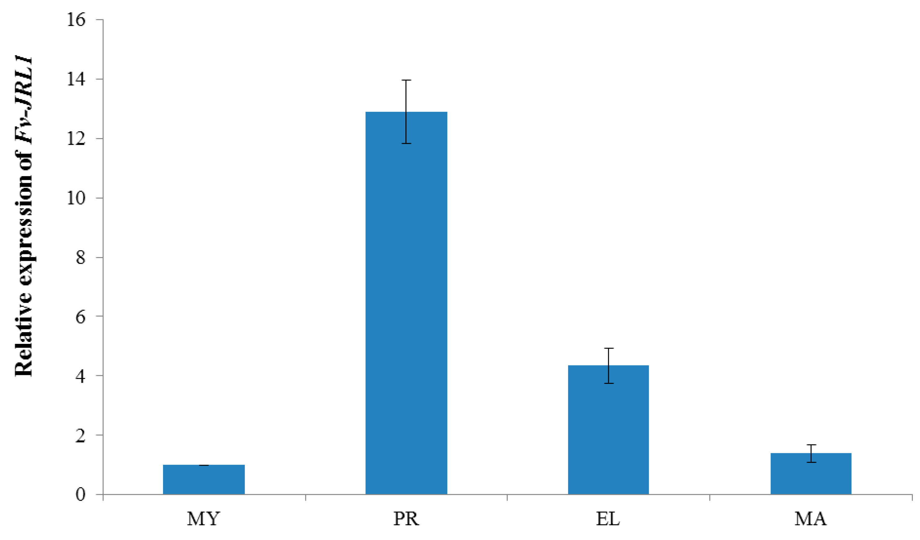
2.2. Expression Patterns Revealed the Potential Function of Fv-JRL1 Protein in the Formation of the Fruiting Body

To investigate the possible roles of Fv-JRL1 protein, we assessed the expression of Fv-JRL1 gene during different development stages using qRT-PCR. The results (Figure 2) indicated that the expression of Fv-JRL1 in the primordial stage was upregulated by approximately 13-fold relative to the mycelial stage (Figure 2). Additionally, Fv-JRL1 transcription was decreased at the elongation and mature stages. These results suggested that Fv-JRL1 proteins are likely related to the formation of the fruiting body.

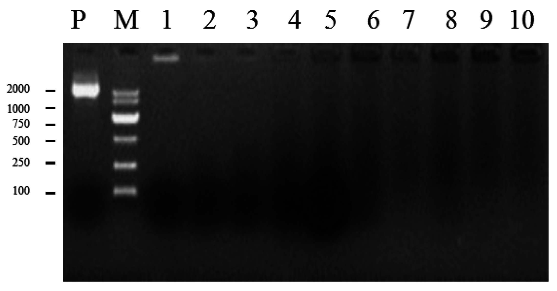
2.3. Confirmation of the Ratio between Plasmid DNA and Liposomes

In the present study, the efficiency of DNA encapsulation was assessed using agarose gel electrophoresis [31,32]. As shown in Figure 3, a DNA band migrated towards the cathode in lane 1, indicating plasmid DNA that was not encapsulated in liposome, as compared to lane P. Thus, the proportion (1 µg plasmid DNA: 1 µL liposomes) was chosen as the optimal ratio for transformation.

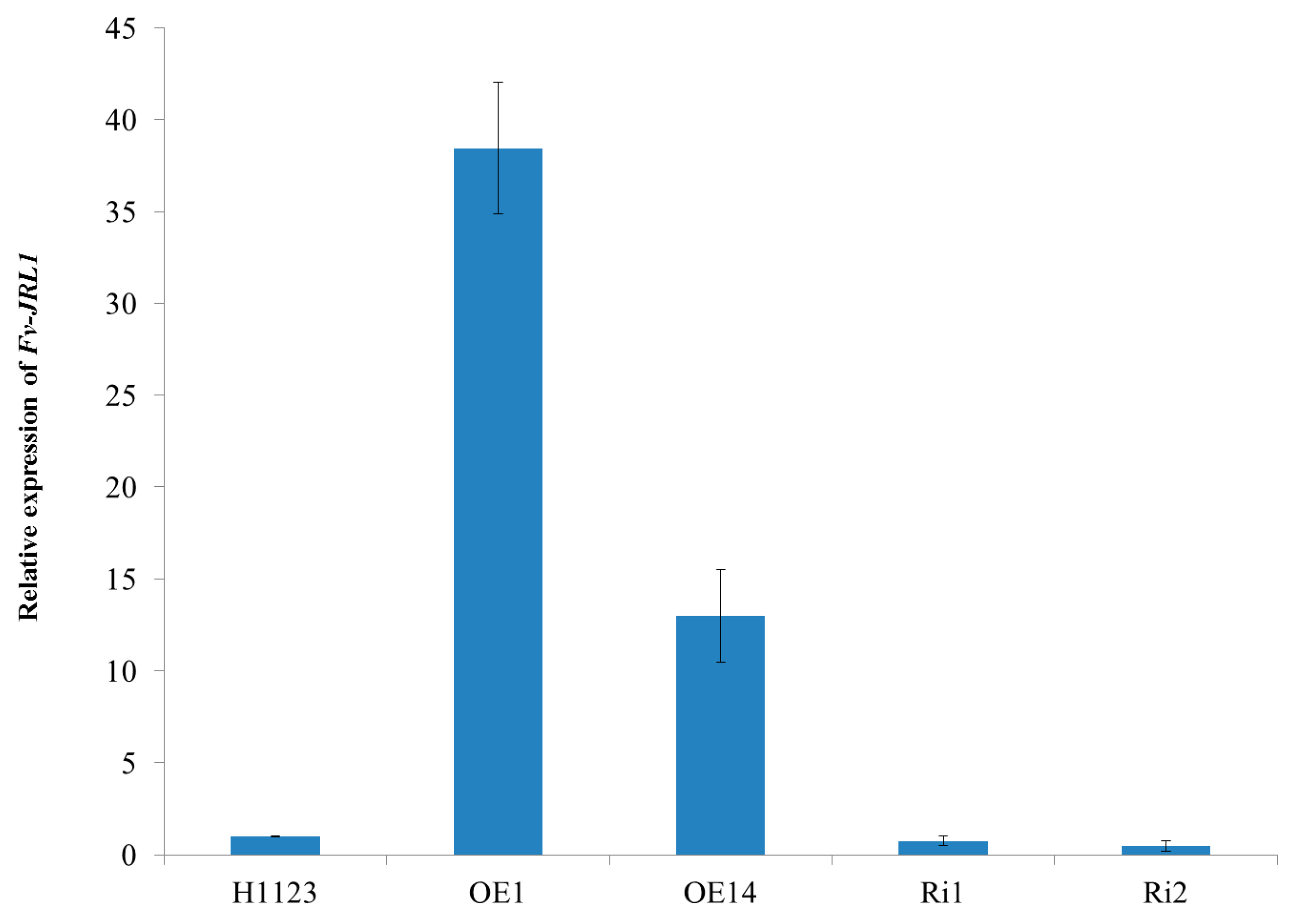
2.4. Generation of Mutants and Fv-JRL1 Gene Expression Analysis

After liposome-mediated transformation, colonies with hygromycin B resistance were obtained and verified by PCR. The presence of the hygromycin B phosphotransferase (hpt) gene (1057 bp), as well as the sequences between the glyceraldehyde-3-phosphate dehydrogenase (gpd) gene promotor (Pgpd) and the Fv-JRL1 gene (1084 bp), was confirmed in overexpression transformants OE1 and OE14 (Figure S1). The presence of the hpt gene and sequences between the Pgpd and the first intron of Fv-JRL1 (733 bp) were identified in RNAi transformants Ri1 and Ri2 (Figure S1).
In order to verify that Fv-JRL1 was silenced or overexpressed, the expression levels of the Fv-JRL1 gene were measured in all transformants. Compared to the wild-type strain H1123, the expression levels of Fv-JRL1 were upregulated approximately 35-fold and 13-fold in transformants OE1 and OE14, respectively (Figure 4). In contrast, Fv-JRL1 gene expression was decreased by 26% and 51% in Ri1 and Ri2, respectively, compared to H1123.

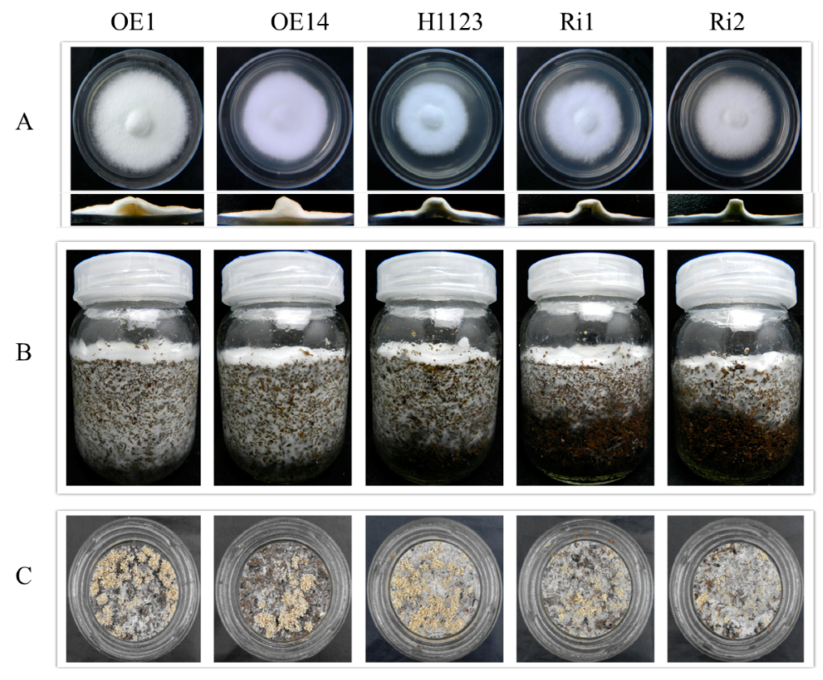
2.5. Phenotypic Characterization of Mutants

To gain insight into the phenotypic alterations, transformants were incubated at 25 °C for 5 days. Experiments carried out on potato dextrose agar (PDA) plates showed that overexpression of Fv-JRL1 resulted in an increase of growth and aerial hyphae compared to wild-type strain H1123. Further, cross-section of the two RNAi transformants indicated a reduction in the amount of hyphae in contrast to overexpression transformants (Figure 5A).
When grown on composted substrate, RNAi-silenced strains Ri1 and Ri2 produced fewer primordia than the wild-type (Figure 5C), exhibited retarded vegetative growth (Figure 5B), and delayed the formation of fruiting bodies. Wild-type F. velutipes H1123 needed 25 days of incubation to expand beyond the glass bottles, while Ri1 and Ri2 needed 29 days of incubation to reach to the bottom of bottles. The fruiting-body initiation appeared 15 days after mycelium stimulation by scratching the substrate surface off, while the fruiting body formation in Ri1 and Ri2 required 18 days when mycelium had covered the composted substrate surface after mycelium stimulation. In contrast, overexpressed strains OE1 and OE14 exhibited an increased growth rate (22 and 23 days, respectively, to reach to the bottom of bottles), producing fruiting bodies 13 days after removing the substrate surface.

3. Discussion

Lectins are also widely known as a class of carbohydrate-binding proteins that are important in a variety of biological processes, including cellular recognition [33], host-pathogen interactions, serum glycoprotein turnover, and innate immune responses [34]. The discovery of lectins was initially reported by Stillmark when studying proteinaceous hemagglutinating factor in Ricinus communis [35]. A variety of lectins have since been identified and purified in other organisms, including edible mushrooms [36], and they are widely used as reagents due to their medicinal properties. While lectins have been extensively studied in plants and animals, little research is available on their in vivo functions in mushrooms.
Previous studies indicate that mushroom lectins take part in the promoting differentiation of fruiting body primordia from the mycelia in Agrocybe aegerita [37]. In the present study, a lectin gene, Fv-JRL1, was identified based on the annotation of genome. Analysis of the deduced protein showed that Fv-JRL1 carried a conserved jacalin-like lectin domain and 33% identity to the jacalin-related lectin GFL (BAE43874.1) from Grifola frondosa, suggesting that Fv-JRL1 from the present study was in the same family of proteins. Phylogenetic analysis provided further evidence that the protein encoded by Fv-JRL1 gene was a jacalin-related lectin.
F. velutipes has been appreciated not only as a useful model system for investigation of matrix degradation [38], but also as model fungal species for biochemical and physiological studies of formation and elongation of fruit body [23]. Fruiting body formation has recently attracted increased attention for many investigators. The present qRT-PCR analysis showed that Fv-JRL1 expression was maximal at the primordia formation stage and then declined throughout the remaining stages of fruiting body development. These results suggested that Fv-mJRL1 could be correlated with fruiting body formation in F. velutipes.
RNAi and overexpression, which can be applied for both down- and upregulation of target genes, have proven to be beneficial and useful tools for the manipulation of gene expression in fungi. With this in mind, RNAi and overexpression plasmids were constructed to contain the inverted-repeat sequences of the Fv-JRL1 gene for silencing, and a fragment of the Fv-JRL1 gene from ATG to TAG for overexpression, respectively. Integration of the Fv-JRL1 silencing cassette led to a reduction in aerial mycelium and the fruiting body, consistent with the expression level of this gene, as well as delayed of fruiting body formation. Contrary to this, overexpression of Fv-JRL1 resulted in an increase in aerial mycelium and promotion of fruiting body formation by reduction of the formation time. Taken together, these results clearly demonstrate that jacalin-related lectin gene Fv-JRL1 in F. velutipes really does play an important regulatory role in the formation of aerial mycelium and fruiting body, thus providing insight into the physiological functions of lectins in vivo.

4. Materials and Methods

4.1. Strains, Vector, and Culture Conditions

Monokaryon strains W23 and dikaryon strain H1123 (a derivative of hybridization of monokaryon strains W23 and L11), provided by the Fujian Edible Fungi Germplasm Resource Collection Center of China [39]. For genomic DNA extraction using a modified CTAB (cetyltrimethyl ammonium bromide) approach [40], strain W23 was grown in liquid potato dextrose broth (PDB) at 25 °C for 10 days, and the mycelia were stored at −80 °C after being harvested.
Escherichia coli DH5α (Tiangen, Beijing, China) was employed for propagation of plasmids. The binary vector pBHg-BCA1 was obtained from the Mycological Research Center of Fujian Agriculture and Forestry University.

4.2. Identification of Fv-JRL1 from F. velutipes

The sequence of the Fv-JRL1 gene was obtained from the genome of F. velutipes strain W23, (GenBank Accession No. APHZ00000000; BioProject: 191864). For analysis of gene structure, all of the transcriptome reads at each developmental stage [39,41] were mapped to the full-length Fv-JRL1 gene, as well as 2 kbp each of upstream and downstream sequences, using Zoom lite with a maximum of 40 mismatched base pairs [39,42]. In addition, comparison of the sequences of Fv-JRL1 in W23 and L11 (GenBank Accession No. APIA00000000; BioProject: 191865) was done using DNAMAN.

4.3. Transcription Pattern Analysis of Fv-JRL1 Gene

F. velutipes strain H1123 was cultured on composted substrate prepared as described by Wang et al. [41]. Collection of mycelia and samples at different developmental stages was performed according to the method of Wang et al. [41], after which all samples were immediately frozen in liquid nitrogen.
Total RNA was isolated with an E.Z.N.A.TM Plant RNA Kit (Omega, Stamford, CT, USA) in accordance with the manufacturer’s instructions. A NanoDrop ND-1000 Spectrophotometer (NanoDrop Technologies, Wilmington, DE, USA) was used to determine RNA concentration. Synthesis of cDNA was carried out using PrimeScript RT reagent Kit (Takara, Tokyo, Japan) according to the manufacturer’s instructions.
Quantitative real-time quantitative PCR (qRT-PCR) was conducted in triplicate on a CFX96 Real-Time PCR Detection System (Bio-Rad, Hercules, CA, USA). Bio-Rad iTaq TM Universal SYBR Green Supermix (Bio-Rad) was used in this study. Twenty-microliter reaction mixtures were prepared in accordance with manufacturer’s instructions using Bio-Rad iTaqTM Universal SYBR Green Supermix (Bio-Rad). The qRT-PCR protocols were as follows: initial denaturation at 94 °C for 30 s; 40 cycles of 5 s at 95 °C, 30 s at 58 °C, and 65 °C for 5 s. Primer Premier 5.0 was applied for design of primers for Fv-JRL1 and reference gene glyceraldehyde-3-phosphate dehydrogenase (gapdh) (Table S1) [39,43,44]. The transcript levels of target genes were calculated using 2−ΔΔCt method [45].

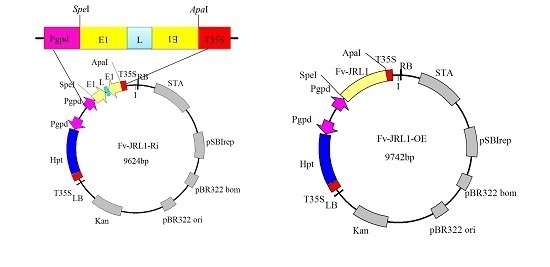
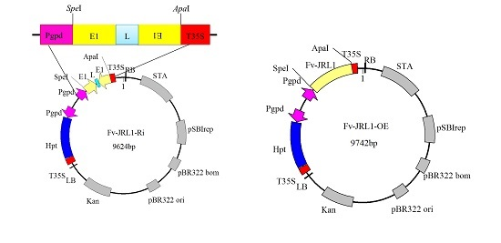
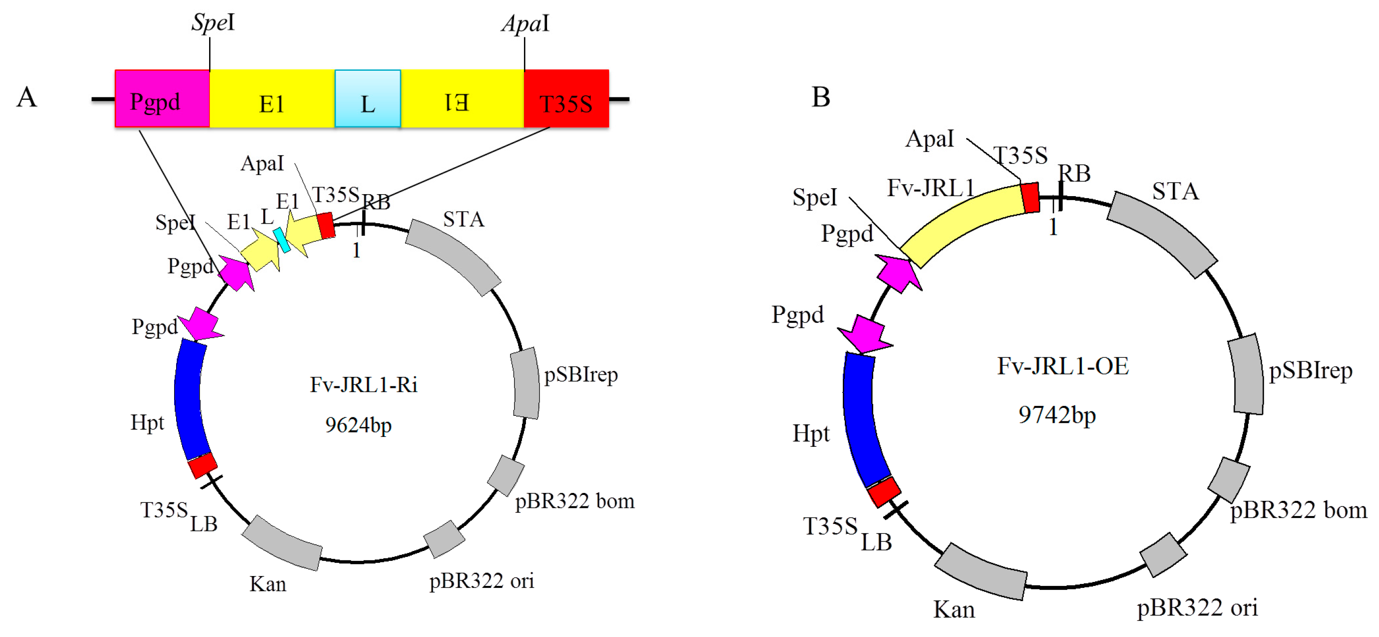
4.4. Construction of Gene Expression Plasmids

The binary vector pBHg-BCA1 [44], encompassing the hygromycin B phosphotransferase (hpt) gene for hygromycin resistance and an Agaricus bisporus glyceraldehyde-3-phosphate dehydrogenase (gpd) gene promoter, was used for constructing an RNAi hairpin vector and overexpression vector.
A fragment of Fv-JRL1 (GenBank Accession No. KU310976) from genomic DNA of F. velutipes strain W23 containing the region from base pairs 25 to 400, corresponding to the 5’ untranslated region, the first exon, and the first intron, was amplified by PCR with primers Fv-JRL1-S-F and Fv-JRL1-S-R. For reverse-orientation cloning, the 5’ untranslated region and the first exon of Fv-JRL1 were amplified with primers Fv-JRL1-A-F and Fv-JRL1-A-R. PCR products were ligated into the SpeI and ApaI sites of plasmid pBHg-BCA1 with the pEASY-Uni Seamless Cloning and Assembly Kit (Transgen Biotech, Beijing, China). Primer design and ligation were carried out according to the manufacturer’s instructions. A new plasmid, designated as Fv-JRL1-RNAi, was obtained (Figure 6) after sequence verification and digestion with SpeI and ApaI (Takara).
For overexpression assays, primers Fv-JRL1-F and Fv-JRL1-R, which introduced SpeI and ApaI recognition sites, respectively, were used to amplify the full-length Fv-JRL1 gene (from ATG to TAG) from F. velutipes W23. The PCR product was digested with SpeI and ApaI, and ligated into the linearized plasmid pBHg-BCA1 with the same two restriction endonucleases. The resulting plasmid Fv-JRL1-RNAi, containing the amplified full-length Fv-JRL1 gene, was transferred into E. coli DH5α. The recombinant plasmid, designated as Fv-JRL1-OE, was subsequently identified using PCR and sequencing.

4.5. Eletrophoretic Mobility of Plasmids DNA: Liposomes Complexes at Different Ratios

To screen the optimal ratio between plasmid DNA and liposomes, a range of liposome volumes (LipofectamineTM 2000, Invitrogen, Carlsbad, CA, USA) was used for assessment of encapsulation of a constant amount of plasmid DNA. Briefly, 5 µL of Fv-JRL1-OE vector (200 ng/µL) was prepared prior to the addition of liposomes. A range of volumes of liposomes was then added, formulated as liposome/plasmid DNA (10 µL:1 µg, 9 µL:1 µg, 8 µL:1 µg, 7 µL:1 µg, 6 µL:1 µg, 5 µL:1 µg, 4 µL:1 µg, 3 µL:1 µg, 2 µL:1 µg, 1 µL:1 µg). The complexes were then incubated on ice for 30 min, followed by agarose gel electrophoresis to determine the optimal ratio of plasmid DNA to liposome by evaluating the ratio at which liposome-encapsulated DNA was distinguishable from plasmid DNA.

4.6. Fungal Transformation and Screening of Transgenic F. velutipes with PCR

The Fv-JRL1-OE and Fv-JRL1-RNAi plasmids were transferred to F. velutipes strain H1123 by liposome-mediated methods. Briefly, liposome/plasmid DNA complexes were prepared by combining liposomes and plasmid DNA at a ratio of 1 µL liposomes per 1 µg plasmid DNA after incubation on ice for 30 min. Mycelium homogenate of strain H1123 was obtained by grinding using a sterilized mortar, in which 1 mL of 0.6 M mannitol was added prior to grinding. The homogenate was then added rapidly to the liposome/plasmid DNA complexes and then incubated on ice for 30 min. Five times the total volume of liquid regeneration complete medium (RCM) (2.0 g tryptone, 2.0 g yeast, 0.5 g MgSO4·7H2O, 0.46 g K2HPO4, 20 g glucose, and 1 g KH2PO4 per liter) was added, and the resulting mixture was incubated at 25 °C and shaking at 150 rpm for 2 days. The mycelia were collected by centrifugation at 5000 rpm for 10 min, and incubated on PDA plates containing 25 μg/mL hygromycin B at 25 °C for 25 days. The hygromycin-resistant transformants, following five successive transfers to PDA plates containing 30 μg/mL hygromycin B, were assumed to be stable and selected for further investigation.
Genomic DNA from mycelia of the putative transformants was extracted as described previously [40]. Transformants were confirmed by PCR to detect the presence of the hpt gene using primers hpt-A and hpt-S, and the presence of sequences between the gpd gene promoter and the Fv-JRL1 gene using primers GBT-F and Fv-JRL1-R for overexpression transformants, or the sequences between the gpd gene promoter and the first intron of Fv-JRL1 using primers GBT-F and Fv-JRL1-S-R for RNAi transformants. PCR was conducted with rTaq Polymerase (Takara, Japan) using the following parameters: 94 °C for 5 min; 35 cycles of 94 °C for 45 s; 58 °C for 45 s; 72 °C for 1 min; and a final extension of 72 °C for 10 min.

4.7. RNA Extraction and qRT-PCR

F. velutipes transformants and wild-type strain H1123 were grown in PDB medium at 25 °C and shaking at 150 rpm for 15 days. The mycelia were harvested, and then frozen in liquid nitrogen. Isolation of total RNA and qRT-PCR were conducted as described above.

4.8. Phenotype of Mutants

For morphology of the colonies, 5 mm mycelial pieces were placed onto the PDA plates and grown in the dark at 25 °C for 5 days. The morphology of the colonies was observed daily, and then photographed on day 5 after incubation. In addition, fruiting trials for transformants and wild-type strains were carried out in glass bottles containing 180 g composted substrate prepared as described by Wang et al. [41]. Experiments were replicated three times.

Supplementary Materials

Supplementary materials can be found at www.mdpi.com/1422-0067/17/11/1884/s1.

Acknowledgments

This work was supported by the National Key Basic Research Program of China (2014CB138302) and Chinese Agriculture Research System (CARS24). The authors are grateful to the Fujian Edible Fungi Engineering Technology Research Center and National Edible Fungi Breeding Center (Fujian Division) for providing experimental facilities.

Author Contributions

Yu-Ji Jiang and Bao-Gui Xie conceived and designed the experiments; Ren-Liang Chen, Yuan-Ping Lu, Ying Long and Xiao Li performed the experiments and analyzed the data; Yuan-Ping Lu and Bao-Gui Xie drafted the manuscript; all of authors reviewed and approved the final manuscript.

Conflicts of Interest

The authors declare no conflict of interest.

References

  1. Goldstein, I.J.; Hayes, C.E. The lectins: Carbohydrate-binding proteins of plants and animals. Adv. Carbohydr. Chen. Biol. 1978, 35, 127–340. [Google Scholar]
  2. Hassan, M.A.A.; Rouf, R.; Tiralongo, E.; May, T.W.; Tiralongo, J. Mushroom lectins: Specificity, structure and bioactivity relevant to human disease. Int. J. Mol. Sci. 2015, 16, 7802–7838. [Google Scholar] [CrossRef] [PubMed]
  3. Mirelman, D.; Galun, E.; Sharon, N.; Lotan, R. Inhibition of fungal growth by wheat germ agglutinin. Nature 1975, 256, 414–416. [Google Scholar] [CrossRef] [PubMed]
  4. Janzen, D.H.; Juster, H.B.; Liener, I.E. Insecticidal action of the phytohemagglutinin in black beans on a bruchid beetle. Science 1976, 192, 795–796. [Google Scholar] [CrossRef] [PubMed]
  5. Gatehouse, A.M.R.; Powell, K.S.; Peumans, W.J.; van Damme, E.J.; Gatehouse, J.A. Insecticidal properties of plant lectins: Their potential in plant protection. In Lectins Biomedical Perspectives; Taylor & Francis e-library: Bristol, PA, USA, 1995; pp. 35–57. [Google Scholar]
  6. Peumans, W.J.; van Damme, E.J. Lectins as plant defense proteins. Plant Physiol. 1995, 109, 347–352. [Google Scholar] [CrossRef] [PubMed]
  7. Brock, T.; Madigan, M.; Martinko, J.; Parker, J. Biology of Microorganisms, 7th ed.; Prentice-HallInternational, Inc.: Englewood Cliffs, NJ, USA, 1994. [Google Scholar]
  8. Sharon, N. Lectin receptors as lymphocyte surface markers. Adv. Immunol. 1983, 34, 213–298. [Google Scholar] [PubMed]
  9. Raz, A.; Lotan, R. Endogenous galactoside-binding lectins: A new class of functional tumor cell surface molecules related to metastasis. Cancer Metastasis Rev. 1987, 6, 433–452. [Google Scholar] [CrossRef] [PubMed]
  10. Lotan, R.; Raz, A. Lectins in cancer cells. Ann. N. Y. Acad. Sci. 1988, 551, 385–398. [Google Scholar] [CrossRef] [PubMed]
  11. Junqueira, L.C.; Carneiro, J.; Kelley, R.O. Basic Histology, 8th ed.; Appleton and Lange: Connecticut, Norwalk, CT, USA, 1995. [Google Scholar]
  12. Chang, S.T.; Miles, P.G. Mushroom biology—A new disciplines. Mycologist 1992, 6, 64–65. [Google Scholar] [CrossRef]
  13. Singh, S.S.; Wang, H.; Chan, Y.S.; Pan, W.; Dan, X.; Yin, C.M.; Akkouh, O.; Ng, T.B. Lectins from edible mushrooms. Molecules 2014, 20, 446–469. [Google Scholar] [CrossRef] [PubMed]
  14. Chang, S.T.; Buswell, J.A. Mushroom nutriceuticals. World J. Microb. Biotechnol. 1996, 12, 473–476. [Google Scholar] [CrossRef] [PubMed]
  15. Kulshreshtha, S.; Mathur, N.; Bhatnagar, P. Mushroom as a product and their role in mycoremediation. AMB Express 2014, 4, 1. [Google Scholar] [CrossRef] [PubMed]
  16. Guillamón, E.; García-Lafuente, A.; Lozano, M.; D’Arrigo, M.; Rostagno, M.A.; Villares, A.; Martínez, J.A. Edible mushrooms: Role in the prevention of cardiovascular diseases. Fitoterapia 2010, 81, 715–723. [Google Scholar] [CrossRef] [PubMed]
  17. Wasser, S.P. Medicinal mushroom science: History, current status, future trends, and unsolved problems. Int. J. Med. Mushrooms 2010, 12. [Google Scholar] [CrossRef]
  18. Xu, X.; Yan, H.; Chen, J.; Zhang, X. Bioactive proteins from mushrooms. Biotechnol. Adv. 2011, 29, 667–674. [Google Scholar] [CrossRef] [PubMed]
  19. Wasser, S.P. Medicinal mushroom science: Current perspectives, advances, evidences, and challenges. Biomed. J. 2014, 37, 345. [Google Scholar] [CrossRef] [PubMed]
  20. Wang, H.; Ng, T.B.; Ooi, V.E.C. Lectins from mushrooms. Mycol. Res. 1998, 102, 897–906. [Google Scholar] [CrossRef]
  21. Wang, H.; Gao, J.; Ng, T.B. A new lectin with highly potent antihepatoma and antisarcoma activities from the oyster mushroom Pleurotus ostreatus. Biochem. Biophys. Res. Commun. 2000, 275, 810–816. [Google Scholar] [CrossRef] [PubMed]
  22. Yang, N.; Liang, Y.; Xiang, Y.; Zhang, Y.; Sun, H.; Wang, D.C. Crystallization and preliminary crystallographic studies of an antitumour lectin from the edible mushroom Agrocybe aegerita. Protein Pept. Lett. 2005, 12, 705–707. [Google Scholar] [CrossRef] [PubMed]
  23. Yatohgo, T.; Nakata, M.; Tsumuraya, Y.; Hashimoto, Y.; Yamamoto, S. Purification and properties of a lectin from the fruitbodies of Flammulina velutipes. Agric. Biol. Chem. 1988, 52, 1485–1493. [Google Scholar] [CrossRef]
  24. Oguri, S.; Ando, A.; Nagata, Y. A novel developmental stage-specific lectin of the basidiomycete Pleurotus cornucopiae. J. Bacteriol. 1996, 178, 5692–5698. [Google Scholar] [CrossRef] [PubMed]
  25. Woffelman, C. DNAMAN for Windows; Version 5.2.10; Lynon Biosoft, Institute of Molecular Plant Sciences, Leiden University: Leiden, The Netherlands, 2004. [Google Scholar]
  26. Gasteiger, E.; Hoogland, C.; Gattiker, A.; Duvaud, S.E.; Wilkins, M.R.; Appel, R.D.; Bairoch, A. Protein Identification and Analysis Tools on the ExPASy Server; Humana Press Inc.: Totowa, NJ, USA, 2005; pp. 571–607. [Google Scholar]
  27. Quevillon, E.; Silventoinen, V.; Pillai, S.; Harte, N.; Mulder, N.; Apweiler, R.; Lopez, R. InterProScan: protein domains identifier. Nucleic Acids Res. 2005, 33, W116–W120. [Google Scholar] [CrossRef] [PubMed]
  28. Thompson, J.D.; Gibson, T.J.; Plewniak, F.; Jeanmougin, F.; Higgins, D.G. The clustal_x windows interface: Flexible strategies for multiple sequence alignment aided by quality analysis tools. Nucleic Acids Res. 1997, 25, 4876–4882. [Google Scholar] [CrossRef] [PubMed]
  29. Tamura, K.; Peterson, D.; Peterson, N.; Stecher, G.; Nei, M.; Kumar, S. MEGA5: Molecular evolutionary genetics analysis using MAXIMUM likelihood, evolutionary distance, and maximum parsimony methods. Mol. Biol. Evol. 2011, 28, 2731–2739. [Google Scholar] [CrossRef] [PubMed]
  30. Meng, L.; Yan, J.; Xie, B.; Li, Y.; Chen, B.; Liu, S.; Li, D.; Yang, Z.; Zeng, X.; Deng, Y.; et al. Genes encoding FAD-binding proteins in Volvariella volvacea exhibit differential expression in homokaryons and heterokaryons. Microbiol. Res. 2013, 168, 533–546. [Google Scholar] [CrossRef] [PubMed]
  31. Xu, Y.; Szoka, F.C. Mechanism of DNA release from cationic liposome/DNA complexes used in cell transfection. Biochemistry 1996, 35, 5616–5623. [Google Scholar] [CrossRef] [PubMed]
  32. Wang, D.; Christopher, M.E.; Nagata, L.P.; Zabielski, M.A.; Li, H.; Wong, J.P.; Samuel, J. Intranasal immunization with liposome-encapsulated plasmid DNA encoding influenza virus hemagglutinin elicits mucosal, cellular and humoral immune responses. J. Clin. Virol. 2004, 31, 99–106. [Google Scholar] [CrossRef] [PubMed]
  33. Lis, H.; Sharon, N. Lectins: Carbohydrate-specific proteins that mediate cellular recognition. Chem. Rev. 1998, 98, 637–674. [Google Scholar] [CrossRef] [PubMed]
  34. Vijayan, M.; Chandra, N. Lectins. Curr. Opin. Struct. Biol. 1999, 9, 707–714. [Google Scholar] [CrossRef]
  35. Stillmark, H. Ueber Ricin ein giftiges Ferment aus den Samen von Ricinus communis L. und einigen anderen Euphorbiaceen. Inaugural-Dissertation, Dorpat, Schnakenburg, Tartu, Estonia, 1888. [Google Scholar]
  36. Singh, R.S.; Bhari, R.; Kaur, H.P. Mushroom lectins: Current status and future perspectives. Crit. Rev. Biotechnol. 2010, 30, 99–126. [Google Scholar] [CrossRef] [PubMed]
  37. Sun, H.; Zhao, C.G.; Tong, X.; Qi, Y.P. A lectin with mycelia differentiation and antiphytovirus activities from the edible mushroom Agrocybe aegerita. J. Biochem. Mol. Biol. 2003, 36, 214–222. [Google Scholar] [CrossRef] [PubMed]
  38. Park, Y.J.; Baek, J.H.; Lee, S.; Kim, C.; Rhee, H.; Kim, H.; Seo, J.S.; Park, H.R.; Yoon, D.E.; Nam, J.Y.; et al. Whole genome and global gene expression analyses of the model mushroom Flammulina velutipes reveal a high capacity for lignocellulose degradation. PLoS ONE 2014, 9, e93560. [Google Scholar] [CrossRef] [PubMed]
  39. Liu, F.; Wang, W.; Chen, B.Z.; Xie, B.G. Homocitrate synthase expression and lysine content in fruiting body of different developmental stages in Flammulina velutipes. Curr. Microbiol. 2015, 70, 821–828. [Google Scholar] [CrossRef] [PubMed]
  40. Porebski, S.; Bailey, L.G.; Baum, B.R. Modification of a CTAB DNA extraction protocol for plants containing high polysaccharide and polyphenol components. Plant Mol. Biol. Rep. 1997, 15, 8–15. [Google Scholar] [CrossRef]
  41. Wang, W.; Liu, F.; Jiang, Y.; Wu, G.; Guo, L.; Chen, R.; Chen, B.; Lu, Y.; Dai, Y.; Xie, B. The multigene family of fungal laccases and their expression in the white rot basidiomycete Flammulina velutipes. Gene 2015, 563, 142–149. [Google Scholar] [CrossRef] [PubMed]
  42. Zhang, Z.; Lin, H.; Ma, B. ZOOM Lite: Next-Generation sequencing data mapping and visualization software. Nucleic Acids Res. 2010, 38, W743–W748. [Google Scholar] [CrossRef] [PubMed]
  43. Wu, G.M.; Yan, J.J.; Yang, Z.Y.; Zhang, T.J.; Xie, B.G. Analysis on structure and differential expression of a laccase gene fv-lac4 that possible involves in development of fruit body of Flammulina velutipes. Genom. Appl. Biol. 2014, 33, 1254–1260. (In Chinese) [Google Scholar]
  44. Shi, L.L.; van Peer, A.F.; Guo, L.X.; Chen, R.L.; Wang, W.; Yan, J.J.; Deng, Y.J.; Xie, B.G. Agrobacterium-mediated transformation of an endogenous HMG-box transcription factor fvhom1 in Flammulina velutipes. Genom. Appl. Biol. 2014, 33, 1268–1274. (In Chinese) [Google Scholar]
  45. Livak, K.J.; Schmittgen, T.D. Analysis of relative gene expression data using real-time quantitative PCR and the 2−ΔΔCt method. Methods 2001, 25, 402–408. [Google Scholar] [CrossRef] [PubMed]
Figure 1. Phylogenetic analysis of Fv-JRL1 with amino acid sequences of JRL1 identified from different organisms. P. platycephala, Parkia platycephala; H. Tuberosus, Helianthus Tuberosus; C. arvensis, Convolvulus arvensis; A. integer, Artocarpus integer; H. russula, Hygrophorus russula; G. trabeum, Gloeophyllum trabeum; G. frondosa, Grifola frondosa; P. expansum, Penicillium expansum; U. virens, Ustilaginoidea virens; C. militaris, Cordyceps militaris. The triangle indicates Flammulina velutipes Fv-JRL1 protein.
Ijms 17 01884 g001
Figure 2. Expression patterns of Fv-JRL1 during different development stages in F. velutipes. MY, mycelium; PR, primordium; EL, elongation stage; MA, mature fruit body.
Ijms 17 01884 g002
Figure 3. Gel electrophoresis of plasmid DNA (lane P) and plasmid DNA–liposome (lanes 1–10) composed of 1 µg plasmid DNA and 1–10 µL liposome. Lane P, naked plasmid DNA. Lane M, Marker DL2000. Lanes 1–10, 1 µg plasmid DNA mixed with 1, 2, 3, 4, 5, 6, 7, 8, 9, and 10 µL liposome, respectively.
Ijms 17 01884 g003
Figure 4. The transcription analysis of Fv-JRL1. The relative expression level of Fv-JRL1 was evaluated as fold changes in comparison with the expression level in the wild-type (WT) strain H1123.
Ijms 17 01884 g004
Figure 5. Phenotypic comparison of wild-type and Fv-JRL1 mutants. (A) Colonies and cross-sections of wild-type and Fv-JRL1 mutants after 5 days of incubation at 25 °C on potato dextrose agar (PDA) plates; (B) Growth after 22 days of incubation on composted substrate; (C) Fruiting trial.
Ijms 17 01884 g005
Figure 6. Binary vector Fv-JRL1-RNAi and Fv-JRL1-OE. Pgpd, A. bisporus glyceraldehyde-3-phosphate dehydrogenase gene promoter; hpt, hygromycin B phosphotransferase gene; Kan, kanamycin resistance gene; LB, left border; RB, right border. (A) Binary vector Fv-JRL1-RNAi; E1, exon1; L, linker; (B) binary vector Fv-JRL1-OE; Fv-JRL1, full-length of Fv-JRL1 gene from F. velutipes.
Ijms 17 01884 g006

