Article

Multi-Approach Analysis for the Identification of Proteases within Birch Pollen

1
Department of Molecular Biology, University of Salzburg, Salzburg 5020, Austria
2
Department of Breeding Informatics, Georg August-Universität Göttingen, Göttingen 37073, Germany
*
Author to whom correspondence should be addressed.
Int. J. Mol. Sci. 2017, 18(7), 1433; https://doi.org/10.3390/ijms18071433
Submission received: 26 May 2017 / Revised: 28 June 2017 / Accepted: 30 June 2017 / Published: 4 July 2017
(This article belongs to the Special Issue Proteolysis in Allergic Sensitization and Th2 Response)

Abstract

:
Birch pollen allergy is highly prevalent, with up to 100 million reported cases worldwide. Proteases in such allergen sources have been suggested to contribute to primary sensitisation and exacerbation of allergic disorders. Until now the protease content of Betula verrucosa, a birch species endemic to the northern hemisphere has not been studied in detail. Hence, we aim to identify and characterise pollen and bacteria-derived proteases found within birch pollen. The pollen transcriptome was constructed via de novo transcriptome sequencing and analysis of the proteome was achieved via mass spectrometry; a cross-comparison of the two databases was then performed. A total of 42 individual proteases were identified at the proteomic level. Further clustering of proteases into their distinct catalytic classes revealed serine, cysteine, aspartic, threonine, and metallo-proteases. Further to this, protease activity of the pollen was quantified using a fluorescently-labelled casein substrate protease assay, as 0.61 ng/mg of pollen. A large number of bacterial strains were isolated from freshly collected birch pollen and zymographic gels with gelatinase and casein, enabled visualisation of proteolytic activity of the pollen and the collected bacterial strains. We report the successful discovery of pollen and bacteria-derived proteases of Betula verrucosa.

1. Introduction

Ubiquitous to all living organisms, proteases function via cleavage of peptide bonds. All proteases act through stabilising the oxygen of the substrate peptide bond in an oxyanion hole, hence polarising the carbonyl group and making the carbon atom highly vulnerable to attack by an activated nucleophile [1]. Classifications of proteases are primarily based on the presence of molecules central to nucleophile formation, hence, deriving the four major catalytic classes of protease, i.e., cysteine, serine, metallo- and aspartic proteases [1,2]. Further detailed classification of proteases are described in the MEROPs database (Available online: http://merops.sanger.ac.uk). Each peptidase is provided an accession number correlating to the catalytic class (e.g., serine, cysteine). Accordingly, every peptidase belongs to a “family”, which represents a homologous group of proteins. Further classification into a “clan” defines evolutionary origins and can contain multiple families and occasionally multiple families of a distinct catalytic class [3].
The endogenous role of proteases in pollen is highly relevant in the process of germination; upon contact with a compatible stigma pollen grains hydrate, initiating a large release of solutes ultimately resulting in plant germination [4]. Proteases, some of which are situated on the exterior layers of pollen, itself, are included in this exudate and are released early in the hydration process [5,6]. Such pollen hydration is imitated upon contact with surface mucosa of the airway epithelium [5,7,8]. As a direct consequence, pollen-associated proteolytic activities have been implicated in the development of allergic diseases, with the capability to induce or enhance immune responses. An example includes the cysteine protease Amb a 11, derived from ragweed pollen, presenting both inducing and enhancing capabilities, as a sensitisation agent with proteolytic activity [9]. Other pollen-associated enzymes have been shown to compromise epithelial function, contributing to pollen allergenicity via the facilitation of allergens crossing such defensive barriers [10] or through direct activation of pattern recognition receptors [11]. Hence, pollen derived proteases pose a relevant source of investigation with relation to allergic sensitisation and development of allergic disease.
An excess of 100 million people worldwide have a reported allergy to birch pollen. Until now the protease content of Betula verrucosa, a species endemic to the northern hemisphere [12], has not been studied in detail. Hence, we aim to identify and characterise pollen and bacterial derived proteases found within Betula verrucosa pollen. We propose to employ a multi-approach analysis for the identification of proteases within commercial Betula verrucosa pollen extracts. Methods include proteomic and transcriptomic cross-referencing of the birch pollen and a protease assay to provide a quantitative measurement of proteolytic activity, with regards to the substrate casein. The application of freshly-collected birch pollen extracts to nutrient agar plates, together with Gram staining and 16S rRNA sequencing enabled identification of several bacterial isolates present in the pollen. For all samples, zymograms were performed for visual interpretation of the proteolytic activity.

2. Results

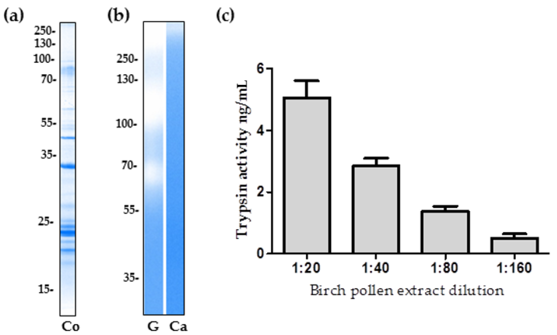
2.1. The Proteolytic Activity of Birch Pollen Extract

To demonstrate the presence of proteins within the birch pollen extract (BPE), SDS-PAGE and Coomassie Brilliant Blue staining was performed (Figure 1a). The total protein concentration of the BPE, as determined by Bradford assay, was measured as 1.8 mg/mL. Gelatinase activity was shown in Figure 1b through the presence of three broad bands with a distribution of >250 kDa, 100–130 kDa, and ~70 kDa. Within the 0.1% casein gel, the visualisation of BPE caseinolytic activity is not as evident, with a faint band positioned above in the high molecular weight range (Figure 1c).
Using fluorescently labelled casein and, in reference to a trypsin standard curve, the protease activity of birch pollen was quantified as 0.61 ng/mg of pollen, with a standard deviation of ±0.09 ng/mg. A serial dilution of the BPE was performed and three independent experiments containing three replicates of each dilution were analysed (Figure 1c) and referenced to trypsin standard curves with R2 values between 0.95 and 0.98. The multiplication of each attained data point with the relevant dilution factor, when averaged, reveals the caseinolytic activity of the undiluted birch pollen extract in relation to trypsin activity, which translates to 0.61 ng/mg of birch pollen.

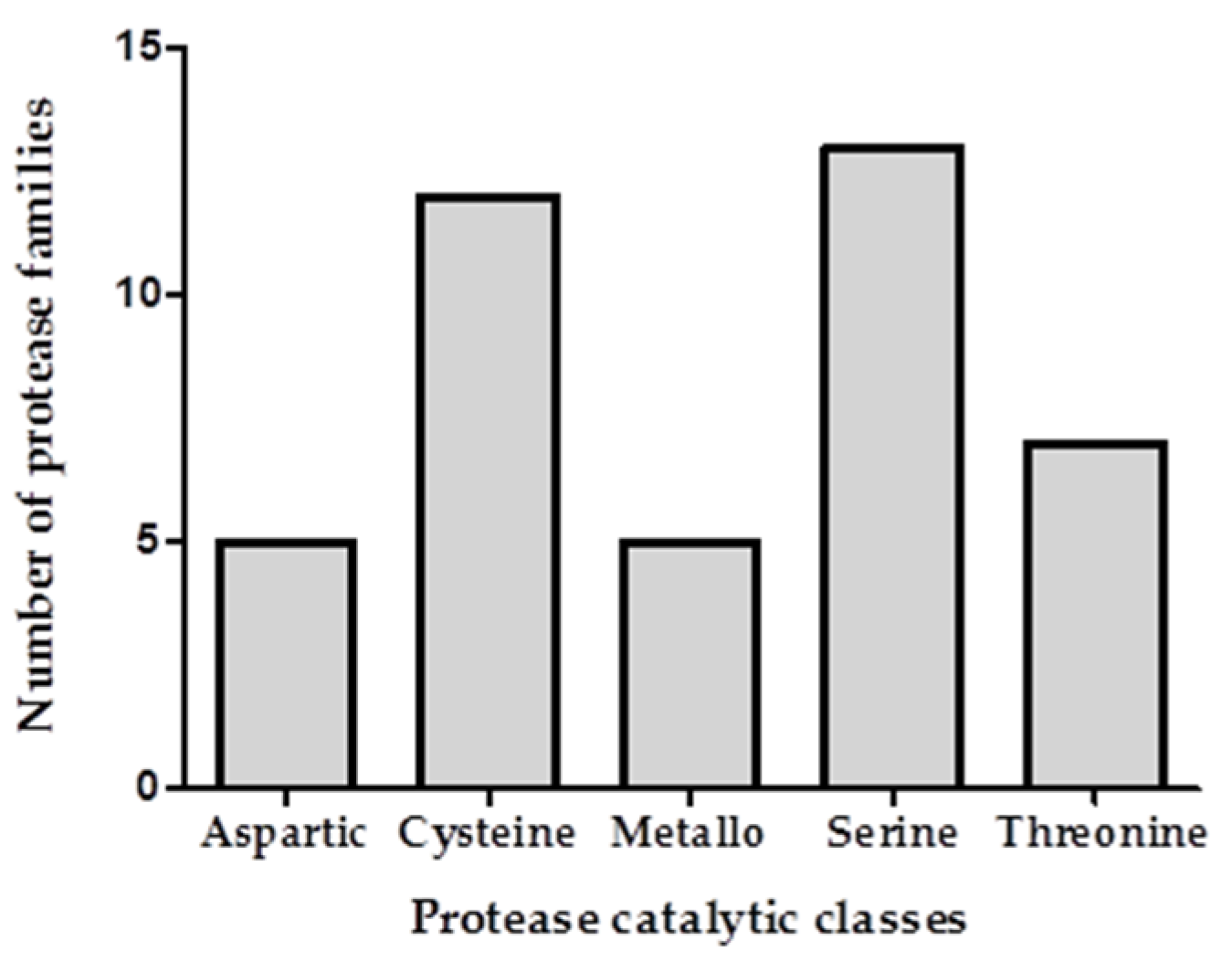
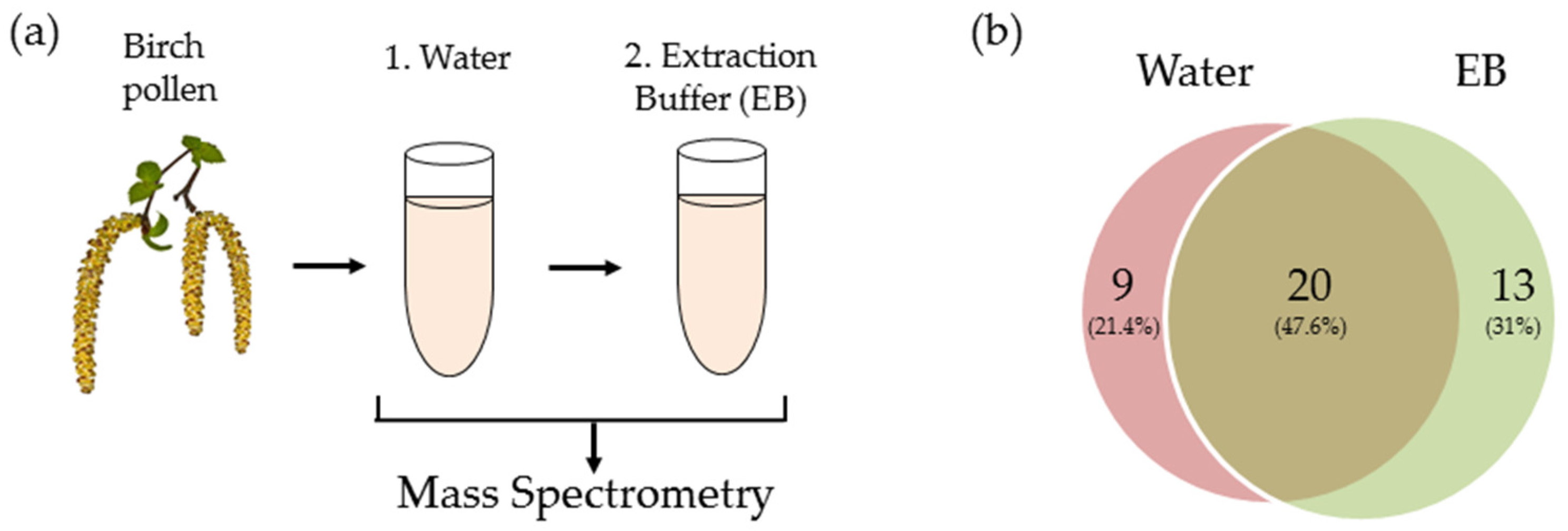
2.2. Transcriptomic and Proteomic Analysis

Transcriptomic analysis revealed proteases from 52 distinct families of the Pfam database. Data was referenced to the known sequence of Arabidopsis thaliana, a model organism widely used in molecular genetics. The NCBI sequence read archive (SRA) accession code for the RNA-sequencing reads of birch pollen is SRS2152558. The transcriptome expression levels for the proteases are provided in Höllbacher et al. [13]. Using a cross-referencing approach and with application to proteases alone, the proteome was compared to the transcriptome in order to identify birch pollen proteases in the extract. Two consecutive treatments (water and trypsin extraction buffer) were carried out on the BPE prior to mass spectrometry analysis and the exudates were analysed (Figure 2a). The collection of proteases obtained from the two different treatments potentially reflects differences in protein solubility. Following water extraction of the BPE, a total of 29 proteases were identified through proteomic and transcriptomic analysis (Figure 2b). Division into catalytic classes revealed serine (n = 8), cysteine (n = 7), aspartic (n = 4), metallo (n = 5), and threonine (n = 5)-type proteases.
Trypsin extraction buffer treatment of the BPE resulted in the identification of 33 proteases. The majority of which were identified as serine proteases (n = 11) along with cysteine (n = 9), threonine (n = 5) and metallo- (n = 5) proteases. An overlap of 20 proteases were identified between two different extractions. Combining the two datasets and removing duplicates revealed a total of 42 identified proteases (Table 1), the categorisation into catalytic classes is depicted in Figure 3. Water treatment resulted in nine distinct proteases, extraction buffer treatment revealed the identification of 13 morphologically distinct proteases. Between the two treatments an overlap of 20 proteases were identified. Within these 20 proteases one metallo- and two threonine proteases are not currently defined in the MEROPS database.

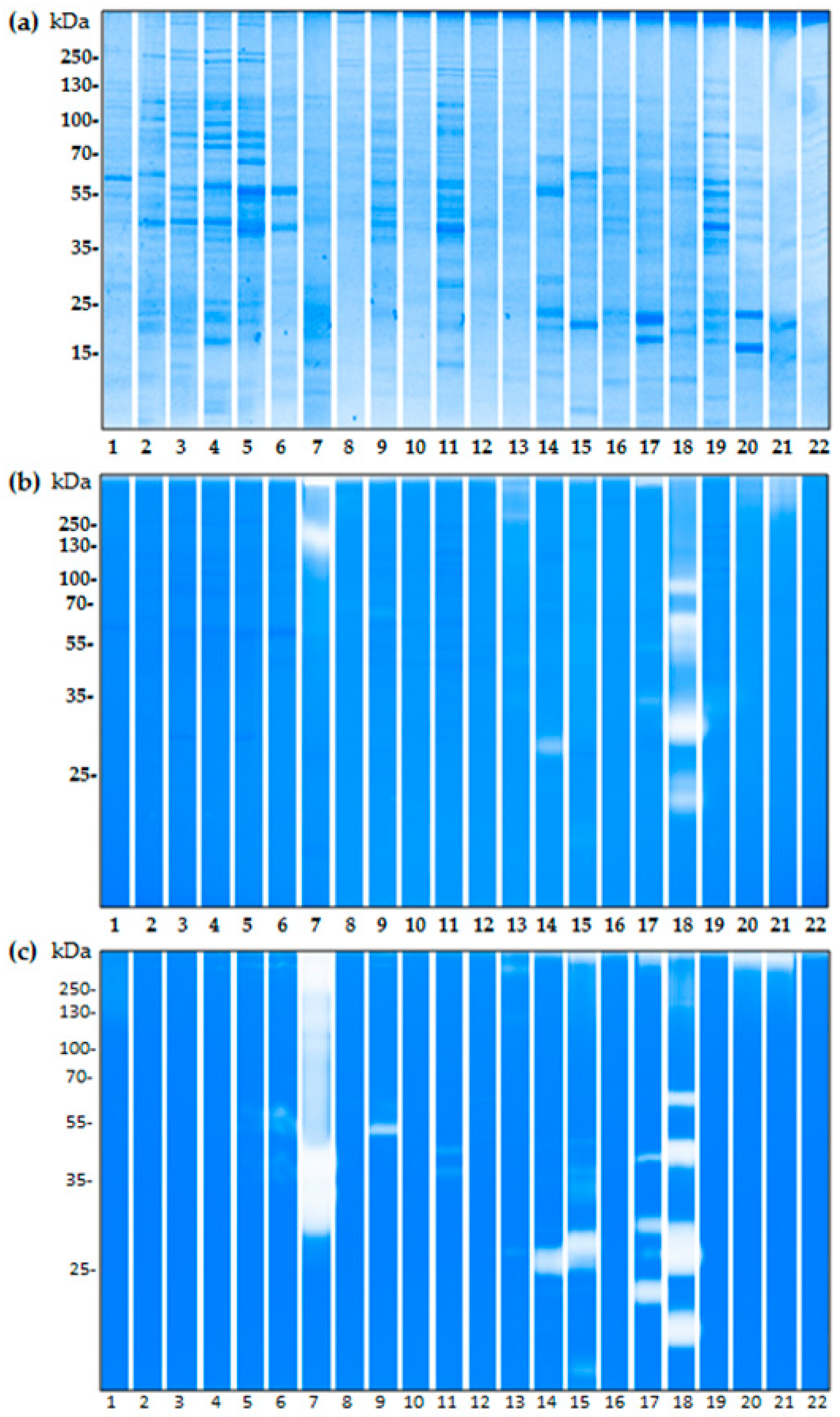
2.3. The Proteolytic Activity of Bacterial Isolates from Birch Pollen

From the fresh pollen extracts we were able to successfully identify 22 bacterial isolates (seven Gram-negative and 15 Gram-positive) (Table 2) of which 17 strains presented with proteolytic activity as visually represented in either casein- or gelatin-based zymographic gels (Figure 4). For the casein substrate (Figure 4b), bacterial isolate number 7 of the Xanthomonadaceae bacterial family has two high molecular weight bands at 250 kDa and above. The isolate number 18 of the Bacillaceae family, is also present with strong proteolytic activity with at least five bands from below 25 to 100 kDa.
Within the casein gel (Figure 4b) the gram positive bacterial strains numbered 9, 13, 14, 15, 17, 21, show faint but existing proteolytic activity. Gelatinase activity is, in general, more prominent within the isolated bacterial strains, than that of casein. Up to ten of the identified bacterial strains present with gelatinase activity (Figure 4c). For isolates of the bacillaceae family numbered 15, 16, 18, and 19 a high gelatinase activity was observed. In particular, isolate number 8 of the Gordoniaceae family, presents a very saturated activity with over half of the lane stained. Sample 19, of the Bacillaceae family, shows at least four distinct bands, ranging from ~60 to 25 kDa. Lower activity is shown for Gram-positive samples 9, 12, 14, 21, 22. All of the bacterial isolates that present proteolytic activity within the gelatin substrate are Gram-positive.

3. Discussion

Whilst some proteases are, themselves, described as allergens [11], it has been heavily suggested that others play an assistive role in the development of allergy [14]. The respiratory epithelium consists of a complex set of epithelial cells which, in healthy individuals, maintains a finely-tuned homeostatic environment between internal and external stimuli, and provides a protective barrier against damaging external stimuli [15]. Proteases have been shown to disrupt the epithelial barrier integrity, through direct degradation of essential tight junction proteins [16] and via innate immune receptors involved in pro-inflammatory responses. In particular, protease activated receptor–2 (PAR-2) activation has been shown to alter the permeability between cells via p38 MAP kinase signalling [17]. Such disruption may promote inflammation, resulting in a primed environment for allergic sensitization to occur [18]. Further to this, the loss of epithelial integrity represents the ideal opportunity for allergens to enter and be detected by cells of the innate immune system [11]. In this context, we investigated the protease content of commercially available birch pollen. Initial zymogram experiments confirmed the presence of proteolytic activity in the birch pollen extracts for casein and gelatin substrates. To provide a quantitative measure of such activity we then performed a fluorescein isothiocyanate labelled casein protease assay. The assay revealed that birch pollen extract has a proteolytic activity of 0.61 ng per mg of pollen, with reference to the proteolytic activity of trypsin. Hence, we aimed to further identify the present proteases via a proteomic approach. The cross comparison of the proteome and transcriptome provided a database of proteases reliably identified to be of birch pollen origin. The analysis led to the successful identification of 42 proteases. The MEROPS database groups proteins of similar evolutionary origins into clans. Of the 54 described on MEROPS, our proteomic dataset describes proteases derived from 13 different clans. For further classification into a family of homologous proteases, our investigation revealed proteases deriving from 18 different families out of a total of 258 [19]. Of note are the five different families of cysteine proteases (C1, C2, C12, C19, and C85) deriving from the CA clan. Proteases of the C1 family (subfamily A) include papain [20], a highly relevant initiator of lung inflammation, as described using a mouse model, [21] and the well characterised proteolytic allergen of house dust mite, Der p 1 [22]. Homology to such proteases could be of significant relevance of understanding the process of allergic sensitization towards birch pollen. Hence, the further investigation of identified cysteine proteinases RD21A and RD19A, could be of high interest. The C2 family contains the calcium dependant enzyme, calpain. In particular, endogenous calpain-1 has been shown to contribute to IgE-mediated mast cell activation [23]. Furthermore, the involvement of calpain in eosinophilic disease is documented in detail [24]. In this context, calpain-type cysteine protease ADL1 also represents an interesting target for further investigation. With regard to their activity, metalloproteases, in particular those of the MA clan, are of relevance to allergy research. As their name suggests, metalloproteases perform their catalytic nucleophilic attack via a metal ion [25]. In particular, thermolysin, described as the prototypic protease of the MA clan, is able to elicit the activation of PAR-2 [26]. Our proteomic analysis recognized organellar oligopeptidase A (M3) and puromycin-sensitive aminopeptidase (M1) from the MA clan. Among all known proteases one third are classified as being serine proteases [27], which is reflective for our data set showing a total of 13 serine proteases of the total 42. Serine proteases are frequently referred to in the context of allergy, either as proteolytic allergens, e.g., Der f 6 [28] and Cur l 1 [29] or found within allergenic sources, for example in the highly-allergenic ragweed pollen [30,31]. In particular, the extracellular alkaline serine proteases and subtilisins, are highly relevant to allergy [32]. Cases of subtilisin allergy linked to the use of cleaning detergents containing bacteria-derived subtilisin enzymes have been reported. The discovery of a major allergen characterised as a subtilisin–like protease within the allergenic plant fungus, Curvularia lunata [33,34], is of interest to our data set with the identification of four subtilisin-like proteases, i.e., SBT1.7, SBT1.8, SBT4.15 and SBT5.4. It has been described in the literature that such proteases have the ability to cross-react and activate PAR-2 receptors [11,35]. The SC clan, family S10, consists primarily of serine carboxypeptidases; we have identified five such proteases, which share homology with the allergen Api m 9, derived from honey bee (Apis mellifera) [36]. Furthermore, the presence of environmental serine proteases has been linked to increased incidence of allergic disease [37], emphasizing the significance of further investigation.
Whilst endogenous birch pollen proteases are of key interest, we also wanted to investigate the potential proteolytic capability of the pollen microbiome. Recent studies exploring the microbiome highlight the potential significance of bacteria in allergen sensitisation [38]. A previous study, centred in Giessen, Germany, identified a complex birch pollen microbiome of 16 bacterial isolates [39]. We aimed to isolate bacteria derived from birch pollen within Salzburg, Austria, and tested for the presence of proteolytic activity. Our study has revealed a total of 22 bacterial isolates on the freshly-collected birch pollen, around half of which presented with proteolytic activity as visualised by gelatin and casein zymography. Eight of the 22 tested isolates belong to the Bacillaceae family which has been previously associated with allergy. In particular Bacillus subtilis of the Bacillaceae family, which excretes enzymes previously shown to elicit allergic responses [40]. Furthermore, the exploitation of their enzyme secreting capability in the detergent industry has posed a risk for occupational allergy and asthma [41,42]. Conversely, the application of Bacillus subtilis as an orally-administered probiotic [43] highlights the diverging outcomes, with regards to the development of allergy, of such protease exposure to differing physiological surfaces. Another well documented and highly relevant bacterial strain of the Bacillaceae family is Lysinibacillus fusiformis. The isolation of a serine metalloprotease from the bacterial strain Lysinibacillus fusiformis AU01 showed roles similar to that of the serine protease trypsin. The application of the yielded protease to adhering cell tissue cultures resulted in the dissociation of monolayers from tissue culture flasks suggesting the ability to disrupt cell–cell interactions [44], whilst the 16s rRNA sequencing did not. Our results show the presence of mild caseinolytic activity, as well as multiple bands of gelatinase activity. Further research is required to understand the impact such proteases have on epithelial cell surfaces.
Of the isolated bacterial strains, Gram-negative bacteria appeared to have less proteolytic power than Gram-positive strains. A difference in the transport mechanisms and the fact that most Gram-negative bacteria have pro-peptides, could provide one explanation as to the lack of activation present in the zymographic gels for Gram-negative bacteria, when compared to the positive strains [45]. In vitro/in vivo experimental conditions could provide a more relevant context, for bacterial protease release and future studies should focus on the in vivo relevance of such released proteases. Proteases are highly evolved; different classes of proteases are able to perform the same reaction via distinct mechanisms. Even within families of related proteases, the endogenous functions can show huge disparity, hence predicting the function of a protease presents as a difficult task [1,46]. Further to this, predictions of proteolytic activity based on homologous proteins, for example those in the same family/subfamily, could prove inaccurate, hence, further investigation into individual proteins is required for a full characterization. Moreover, the role proteases play in producing the phenotype of the pollen itself, is an interesting context in which to explore pollen and their associated enzymes. Our analysis has identified several highly relevant birch pollen associated proteases; the further study of which should contribute to the understanding of birch pollen sensitization and allergy development.

4. Materials and Methods

4.1. Pollen Extract Preparation

Betula verrucosa pollen (allergon AB, Thermo Fisher Scientific, Ängelholm, Sweden, Batch 012510101) prepared in water (180 mg/mL) was shaken for 12 h at 4 °C. Centrifugation at 13,000× g for 15 min at 4 °C enabled procurement of the supernatant. Protein concentration was then determined using Pierce Coomassie Brilliant Blue G-250 (Thermo Scientific, Vienna, Austria) according to manufacturer’s instructions. Using a spectrophotometer, absorbance was measured at 595 nm.

4.2. Isolation of Bacteria on Pollen Grains

Betula verrucosa pollen was collected from five different trees in Salzburg, Austria. Ten milligrams of pollen was dissolved in 1 mL PBS. GC-agar plates (containing 5% FCS and 1 µg/mL Nystatin) and nutrient agar plates (containing 1 µg/mL Nystatin) were incubated at 20 °C and 37 °C together with 100 µL of the pollen suspension. Gram staining was performed on pure cultures of selected colonies and bacterial families were identified by 16S rRNA sequencing. Briefly, 16S RNA was amplified using 16S fwd (AGAGTTTGATCCTGGCTCAG) and 16S rev (AGGAGGTGATCCAACCGCA) primers. The resulting products had a size of approx. 1.5 kb and were subjected to DNA sequencing (MWG Eurofins, Ebersberg, Germany). Sequences were analysed using the NCBI-Basic Local Alignment Search Tool (BLAST; http://blast.ncbi.nlm.nih.gov 26.07.11) with the following parameters: Database: nr/nt; exclude uncultured/environmental sample sequences; megablast. Due to the high similarities in 16S sequences, the highest degree of qualitative analysis was the level of bacterial families. The selected bacteria were then cultured in nutrient broth and sonicated in lysis buffer (20 mM Tris pH 7.5, 1 mM EDTA, 100 mM NaCl, 1% Triton X-100, 0.5% DOC, 0.1% SDS, 0.5% NP-40). Samples were centrifuged for 15 min at 15,000× g to remove debris.

4.3. SDS-PAGE

Birch pollen extract and bacterial samples were mixed with 4× reducing sample buffer (125 mM Tris pH 6.8, 20% glycerol, 6% SDS, 0.02% bromophenol blue, 10% β-mercaptoethanol) and loaded with 15 µg of protein per well. Samples were heated for 5 min at 95 °C and run in 10% (w/v) polyacrylamide separation gels at 120 V for 150 min in SDS-Tris-glycine buffer, pH 8.05, and stained with Coomassie blue R-250 (Biorad, Munich, Germany).

4.4. Zymography

Ten percent (w/v) polyacrylamide gels were co-polymerised with either 0.1% type A gelatin from porcine skin (Sigma-Aldrich, Schnelldorf, Germany) or 0.1% casein from bovine milk (Sigma-Aldrich, Schnelldorf, Germany). Birch pollen extract and bacterial strains (with protein amounts of 5 µg for gelatin and 15 µg for casein per well) were added to 4× non-reducing sample buffer (125 mM Tris pH 6.8, 20% glycerol, 6% SDS, 0.02% bromophenol blue). Samples were incubated at 25 °C for 5 min before being loaded into gels. Following electrophoresis at constant 100 V at room temperature. Gels were incubated two times for 30 min in wash buffer (2.5% Triton X-100 in H2O) solution at room temperature, then incubated at 37 °C for 16 h in developing buffer (10 mM Tris pH 7.5 with 5 mM CaCl2, 1 µM ZnCl2). Thereafter, gels were stained with 30% (v/v) methanol and 10% (v/v) acetic acid, containing 0.5% (w/v) Coomassie brilliant blue R-250 (Biorad, Vienna, Austria). De-staining was performed with 50% (v/v) methanol and 10% (v/v) acetic acid. Gelatinase activity was visualized as unstained bands on a blue background, representing areas of proteolysis of the substrate protein.

4.5. Protease Activity Assay

Quantification of birch pollen proteolytic activity was performed using a fluorescein isothiocyanate labelled casein protease assay kit (Pierce, Thermo Scientific, Vienna, Austria). Trypsin was used as a standard, ranging from 0.008 to 0.5 µg/mL. Birch pollen extract was prepared freshly in a two-fold serial dilution and CaCl2 was added to a final concentration of 100 µM. The measurements were performed in a white flat bottom 96-well plate (Nunc, Roskilde, Denmark). The fluorescence was measured in a Tecan Infinite 200 PRO (Tecan Group Ltd., Männedorf, Switzerland) plate reader with the filter setting 485/535 nm (Ex/Em). Three independent experiments containing three replicates of each dilution were analysed. The mean and standard deviation of the data points were calculated.

4.6. RNA Sequencing

Total RNA was isolated from the pollen of Betula verrucosa (allergon AB, Thermo Fisher Scientific, Ängelholm, Sweden, batch 012510101). Five hundred milligrams of pollen were homogenised and resuspended in 4.2 M of guanidine thiocyanate, 50 mM of BES pH 7.2, 4 mM Ethylenediaminetetraacetic acid (EDTA), supplemented with 1% (v/v) β-mercaptoethanol, 1% (w/v) N-lauryl-sarkosyl, and 1% (v/v) n-butanol. Following centrifugation at 15,000× g for 15 min, RNA was extracted from the supernatant using TRIzol LS reagent (Invitrogen, Carlsbad, CA, USA), according to the manufacturers’ manual.
RNA-sequencing was performed with Illumina’s HiSeq 2500 system and delivered 220 m paired reads. Per base sequence quality was encoded in the Phred +33 format and assessed using FastQC: A quality control tool for high throughput sequence data (Avaialbe online: http://www.bioinformatics.babraham.ac.uk/projects/fastqc). Using Trimmomatic [47], low quality bases from the ends of reads were removed and pairs where both reads passed the checkpoint were processed. As the full genome for Betula verrucosa is not currently available, the Trinity [48] pipeline was used to sequentially apply the three tools; Inchworm, Chrysalis, and Butterfly for de novo transcriptome assembly. Quantification of the assembly was performed with Kallisto.
Quality parameters and general statistics were obtained by FastQC, the fastqutils from NGSUtils [48] and from the RNA-Seq provider’s report. The transcripts were then functionally annotated using blastx and UniProt/SwissProt (version 11_2016). As databases. The e-value cutoff for blastx was set to 0.001. The NCBI SRA accession code for the RNA-seq reads is SRX2769122. The expression levels for the proteases are given in Höllbacher et al. [13].

4.7. Mass Spectrometry Analysis

For mass spectrometric analysis, the extracts were digested with the ProteoExtract All-in-One Tryps For mass spectrometric analysis, the extracts were digested with the ProteoExtract All-in-One Trypsin Digestion Kit (EMD Millipore, Billerica, MA, USA). Resulting peptides were desalted using C18 ZipTips (EMD Millipore) and separated by reverse-phase nano-HPLC (Dionex Ultimate 3000, Thermo Fisher Scientific, Bremen, Germany, column: PepSwift Monolithic Nano Column, 100 µm × 25 cm, Dionex). The column was eluted with an acetonitrile gradient (Solvent A: 0.1% (v/v) FA/0.01% (v/v) TFA/5% (v/v) DMSO; solvent B: 0.1% (v/v) FA/0.01% (v/v) TFA/90% (v/v) ACN/5% (v/v) DMSO; 5–45% B in 60 min) at a flow rate of 0.8 µL/min at 55 °C. Peptides were analysed with a Q Exactive Orbitrap mass spectrometer (Thermo Fisher Scientific, Bremen, Germany,) directly coupled to the HPLC. Capillary voltage at the nano electrospray head was 2 kV, the instrument was tuned for maximum sensitivity. For peptide assignments, a top 12 method was used with a normalized fragmentation energy at 27%. Protein assignment was done with PEAKS Studio 8 (Bioinformatics Solutions, Waterloo, Canada). As a sequence database we took the annotated transcripts as described above, and translated to all six protein sequence reading frame. Only peptide hits with a probability score (−10logP) ≥ 35 were used for protein identification.
We took the annotated transcripts as described above, and translated to all six protein sequence reading frames.

4.8. Further Analysis

Analysis of the cross referenced proteome/transcriptome data was carried out manually.

Acknowledgments

This work was supported by the Austrian Science Funds (FWF) International PhD Program “Immunity in Cancer and Allergy”, ICA (Project W1213) and by the project TransBetula of the Free University of Bozen/Bolzano, Italy. We thank Heidi Hofer for her contribution in preparing the isolation of birch pollen RNA.

Author Contributions

Fatima Ferreira, Silja Wessler, Gabriele Gadermaier, and Olivia E. McKenna conceived and designed the experiments; Peter Briza performed the MS measurements; Armin O. Schmitt constructed the transcriptome; Peter Lackner analyzed the transcriptomics data; Gernot Posselt performed all bacterial experiments, Olivia E. McKenna performed the rest of the experiments; and Olivia E. McKenna prepared the manuscript.

Conflicts of Interest

The authors declare no conflict of interest.

References

  1. Van der Hoorn, R.A.L. Plant proteases: From phenotypes to molecular mechanisms. Annu. Rev. Plant Biol. 2008, 59, 191–223. [Google Scholar] [CrossRef] [PubMed]
  2. Rao, M.B.; Tanksale, A.M.; Ghatge, M.S.; Deshpande, V.V. Molecular and biotechnological aspects of microbial proteases. Microbiol. Mol. Biol. Rev. 1998, 62, 597–635. [Google Scholar] [PubMed]
  3. Rawlings, N.D.; Barrett, A.J.; Finn, R. Twenty years of the merops database of proteolytic enzymes, their substrates and inhibitors. Nucleic Acids Res. 2016, 44, D343–D350. [Google Scholar] [CrossRef] [PubMed]
  4. Krichevskya, A.; Kozlovsky, S.V.; Tian, G.W.; Chen, M.H.; Zaltsmana, A.; Citovskya, V. How pollen tubes grow. Dev. Biol. 2007, 303, 405–420. [Google Scholar] [CrossRef] [PubMed]
  5. Widmer, F.; Hayes, P.J.; Whittaker, R.G.; Kumar, R.K. Substrate preference profiles of proteases released by allergenic pollens. Clin. Exp. Allergy 2000, 30, 571–576. [Google Scholar] [CrossRef] [PubMed]
  6. Blanchard, G.C.; Gardner, R. Characterization of some of enzymes in ragweed pollen. Ann. Allergy 1976, 36, 410–418. [Google Scholar] [PubMed]
  7. Hassim, Z.; Maronese, S.E.; Kumar, R.K. Injury to murine airway epithelial cells by pollen enzymes. Thorax 1998, 53, 368–371. [Google Scholar] [CrossRef] [PubMed]
  8. Baraniuk, J.N.; Esch, R.E.; Buckley, C.E. Pollen grain column chromatography—Quantitation and biochemical-analysis of ragweed-pollen solutes. J. Allergy Clin. Immun. 1988, 81, 1126–1134. [Google Scholar] [CrossRef]
  9. Bouley, J.; Groeme, R.; le Mignon, M.; Jain, K.; Chabre, H.; Bordas-Le Floch, V.; Couret, M.N.; Bussieres, L.; Lautrette, A.; Naveau, M.; et al. Identification of the cysteine protease amb a 11 as a novel major allergen from short ragweed. J. Allergy Clin. Immun. 2015, 136, 1055–1064. [Google Scholar] [CrossRef] [PubMed]
  10. Vinhas, R.; Cortes, L.; Cardoso, I.; Mendes, V.M.; Manadas, B.; Todo-Bom, A.; Pires, E.; Verissimo, P. Pollen proteases compromise the airway epithelial barrier through degradation of transmembrane adhesion proteins and lung bioactive peptides. Allergy 2011, 66, 1088–1098. [Google Scholar] [CrossRef] [PubMed]
  11. Matsumura, Y. Role of allergen source-derived proteases in sensitization via airway epithelial cells. J. Allergy 2012, 2012, 903659. [Google Scholar] [CrossRef] [PubMed]
  12. Ashburner, K.; McAllister, H.A. Botanical Magazine Monograph. In The Genus Betula: A Taxonomic Revision of Birches; Illustrated Reprint; Royal Botanic Gardens, Kew publishing: Kew, UK, 2013; Volume 5, p. 432. [Google Scholar]
  13. Hollbacher, B.; Schmitt, A.O.; Hofer, H.; Ferreira, F.; Lackner, P. Identification of proteases and protease inhibitors in allergenic and non-allergenic pollen. Int. J. Mol. Sci. 2017, 18, 1199. [Google Scholar] [CrossRef] [PubMed]
  14. Reed, C.E.; Kita, H. The role of protease activation of inflammation in allergic respiratory diseases. J. Allergy Clin. Immun. 2004, 114, 997–1008. [Google Scholar] [CrossRef] [PubMed]
  15. Knight, D.A.; Holgate, S.T. The airway epithelium: Structural and functional properties in health and disease. Respirology 2003, 8, 432–446. [Google Scholar] [CrossRef] [PubMed]
  16. Runswick, S.; Mitchell, T.; Davies, P.; Robinson, C.; Garrod, D.R. Pollen proteolytic enzymes degrade tight junctions. Respirology 2007, 12, 834–842. [Google Scholar] [CrossRef] [PubMed]
  17. Enjoji, S.; Ohama, T.; Sato, K. Regulation of epithelial cell tight junctions by protease-activated receptor 2. J. Vet. Med. Sci. 2014, 76, 1225–1229. [Google Scholar] [CrossRef] [PubMed]
  18. Zhang, H.; Zeng, X.; He, S. Evaluation on potential contributions of protease activated receptors related mediators in allergic inflammation. Mediators Inflamm. 2014, 2014, 829068. [Google Scholar] [CrossRef] [PubMed]
  19. Rawlings, N.D.; Barrett, A.J.; Bateman, A. Merops: The peptidase database. Nucleic Acids Res. 2010, 38, D227–D233. [Google Scholar] [CrossRef] [PubMed]
  20. Kamphuis, I.G.; Kalk, K.H.; Swarte, M.B.A.; Drenth, J. Structure of papain refined at 1.65 Å resolution. J. Mol. Biol. 1984, 179, 233–256. [Google Scholar] [CrossRef]
  21. Vannella, K.M.; Ramalingam, T.R.; Hart, K.M.; Prado, R.D.; Sciurba, J.; Barron, L.; Borthwick, L.A.; Smith, A.D.; Mentink-Kane, M.; White, S.; et al. Acidic chitinase primes the protective immune response to gastrointestinal nematodes. Nat. Immunol. 2016, 17, 538. [Google Scholar] [CrossRef] [PubMed]
  22. Furmonaviciene, R.; Ghaemmaghami, A.M.; Boyd, S.E.; Jones, N.S.; Bailey, K.; Willis, A.C.; Sewell, H.F.; Mitchell, D.A.; Shakib, F. The protease allergen Der p 1 cleaves cell surface DC-SIGN and DC-SIGNR: Experimental analysis of in silico substrate identification and implications in allergic responses. Clin. Exp. Allergy 2007, 37, 231–242. [Google Scholar] [CrossRef] [PubMed]
  23. Wu, Z.L.; Chen, X.C.; Liu, F.; Chen, W.; Wu, P.; Wieschhaus, A.J.; Chishti, A.H.; Roche, P.A.; Chen, W.M.; Lin, T.J. Calpain-1 contributes to ige-mediated mast cell activation. J. Immunol. 2014, 192, 5130–5139. [Google Scholar] [CrossRef] [PubMed]
  24. Litosh, V.A.; Rochman, M.; Rymer, J.K.; Porollo, A.; Kottyan, L.C.; Rothenberg, M.E. Calpain-14 and its association with eosinophilic esophagitis. J. Allergy Clin. Immunol. 2017, 139, 1762–1771. [Google Scholar] [CrossRef] [PubMed]
  25. Nagase, H.; Visse, R.; Murphy, G. Structure and function of matrix metalloproteinases and timps. Cardiovasc. Res. 2006, 69, 562–573. [Google Scholar] [CrossRef] [PubMed]
  26. Ubl, J.J.; Grishina, Z.V.; Sukhomlin, T.K.; Welte, T.; Sedehizade, F.; Reiser, G. Human bronchial epithelial cells express PAR-2 with different sensitivity to thermolysin. Am. J. Physiol. Lung Cell. Mol. Physiol. 2002, 282, L1339–L1348. [Google Scholar] [CrossRef] [PubMed]
  27. Cera, E.D. Serine proteases. IUBMB Life 2009, 61, 510–515. [Google Scholar] [CrossRef] [PubMed]
  28. Kawamoto, S.; Mizuguchi, Y.; Morimoto, K.; Aki, T.; Shigeta, S.; Yasueda, H.; Wada, T.; Suzuki, O.; Jyo, T.; Ono, K. Cloning and expression of Der f 6, a serine protease allergen from the house dust mite, dermatophagoides farinae. BBA-Mol. Basis Dis. 1999, 1454, 201–207. [Google Scholar] [CrossRef]
  29. Gupta, R.; Sharma, V.; Sridhara, S.; Singh, B.P.; Arora, N. Identification of serine protease as a major allergen of curvularia lunata. Allergy 2004, 59, 421–427. [Google Scholar] [CrossRef] [PubMed]
  30. Bagarozzi, D.A.; Potempa, J.; Travis, J. Purification and characterization of an arginine-specific peptidase from ragweed (Ambrosia artemisiifolia) pollen. Am. J. Resp. Cell. Mol. 1998, 18, 363–369. [Google Scholar] [CrossRef] [PubMed]
  31. Bagarozzi, D.A., Jr.; Pike, R.; Potempa, J.; Travis, J. Purification and characterization of a novel endopeptidase in ragweed (Ambrosia artemisiifolia) pollen. J. Biol. Chem. 1996, 271, 26227–26232. [Google Scholar] [CrossRef] [PubMed]
  32. Florsheim, E.; Yu, S.; Bragatto, I.; Faustino, L.; Gomes, E.; Ramos, R.N.; Barbuto, J.A.; Medzhitov, R.; Russo, M. Integrated innate mechanisms involved in airway allergic inflammation to the serine protease subtilisin. J. Immunol. 2015, 194, 4621–4630. [Google Scholar] [CrossRef] [PubMed]
  33. Tripathi, P.; Nair, S.; Singh, B.P.; Arora, N. Molecular and immunological characterization of subtilisin like serine protease, a major allergen of curvularia lunata. Immunobiology 2011, 216, 402–408. [Google Scholar] [CrossRef] [PubMed]
  34. Gupta, R.; Beg, Q.K.; Lorenz, P. Bacterial alkaline proteases: Molecular approaches and industrial applications. Appl. Microbiol. Biotechnol. 2002, 59, 15–32. [Google Scholar] [PubMed]
  35. Poll, V.; Denk, U.; Shen, H.D.; Panzani, R.C.; Dissertori, O.; Lackner, P.; Hemmer, W.; Mari, A.; Crameri, R.; Lottspeich, F.; et al. The vacuolar serine protease, a cross-reactive allergen from cladosporium herbarum. Mol. Immunol. 2009, 46, 1360–1373. [Google Scholar] [CrossRef] [PubMed]
  36. Holgate, S.T.; Church, M.K.; Broide, D.H.; Martinez, F.D. Allergy, 4th ed.; Elsevier: Amsterdam, The Netherlands, 2012. [Google Scholar]
  37. Montealegre, F.; Fernandez, B.; Delgado, A.; Fernandez, L.; Roman, A.; Chardon, D.; Rodriguez-Santana, J.; Medina, V.; Zavala, D.; Bayona, M. Exposure levels of asthmatic children to allergens, endotoxins, and serine proteases in a tropical environment. J. Asthma 2004, 41, 485–496. [Google Scholar] [CrossRef] [PubMed]
  38. Obersteiner, A.; Gilles, S.; Frank, U.; Beck, I.; Haring, F.; Ernst, D.; Rothballer, M.; Hartmann, A.; Traidl-Hoffmann, C.; Schmid, M. Pollen-associated microbiome correlates with pollution parameters and the allergenicity of pollen. PLoS ONE 2016, 11, e0149545. [Google Scholar] [CrossRef] [PubMed]
  39. Manirajan, B.A.; Ratering, S.; Rusch, V.; Schwiertz, A.; Geissler-Plaum, R.; Cardinale, M.; Schnell, S. Bacterial microbiota associated with flower pollen is influenced by pollination type, and shows a high degree of diversity and species-specificity. Environ. Microbiol. 2016, 18, 5161–5174. [Google Scholar] [CrossRef] [PubMed]
  40. Pepys, J.; Longbottom, J.L.; Hargreave, F.E.; Faux, J. Allergic reactions of the lungs to enzymes of Bacillus subtilis. Lancet 1969, 1, 1181–1184. [Google Scholar] [CrossRef]
  41. Dolovich, J.; Little, D.C. Correlates of skin-test reactions to Bacillus-subtilis enzyme preparations. J. Allergy Clin. Immun. 1972, 49, 43–53. [Google Scholar] [CrossRef]
  42. Basketter, D.A.; Kruszewski, F.H.; Mathieu, S.; Kirchner, D.B.; Panepinto, A.; Fieldsend, M.; Siegert, V.; Barnes, F.; Bookstaff, R.; Simonsen, M.; et al. Managing the risk of occupational allergy in the enzyme detergent industry. J. Occup. Environ. Hyg. 2015, 12, 431–437. [Google Scholar] [CrossRef] [PubMed]
  43. Khatri, I.; Sharma, S.; Ramya, T.N.C.; Subramanian, S. Complete genomes of Bacillus coagulans S-lac and Bacillus subtilis TO-A JPC, two phylogenetically distinct probiotics. PLoS ONE 2016, 11, e0156745. [Google Scholar] [CrossRef] [PubMed]
  44. Prabha, M.S.; Divakar, K.; Priya, J.D.A.; Selvam, G.P.; Balasubramanian, N.; Gautam, P. Statistical analysis of production of protease and esterase by a newly isolated lysinibacillus fusiformis AU01: Purification and application of protease in sub-culturing cell lines. Ann. Microbiol. 2015, 65, 33–46. [Google Scholar] [CrossRef]
  45. Wandersman, C. Secretion, processing and activation of bacterial extracellular proteases. Mol. Microbiol. 1989, 3, 1825–1831. [Google Scholar] [CrossRef] [PubMed]
  46. Jackson, R.M.; Russell, R.B. Predicting function from structure: Examples of the serine protease inhibitor canonical loop conformation found in extracellular proteins. Comput. Chem. 2001, 26, 31–39. [Google Scholar] [CrossRef]
  47. Bolger, A.M.; Lohse, M.; Usadel, B. Trimmomatic: A flexible trimmer for illumina sequence data. Bioinformatics 2014, 30, 2114–2120. [Google Scholar] [CrossRef] [PubMed]
  48. Grabherr, M.G.; Haas, B.J.; Yassour, M.; Levin, J.Z.; Thompson, D.A.; Amit, I.; Adiconis, X.; Fan, L.; Raychowdhury, R.; Zeng, Q.D.; et al. Full-Length transcriptome assembly from RNA-Seq data without a reference genome. Nat. Biotechnol. 2011, 29, 644–652. [Google Scholar] [CrossRef] [PubMed]
Figure 1. Protein separation and proteolytic activity of birch pollen extract. (a) Protein separation of 2 µg birch pollen extract was performed via SDS-PAGE under non-reducing conditions and stained with Coomassie Brilliant Blue (Co). The proteolytic activity of 5 µg of birch pollen extract was visualised via zymographic gels (b) using the substrates 0.1% gelatin (G) and 0.1% casein (Ca). Furthermore, a quantitative measure of the proteolytic content of birch pollen was determined using FITC-labelled casein using serial dilutions of the pollen extract as compared to a trypsin standard curve (c). Bars represent the mean values ± standard deviation (SD), for the three independent measurements.
Ijms 18 01433 g001
Figure 2. Sample preparation for mass spectrometry, consisted of two consecutive steps: (a) 1. Water extraction; 2. Trypsin extraction buffer (EB). The results of mass spectrometry analysis (cross-verified with transcriptomic data), identified a total of 42 proteases for both solutions, distributed as shown in (b). Birch pollen image adapted from image purchased from foltolia.com.
Ijms 18 01433 g002
Figure 3. Total number of identified protease families for each identified catalytic class of proteases (aspartic, cysteine, metallo, serine, and threonine proteases).
Ijms 18 01433 g003
Figure 4. Gel electrophoresis was performed for (a) Coomassie stained SDS-PAGE gel and zymographic methods using (b) 0.1% casein (c) 0.1% gelatin for birch pollen derived bacterial isolates described by bacterial family in Table 2, numbered 1–22. (Gram negative: 1–7; Gram-positive: 8–22).
Ijms 18 01433 g004
Table 1. Proteases identified via proteome and transcriptome cross-analysis of commercial birch pollen extracts.
Catalytic TypeDescriptionMEROPS AccessionClan (Subclan)Family (Subfamily)Ext.
AsparticAspartic proteinase A1A01.A02AAA1 (A)EB, W
AsparticAspartic proteinase CDR1A01.069AAA1 (B)EB, W
AsparticAspartic proteinase oryzasin-1A01.020AAA1 (A)EB
Asparticβ-secretase 2A01.041AAA1 (A)W
AsparticSignal peptide peptidaseA22.A15ADA22 (B)W
CysteineCalpain-type cysteine protease ADL1C02.019CAC2 (A)EB
CysteineCysteine protease RD19AC01.022CAC1 (A)W
CysteineCysteine proteinase RD21AC01.064CAC1 (A)EB, W
CysteineDesumoylating isopeptidase 1C97.001CPC97EB
CysteineOTU domain-containing protein 5C85.001CAC85 (B)EB
CysteineProtein DJ-1 homolog DC56.A01PC (C)C56EB, W
CysteineThiol protease aleurain-likeC01.162CAC19 (A)W
CysteineUbiquitin carboxyl-terminal hydrolaseC12.A03CAC12EB, W
CysteineUbiquitin carboxyl-terminal hydrolaseC19.094CAC19EB, W
CysteineUbiquitin carboxyl-terminal hydrolaseC19.068CAC19W
CysteineUbiquitin thioesterase OTU1C85.007CAC85 (B)EB
CysteineUbiquitin-like-specific protease 1DC48.A04CEC48EB
MetalloLeucine aminopeptidase 2n/aMFM17EB, W
MetalloMitochondrial-peptidase subunit α 2n/an/an/aEB, W
MetalloOrganellar oligopeptidase AM03.A01MA (E)M3 (A)EB, W
MetalloProbable Xaa-Pro aminopeptidase PM24.A10MGM24 (B)EB, W
MetalloPuromycin-sensitive aminopeptidaseM01.029MA (E)M1EB, W
SerineATP-dependent Clp protease subunit 5S14.A01SKS14EB
SerineDipeptidyl peptidase family member 6S09.A77SCS9EB, W
SerineProbable glutamyl endopeptidase,S09.021SCS9 (D)EB
SerineSerine carboxypeptidase-like 20S10.A11SCS10EB, W
SerineSerine carboxypeptidase-like 40S10.A41SCS10W
SerineSerine carboxypeptidase-like 42S10.A21SCS10W
SerineSerine carboxypeptidase-like 48S10.A46SCS10EB, W
SerineSerine carboxypeptidase-like 49S10.A45SCS10EB, W
SerineSubtilisin-like protease SBT1.7S08.112SBS8 (A)EB, W
SerineSubtilisin-like protease SBT1.8S08.A24SBS8 (A)EB
SerineSubtilisin-like protease SBT4.15S08.A13SBS8 (A)EB
SerineSubtilisin-like protease SBT5.4S08.A26SBS8 (A)EB, W
SerineTripeptidyl-peptidase 2S08.A56SBS8 (A)EB
ThreonineProteasome subunit α type-3n/an/an/aW
ThreonineProteasome subunit α type-5-BT01.995PB (T)T1 (A)EB, W
ThreonineProteasome subunit α type-6T01.971PB (T)T1 (A)EB, W
ThreonineProteasome subunit α type-6-Bn/an/an/aEB
ThreonineProteasome subunit α type-7n/aPB (T)T1 (A)EB, W
ThreonineProteasome subunit β type-4T01.987PB (T)T1 (X)W
ThreonineProteasome subunit β type-5-BT01.A10PB (T)T1 (A)EB
Abbreviations: Ext. = Extraction method, EB = extraction buffer (trypsin based), W = water, n/a = not available.
Table 2. Identified bacterial strains from freshly collected birch pollen.
Gram StainNo. in GelBacterial OrderBacterial FamilyCG
negative1CaulobacteralesCaulobacteraceae+
2EnterobacterialesNoctuoideaceae
3PseudomonadalesPseudomonadaceae
4PseudomonadalesPseudomonadaceae
5PseudomonadalesPseudomonadaceae
6SphingomonadalesSphingomonadaceae+
7XanthomonadalesXanthomonadaceae+++++
positive8ActinomycetalesGordoniaceae
9ActinomycetalesMicrobacteriaceae++
10ActinomycetalesMicrococcaceae
11ActinomycetalesMicrococcaceae+
12ActinomycetalesNocardioidaceae
13ActinomycetalesStreptomycetaceae++
14BacillalesBacillaceae+++
15BacillalesBacillaceae++
16BacillalesBacillaceae
17BacillalesBacillaceae+++
18BacillalesBacillaceae+++++
19BacillalesBacillaceae+
20BacillalesBacillaceae+
21BacillalesBacillaceae++
22BacillalesPaenibacillaceae
Abbreviations: C = casein, G = gelatin, (proteolytic activity ranging from high = +++ to none = –).

