Article

Overexpression of a SBP-Box Gene (VpSBP16) from Chinese Wild Vitis Species in Arabidopsis Improves Salinity and Drought Stress Tolerance

1
State Key Laboratory of Crop Stress Biology in Arid Areas, College of Horticulture, Northwest A&F University, Yangling 712100, China
2
Key Laboratory of Horticultural Crop Biology and Germplasm Innovation in Northwest China, Ministry of Agriculture, Yangling 712100, China
3
College of Horticulture, Qingdao Agricultural University, Qingdao 266109, China
4
Qingdao Key Laboratory of Genetic Development and Breeding in Horticultural Plants, Qingdao Agricultural University, Qingdao 266109, China
*
Author to whom correspondence should be addressed.
Int. J. Mol. Sci. 2018, 19(4), 940; https://doi.org/10.3390/ijms19040940
Submission received: 10 February 2018 / Revised: 15 March 2018 / Accepted: 19 March 2018 / Published: 22 March 2018
(This article belongs to the Section Molecular Plant Sciences)

Abstract

:
Salinity and drought are two major abiotic stresses that limit grape productivity. Responses to stress in grape are known to be regulated by several families of transcription factors. However, little is known about the role of grape Squamosa promoter binding protein (SBP)-box transcription factor genes in response to abiotic stress. To better understand the functions of the grape SBP-box genes in abiotic stress tolerance, a full-length complementary DNA (cDNA) sequence of the putative SBP-box transcription factor gene, VpSBP16 was amplified from Chinese wild grapevine Vitis pseudoreticulata clone “Baihe-35-1”. We observed that the VpSBP16 protein fused to the green fluorescent protein (GFP) reporter accumulated in the nucleus when transiently expressed in onion epidermal cells. Moreover, VpSBP16 was shown to have transcriptional activation activity using a yeast trans-activation assay. We performed a VpSBP16 functional analysis through the characterization of transgenic Arabidopsis thaliana plants constitutively over-expressing VpSBP16. The transgenic lines had longer roots and the seeds had a higher germination rate than the wild type (WT) under osmotic stress. In addition, the accumulation of reactive oxygen species (ROS) of transgenic seedlings was significantly lower than WT in the transgenic lines, as was electrolyte leakage. VpSBP16 overexpression also elevated expression levels of stress-response genes involved in the salt overly sensitive (SOS) pathway. These results indicate that overexpression VpSBP16 in A. thaliana enhances tolerance of salt and drought stress during seed germination, as well in seedlings and mature plants, by regulating SOS and ROS signaling cascades.

Graphical Abstract

1. Introduction

Plants frequently encounter stressful environmental conditions, such as extreme temperatures, drought, and high salinity, which can severely limit growth and development and greatly reduce the quality and yield of crops. To prevent the potentially harmful effects of such stresses, plants have evolved a range of physiological, biochemical, molecular, and cellular processes that represent tolerance or avoidance mechanisms.
Transcriptional control of the expression of stress-responsive genes is a crucial part of plant responses to abiotic stress and many transcription factors have been identified that regulate responses to environmental cues by activating or repressing multiple target genes. For example, MYB2, MYB15 [1,2]; bZIP24 [3]; WRKY25, WRKY33, WRKY63/ABO3, WRKY18, WRKY60 [4,5,6] from Arabidopsis thaliana, OsMPS [7] from rice (Oryza sativa) and SlAREB [8] from tomato (Solanum lycopersicum) have all been reported to regulate responses to some abiotic stresses by activating or repressing transcription of multiple target genes, such as RD29A, COR15, KIN1.
Squamosa promoter binding protein (SBP)-box genes encode a family of transcription factors that are exclusively present in plants [9,10,11]. A common feature of SBP-box genes is that the corresponding proteins contain a highly conserved SBP-domain, an assembly of approximately 76 amino acid residues that includes two zinc fingers and a nuclear localization signal [9,12,13]. To date, several important and divergent biological processes have been reported to be regulated by SBP-box genes. However, only a few SBP genes have been shown to play a role in responses to abiotic and biotic stresses. For example, SBP genes in Arabidopsis are responsive to various biotic and abiotic stress, and interact with genes involved in the defense response pathway [14]. Arabidopsis AtSPL14 has been associated with programmed cell death and to play a role in sensitivity to the fungal toxin fumonisin B1 [15]. The birch BpSPL9 gene was reported to increase resistance to abiotic stress by enhancing the activities of superoxide dismutase (SOD) and peroxidase (POD) in transgenic lines [16]. Furthermore, preliminary expression profiling of 31 maize ZmSPL genes showed that some genes were influenced by several environmental stimuli, including drought, cold, salinity, and abscisic acid exposure [17]. It has also been reported that responses to heat stress mediated by the micro-RNA, miR156, operate through SPL transcription factors (SPL2, SPL9 and SPL11) in A. thaliana [18]. Overexpression of OsmiR156k in rice was reported to reduce tolerance to cold stress by down-regulating SPL3, SPL14 and SPL17 [19], while SPL9, SPL10 and SOC1 from A. thaliana were downregulated under stress conditions and upregulated after recovery from stress conditions [20]. Lastly, miR156 was shown to improve drought tolerance in alfalfa, at least in part by silencing SPL13 [21].
In contrast to the extensive studies in model species such as A. thaliana, relatively little is known about SBP-box genes from grape. We previously found that VvSBP17, the ortholog of A. thaliana AtSPL14, was up-regulated after infection with the phytoplasma Bois Noir in the susceptible V. vinifera cultivar ‘Chardonnay’ [22]. Moreover, it has been suggested that VpSBP5 from V. pseudoreticulata participates in the regulation of resistance to Erisyphe necator by inducing salicylic acid (SA) and methyl jasmonate (MeJA) molecular signals [23]. However, a role for SBP-box genes in responses to abiotic stresses in grape has yet to be demonstrated. In the present study, we describe the functional evaluation of an SBP gene, VpSBP16, from a Vitis wild species following its over expression in A. thaliana, and present data that support a role of its role enhancing abiotic stress resistance.

2. Results

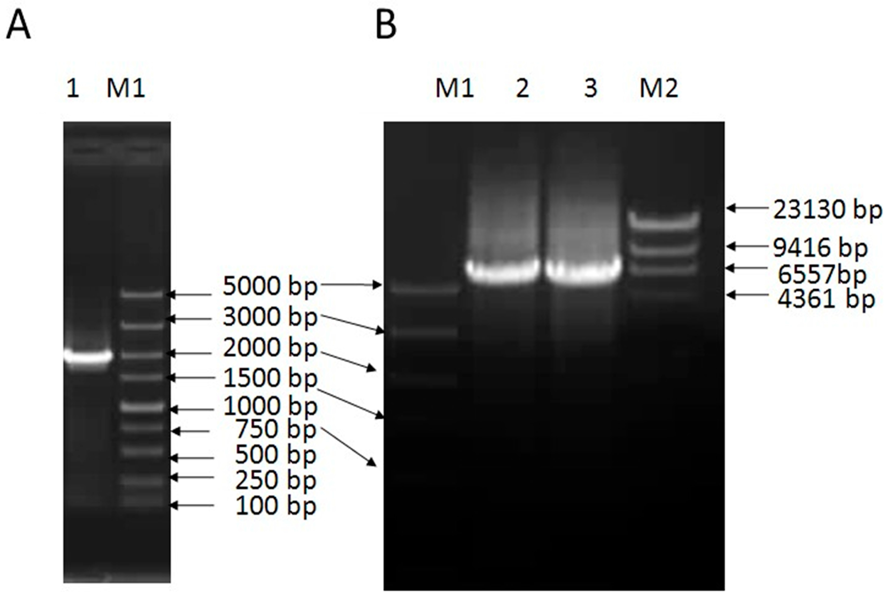
2.1. Cloning and Sequence Analysis of VpSBP16

A 5984 base pair (bp) full-length VpSBP16 DNA sequence, including a 1647 bp open reading frame (ORF), was amplified from complementary DNA (cDNA) (Figure 1A) or genomic DNA (Figure 1B) extracted from the leaves of V. pseudoreticulata W. T. Wang clone ‘Baihe-35-1’, using the primers based on the cDNA sequence of VvSBP16 obtained from the Grape Genome Database (http://www.genoscope.cns.fr). A comparison between the genomic DNA and cDNA sequence revealed that the coding region of VpSBP16 has two introns (557 bp and 3662 bp), the relative positions of which are shown in Figure 2. The predicted VpSBP16 protein contains a highly conserved SBP-domain with two zinc-binding sites of the C2HCH type (zinc finger 1 and zinc finger 2), together with a nuclear localization signal (NLS) (Figure 2A).

2.2. Subcellular Localization and Function of VpSBP16 in Transcriptional Activation

Sequence analysis of the grape SBP-box genes revealed that their deduced protein sequences contained putative NLS regions (Figure 2A). To confirm targeting of VpSBP16 to the nucleus, the VpSBP16 coding sequence (CDS) without the C terminal termination codon was translationally fused to green fluorescent protein (GFP) coding sequence in the pBI221-GFP vector to generate the pBI221-VpSBP16-GFP vector. This vector and a pBI221-GFP negative control vector were transformed into onion epidermal cells using particle bombardment [24] for transient expression analysis. The VpSBP16-GFP fusion protein was observed by confocal microscopic analysis to accumulate in the nucleus of onion epidermal cells, whereas the GFP control protein was distributed throughout the cell (Figure 3A), consistent with the prediction that VpSBP16 is a nuclear protein that functions as a transcription factor.
To investigate whether VpSBP16 has transcriptional activation activity, the full-length VpSBP16 CDS and the yeast GAL4 cDNA were separately fused to the GAL4 DNA-binding domain in the pGBKT7 vector. Yeast cells transformed with the pGBKT7 control vector only grew on Single Dropout (-Trp) (SD/-Trp) medium, while those transformed with the VpSBP16 and GAL4 plasmids were able to grow on both SD/-Trp and SD/-Trp/-Ade/-His media, and exhibited blue staining in X-α-gal solution (Figure 3B), demonstrating that the VpSBP16 protein can function as a transcriptional activator.

2.3. Effect of Osmotic Stress on Seed Germination in Transgenic VpSBP16 Arabidopsis Lines

We generated transgenic A. thaliana plants expressing the VpSBP16 ORF under the regulation of the constitutive CaMV 35S promoter to determine whether the gene confers abiotic stress tolerance. A total of 89 independent transgenic lines were obtained and the presence of the transgene confirmed by PCR and kanamycin antibiotic selection. The expression level of VpSBP16 in twelve transgenic lines with the better performance under salt stress was verified by semi-quantitative RT-PCR with transcript-specific primers VpSBP16-F and VpSBP16-R (Table 1).The three lines (VpSBP16-1, VpSBP16-14, and VpSBP16-47) with the best performance and the highest levels of expression were selected for further study (Figure S1), and grown in the same Petri dish or pot with different stress conditions as WT plants (Figure 4 and Figure 5).
We measured the germination rates of seeds from the three transgenic Arabidopsis lines and WT plants placed on Murashige and Skoog (MS) medium, MS medium with 150 mM NaCl and MS medium with 400 mM mannitol (Figure 4). Two days after sowing, seeds from the three transgenic lines (VpSBP16-1, VpSBP16-14 and VpSBP16-47) and wild type (WT) began to germinate on MS medium, while at 4 days after germination the rate nearly was 100% (Figure 4E). The germination rate of the transgenic lines (VpSBP16-1, VpSBP16-14 and VpSBP16-47) on 150 mM NaCl medium was 81.5%, 86.7% and 75.0% respectively, 14 days after culturing. However, the germination rate of wild type was only 56.4%. On the MS medium with 400 mM mannitol, the result was similar, with the WT showing a lower germination rate (Figure 4E). In general, compared to MS medium, the germination of all seeds on the MS medium with 150 mM NaCl or 400 mM mannitol was lower, while the transgenic seeds exhibited 17–32% higher germination rates than WT seeds sown on the same media (Figure 4E).

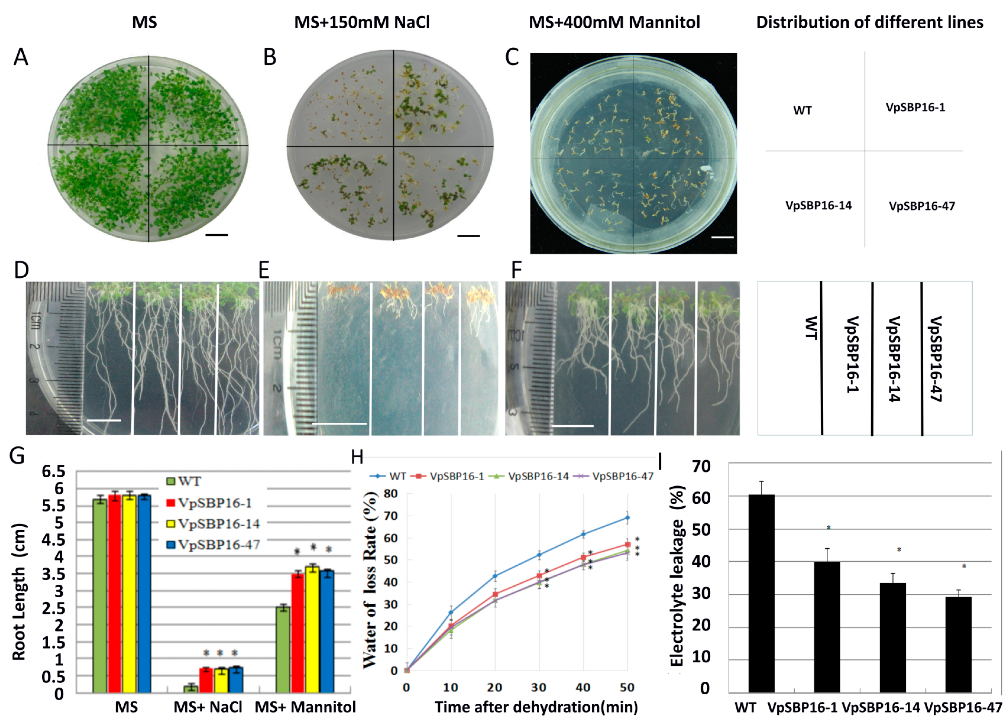
2.4. VpSBP16 Transgenic Arabidopsis Seedling Resistance to Osmotic Stress

To assess whether VpSBP16 confers resistance to osmotic stress, the seedling of three transgenic lines (VpSBP16-1, VpSBP16-14 and VpSBP16-47) and WT were grown on MS medium containing 150 mM NaCl or 400 mM mannitol. Both the WT and transgenic seedling displayed similar growth characteristics on MS basal medium (Figure 5A). However, upon exposure to 150 mM NaCl, their growth was strikingly different after 35 days. Three transgenic lines had not only a higher seed germination rate, but also larger root systems and leaves that were greener (Figure 5B,E). The WT seedlings had withered yellow leaves, grew slower and did not form normal cotyledons (Figure 5B). Moreover, the root lengths of the WT seedlings were 0.15–0.2 cm, while those of the transgenic seedlings were approximately 4-fold greater (Figure 5D,E,G), indicating that overexpression of the VpSBP16 gene offsets some of the inhibitory effects of salt stress on root elongation. The seedlings grown on medium with 400 mM mannitol had the same growth characteristics as those grown under salt stress (Figure 5C,F,G).
We also measured the transpirational water loss of seedlings from 10-day-old WT and transgenic plants left on the laboratory bench at room temperature with a humidity of 40–45%, every 10 min over a 50 min period. Less water loss was detected in the three transgenic lines than in the WT control (Figure 5H). We next measured changes in relative electrolyte leakage in the transgenic and WT seedlings to determine whether there was a correlation with the improved osmotic stress tolerance of the VpSBP16 transgenic seedlings. The relative electrolyte leakage was also significantly lower in transgenic lines than in WT (Figure 5I). These results suggest that osmotic damage in the transgenic lines was significantly lower than in the control and overexpression of VpSBP16 significantly improved resistance to osmotic stress.

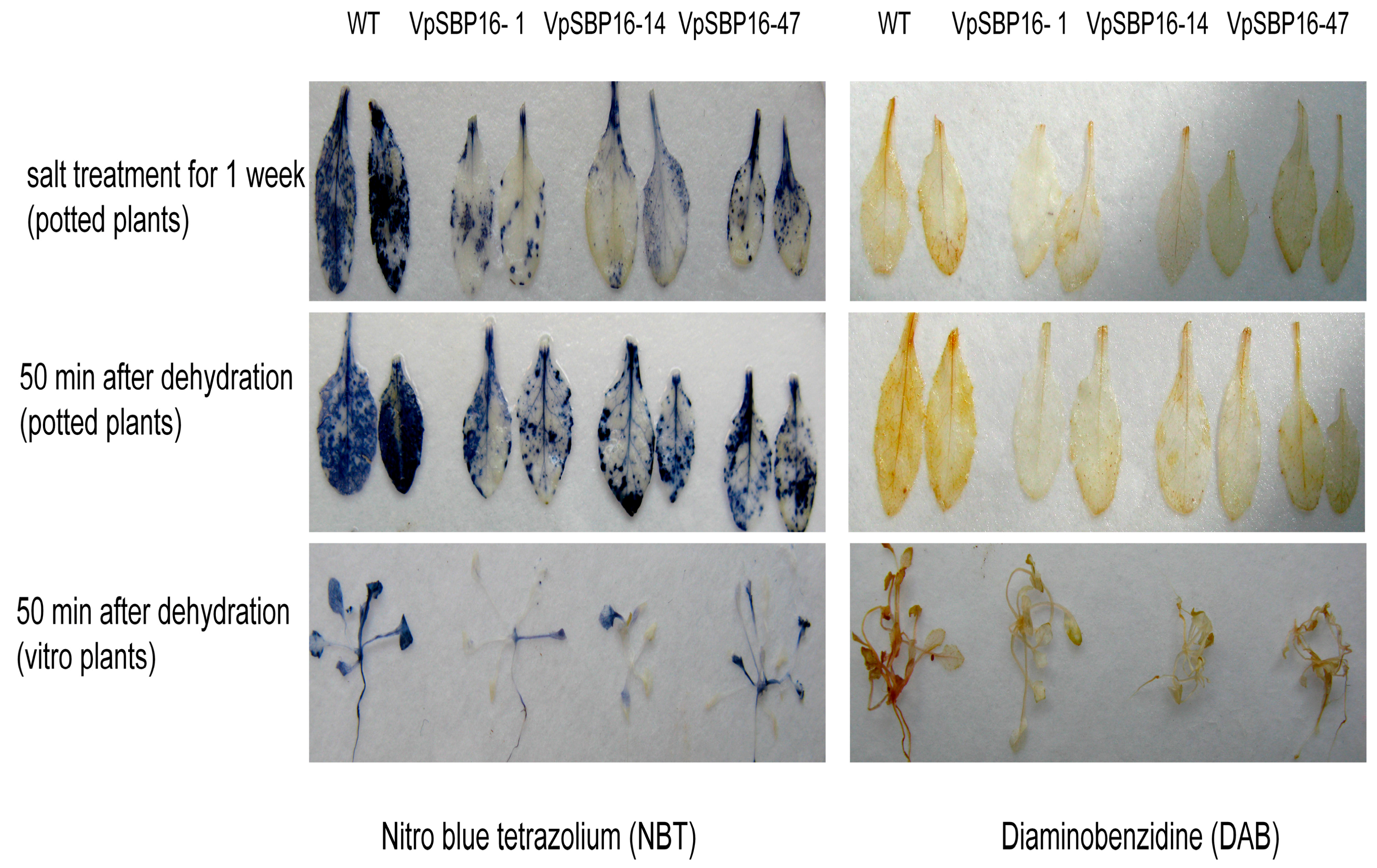
2.5. Response to Abiotic Stresses of VpSBP16 Transgenic Arabidopsis Plants

The results described above indicated that overexpression of VpSBP16 involved resistance of seedlings to salt stress and short-term drought stress. We next examined the responses of adult plants. Three-week-old transgenic and WT plants grown in the same pot were irrigated with 300 mM NaCl solution. Most leaves of the WT plants showed etiolated and wilted symptoms after 1 week of treatment. In contrast, those of the transgenic plants remained green or showed only mild etiolation under the same treatments (Figure 6A). The growth and development of 2-week-old transgenic lines and WT grown in the same pot following the drought treatment for 18 days was then investigated. All WT and transgenic plants obviously withered and exhibited severe water loss related symptoms, and after rewatering for 3 days, most of transgenic plants had resumed normal growth (73–80% recovery rate), but almost all the WT plants had died (2% recovery rate, Figure 6B,C).
Substantial accumulation of reactive oxygen species (ROS) because of exposure to stress can cause progressive oxidative damage, ultimately leading to cell injury and even death [25]. We measured levels of the ROS species O2 and H2O2 in leaves of plants grown under water loss and salt stress by NBT and DAB histochemical staining, respectively. We observed that leaves of the transgenic lines accumulated much lower levels of O2 or H2O2 than those of WT plants (Figure 7).

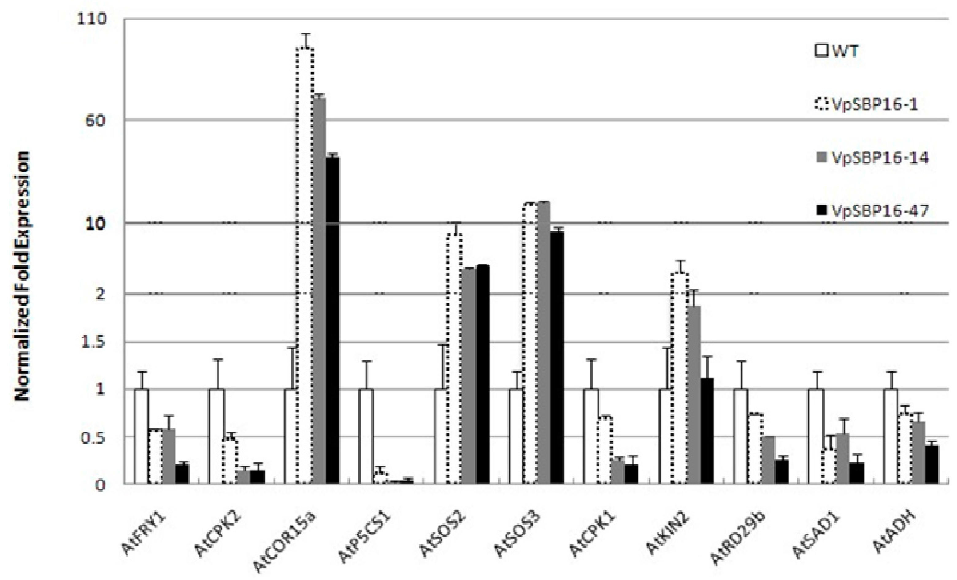
2.6. Altered Expression of Abiotic Stress Responsive Genes in Transgenic VpSBP16 Plants

Drought and high salinity are common stress conditions that adversely affect plant growth and crop productivity. In order to better understand the mechanistic basis of the improved abiotic stress tolerance exhibited by the transgenic VpSBP16 plants, we measured the expression profiles of a number of abiotic stress-responsive genes (AtSOS2, AtSOS3, AtFRY1, AtSAD1, AtADH, AtCOR15a, AtKIN2, AtP5CS1, AtRD29B, AtCDPK1 and AtCDPK2) in 3-week-old WT and transgenic plants using quantitative real-time PCR (qRT-PCR) analysis. We saw that the transgenic VpSBP16 overexpressing lines had higher expression levels of AtSOS2, AtSOS3, AtCOR15a and AtKIN2. In three transgenic lines VpSBP16-1, VpSBP16-14 and VpSBP16-47, the expression of AtCOR15a gene was 80, 65 and 35 times that of WT, respectively, while the expression of AtSOS2 and AtSOS3 was approximately 6-fold and 10-fold that of WT. The expression of AtKIN2 gene in the transgenic lines was 2-fold greater than that of WT (Figure 8). However, the expression levels of AtFRY1, AtSAD1, AtADH, AtP5CS1, AtRD29B, AtCDPK1 and AtCDPK2 were lower than those in WT to varying degrees (Figure 8).

3. Discussion

Plants are exposed to a variety of biotic and abiotic stresses, such as cold, drought, salt stress and pathogens, during their life cycle, many of which can have an irreversible effect on growth and development. Many studies have identified transcription factors that are important for regulating plant responses to stress, including members of the WRKY, bZIP, AP2/ERF and MYB. In this regard, the functions of SBP family genes have been investigated in several plant species, but to date nothing has been reported regarding the role of SBP-box genes in grape responses to abiotic stress.
Members of the SBP-box gene family encode DNA binding proteins that function as transcription factors [26], and have been identified only in green plants, including taxa from single-cell algae to later-diverging land plants. Characteristics of a SBP domain are a ~74 amino acid (aa) residue region, a nuclear localization signal and the ability to bind DNA involving two zinc-finger domains [13]. In all cases, the SBP-domains are very similar, with a high sequence conservation at certain positions and the best-conserved aa residues are cysteines and histidines that are used to coordinate two zinc ions. Similarly well conserved are the basic amino acid residues that are thought to be involved in general and specific DNA binding, and in nuclear translocation of the proteins [9,13]. The VpSBP16 amino acid sequence contains all the above structural features, including the SBP-box and a putative nuclear localization signal (Figure 2). We confirmed that VpSBP16 is targeted to the nucleus and possesses transcriptional activation activity (Figure 3).
We studied the role of VpSBP16 in A. thaliana in the context of salinity and drought stress. Over-expression of VpSBP16 in A. thaliana yielded transgenic lines with enhanced resistance to high osmoticum, dehydration, long-term drought, and salt stress compared with the WT.
The semi- and selective permeability of the plasma membrane are fundamentally important characteristics for maintaining normal plant physiological function. However, these permeability characteristics can be perturbed in response to stress, which can result in electrolyte leakage. We inferred the extent of the cellular injury, and by extension resistance to stress, by measuring electrolyte leakage. We found that the electrolyte leakage in WT was significantly higher than in the transgenic lines after the water loss treatment. The results were consistent with the improved tolerance to stress shown by the VpSBP16 transgenic lines.
Stress occurs when the root system cannot supply enough water to transpiring leaves to maintain a proper water balance. Accordingly, the ability of plant to overcome environmental stresses is influenced by its root system size and distribution. We found that the VpSBP16 transgenic lines had longer and a larger root system compared to the WT under the stresses, which likely helped in the tolerance of salt and drought stress by increasing water uptake. Under high saline condition, salt imposes an oxidative stress caused by the generation of ROS, such as singlet oxygen, H2O2 and O2 [27]. Usually, this source of ROS does not result in serious harm to the plant, but under high salt and drought conditions, excess ROS is not eliminated rapidly enough, causing severe oxidative damage, and affecting physiology [28]. Therefore, the removal of excess ROS and the maintenance of ROS homeostasis are crucial for mitigating ROS toxicity and improving plant stress resistance.
Diverse transcription factors have been shown to contribute to the regulation of active oxygen scavenging. For example, overexpression of OsMYB2 in rice was reported to reduce the content of ROS and enhance the drought tolerance [29], and under H2O2 stress, the expression of the banana WRKY family gene, MusaWRKY71, resulted in increased expression of peroxidase genes, which are part of the ROS scavenging system in banana seedlings [7]. Moreover, overexpression JERF3 gene and SlERF3 gene from Solanum lycopersicum in tobacco, and TERF2 from Solanum lycopersicum and SUB1A gene from Oryza sativa in rice all improved the stress tolerance by enhancing the scavenging of ROS [30,31,32,33]. We observed that the levels of ROS in the VpSBP16 transgenic lines were significantly lower than in WT plants, following exposure to the abiotic stresses.
We also examined the expression of some stress-responsive genes, such as AtSOS2, AtSOS3, AtFRY1, AtSAD1, AtADH, AtCOR15a, AtKIN2, AtP5CS1, AtRD29B, AtCDPK1 and AtCDPK2 to investigate the potential mechanisms of VpSBP16 in stress responses. The stress-responsive gene candidates COR15b, SOS2, SOS3, exhibited a significantly higher expression level in the VpSBP16 overexpression plants than in WT. Of these genes, SOS3 was identified through genetic screening of salt-sensitive mutants and SOS2 has been shown to function as a point of crosstalk between SOS and other signaling pathways [34]. Additionally, overexpression of SlSOS2 (SlCIPK24) in tomato, MdSOS2L1 in apple or PtSOS2 genes (PtSOS2.1, PtSOS2.2, PtSOS2.3) in poplar was reported to confer salt tolerance to transgenic tomato, apple calli or poplar, respectively [35,36,37], further indicating its importance in abiotic stress resistance. However, the expression levels of AtP5CS1, AtCDPK1, AtCDPK2, AtRD29B, AtADH and AtFRY1 were down-regulated in the transgenic lines, contradicts previous research. The key enzyme in proline synthesis (P5CS1) is involved in the natural adaptation among Arabidopsis accessions in differential responses to osmotic stress [38,39]. Calcium-dependent protein kinases (CDPK) as the well-known Ca2+-sensor protein, play important roles in the plant response to salt stress [40]. The RD29B promoter region carrying several ABRE sequences and one DRE is controlled mainly by ABA and the -214 G-box in the AtAdh promoter also is the ABA response element [41,42]. AtRD29B, AtADH, AtFRY1 and AtSAD1 all confer both abiotic or biotic stress resistance in A. thaliana by involving in ABA signal transduction pathways [43,44,45,46]. Therefore, we speculate the resistance of the transgenic VpSBP16 plants against salt and drought stress have nothing to do with ABA signal transduction pathway and Ca2+-dependent signal network. However, further studies are necessary to unravel the reason of why these genes were down-regulated.
Based on these results we propose that overexpression of VpSBP16 may increase resistance to salt and drought stress by regulating the SOS signaling cascade and ROS signaling. However, further studies will be needed to identify the exact molecular mechanisms by which the VpSBP16 mediate responses to abiotic stresses. We noted that AtCOR15a was significantly induced in the VpSBP16 overexpression plants, suggesting that VpSBP16 may also be associated with cold stress responses. This will be a target of future research.

4. Materials and Methods

4.1. Plant Materials and Growth Conditions

Chinese wild grape, Vitis pseudoreticulata “Baihe-35-1”, used for cloning of VpSBP16, was grown in the grape repository of Northwest A&F University, Yangling, Shaanxi, China (34°20′ N, 108°24′ E). A. thaliana plants (T3 transgenic lines and wild-type (WT) ecotype Columbia-0) were grown at 22 °C, 70% relative humidity and long day (8 h dark, 16 h light) conditions. All experiments were repeated in triplicate and all samples were immediately frozen in liquid nitrogen and stored at −80 °C until further use.

4.2. Isolation and Analysis of the VpSBP16 cDNA

The cDNA was synthesized from total RNA samples extracted from V. pseudoreticulata ‘Baihe-35-1’ leaves. The experiments were performed essentially as previously described [47]. A pair of gene-specific primers (VpSBP16-F1 and VpSBP16-R1) (Table 1) were used to amplify the predicted VpSBP16 ORF from the cDNA template with Taq DNA polymerase (TaKaRa Biotechnology, Dalian, China) and the following cycling program: 94 °C for 3 min, 35 cycles at 94 °C for 30 s, 58 °C for 30 s and 72 °C for 2 min; and extension at 72 °C for 10 min. The amplified products were cloned into the pGEM-Teasy vector (Promega, Madison, WI, USA) to generate pGEM-Teasy-VpSBP16, and transformed into E. coli strain DH5α. Positive clones were sequenced at TaKaRa Biotechnology. The sequences were analyzed to identify conserved regions using Conserved Domains (http://www.ncbi.nlm.nih.gov/Structure/cdd/wrpsb.cgi). The subcellular localization of VpSBP16 was predicted using the Center for Biological Sequence analysis software (http://genome.cbs.dtu.dk/services/TargetP; http://genome.cbs.dtu. dk/services/SignalP/).

4.3. Subcellular Localization and Trans-Activation Assay

The VpSBP16 CDS without the termination codon was amplified using VpSBP16-F2 and R2 with XbaI and KpnI sites (Table 1) from the pGEM-Teasy-VpSBP16 plasmid template, using Taq DNA polymerase (TaKaRa Biotechnology, Tokyo, Japan). The amplified product was inserted immediately upstream of the green fluorescent protein (GFP) coding sequence in the pBI221-GFP vector (Clontech Laboratories, Mountain View, CA, USA) and digested with the same restriction enzymes to generate pBI221-VpSBP16-GFP. Both the SBP16-containing vector and a background control vector with no insert were delivered into onion epidermal cells using a PDS-1000/He gene gun (Bio-Rad Laboratories Inc., Hercules, CA, USA) at 1100 psi as previously described [24], and then the cells were cultured on MS media in the dark at 22 °C for 18 h. Following cultivation, GFP signal was visualized using a Zeiss confocal microscope (LSM510; Carl Zeiss; Thornwood, NY, USA) with an excitation wavelength of 480 ± 20 nm and an emission wavelength of 510 ± 20 nm.
The coding regions of yeast GAL4 and grape VpSBP16 were separately ligated into the NcoI/BamHI and XmaI/BamHI sites of the GAL4 DNA-binding domain of the pGBKT7 vector (Clontech Laboratories), generating the plasmids pGBKT7-Gal4 (positive control) and pGBKT7-VpSBP16, respectively, using DNA fragments amplified with the gene-specific primers Gal4-F and R, and VpSBP16 F3 and R3 (Table 1). The vectors were used in a trans-activation assay with a yeast assay system as described previously [47]. The resulting plasmids, pGBKT7-Gal4 and pGBKT7-VpSBP16, as well as the empty vector pGBKT7 (negative control), were transformed into yeast (Saccharomyces cerevisiae) AH109 cells, which were then streaked on SD/-Trp and SD/-Trp/-Ade/-His/X-α-Gal plates to observe yeast growth at 30 °C for 3–4 days.

4.4. Generation of Transgenic A. thaliana Plants Over-Expressing VpSBP16

The CDS of VpSBP16 was amplified from the pGEM-Teasy-VpSBP16 plasmid template using the gene-specific primers VpSBP16-F4 and R4 (Table 1), then inserted immediately downstream of the CaMV 35S promoter in the plant overexpression vector, pCambia2300 (Clontech Laboratories) to produce the plasmid pCambia2300-35S-VpSBP16. This construct was then introduced into Agrobacterium tumefaciens strain EHA105, which was in turn used to transform A. thaliana via the floral dip method [48]. Transgenic seeds (T1) were selected on MS agar medium supplemented with 60 mg L−1 kanamycin and T2 transgenic lines were selected on MS agar medium supplemented with 60 mg L−1 kanamycin and 150 mM NaCl. Three of the resulting T3 homozygous lines with most obvious phenotypes (SBP16-1, SBP16-14 and SBP16-47) and highest levels of expression were selected for further study.

4.5. Semi-Quantitative RT-PCR Analysis

Total A. thaliana RNA was extracted from entire WT and twelve transgene line (SBP16-1, SBP16-8, SBP16-14, SBP16-18, SBP16-23, SBP16-32, SBP16-35, SBP16-40, SBP16-47, SBP16-57, SBP16-61 and SBP16-67) plants using the E.Z.N.A.® Plant RNA Kit (Omega Bio-tek, Norcross, GA, USA). Cycling parameters were as follows: 94 °C for 3 min, 25 cycles at 94 °C for 30 s, 60 °C for 30 s and 72 °C for 30 s, with a final elongation step at 72 °C for 10 min. An 8 μL sample of each PCR product was subsequently separated on a 1.2% (w/v) agarose gel, then stained with ethidium bromide and photographed under UV light. Atactin1 (At2g37620) was amplified for use as an internal control. VpSBP16-F, VpSBP16-R and AtActin1-F, AtActin1-R specific primer sequences are listed in Table 1.

4.6. Germination Assays

For the seed germination assays, 100–150 seeds from each of three selected T3 homozygous lines and from WT plants were vernalized for 3 days at 4 °C, and then sown on MS medium or MS medium supplemented with 150 mM NaCl or 400 mM mannitol. The percentage of germinated seeds was calculated based on the number of seedlings that had reached the cotyledon stage at 2 weeks [49]. All seeds used for the germination analysis were harvested and stored at the same time under the same conditions. All germination assays were performed in triplicate.

4.7. Salt and Drought Treatments for Wild Type (WT) and T3 Transgenic Lines

Seeds from WT and T3 transgenic lines were sterilized and vernalized for 3–4 days at 4 °C on the same MS medium, supplied with 150 mM NaCl or 400 mM mannitol, and then transferred to growth chambers. For the potted plants, three-week-old A. thaliana and transgenic lines were irrigated with 300 mM NaCl at 2-day intervals for 15 days or not watered, corresponding to salt and drought treatments, respectively. Plants that were well watered were used as the negative control. Following the drought treatment for 18 days, plants were re-watered for 3 days.

4.8. Determination of the Water Loss Rate and Electrolyte Leakage

For the determination of water loss, whole plants of 2-week-old transgenic and WT plants grown on the same MS medium were placed on dry filter paper and at 40–45% relative humidity at room temperature and weighed at the indicated times. The water loss rate was calculated based on the initial fresh weight of the samples and the experiment was repeated three times [50]. All plants were sampled after dehydration for 50 min and were vacuum-infiltrated with deionized water for 20 min. After 2 h, the conductivities (C1) of the solutions were determined using a conductivity detector. Subsequently, the seedlings were boiled for 20 min in deionized water and cooled to room temperature. The conductivities (C2) of the solutions was then determined. The C1 to C2 (C1/C2) ratios were calculated and used as a measure of the relative electrolyte leakage [51].

4.9. Detection of H2O2 and O2

Excised leaves or whole plants were placed in 1 mg mL1 diaminobenzidine (DAB) solution (Sigma, Steinheim, Germany) for 8 h to monitor H2O2 production, or were incubated in HEPES buffer (pH 7.5) containing 6 mM nitro blue tetrazolium(NBT) for 2 h to detect O2 production. The samples were then cleared to remove chlorophyll at 80 °C in 80% (v/v) ethanol for 2 h and immersed in 10% (v/v) glycerol for observations [52,53].

4.10. Quantitative Real-Time RT-PCR Analysis

Total A. thaliana RNA was extracted from entire WT and three transgene line (SBP16-1, SBP16-14 and SBP16-69) plants using the E.Z.N.A.® Plant RNA Kit (Omega Bio-tek, USA, R6827-01). First-strand cDNA for expression analysis was synthesized from 1 μg of DNase-treated total RNA using PrimeScript™ RTase (TaKaRa Biotechnology). Each reaction was done in triplicates with a reaction volume of 25 μL. Cycling parameters were 95 °C for 30 s, 40 cycles of 95 °C for 5 s, and 60 °C for 30 s. For dissociation curve analysis, a program including 95 °C for 15 s, followed by a constant increase from 60 °C to 95 °C, was included after the PCR cycles. Quantitative RT-PCR was conducted using SYBR green (Takara Biotechnology) with an IQ5 real time PCR machine (Bio-Rad, Hercules, CA, USA). Each reaction was performed in triplicate and data were analyzed as previously described [54]. The expression of stress-related genes such as AtFRY1, AtSAD1, AtADH, AtP5CS1, AtRD29B, AtCDPK1 and AtCDPK2 were measured by RT-PCR in WT and T3 transgenic lines. Atactin1 (At2g37620) was amplified for use as an internal control. The sequences of the quantitative PCR primers are listed in Table 1.

5. Conclusions

Overexpression of VpSBP16 may increase resistance to salt and drought stress by regulating the SOS signaling cascade and ROS signaling.

Supplementary Materials

Supplementary materials can be found at https://www.mdpi.com/1422-0067/19/4/940/s1.

Acknowledgments

This work was supported by the National Natural Science Foundation of China (31572110), the Scientific Research Foundation for Outstanding Young Scientists of Shandong Province (BS2014SW027), as well as the Scientific Research Funds for High-level Personnel of Qingdao Agricultural University (663/1114338, 661/1116009). We thank PlantScribe (www.plantscribe.com) for editing this manuscript.

Author Contributions

Xiping Wang, Hongmin Hou designed the study. Hongmin Hou, Hui Jia and Qin Yan performed data analysis. Xiping Wang provided guidance for the whole study. Hongmin Hou and Xiping Wang wrote and revised the manuscript. All authors approved the final manuscript.

Conflicts of Interest

The authors declare no conflict of interest.

Abbreviations

CaMVCauliflower mosaic virus
ORFOpen reading frame
RT-PCRReverse transcription-polymerase chain reaction
MSMurashige and Skoog
NBTNitro blue tetrazolium
DABDiaminobenzidine
ROSReactive oxygen species
H2O2Hydrogen peroxide
O2Superoxide anion
SOSSalt overly sensitive
SBPSquamosa promoter binding protein
NLSNuclear localization signal
WTWild-type

References

  1. Abe, H.; Urao, T.; Ito, T.; Seki, M.; Shinozaki, K.; Yamaguchi-Shinozaki, K. Arabidopsis AtMYC2 (bHLH) and AtMYB2 (MYB) Function as Transcriptional Activators in Abscisic Acid Signaling. Plant Cell 2003, 15, 63–78. [Google Scholar] [CrossRef] [PubMed]
  2. Ding, Z.; Li, H.; X, A.; X, L.; H, Q.; D, W. Transgenic expression of MYB15 confers enhanced sensitivity to abscisic acid and improved drought tolerance in Arabidopsis thaliana. J. Genet. Genom. 2009, 36, 17–29. [Google Scholar] [CrossRef]
  3. Yang, O.S.; Popova, O.V.; Süthoff, U.; Lüking, I.; Dietz, K.J.; Golldack, D. The Arabidopsis basic leucine zipper transcription factor AtbZIP24 regulates complex transcriptional networks involved in abiotic stress resistance. Gene 2009, 436, 45–55. [Google Scholar] [CrossRef] [PubMed]
  4. Han, C.; Lai, Z.; Shi, J.; Yong, X.; Chen, Z.; Xu, X. Roles of arabidopsis WRKY18, WRKY40 and WRKY60 transcription factors in plant responses to abscisic acid and abiotic stress. BMC Plant Biol. 2010, 10, 281–296. [Google Scholar]
  5. Jiang, Y.; Deyholos, M.K. Functional characterization of Arabidopsis NaCl-inducible WRKY25 and WRKY33 transcription factors in abiotic stresses. Plant Mol. Biol. 2009, 69, 91–105. [Google Scholar] [CrossRef] [PubMed]
  6. Ren, X.; Chen, Z.; Liu, Y.; Zhang, H.; Zhang, M.; Liu, Q.; Hong, X.; Zhu, J.K.; Gong, Z. ABO3, a WRKY transcription factor, mediates plant responses to abscisic acid and drought tolerance in Arabidopsis. Plant J. 2010, 63, 417–429. [Google Scholar] [CrossRef] [PubMed]
  7. Schmidt, R.; Schippers, J.H.; Mieulet, D.; Obata, T.; Fernie, A.R.; Guiderdoni, E.; Mueller-Roeber, B. MULTIPASS, a rice R2R3-type MYB transcription factor, regulates adaptive growth by integrating multiple hormonal pathways. Plant J. 2013, 76, 258–273. [Google Scholar] [CrossRef] [PubMed]
  8. Hsieh, T.H.; Li, C.W.; Su, R.C.; Cheng, C.P.; Tsai, Y.C.; Chan, M.T. A tomato bZIP transcription factor, SlAREB, is involved in water deficit and salt stress response. Planta 2010, 231, 1459–1473. [Google Scholar] [CrossRef] [PubMed]
  9. Klein, J.; Saedler, H.; Huijser, P. A new family of DNA binding proteins includes putative transcriptional regulators of the Antirrhinum majus floral meristem identity gene SQUAMOSA. Mol. Genet. Genom. 1996, 250, 7–16. [Google Scholar]
  10. Xie, K.; Wu, C.; Xiong, L. Genomic Organization, Differential Expression, and Interaction of SQUAMOSA Promoter-Binding-Like Transcription Factors and microRNA156 in Rice. Plant Physiol. 2006, 142, 280–293. [Google Scholar] [CrossRef] [PubMed]
  11. Preston, J.C.; Hileman, L.C. Functional Evolution in the Plant SQUAMOSA-PROMOTER BINDING PROTEIN-LIKE (SPL) Gene Family. Front. Plant Sci. 2013, 4, 1–13. [Google Scholar] [CrossRef] [PubMed]
  12. Birkenbihl, R.P.; Jach, G.; Saedler, H.; Huijser, P. Functional Dissection of the Plant-specific SBP-Domain: Overlap of the DNA-binding and Nuclear Localization Domains. J. Mol. Biol. 2005, 352, 585–596. [Google Scholar] [CrossRef] [PubMed]
  13. Yamasaki, K.; Kigawa, T.; Inoue, M.; Yamasaki, T.; Yabuki, T.; Aoki, M.; Seki, E.; Matsuda, T.; Tomo, Y.; Terada, T. An Arabidopsis SBP-domain fragment with a disrupted C-terminal zinc-binding site retains its tertiary structure. FEBS Lett. 2006, 580, 2109–2116. [Google Scholar] [CrossRef] [PubMed]
  14. Wang, Y.; Hu, Z.; Yang, Y.; Chen, X.; Chen, G. Function annotation of an SBP-box gene in Arabidopsis based on analysis of co-expression networks and promoters. Int. J. Mol. Sci. 2009, 10, 116–132. [Google Scholar] [CrossRef] [PubMed]
  15. Stone, J.M.; Liang, X.; Nekl, E.R.; Stiers, J.J. Arabidopsis AtSPL14, a plant-specific SBP-domain transcription factor, participates in plant development and sensitivity to fumonisin B1. Plant J. 2005, 41, 744–754. [Google Scholar] [CrossRef] [PubMed]
  16. Ning, K.; Chen, S.; Huang, H.; Jiang, J.; Yuan, H.; Li, H. Molecular characterization and expression analysis of the SPL gene family with BpSPL9 transgenic lines found to confer tolerance to abiotic stress in Betula platyphylla Suk. Plant Cell Tissue Organ Cult. 2017, 130, 1–13. [Google Scholar] [CrossRef]
  17. Mao, H.D.; Yu, L.J.; Li, Z.J.; Yan, Y.; Han, R.; Liu, H.; Ma, M. Genome-wide analysis of the SPL family transcription factors and their responses to abiotic stresses in maize. Plant Gene 2016, 6, 1–12. [Google Scholar] [CrossRef]
  18. Stief, A.; Altmann, S.; Hoffmann, K.; Pant, B.D.; Scheible, W.; Bäurle, I. Arabidopsis miR156 Regulates Tolerance to Recurring Environmental Stress through SPL Transcription Factors. Plant Cell 2014, 26, 1792–1807. [Google Scholar] [CrossRef] [PubMed]
  19. Cui, N.; Sun, X.; Sun, M.; Jia, B.; Duanmu, H.; Lv, D.; Duan, X.; Zhu, Y. Overexpression of OsmiR156k leads to reduced tolerance to cold stress in rice (Oryza Sativa). Mol. Breed. 2015, 35, 214–225. [Google Scholar] [CrossRef]
  20. Cui, L.G.; Shan, J.X.; Shi, M.; Gao, J.P.; Lin, H.X. The miR156-SPL9-DFR pathway coordinates the relationship between development and abiotic stress tolerance in plants. Plant J. 2014, 80, 1108–1117. [Google Scholar] [CrossRef] [PubMed]
  21. Arshad, M.; Feyissa, B.A.; Amyot, L.; Aung, B.; Hannoufa, A. MicroRNA156 improves drought stress tolerance in alfalfa (Medicago sativa) by silencing SPL13. Plant Sci. 2017, 258, 122–136. [Google Scholar] [CrossRef] [PubMed]
  22. Hou, H.; Li, J.; Gao, M.; Singer, S.D.; Wang, H.; Mao, L.; Fei, Z.; Wang, X. Genomic Organization, Phylogenetic Comparison and Differential Expression of the SBP-Box Family Genes in Grape. PLoS ONE 2013, 8, e59358. [Google Scholar] [CrossRef] [PubMed]
  23. Hou, H.; Yan, Q.; Wang, X.; Xu, H. A SBP-Box Gene VpSBP5 from Chinese Wild Vitis Species Responds to Erysiphe necator and Defense Signaling Molecules. Plant Mol. Biol. Rep. 2013, 31, 1261–1270. [Google Scholar] [CrossRef]
  24. Marè, C.; Mazzucotelli, E.; Crosatti, C.; Francia, E.; Stanca, A.M.; Cattivelli, L. Hv-WRKY38: A new transcription factor involved in cold- and drought-response in barley. Plant Mol. Biol. 2004, 55, 399–416. [Google Scholar] [CrossRef] [PubMed]
  25. Liu, Y.; He, C. Regulation of plant reactive oxygen species (ROS) in stress responses: Learning from AtRBOHD. Plant Cell Rep. 2016, 35, 995–1007. [Google Scholar] [CrossRef] [PubMed]
  26. Cardon, G.; Höhmann, S.; Klein, J.; Nettesheim, K.; Saedler, H.; Huijser, P. Molecular characterisation of the Arabidopsis SBP-box genes. Gene 1999, 237, 91–104. [Google Scholar] [CrossRef]
  27. And, P.M.H.; Bressan, R.A.; Zhu, J.K.; Bohnert, H.J. Plant cellular and molecular responses to high salinity. Annu. Rev. Plant Biol. 2000, 51, 463–499. [Google Scholar] [CrossRef]
  28. Mittova, V.; Tal, M.; Volokita, M.; Guy, M. Salt stress induces up-regulation of an efficient chloroplast antioxidant system in the salt-tolerant wild tomato species Lycopersicon pennellii but not in the cultivated species. Physiol. Plant. 2002, 115, 393–400. [Google Scholar] [CrossRef] [PubMed]
  29. Yang, A.; Dai, X.; Zhang, W. A R2R3-type MYB gene, OsMYB2, is involved in salt, cold, and dehydration tolerance in rice. J. Exp. Bot. 2012, 63, 2541–2556. [Google Scholar] [CrossRef] [PubMed]
  30. Fukao, T.; Bailey-Serres, J. The submergence tolerance regulator SUB1A mediates crosstalk between submergence and drought tolerance in rice. Plant Cell 2011, 23, 412–427. [Google Scholar] [CrossRef] [PubMed]
  31. Pan, I.; Li, C.W.; Su, R.C.; Cheng, C.P.; Lin, C.S.; Chan, M.T. Ectopic expression of an EAR motif deletion mutant of SlERF3 enhances tolerance to salt stress and Ralstonia solanacearum in tomato. Planta 2010, 232, 1075–1086. [Google Scholar] [CrossRef] [PubMed]
  32. Tian, Y.; Zhang, H.; Pan, X.; Chen, X.; Zhang, Z.; Lu, X.; Huang, R. Overexpression of ethylene response factor TERF2 confers cold tolerance in rice seedlings. Transgenic Res. 2011, 20, 857–866. [Google Scholar] [CrossRef] [PubMed]
  33. Wu, L.; Zhang, Z.; Zhang, H.; Wang, X.C.; Huang, R. Transcriptional modulation of ethylene response factor protein JERF3 in the oxidative stress response enhances tolerance of tobacco seedlings to salt, drought, and freezing. Plant Physiol. 2008, 148, 1953–1963. [Google Scholar] [CrossRef] [PubMed]
  34. Ji, H.; Pardo, J.M.; Batelli, G.; Van Oosten, M.J.; Bressan, R.A.; Li, X. The Salt Overly Sensitive (SOS) Pathway: Established and Emerging Roles. Mol. Plant 2013, 6, 275–286. [Google Scholar] [CrossRef] [PubMed]
  35. Hu, D.G.; Ma, Q.J.; Sun, C.H.; Sun, M.H.; You, C.X.; Hao, Y.J. Overexpression of MdSOS2L1, a CIPK protein kinase, increases the antioxidant metabolites to enhance salt tolerance in apple and tomato. Physiol. Plant. 2015, 156, 201–214. [Google Scholar] [CrossRef] [PubMed]
  36. Huertas, R.; Olías, R.; Eljakaoui, Z.; Gálvez, F.J.; Li, J.; De Morales, P.A.; Belver, A.; Rodríguezrosales, M.P. Overexpression of SlSOS2 (SlCIPK24) confers salt tolerance to transgenic tomato. Plant Cell Environ. 2012, 35, 1467–1482. [Google Scholar] [CrossRef] [PubMed]
  37. Zhou, J.; Wang, J.; Bi, Y.; Wang, L.; Tang, L.; Xiang, Y.; Ohtani, M.; Demura, T.; Qiang, Z. Overexpression of PtSOS2 enhances salt tolerance in transgenic poplars. Plant Mol. Biol. Rep. 2014, 32, 185–197. [Google Scholar]
  38. Kesari, R.; Lasky, J.R.; Villamor, J.G.; Des Marais, D.L.; Chen, Y.J.; Liu, T.W. Intron-mediated alternative splicing of arabidopsis p5cs1 and its association with natural variation in proline and climate adaptation. Proc. Natl. Acad. Sci. USA 2012, 109, 9197–9202. [Google Scholar] [CrossRef] [PubMed]
  39. Des Marais, D.L.; Mckay, J.K.; Richards, J.H.; Sen, S.; Wayne, T.; Juenger, T.E. Physiological genomics of response to soil drying in diverse Arabidopsis accessions. Plant Cell 2012, 24, 893–914. [Google Scholar] [CrossRef] [PubMed]
  40. Ma, S.Y.; Wu, W.H. Atcpk23 functions in Arabidopsis responses to drought and salt stresses. Plant Mol. Biol. 2007, 65, 511–518. [Google Scholar] [CrossRef] [PubMed]
  41. Sakuma, Y.; Maruyama, K.; Osakabe, Y.; Qin, F.; Seki, M.; Shinozaki, K. Functional analysis of an arabidopsis transcription factor, dreb2a, involved in drought-responsive gene expression. Plant Cell 2006, 18, 1292–1309. [Google Scholar] [CrossRef] [PubMed]
  42. Guiltinan, M.J.; Marcotte, W.R., Jr.; Quatrano, R.S. A plant leucine zipper protein that recognizes an abscisic acid response element. Science 1990, 250, 267–271. [Google Scholar] [CrossRef] [PubMed]
  43. Msanne, J.; Lin, J.; Stone, J.M.; Awada, T. Characterization of abiotic stress-responsive Arabidopsis thaliana rd29a and rd29b genes and evaluation of transgenes. Planta 2011, 234, 97–107. [Google Scholar] [CrossRef] [PubMed]
  44. Xiong, L.M.; Gong, Z.Z.; Christopher, D.R.; Senthil, S.; Guo, Y.; Xu, W.Y.; Galbraith, D.; Zhu, J.K. Modulation of abscisic acid signal transduction and biosynthesis by an Sm-like protein in Arabidopsis. Dev. Cell 2001, 1, 771–781. [Google Scholar] [CrossRef]
  45. Lu, G.; Paul, A.L.; Mccarty, D.R.; Ferl, R.J. Transcription factor veracity: Is GBF3 responsible for aba-regulated expression of Arabidopsis Adh? Plant Cell 1996, 8, 847–857. [Google Scholar] [CrossRef] [PubMed]
  46. Xiong, L.M.; Lee, B.; Ishitani, M.; Lee, H.; Zhang, C.; Zhu, J.K. Fiery1 encoding an inositol polyphosphate 1-phosphatase is a negative regulator of abscisic acid and stress signaling in Arabidopsis. Gene Dev. 2001, 15, 1971–1984. [Google Scholar] [CrossRef] [PubMed]
  47. Fujita, M.; Fujita, Y.; Maruyama, K.; Seki, M.; Hiratsu, K.; Ohme-Takagi, M.; Tran, L.S.P.; Yamaguchi-Shinozaki, K.; Shinozaki, K. A dehydration-induced NAC protein, RD26, is involved in a novel ABA-dependent stress-signaling pathway. Plant J. 2004, 39, 863–876. [Google Scholar] [CrossRef] [PubMed]
  48. Clough, S.; Bent, A. Floral dip: A simplified method for Agrobacterium-mediated transformation of Arabidopsis thaliana. Plant J. 1998, 16, 735–743. [Google Scholar] [CrossRef] [PubMed]
  49. Saleki, R.; Young, P.G.; Lefebvre, D.D. Mutants of Arabidopsis thaliana Capable of Germination under Saline Conditions. Plant Physiol. 1993, 101, 839–845. [Google Scholar] [CrossRef] [PubMed]
  50. Zhang, L.; Zhao, G.; Xia, C.; Jia, J.; Liu, X.; Kong, X. A wheat R2R3-MYB gene, TaMYB30-B, improves drought stress tolerance in transgenic Arabidopsis. J. Exp. Bot. 2012, 63, 5873–5885. [Google Scholar] [CrossRef] [PubMed]
  51. Cao, W.H.; Liu, J.; He, X.J.; Mu, R.L.; Zhou, H.L.; Chen, S.Y. Modulation of ethylene responses affects plant salt-stress responses. Plant Physiol. 2007, 143, 707–719. [Google Scholar] [CrossRef] [PubMed]
  52. Kotchoni, S.O.; Kuhns, C.; Ditzer, A.; Kirch, H.H.; Bartels, D. Over-expression of different aldehyde dehydrogenase genes in Arabidopsis thaliana confers tolerance to abiotic stress and protects plants against lipid peroxidation and oxidative stress. Plant Cell Environ. 2006, 29, 1033–1048. [Google Scholar] [CrossRef] [PubMed]
  53. Kim, S.H.; Woo, D.H.; Kim, J.M.; Lee, S.Y.; Chung, W.S.; Moon, Y.H. Arabidopsis MKK4 mediates osmotic-stress response via its regulation of MPK3 activity. Biochem. Biophys. Res. Commun. 2011, 412, 150–154. [Google Scholar] [CrossRef] [PubMed]
  54. Gao, M.; Niu, J.; Zhao, S.; Jiao, C.; Xu, W.; Fei, Z.; Wang, X. Characterization of Erysiphe necator-Responsive Genes in Chinese Wild Vitis quinquangularis. Int. J. Mol. Sci. 2012, 13, 11497–11519. [Google Scholar] [CrossRef] [PubMed]
Figure 1. Cloning of VpSBP16 from V. pesudoreticulata. (A) PCR amplification of the full-length VpSBP16 cDNA and (B) DNA from Vitis pseudoreticulata. M1: DNA marker DL5000; M2: DNA marker λ-Hind III; 1: VpSBP16 PCR product from cDNA; 2 and 3: VpSBP16 PCR product.
Ijms 19 00940 g001
Figure 2. The DNA, cDNA nucleotide sequence and deduced amino acid sequence of VpSBP16 from V. pesudoreticulata (A) and Exon-intron structures of VpSBP16 (B). The SBP domain is shown in red and the two zinc-binding sites of the C2HCH type (zinc finger 1 and zinc finger 2) are indicated with green and yellow boxes. The conserved basic amino acids of the nuclear location signal are shaded in dark grey.
Ijms 19 00940 g002
Figure 3. Subcellular localization and transcriptional activation function of VpSBP16. (A) Subcellular localization of the VpSBP16-GFP (bottom row) fusion protein (top row) in onion epidermal cells. White arrowheads indicate the location of the nucleus in onion epidermal cell, scale bars: 50 μm. (B) Transcriptional activation function of VpSBP16 in yeast. Yeast cells containing the different plasmids grown on SD/-Trp select medium (left). Yeast cells containing the different plasmids grown on SD/-Trp-His-Ade+X-α-gal selection medium (right). 1: Positive control. (pGBKT7-Gal4); 2: pGBKT7-VpSBP16; 3: Negative control (pGBKT7). The experiments were repeated three times with consistent results.
Ijms 19 00940 g003
Figure 4. Phenotypes of wild type (WT) and VpSBP16 transgenic Arabidopsis thaliana lines at the seed germination stage placed under osmotic stress (AD) Photographs of seed germination in WT and transgenic lines 14 days after seeds were cultivated on Murashige-Skoog (MS) basal medium, MS basal medium supplemented with 150 mM NaCl or 400 mM mannitol. The experiments were repeated three times with consistent results, scale bars: 1 cm. (E) Seed germination rates of WT and transgenic lines cultivated on MS basal medium, MS basal medium supplemented with 150 mM NaCl or 400 mM mannitol for 14 days, respectively. Each data point is the mean of three replicates of 100–150 seeds. The error bars indicate the SD.
Ijms 19 00940 g004
Figure 5. Analysis of the osmotic tolerance of WT and VpSBP16 transgenic A. thaliana seedlings. (AC) Photographs of seedlings sown on MS basal medium, MS basal medium supplemented with 150 mM NaCl or 400 mM mannitol for 14, 35 and 20 days, respectively. The experiments were repeated three times with consistent results, scale bars: 1 cm. (DG) Root growth (DF) and root length (G) of WT and transgenic lines grown on MS basal medium, MS basal medium supplemented with 150 mM NaCl or 400 mM mannitol for 14, 35 and 20days, respectively. The experiments were repeated three times with consistent results, scale bars: 1 cm. (H) Water loss rate from 2-week-old detached transgenic and WT plants grown on the same MS basal medium, measured over a 50 min experimentation period. (I) Electrolyte leakage of seedlings of WT and the VpSBP16-1, VpSBP16-14 and VpSBP16-47 transgenic plants sampled at the last time point of dehydration. Asterisks indicated values that are significantly different from WT (n = 50 for each genotype, * 0.01< p < 0.05, one-way ANOVA). Each data point is the mean of three replicates of ten detached plants.
Ijms 19 00940 g005
Figure 6. Growth of WT and VpSBP16 transgenic A. thaliana plants in pots under non-stress or osmotic stress conditions. (A) Representative photographs of 3-week-old WT and transgenic lines before salt treatment and after salt treatment for 1 week. The experiments were repeated three times with consistent results, scale bars: 1 cm.(B) Survival rates of WT and transgenic lines 3 days after re-watering. Each data point is the mean of three replicates. The error bars indicate the SD. (n = 50 for each genotype, * 0.01< p < 0.05, one-way ANOVA). (C) Representative photographs showing the phenotype of plants before drought, 18 days after drought treatment and 3 days after re-watering. The experiments were repeated three times with consistent results, scale bars: 1 cm.
Ijms 19 00940 g006
Figure 7. Histochemical staining assay of ROS accumulation with nitro blue tetrazolium (NBT) and diaminobenzidine (DAB) in 2-week-old wild type (WT), or VpSBP16-1, VpSBP16-14 and VpSBP16-47 transgenic lines after salt treatment for 1 week or after the transpirational water loss from 50 min. The experiment was repeated three times with 5–10 leaves or plants.
Ijms 19 00940 g007
Figure 8. Expression levels of abiotic stress responsive genes in 3-week-old WT and VpSBP16 transgenic A. thaliana plants. AtActin1 was used as internal control for qRT-PCR. Mean values and SDs were obtained from three technical and three biological.
Ijms 19 00940 g008
Table 1. The information of primers used in this paper. F indicates forward primer, R indicates reversed primer and the underline indicates restriction sites.
Primer PairsForward and Reverse Primers (5′-3′)Restriction Enzyme Cutting Site
VpSBP16-FF: GCCCAATTCGTCTGCAAGAAGTnone
VpSBP16-RR: CACCACCCTTGCCACATGAAACAnone
VpSBP16-F1F: ATGGGGTCTTGGAGCTACGnone
VpSBP16-R1R: TTACGTTATCCGGAGGCnone
AtSOS2-FF: ATTGAGGCTGTAGCGAACnone
AtSOS2-RR: GGTATTCCTTCTGTTGCCnone
AtSOS3-FF: GGAGGAATCTCTTCGCTGnone
AtSOS3-RR: CACGAAAGCCTTATCCACCnone
AtFRY1-FF: CGCAGTAGCACTAGGATTGnone
AtFRY1-RR: TTGACACCGAGTTTATTGGnone
AtADH-FF: CTCTTGGTGCTGTTGGTTTAGGnone
AtADH-RR: AATTGGCTTGTCATGGTCTTTCnone
AtCOR15a-FF: GCGAACAATCCTTCACAGnone
AtCOR15a-RR: CTTCGGGAGACCCACCTnone
AtKIN2-FF: GTCAGAGACCAACAAGAATGCCnone
AtKIN2-RR: TGACTCGAATCGCTACTTGTTCnone
AtP5CS1-FF: TTCTCAGATGGTTTCCAGGTTGnone
AtP5CS1-RR: TGGGAATGTCCTGATGGGTGnone
AtRD29B-FF: GTGAAGATGACTATCTCGGTGGTCnone
AtRD29B-RR: TACCAAGAGACTCAGCAATCTCTGnone
AtCDPK1-FF: CCGTTTTGGGCTGAGACTGAAnone
AtCDPK1-RR: CCATGGGTGAGCTAACACTTGCnone
AtCDPK2-FF: GTCCATTACCTTCCCGGCATATnone
AtCDPK2-RR: CAGCAATTACCCGTAATGCCATTnone
Atactin1-FF: AGGCACCTCTTAACCCTAAAGCnone
Atactin1-RR: GGACAACGGAATCTCTCAGCnone
VpSBP16-F2F: CGCTCTAGAATGGGGTCTTGGAGCTACGXbaI site underlined
VpSBP16-R2R: ATAGGTACCCGTTATCCGGAGGCAGAAAGKpnI site underlined
VpSBP16-F3F: TATCCCGGGATGGGGTCTTGGAGCTACGXmaI site underlined
VpSBP16-R3R: GGCGGATCCTTACGTTATCCGGAGGCAGABamHI site underlined
Gal4-FF: CCATGGTAATGAAGCTACTGTCTTCTATNcoI site underlined
Gal4-RR: GGATCCTTACTCTTTTTTTGGGTTTGBamHI site underlined
VpSBP16-F4F: CACGGATCCATGGGGTCTTGGAGCTABamHI site underlined
VpSBP16-R4R: GGCGGTACCTTACGTTATCCGGAGGCKpnI site underlined

