HER2 Directed Antibody-Drug-Conjugates beyond T-DM1 in Breast Cancer


Abstract
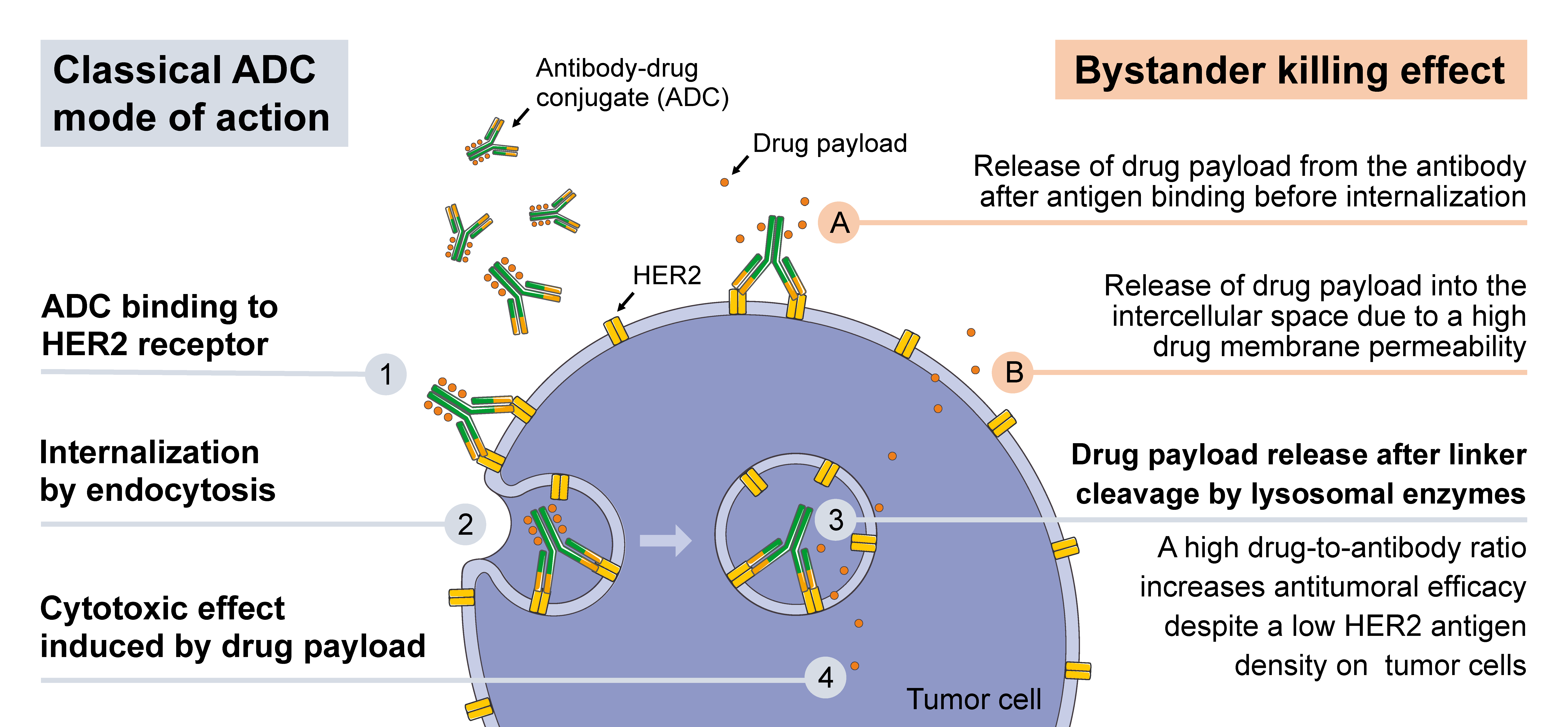
:1. Introduction
2. A166
2.1. ADC Constituents
2.2. Ongoing Trials without Published Results
3. ALT-P7 (HM2/MMAE)
3.1. ADC Constituents
3.2. Ongoing Trials without Published Results
4. ARX-788 (ARX788)
4.1. ADC Constituents
4.2. Preclinical Data
4.3. Ongoing Trials without Published Results
5. DHES0815A (Anti-HER2/PBD-MA)
5.1. ADC Constituents
5.2. Ongoing Trials without Published Results
6. DS-8201a (Trastuzumab Deruxtecan)
6.1. ADC Constituents
6.2. Preclinical Data
6.3. Clinical Data
6.4. Ongoing Trials without Published Results
7. MEDI4276
7.1. ADC Constituents
7.2. Preclinical Data
7.3. Clinical Data
8. RC48 (RC48-ACD, Hertuzumab-vc-MMAE)
8.1. ADC Constituents
8.2. Preclinical Data
8.3. Clinical Data
8.4. Ongoing Trials without Published Results
9. SYD985 ([vic-]Trastuzumab Duocarmazine)
9.1. ADC Constituents
9.2. Preclinical Data
9.3. Clinical Data
9.4. Ongoing Trials without Published Results
10. XMT-1522 (TAK-522)
10.1. ADC Constituents
10.2. Preclinical Data
10.3. Clinical Data
11. Discussion
Author Contributions
Funding
Conflicts of Interest
Abbreviations
| ABC | advanced breast cancer |
| ADC | antibody-drug-conjugates |
| AE | adverse event |
| AF-HPA | auristatin f-hydroxypropylamide |
| ALT | alanine transaminase |
| AST | aspartate transaminase |
| CBR | clinical benefit rate |
| CI | confidence interval |
| CR | complete response |
| DAR | drug antibody ratio |
| DCR | disease control rate |
| DFS | disease-free survival |
| DOR | duration of response |
| EGF | epidermal growth factor |
| EGFR | epidermal growth factor receptor |
| HER2 | human epidermal growth factor receptor 2 |
| HER3 | human epidermal growth factor receptor 3 |
| HER4 | human epidermal growth factor receptor 4 |
| ISH | in situ hybridization |
| IHC | immunohistochemistry |
| ILD | interstitial lung disease |
| MBC | metastatic breast cancer |
| MDT | maximum tolerated dose |
| MMAE | monomethyl auristatin E |
| MMAF | monomethyl auristatin F |
| ORR | overall response rate |
| PFS | progression-free survival |
| PBD-MA | pyrrolo[2,1-c][1,4]benzodiazepine monoamide |
| PR | partial response |
| Q2W | every 2 weeks |
| Q3W | every 3 weeks |
| ROR2 | receptor tyrosine kinase-like orphan receptor 2 |
| RP2D | recommended phase 2 dose |
| SAE | serious adverse event |
| SD | stable disease |
References
- Moasser, M.M. The oncogene HER2: Its signaling and transforming functions and its role in human cancer pathogenesis. Oncogene 2007, 26, 6469–6487. [Google Scholar] [CrossRef] [PubMed]
- Iqbal, N.; Iqbal, N. Human epidermal growth factor receptor 2 (HER2) in cancers: Overexpression and therapeutic implications. Mol. Biol. Int. 2014, 2014, 852748. [Google Scholar] [CrossRef] [PubMed]
- Uhlen, M.; Fagerberg, L.; Hallstrom, B.M.; Lindskog, C.; Oksvold, P.; Mardinoglu, A.; Sivertsson, A.; Kampf, C.; Sjostedt, E.; Asplund, A.; et al. Proteomics. Tissue-based map of the human proteome. Science 2015, 347, 1260419. [Google Scholar] [CrossRef] [PubMed]
- Press, M.F.; Cordon-Cardo, C.; Slamon, D.J. Expression of the HER-2/neu proto-oncogene in normal human adult and fetal tissues. Oncogene 1990, 5, 953–962. [Google Scholar] [PubMed]
- Yan, M.; Schwaederle, M.; Arguello, D.; Millis, S.Z.; Gatalica, Z.; Kurzrock, R. HER2 expression status in diverse cancers: Review of results from 37,992 patients. Cancer Metastasis Rev. 2015, 34, 157–164. [Google Scholar] [CrossRef] [PubMed]
- Wolff, A.C.; Hammond, M.E.; Hicks, D.G.; Dowsett, M.; McShane, L.M.; Allison, K.H.; Allred, D.C.; Bartlett, J.M.; Bilous, M.; Fitzgibbons, P.; et al. Recommendations for human epidermal growth factor receptor 2 testing in breast cancer: American society of clinical oncology/college of american pathologists clinical practice guideline update. J. Clin. Oncol. 2013, 31, 3997–4013. [Google Scholar] [CrossRef] [PubMed]
- Moja, L.; Tagliabue, L.; Balduzzi, S.; Parmelli, E.; Pistotti, V.; Guarneri, V.; D’Amico, R. Trastuzumab containing regimens for early breast cancer. Cochrane Database Syst. Rev. 2012, CD006243. [Google Scholar] [CrossRef] [PubMed]
- Ponde, N.; Brandao, M.; El-Hachem, G.; Werbrouck, E.; Piccart, M. Treatment of advanced HER2-positive breast cancer: 2018 and beyond. Cancer Treat. Rev. 2018, 67, 10–20. [Google Scholar] [CrossRef] [PubMed]
- Wolff, A.C.; Hammond, M.E.H.; Allison, K.H.; Harvey, B.E.; Mangu, P.B.; Bartlett, J.M.S.; Bilous, M.; Ellis, I.O.; Fitzgibbons, P.; Hanna, W.; et al. Human epidermal growth factor receptor 2 testing in breast cancer: American society of clinical oncology/college of american pathologists clinical practice guideline focused update. J. Clin. Oncol. 2018, 36, 2105–2122. [Google Scholar] [CrossRef] [PubMed]
- Schalper, K.A.; Kumar, S.; Hui, P.; Rimm, D.L.; Gershkovich, P. A retrospective population-based comparison of HER2 immunohistochemistry and fluorescence in situ hybridization in breast carcinomas: Impact of 2007 american society of clinical oncology/college of american pathologists criteria. Arch. Pathol. Lab. Med. 2014, 138, 213–219. [Google Scholar] [CrossRef] [PubMed]
- Paik, S.; Kim, C.; Jeong, J.; Geyer, C.E.; Romond, E.H.; Mejia-Mejia, O.; Mamounas, E.P.; Wickerham, D.; Costantino, J.P.; Wolmark, N. Benefit from adjuvant trastuzumab may not be confined to patients with IHC 3+ and/or FISH-positive tumors: Central testing results from NSABP B-31. J. Clin. Oncol. 2007, 25, 511. [Google Scholar]
- Perez, E.A.; Reinholz, M.M.; Hillman, D.W.; Tenner, K.S.; Schroeder, M.J.; Davidson, N.E.; Martino, S.; Sledge, G.W.; Harris, L.N.; Gralow, J.R.; et al. HER2 and chromosome 17 effect on patient outcome in the n9831 adjuvant trastuzumab trial. J. Clin. Oncol. 2010, 28, 4307–4315. [Google Scholar] [CrossRef] [PubMed]
- Fehrenbacher, L.; Cecchini, R.; Geyer, C.; Rastogi, P.; Costantino, J.; Atkins, J.; Polikoff, J.; Boileau, J.-F.; Provencher, L.; Stokoe, C.; et al. Abstract GS1-02: NSABP B-47 (NRG oncology): Phase III randomized trial comparing adjuvant chemotherapy with adriamycin (A) and cyclophosphamide (C) → weekly paclitaxel (WP), or docetaxel (T) and c with or without a year of trastuzumab (H) in women with node-positive or high-risk node-negative invasive breast cancer (IBC) expressing HER2 staining intensity of IHC 1+ or 2+ with negative FISH (HER2-low IBC). Cancer Res. 2018, 78, GS1-02. [Google Scholar]
- Domenyuk, V.; Gatalica, Z.; Santhanam, R.; Wei, X.; Stark, A.; Kennedy, P.; Toussaint, B.; Levenberg, S.; Wang, J.; Xiao, N.; et al. Poly-ligand profiling differentiates trastuzumab-treated breast cancer patients according to their outcomes. Nat. Commun. 2018, 9, 1219. [Google Scholar] [CrossRef] [PubMed]
- Beck, A.; Goetsch, L.; Dumontet, C.; Corvaia, N. Strategies and challenges for the next generation of antibody-drug conjugates. Nat. Rev. Drug Discov. 2017, 16, 315–337. [Google Scholar] [CrossRef] [PubMed]
- Staudacher, A.H.; Brown, M.P. Antibody drug conjugates and bystander killing: Is antigen-dependent internalisation required? Br. J. Cancer 2017, 117, 1736–1742. [Google Scholar] [CrossRef] [PubMed]
- Verma, S.; Miles, D.; Gianni, L.; Krop, I.E.; Welslau, M.; Baselga, J.; Pegram, M.; Oh, D.Y.; Dieras, V.; Guardino, E.; et al. Trastuzumab emtansine for HER2-positive advanced breast cancer. N. Engl. J. Med. 2012, 367, 1783–1791. [Google Scholar] [CrossRef] [PubMed]
- Krop, I.E.; Kim, S.B.; Gonzalez-Martin, A.; LoRusso, P.M.; Ferrero, J.M.; Smitt, M.; Yu, R.; Leung, A.C.; Wildiers, H.; TH3RESA Study Collaborators. Trastuzumab emtansine versus treatment of physician’s choice for pretreated her2-positive advanced breast cancer (TH3RESA): A randomised, open-label, phase 3 trial. Lancet Oncol. 2014, 15, 689–699. [Google Scholar] [CrossRef]
- Von Minckwitz, G.; Huang, C.S.; Mano, M.S.; Loibl, S.; Mamounas, E.P.; Untch, M.; Wolmark, N.; Rastogi, P.; Schneeweiss, A.; Redondo, A.; et al. Trastuzumab emtansine for residual invasive HER2-positive breast cancer. N. Engl. J. Med. 2019, 380, 617–628. [Google Scholar] [CrossRef] [PubMed]
- Lewis Phillips, G.D.; Li, G.; Dugger, D.L.; Crocker, L.M.; Parsons, K.L.; Mai, E.; Blattler, W.A.; Lambert, J.M.; Chari, R.V.; Lutz, R.J.; et al. Targeting HER2-positive breast cancer with trastuzumab-DM1, an antibody-cytotoxic drug conjugate. Cancer Res. 2008, 68, 9280–9290. [Google Scholar] [CrossRef] [PubMed]
- NCI. NCI Drug Dictionary a166. Available online: https://www.cancer.gov/publications/dictionaries/cancer-drug/def/795827 (accessed on 23 January 2019).
- NCI. NCI Drug Dictionary alt-p7. Available online: https://www.cancer.gov/publications/dictionaries/cancer-drug/def/793586 (accessed on 23 January 2019).
- Doronina, S.O.; Toki, B.E.; Torgov, M.Y.; Mendelsohn, B.A.; Cerveny, C.G.; Chace, D.F.; DeBlanc, R.L.; Gearing, R.P.; Bovee, T.D.; Siegall, C.B.; et al. Development of potent monoclonal antibody auristatin conjugates for cancer therapy. Nat. Biotechnol. 2003, 21, 778–784. [Google Scholar] [CrossRef] [PubMed]
- Humphreys, C.R.C.; Kirtely, J.; Hewit, A.; Biroc, S.; Knudsen, N.; Skidmore, L.; Wahl, A. Abstract 639: Site specific conjugation of ARX-788, an antibody drug conjugate (ADC) targeting HER2, generates a potent and stable targeted therapeutic for multiple cancers. Cancer Res. 2015, 75, 639. [Google Scholar] [CrossRef]
- NCI. Nci Drug Dictionary dhes0815a. Available online: https://www.cancer.gov/publications/dictionaries/cancer-drug/def/795265 (accessed on 31 January 2019).
- Ogitani, Y.; Aida, T.; Hagihara, K.; Yamaguchi, J.; Ishii, C.; Harada, N.; Soma, M.; Okamoto, H.; Oitate, M.; Arakawa, S.; et al. DS-8201a, a novel HER2-targeting ADC with a novel DNA topoisomerase I inhibitor, demonstrates a promising antitumor efficacy with differentiation from T-DM1. Clin. Cancer Res. 2016, 22, 5097–5108. [Google Scholar] [CrossRef] [PubMed]
- Pommier, Y. Topoisomerase I inhibitors: Camptothecins and beyond. Nat. Rev. Cancer 2006, 6, 789–802. [Google Scholar] [CrossRef] [PubMed]
- Kumazawa, E.; Jimbo, T.; Ochi, Y.; Tohgo, A. Potent and broad antitumor effects of DX-8951f, a water-soluble camptothecin derivative, against various human tumors xenografted in nude mice. Cancer Chemother. Pharmacol. 1998, 42, 210–220. [Google Scholar] [CrossRef] [PubMed]
- Ogitani, Y.; Hagihara, K.; Oitate, M.; Naito, H.; Agatsuma, T. Bystander killing effect of DS-8201a, a novel anti-human epidermal growth factor receptor 2 antibody-drug conjugate, in tumors with human epidermal growth factor receptor 2 heterogeneity. Cancer Sci. 2016, 107, 1039–1046. [Google Scholar] [CrossRef] [PubMed]
- Doi, T.; Iwata, H.; Tsurutani, J.; Takahashi, S.; Park, H.; Redfern, C.H.; Shitara, K.; Shimizu, C.; Taniguchi, H.; Iwasa, T.; et al. Single agent activity of DS-8201a, a her2-targeting antibody-drug conjugate, in heavily pretreated HER2 expressing solid tumors. J. Clin. Oncol. 2017, 35, 108. [Google Scholar] [CrossRef]
- Iwata, H.; Tamura, K.; Doi, T.; Tsurutani, J.; Modi, S.; Park, H.; Krop, I.E.; Sagara, Y.; Redfern, C.H.; Murthy, R.K.; et al. Trastuzumab deruxtecan (DS-8201a) in subjects with HER2-expressing solid tumors: Long-term results of a large phase 1 study with multiple expansion cohorts. J. Clin. Oncol. 2018, 36, 2501. [Google Scholar] [CrossRef]
- Modi, S.; Tsurutani, J.; Tamura, K.; Park, H.; Sagara, Y.; Murthy, R.; Iwata, H.; Krop, I.; Doi, T.; Redfern, C.; et al. Trastuzumab deruxtecan (DS-8201a) in subjects with HER2-low expressing breast cancer: Updated results of a large phase 1 study. Cancer Res. 2019, 79. [Google Scholar] [CrossRef]
- Li, J.Y.; Perry, S.R.; Muniz-Medina, V.; Wang, X.; Wetzel, L.K.; Rebelatto, M.C.; Hinrichs, M.J.; Bezabeh, B.Z.; Fleming, R.L.; Dimasi, N.; et al. A biparatopic HER2-targeting antibody-drug conjugate induces tumor regression in primary models refractory to or ineligible for HER2-targeted therapy. Cancer Cell 2016, 29, 117–129. [Google Scholar] [CrossRef] [PubMed]
- Oganesyan, V.; Peng, L.; Bee, J.S.; Li, J.; Perry, S.R.; Comer, F.; Xu, L.; Cook, K.; Senthil, K.; Clarke, L.; et al. Structural insights into the mechanism of action of a biparatopic anti-HER2 antibody. J. Biol. Chem. 2018, 293, 8439–8448. [Google Scholar] [CrossRef] [PubMed]
- Pegram, M.; Hamilton, E.; Tan, A.R.; Storniolo, A.M.; Elgeioushi, N.; Abdullah, S.; Marshall, S.; Patel, M. 47o—Phase 1 study of bispecific HER2 antibody-drug conjugate medi4276 in patients with advanced HER2-positive breast or gastric cancer. Ann. Oncol. 2018, 29, mdy048.005. [Google Scholar] [CrossRef]
- Yao, X.; Jiang, J.; Wang, X.; Huang, C.; Li, D.; Xie, K.; Xu, Q.; Li, H.; Li, Z.; Lou, L.; et al. A novel humanized anti-HER2 antibody conjugated with mmae exerts potent anti-tumor activity. Breast Cancer Res. Treat. 2015, 153, 123–133. [Google Scholar] [CrossRef] [PubMed]
- Wang, J.; Xu, B.; Wang, W.; Fang, J. An open-label, dose-escalation phase i study to evaluate RC48-ADC, a novel antibody-drug conjugate, in patients with HER2-positive metastatic breast cancer. J. Clin. Oncol. 2018, 36, 1030. [Google Scholar] [CrossRef]
- Gong, J.; Shen, L.; Wang, W.; Fang, J. Safety, pharmacokinetics and efficacy of RC48-ADC in a phase i study in patients with her2-overexpression advanced solid cancer. J. Clin. Oncol. 2018, 36, e16059. [Google Scholar] [CrossRef]
- Xu, B.; Wang, J.; Zhang, Q.; Liu, Y.; Feng, J.F.; Wang, W.; Fang, J. An open-label, multicenter, phase Ib study to evaluate RC48-ADC in patients with HER2-positive metastatic breast cancer. J. Clin. Oncol. 2018, 36, 1028. [Google Scholar] [CrossRef]
- Elgersma, R.C.; Coumans, R.G.; Huijbregts, T.; Menge, W.M.; Joosten, J.A.; Spijker, H.J.; de Groot, F.M.; van der Lee, M.M.; Ubink, R.; van den Dobbelsteen, D.J.; et al. Design, synthesis, and evaluation of linker-duocarmycin payloads: Toward selection of HER2-targeting antibody-drug conjugate SYD985. Mol. Pharm. 2015, 12, 1813–1835. [Google Scholar] [CrossRef] [PubMed]
- Dokter, W.; Ubink, R.; van der Lee, M.; van der Vleuten, M.; van Achterberg, T.; Jacobs, D.; Loosveld, E.; van den Dobbelsteen, D.; Egging, D.; Mattaar, E.; et al. Preclinical profile of the HER2-targeting ADC SYD983/SYD985: Introduction of a new duocarmycin-based linker-drug platform. Mol. Cancer Ther. 2014, 13, 2618–2629. [Google Scholar] [CrossRef] [PubMed]
- Van der Lee, M.M.; Groothuis, P.G.; Ubink, R.; van der Vleuten, M.A.; van Achterberg, T.A.; Loosveld, E.M.; Damming, D.; Jacobs, D.C.; Rouwette, M.; Egging, D.F.; et al. The preclinical profile of the duocarmycin-based HER2-targeting ADC SYD985 predicts for clinical benefit in low HER2-expressing breast cancers. Mol. Cancer Ther. 2015, 14, 692–703. [Google Scholar] [CrossRef] [PubMed]
- Aftimos, P.; van Herpen, C.; Mommers, E.; Koper, N.; Goedings, P.; Oesterholt, M.; Awada, A.; Desar, I.; Lim, J.; Dean, E.; et al. Abstract p6-12-02: SYD985, a novel anti-HER2 adc, shows promising activity in patients with HER2-positive and HER2-negative metastatic breast cancer. Cancer Res. 2017, 77. [Google Scholar] [CrossRef]
- Saura, C.; Thistlethwaite, F.; Banerji, U.; Lord, S.; Moreno, V.; MacPherson, I.; Boni, V.; Rolfo, C.D.; Vries, E.G.E.d.; Herpen, C.M.L.-V.; et al. A phase i expansion cohorts study of SYD985 in heavily pretreated patients with HER2-positive or HER2-low metastatic breast cancer. J. Clin. Oncol. 2018, 36, 1014. [Google Scholar] [CrossRef]
- Bergstrom, D.; Bodyak, N.; Park, P.; Yurkovetskiy, A.; DeVit, M.; Yin, M.; Poling, L.; Thomas, J.; Gumerov, D.; Xiao, D.; et al. Abstract p4-14-28: XMT-1522 induces tumor regressions in pre-clinical models representing HER2-positive and HER2 low-expressing breast cancer. Cancer Res. 2016, 76. [Google Scholar] [CrossRef]
- Yurkovetskiy, A.; Gumerov, D.; Ter-Ovanesyan, E.; Conlon, P.; Devit, M.; Bu, C.; Bodyak, N.; Lowinger, T.; Bergstrom, D. Abstract 48: Non-clinical pharmacokinetics of XMT-1522, a HER2 targeting auristatin-based antibody drug conjugate. Cancer Res. 2017, 77, 48. [Google Scholar] [CrossRef]
- Bergstrom, D.A.; Bodyak, N.; Yurkovetskiy, A.; Park, P.U.; DeVit, M.; Yin, M.; Poling, L.; Thomas, J.D.; Gumerov, D.; Xiao, D.; et al. Abstract lb-231: A novel, highly potent HER2-targeted antibody-drug conjugate (ADC) for the treatment of low HER2-expressing tumors and combination with trastuzumab-based regimens in HER2-driven tumors. Cancer Res. 2015, 75. [Google Scholar] [CrossRef]
- Bodyak, N.; Yurkovetskiy, A.; Park, P.U.; Gumerov, D.R.; DeVit, M.; Yin, M.; Thomas, J.D.; Qin, L.; Lowinger, T.B.; Bergstrom, D.A. Abstract 641: Trastuzumab-dolaflexin, a highly potent fleximer-based antibody-drug conjugate, demonstrates a favorable therapeutic index in exploratory toxicology studies in multiple species. Cancer Res. 2015, 75, 641. [Google Scholar] [CrossRef]
- Traore, T.; Khattar, M. Abstract lb-294: Synergy of an anti-HER2 ADC TAK-522 (XMT-1522) in combination with anti-PD1 monoclonal antibody (MAB) in a syngeneic breast cancer model expressing human HER2. Cancer Res. 2018, 78. [Google Scholar] [CrossRef]
- Hamilton, E.P.; Barve, M.A.; Bardia, A.; Beeram, M.; Bendell, J.C.; Mosher, R.; Hailman, E.; Bergstrom, D.A.; Burris, H.A.; Soliman, H.H. Phase 1 dose escalation of xmt-1522, a novel HER2-targeting antibody-drug conjugate (ADC), in patients (pts) with HER2-expressing breast, lung and gastric tumors. J. Clin. Oncol. 2018, 36, 2546. [Google Scholar] [CrossRef]
- Diamantis, N.; Banerji, U. Antibody-drug conjugates—An emerging class of cancer treatment. Br. J. Cancer 2016, 114, 362–367. [Google Scholar] [CrossRef] [PubMed]


| Drug Name | Cytotoxic Payload | Reported Efficacy in HER2-Low | Phases (Number of Trials, NCT Identifier) | Company |
|---|---|---|---|---|
| A166 | NA | no | Phase 1/2: 1 (NCT03602079) | Klus Pharma, Inc. |
| ALT-P7 (HM2-MMAE) | monomethyl auristatin E | no | 2: 1 (NCT03281824) | Alteogen, Inc. |
| ARX788 | monomethyl auristatin F | no | 1: 2 (NCT02512237, NCT03255070) | Ambrx, Inc. |
| DHES0815A (anti-HER2/PBD-MA) | PBD-MA | no | 1: 1 (NCT03451162) | Genentech, Inc. |
| DS-8201a (Trastuzumab deruxtecan) | DXd | yes | 1: 3 (NCT03523572, NCT03368196, NCT03366428) 2: 1 (NCT03248492) 3: 3 (NCT03734029, NCT03523585, NCT03529110) | Daiichi Sankyo, Inc. |
| MEDI4276 | AZ13599185 | yes | - | MedImmune, LLC |
| RC48 | monomethyl auristatin E | no | 1b/2: 1 (NCT03052634) 2: 1 (NCT03500380) | RemeGen |
| SYD985 ([vic-]trastuzumab duocarmazine) | seco-DUBA | yes | 3: 1 (NCT03262935) | Synthon Biopharmaceuticals BV |
| T-DM1 (Trastuzumab emtansine) | DM1 | no | 1: 3 (NCT02073916, NCT02038010, NCT03364348) 2: 3 (NCT03587740, NCT02073487, NCT02414646) | Roche |
| XMT-1522 (TAK-522) | AF-HPA | yes | 1: 1 (NCT02952729) | Mersana Therapeutics |
| Drug | Target | Running Trials (Number of Trials, NCT Identifier) | Company |
|---|---|---|---|
| U3-1402 | HER3 | Phase 1/2: 1 (NCT02980341) | Daiichi Sankyo, Inc. |
| SGN-LIV1A | LIV1 | Phase 1: 1 (NCT01969643) | Seattle Genetics, Inc. |
| CAB-ROR2-ADC | ROR2 | Phase 1/2: 1 (NCT03504488) | BioAtla, LLC |
| Sacituzumab govitecan (IMMU-132) | Trop-2 | Phase 1/2: 1 (NCT01631552) Phase 2: 1 (NCT02161679) | Immunomedics, Inc. |
© 2019 by the authors. Licensee MDPI, Basel, Switzerland. This article is an open access article distributed under the terms and conditions of the Creative Commons Attribution (CC BY) license (http://creativecommons.org/licenses/by/4.0/).
Share and Cite
Rinnerthaler, G.; Gampenrieder, S.P.; Greil, R. HER2 Directed Antibody-Drug-Conjugates beyond T-DM1 in Breast Cancer. Int. J. Mol. Sci. 2019, 20, 1115. https://doi.org/10.3390/ijms20051115
Rinnerthaler G, Gampenrieder SP, Greil R. HER2 Directed Antibody-Drug-Conjugates beyond T-DM1 in Breast Cancer. International Journal of Molecular Sciences. 2019; 20(5):1115. https://doi.org/10.3390/ijms20051115
Chicago/Turabian StyleRinnerthaler, Gabriel, Simon Peter Gampenrieder, and Richard Greil. 2019. "HER2 Directed Antibody-Drug-Conjugates beyond T-DM1 in Breast Cancer" International Journal of Molecular Sciences 20, no. 5: 1115. https://doi.org/10.3390/ijms20051115
APA StyleRinnerthaler, G., Gampenrieder, S. P., & Greil, R. (2019). HER2 Directed Antibody-Drug-Conjugates beyond T-DM1 in Breast Cancer. International Journal of Molecular Sciences, 20(5), 1115. https://doi.org/10.3390/ijms20051115