Article

Inhibitory Effects of Menadione on Helicobacter pylori Growth and Helicobacter pylori-Induced Inflammation via NF-κB Inhibition

1
Department of Biomedical Laboratory Science, College of Health Sciences, Yonsei University, Wonju 26493, Korea
2
Forensic DNA Division, National Forensic Service, Wonju 26460, Korea
3
Department of Clinical Laboratory Science, College of Medical Sciences, Daegu Haany University, Gyeongsan 38610, Korea
4
Department of Biomedical Laboratory Science, Daekyeung University, Gyeongsan 38547, Korea
5
Department of Clinical Laboratory Science, Semyung University, Jecheon 27136, Korea
6
Natural Products Research Center, Korea Institute of Science and Technology (KIST) Gangneung 25451, Korea
7
National Institute of Crop Science (NICS), Rural Development Administration (RDA), Wanju-Gun 55365, Korea
*
Authors to whom correspondence should be addressed.
These authors contributed equally to this work.
Int. J. Mol. Sci. 2019, 20(5), 1169; https://doi.org/10.3390/ijms20051169
Submission received: 28 January 2019 / Revised: 1 March 2019 / Accepted: 4 March 2019 / Published: 7 March 2019

Abstract

:
H. pylori is classified as a group I carcinogen by WHO because of its involvement in gastric cancer development. Several reports have suggested anti-bacterial effects of menadione, although the effect of menadione on major virulence factors of H. pylori and H. pylori-induced inflammation is yet to be elucidated. In this study, therefore, we demonstrated that menadione has anti-H. pylori and anti-inflammatory effects. Menadione inhibited growth of H. pylori reference strains and clinical isolates. Menadione reduced expression of vacA in H. pylori, and translocation of VacA protein into AGS (gastric adenocarcinoma cell) was also decreased by menadione treatment. This result was concordant with decreased apoptosis in AGS cells infected with H. pylori. Moreover, cytotoxin-associated protein A (CagA) translocation into H. pylori-infected AGS cells was also decreased by menadione. Menadione inhibited expression of several type IV secretion system (T4SS) components, including virB2, virB7, virB8, and virB10, that are responsible for translocation of CagA into host cells. In particular, menadione inhibited nuclear factor kappa-light-chain-enhancer of activated B cell (NF-κB) activation and thereby reduced expression of the proinflammatory cytokines such as IL-1β, IL-6, IL-8, and TNF-α in AGS as well as in THP-1 (monocytic leukemia cell) cell lines. Collectively, these results suggest the anti-bacterial and anti-inflammatory effects of menadione against H. pylori.

1. Introduction

Helicobacter pylori (H. pylori) is a Gram-negative bacterium possessing a characteristic helical appearance. H. pylori primarily colonizes in the human stomach and has been reported to infect approximately half of the world population [1]. Infection of H. pylori on gastric mucosa is associated with various gastric diseases including inflammation, chronic gastritis, peptic ulcers, and gastric adenocarcinoma [2]. The World Health Organization classified H. pylori as a class I carcinogen because of its involvement in gastric cancer development [3]. It is estimated that more than half of the adult population is infected with H. pylori worldwide, and its infection is responsible for 75% of all gastric cancer cases [4]. Therefore, a concerted effort for eradication of H. pylori infection is necessary for health promotion worldwide.
The most studied virulence factors of H. pylori are cytotoxin-associated protein A (CagA) and vacuolating cytotoxin A (VacA). CagA translocates to the host cells by the type IV secretion system (T4SS) [5]. The T4SS of H. pylori consists of up to 32 proteins, but many of their functions are yet unknown [6]. However, the functions of 11 VirB proteins (VirB1–11) and a VirD4 protein have been studied based on their homology to the agrobacterial T4SS proteins [6,7,8,9]. The VirB and VirD4 proteins assemble to form three subparts consisting of a cytoplasmic/inner membrane complex (VirB4, VirB6, VirB8, VirB11, and VirD4), a double membrane-spanning channel (VirB7, VirB9, and VirB10), and an external pilus (VirB2 and VirB5), and these three subparts are interlinked [6,7]. Kwok et al. demonstrated that α5β1 integrin is a host cell receptor that directly binds to VirB5 (CagL) proteins of H. pylori [6]. Once CagA is injected, host cell Src kinases phosphorylate the EPIYA motif of CagA proteins and subsequently deregulate intracellular signaling transduction pathways, disrupt epithelial cell junctions, and induce inflammation [10,11,12,13]. VacA has been known to induce cytoplasmic vacuole formation [14]. VacA protein secretion is associated with the type Va system [14,15,16]. Translocation across the inner-membrane is mediated by Sec-related proteins [15,16]. The signal peptide region of the VacA protein is recognized by SecYEG for the translocation through the inner-membrane [15,16]. SecA is an especially important regulatory protein because it is an ATPase that provides energy necessary for translocation of the proteins by Sec-related proteins [15,16,17]. VacA, which translocates to the host cells, interacts with host cell mitochondria, resulting in apoptosis via activation of the intrinsic caspase cascade [18,19,20,21,22].
One of the mechanisms by which H. pylori infection progresses to gastric carcinogenesis is the persistent presence of the pathogen, which leads to the development of chronic inflammation accompanied by infiltration of neutrophils and lymphocytes as well as the production of proinflammatory cytokines [23]. The gastric mucosal levels of the proinflammatory cytokines are increased in H. pylori-infected subjects, and these include IL-1β, IL-6, IL-8, and TNF-α [24,25]. Especially, H. pylori-mediated IL-8 secretion in gastric epithelial cells requires activation of the nuclear factor kappa-light-chain-enhancer of activated B cells (NF-κB), while H. pylori strains that fail to induce IL-8 secretion do not activate NF-κB [26,27]. In the absence of an activating stimuli, NF-κB remains inactive in the cytoplasm bound to a family of inhibitory proteins known as inhibitors of NF-κB (IκBs). Activated NF-κB forms a homo- or heterodimer and translocates to the nucleus to function as a transcription factor [28]. In particular, NF-κB is clearly one of the most important regulators for expression of proinflammatory cytokines [29]. Activation of NF-κB by H. pylori induces nuclear translocation, which causes increased expression of NF-κB responsive genes including TNF-α, IL-1β, IL-6, and IL-8 [27]. NF-κB activation is also known to regulate cellular growth responses including apoptosis and is required for the induction of inflammatory and tissue-repair genes [23,27,30].
Menadione (2-methyl-1,4-naphthoquinone) is a synthetic form of vitamin K. It is also called vitamin K3. Menadione has a higher anti-hemorrhagic activity than the naturally occurring vitamin K (VitK1 and VitK2) [31]. The generally known roles of vitamin K are the maintenance of blood clotting and bone formation [31,32]. There are several reports demonstrating the anti-bacterial effect of menadione [33,34,35,36]. Andrade et al. reported the antibiotic-modifying activity of menadione in multi-resistant strains of Staphylococcus aureus, Pseudomonas aeruginosa, and Escherichia coli. They suggested that menadione potentiated aminoglycosides against multi-resistant bacteria since it lowered the minimal inhibitory concentration (MIC) of the antibiotics tested in their experiments [37]. In particular, Park et al. described the inhibitory effect of menadione against H. pylori in the screening of various naphthoquinones by a disk diffusion assay [34].
Antibiotic resistance of H. pylori is continuously increasing, and the development of a new therapeutic agent to support H. pylori treatment is necessary. Menadione was reported to have anti-bacterial activity. Therefore, an inhibitory effect of menadione on H. pylori growth and the effects of menadione on CagA and VacA, major virulence factors of H. pylori, were investigated in this study. Furthermore, menadione inhibited NF-κB activation and possessed anti-inflammatory activity according to the previous reports [38]. Thus, the effects of menadione on the expression of the inflammatory cytokines and the NF-κB-mediated signaling pathway during H. pylori-induced inflammatory response in vitro was investigated.

2. Results

2.1. The Inhibitory Effect of Menadione on the Growth of H. pylori

An agar dilution test was performed to determine the MIC of menadione against H. pylori. Mueller–Hinton agar containing various concentrations of menadione was prepared and H. pylori reference strains (ATCC 49503, ATCC 26695, SS1, and HP51) were grown on agar plates for 72 h. According to the agar dilution test, the MIC of menadione against H. pylori was 8 μM (Figure 1). Clinical isolates of H. pylori were collected from gastric biopsies, and the MIC of menadione was determined to confirm whether menadione can inhibit growth of H. pylori clinical isolates as well as the reference strains. Among the 38 clinical isolates, the MIC of 57.9% (22/38) was 8 μM, 21.1% (8/38) was 4 μM, and 10.5% (4/38) was 2 μM (Table 1). These results showed that menadione has an anti-bacterial effect on the clinical isolates of H. pylori as well as the reference strains.

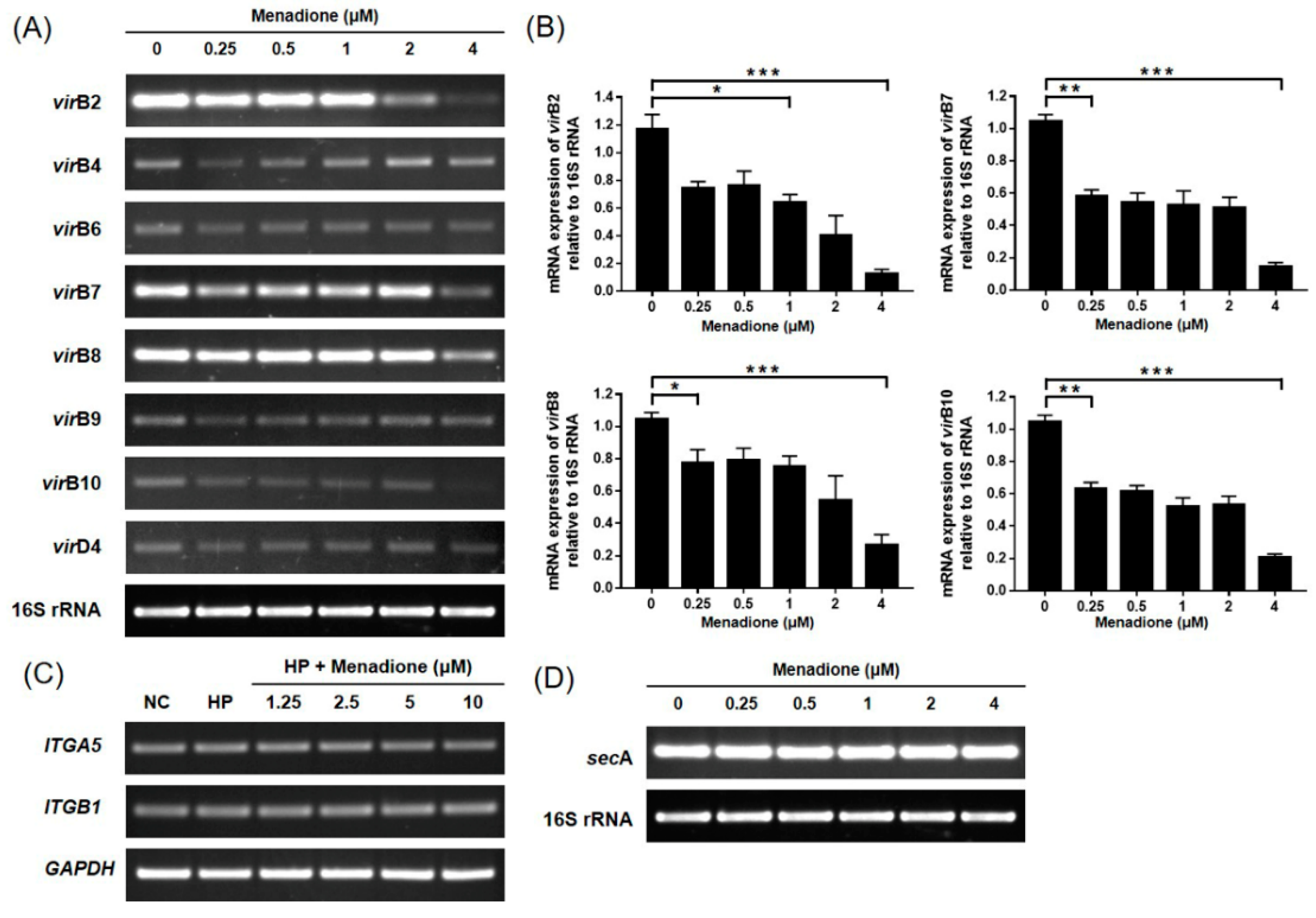
2.2. Menadione Reduced CagA and VacA Translocation to AGS Cells and Recovered Morphological Changes Caused by H. pylori Infection

CagA and VacA proteins are the most well-known cytotoxic proteins secreted by H. pylori. A notable feature appearing after CagA translocation to host cells is the rearrangement of the host cell actin cytoskeleton leading to the change of cell morphology, the so-called hummingbird phenotype [39]. The VacA protein internalizes into the host cell and leads to vacuolation, which is characterized by the accumulation of large vesicles [14]. In the experiment, it was also found that the morphological changes in the H. pylori-infected AGS cells were alleviated by menadione treatment in a dose-dependent manner (Figure 2A). It was presumed that the translocation of the CagA and VacA proteins to AGS cells may have decreased by menadione treatment. Thus, the H. pylori- and menadione-treated AGS cells were harvested and subjected to Western blotting to investigate protein levels of the CagA and VacA in the AGS cells. The CagA and VacA proteins were translocated to the infected AGS cells, and translocation of both proteins was decreased by menadione treatment as predicted (Figure 2B). In particular, the CagA protein level dramatically decreased even by a low dose of menadione treatment (Figure 2B,C). These results indicated that menadione reduced translocation of CagA and VacA proteins to AGS cells and inhibited H. pylori-induced morphological changes (hummingbird phenotype and vacuolations) of AGS cells. To investigate whether menadione directly reduced the expression of CagA and VacA in the bacteria, the mRNA levels of CagA and VacA in H. pylori treated with menadione were assessed by RT-PCR. Menadione reduced the expression of VacA, but it did not reduce the expression of CagA in H. pylori (Figure 3).
CagA and VacA proteins are secreted by type IV and type V secretion systems, respectively. Therefore, whether menadione affected the secretion system of H. pylori was investigated. Specific primers were designed for targeting components of the secretion systems, and the expressions were examined using RT-PCR. In the result, mRNA expressions of virB2, virB7, virB8, and virB10 were decreased by menadione treatment (Figure 4A,B). Moreover, RT-PCR was performed targeting for integrin α5β1, which acts as a receptor for the T4SS on the host cell surface, but expression of this integrin was not changed (Figure 4C). RT-PCR was performed to investigate whether menadione has an effect on the mRNA expression of secA, which is a regulator of the T5SS. However, no change was observed in this experiment regarding the expression of secA (Figure 4D). Collectively, these data indicate that menadione reduced the translocation of VacA to the host cells by the downregulation of vacA mRNA expression in H. pylori, and reduced the translocation of CagA by the downregulation of T4SS components necessary for CagA secretion. By inhibiting the translocation of CagA and VacA proteins, menadione alleviated morphological changes of AGS cells induced by H. pylori infection.

2.3. Menadione Alleviated H. pylori-Induced Apoptosis and Death of AGS Cells

H. pylori infection induces apoptosis of gastric epithelial cells [19,20,21,22]. In this study, it was investigated whether menadione can inhibit apoptosis induced by H. pylori in AGS cells. AGS cells were infected with H. pylori and treated with menadione for 48 h. After incubation, the cells were observed by an inverted microscope. It was found that cell confluency was decreased by H. pylori infection, but the decrease of cell confluency was inhibited by menadione treatment in a dose-dependent manner (Figure 2A). Additionally, cell viability was measured by a WST assay, and it was found that H. pylori infection (200 MOI) reduced cell viability of AGS cells (46.57%) but that the cell death was partially inhibited by menadione treatment (5 μM) (Figure 5A). The dose of menadione used in this experiment was enough to inhibit H. pylori growth without inhibiting the growth of AGS cells. This result showed that menadione rescued AGS cells from H. pylori-induced cell death. Since menadione reduced the translocation of VacA proteins in AGS cells in this study, it was hypothesized that menadione may inhibit H. pylori-induced apoptosis in gastric epithelial cells. In the Western blotting result, poly (ADP-ribose) polymerase (PARP) was cleaved by H. pylori infection, but it was inhibited by menadione treatment (Figure 5B,C). Menadione alone had no effect on the level of PARP in the range of concentration (Figure S1). Based on these results, it could be inferred that menadione inhibited H. pylori-induced apoptotic cell death in AGS cells by inhibiting VacA translocation and H. pylori growth.

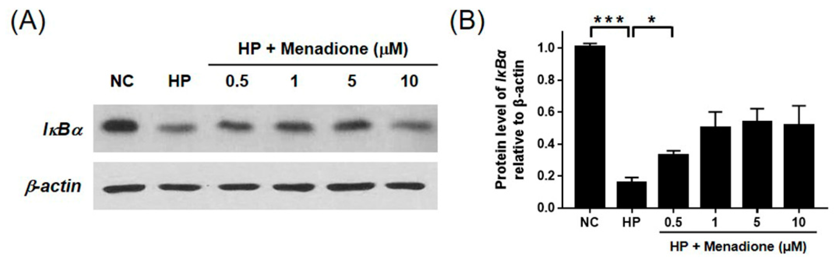
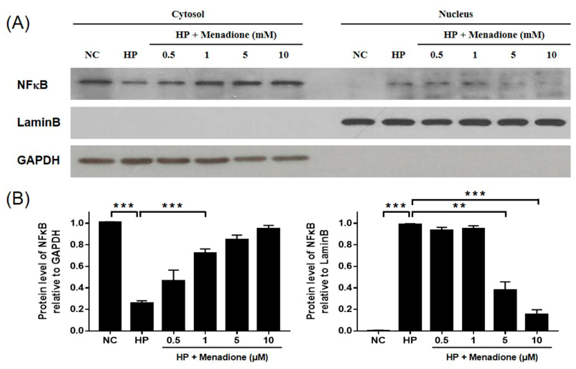
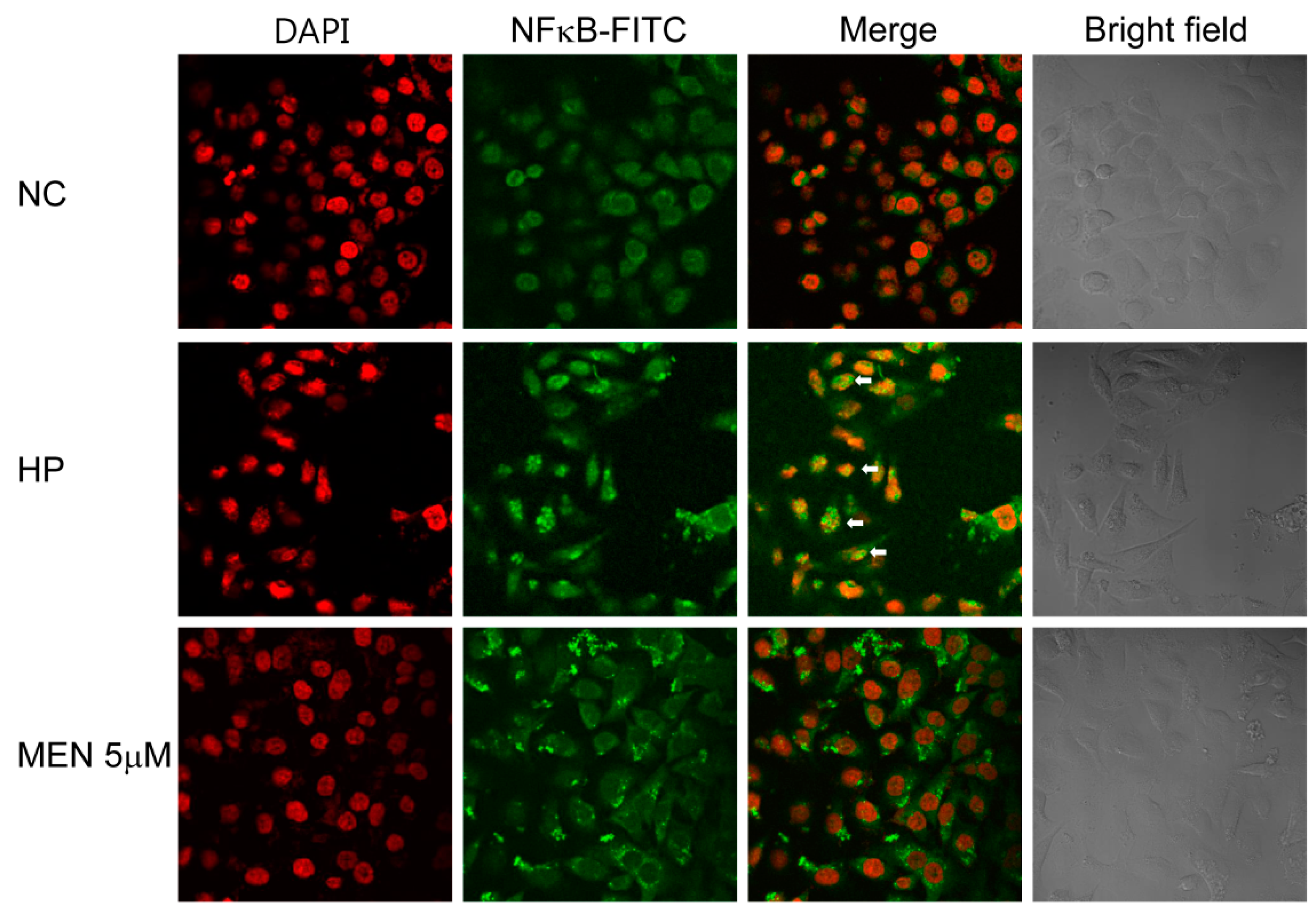
2.4. Menadione Reduces Expression of Inflammatory Cytokines Induced by H. pylori-Induced Infection via Inhibition of NF-κB Activation

It has been reported that H. pylori activates NF-κB, which is one of the most important regulators for expression of proinflammatory cytokines [27,29]. To investigate whether menadione inhibits H. pyori-induced activation of NF-κB and inflammatory responses, AGS cells infected with H. pylori were treated with menadione, and Western blotting was then conducted to detect IκBα. H. pylori infection reduced the IκBα protein level, and menadione treatment partially recovered the reduced IκBα protein level (Figure 6). This result indirectly indicates that menadione reduced NF-κB activation by H. pylori. Activated NF-κB translocates to the nucleus, so nuclear translocation of NF-κB should be observed to precisely investigate the activation of NF-κB. Protein levels of NF-κB in cytosol and the nucleus were measured to confirm the inhibitory effect of menadione on NF-κB activation. AGS cells were infected with H. pylori and treated with menadione, and the cell lysates were then separated into cytosol fraction and nuclear fraction. Western blotting results for NF-κB in each fraction showed that NF-κB in the nucleus was increased by H. pylori infection, but it was reduced by menadione treatment (Figure 7). AGS cells without H. pylori infections were treated with menadione, and there was no variation in IκBα and NF-κB (Figure S1). Furthermore, confocal microscopy shows that FITC-labeled NF-κB was increased in the nucleus after H. pylori infection but it was reduced by menadione treatment (Figure 8). These results have a confirmed inhibitory effect of menadione on NF-κB activation and nuclear translocation by H. pylori.
H. pylori infection increases the levels of the proinflammatory cytokines such as IL-1β, IL-6, IL-8, and TNF-α in gastric mucosa [24,25]. Especially, IL-8 secretion from gastric epithelial cells by H. pylori infection is closely associated with NF-κB activation [26,27]. Therefore, expression of the proinflammatory cytokines in the AGS cells was investigated after treatment with H. pylori and menadione. The RT-PCR results showed that IL-1β, IL-8, and TNF-α mRNA levels were increased by H. pylori infection, and menadione treatment reduced their expressions in AGS gastric epithelial cells (Figure 9A,C). Moreover, the anti-inflammatory effect of menadione was also investigated in a macrophage cell line THP-1. THP-1 monocytic leukemia cells were differentiated into macrophages by PMA treatment. The cells were then infected with H. pylori and treated with menadione. The RT-PCR results also showed a reduction in proinflammatory cytokine expressions by menadione treatment (Figure 9B,C). Collectively, these results indicate that menadione reduces the expression of proinflammatory cytokines that are induced by H. pylori infection in gastric epithelial cells and macrophages via inhibition of NF-κB activation and nuclear translocation.

3. Discussion

H. pylori infection is responsible for 75% of all the gastric cancer cases, and more than half of the adult population is infected with H. pylori worldwide [4]. Numerous reports have suggested the prevalence of clarithromycin resistance and the limitation of current first-line therapy [40,41,42]. Therefore, development of a new therapeutic or supportive agent to help the eradication of H. pylori is necessary.
In this study, the inhibitory effect of menadione against H. pylori growth and H. pylori-induced inflammation is elucidated. The MIC of menadione was demonstrated against H. pylori reference strains and clinical isolates. The MIC of menadione was 8 μM (1.38 μg/mL), which was similar to the MIC of the antibiotics clinically administered to treat H. pylori infection. In our previous report, we isolated 165 H. pylori clinical strains and evaluated the MIC of five antibiotics commonly used for the eradication of H. pylori [43]. The MICs of the clarithromycin, amoxicillin, tetracycline, metronidazole, and levofloxacin against the susceptible H. pylori strains were 0.008–0.125, 0.008–0.5, 0.031–2, 0.063–4, and 0.008–1 μg/mL [43].
Menadione inhibited the H. pylori growth via the downregulation of replication and transcription machinery of H. pylori. Menadione also downregulated virulence factors such as urease and VacA proteins, which are necessary for the successful colonization and pathogenesis of H. pylori. By using in vitro infection model, it was found that menadione reduced H. pylori-induced apoptosis and inflammation. In particular, menadione inhibited NF-κB activation, thereby reducing expression of proinflammatory cytokines.
The MIC of menadione against H. pylori was 8 μM (1.38 μg/mL) in the agar dilution test (Figure 1). Various phytochemicals and plant extracts, including panaxytriol [44], anacardic acids [45], six quinolone alkaloids including evocarpine [46], cabreuvin [47], and catechins [48], have been reported to inhibit H. pylori growth. The MIC of these molecules against H. pylori ranged from 10 to 200 μg/mL. Based on this result, the anti-H. pylori activity of menadione was more effective than those of other phytochemicals previously reported. Moreover, the anti-H. pylori activity of menadione was also demonstrated in the H. pylori clinical isolates (Table 1).
In the current study, menadione treatment reduced the expression of T4SS components as well as the translocation of CagA to AGS cells. The mRNA expressions of virB2, virB7, virB8, and virB10 in AGS cells were decreased by menadione treatment. Although cagA expression was not changed by menadione, decreased expression of the T4SS components can explain how menadione inhibited translocation of CagA protein into AGS cells. The periplasmic core of the T4SS consists of VirB8, VirB7, VirB9, and VirB10, and these proteins form a channel necessary for passenger proteins to pass through the periplasmic space. T4SS pili are generally composed of VirB2 and VirB5, and VirB2 is the major pilin subunit [7].
Inhibition of T4SS expression and CagA translocation was followed by the inhibition of NF-κB activation. Menadione treatment recovered the reduced IκBα protein level and inhibited the nuclear translocation of NF-κB in AGS cells infected with H. pylori, and this was also shown in the Western blotting and confocal microscopy results in this study. NF-κB exists in the cytoplasm in an inactivated form bound to a family of inhibitory proteins known as inhibitors of NF-κB (IκBs) [28]. Phosphorylation of IκB by IκB kinase (IKK) results in the ubiquitin-mediated degradation of IκB followed by activation and translocation of NF-κB to nucleus [28,29]. Therefore, these results collectively indicated the inhibitory effect of menadione on the NF-κB activation by H. pylori.
Chronic gastritis induced by H. pylori is characterized by the cagPAI-dependent expression of proinflammatory cytokines, which is largely mediated by the transcription factor NF-κB [49]. The transcription factor NF-κB regulates various genes that control the initiation of mucosal inflammatory responses induced by bacterial infection in human intestinal epithelial cells [50,51]. Activation of NF-κB by H. pylori induces nuclear translocation, which induces expression of NF-κB responsive genes including TNF-a, IL-1b, IL-6, and IL-8 [27]. Several reports have suggested the importance of CagA protein in the course of H. pylori-induced inflammation in infected gastric epithelial cells. Brandt et al. showed that CagA predominantly triggered NF-κB-mediated IL-8 production [52]. The Ras-Raf-Mek-Erk signaling cascade was involved in the CagA-induced NF-κB activation according to their study [52]. Lamb et al. also suggested the involvement of CagA in the activation of NF-κB. CagA contributed to the ubiquitination of TAK1 by TRAF6 and subsequently activated NF-κB [49]. These reports indicate that CagA acts as a multifunctional protein capable of triggering actin-cytoskeletal rearrangements, cell scattering by E-cadherin cleavage, and NF-κB activation and the production of IL-8 [49,52,53]. Furthermore, Viala et al. showed that H. pylori activates NF-κB via translocation of peptidoglycan by the T4SS in HEK293 cells. The translocation of peptidoglycan activated Nod1 and subsequently activated NF-κB. HEK293 cells do not have TLR2 or TLR4, so they are nonresponsive to LPS [54].
Subsequently, menadione treatment inhibited the expression of proinflammatory cytokines including IL-1β, IL-6, IL-8, and TNF-α induced by NF-κB. Several reports have suggested the anti-inflammatory effect of menadione [38,55,56,57]. Menadione inhibited concanavalin A-induced proliferation and cytokine production in lymphocytes and CD4 T cells. Menadione suppressed ERK, JNK, and NF-κB activation induced by concanavalin A in lymphocytes [56]. Menadione inhibited the TNF-α-induced nuclear translocation of NF-κB in HEK293 cells [38]. Menadione also inhibited the LPS-induced nuclear translocation of NF-κB and the production of TNF-α in a mouse macrophage cell line (RAW264.7) [38]. Menadione derivatives were also reported to have an anti-inflammatory effect on the mouse macrophage cell line, suppressing TNF-α production [55].
VacA causes alterations in the mitochondrial membrane, which changes transmembrane potential and results in the release of cytochrome c [21,58,59]. Released cytochrome c triggers an intrinsic apoptotic cascade [20,22]. Menadione reduced the translocation of VacA to AGS cells, which is correlated with the decrease of vacuolation in the cells. Moreover, inhibited PARP cleavage suggests that H. pylori-induced apoptosis was also reduced by menadione treatment, presumably by decreased VacA translocation. VacA was also implicated in the proinflammatory effect to immune cells. VacA stimulated production of TNF-α and IL-6 in mast cells, and stimulated COX2 expression in neutrophils and macrophages [14,60,61]. Therefore, it is presumed that decreased VacA translocation may also contribute to the anti-inflammatory effect of menadione.
In this study, we confirmed the anti-bacterial activities of menadione against H. pylori. Furthermore, we demonstrated that menadione had anti-inflammatory effects by decreasing the injection of virulence factors into host cells. Further studies are required to fully elucidate the anti-inflammatory and anti-bacterial mechanism of menadione against H. pylori. Previous studies that illuminated the anti-inflammatory effect of menadione used various types of cells including epithelial cells, macrophages, and T cells. Thus, it would be interesting to determine the anti-inflammatory effect of menadione during H. pylori infection in lymphocyte cell lines and primary immune cells. In addition, the use of animal models seems to be necessary to evaluate the safety and effectiveness of menadione in vivo.

4. Materials and Methods

4.1. Materials

The H. pylori reference strain ATCC 49503 was purchased from the American Type Culture Collection (ATCC, Manassas, VA, USA), and the H. pylori SS1 strain and the HP51 strain were obtained from Helicobacter pylori from the Korean Type Culture Collection, Gyeongsang National University (Jinju, Korea). Mueller–Hinton broth, Mueller–Hinton agar, and Brucella agar were purchased from Becton-Dickinson (Braintree, MA, USA) for cultivation of H. pylori. DMEM medium, fetal bovine serum, bovine serum, streptomycin-penicillin, and trypsin-EDTA were purchased from BRL Life Technologies (Grand Island, NY, USA) for mammalian cell culture. Trizol reagent, random hexamer, and Moloney murine leukemia virus reverse-transcriptase (MMLV-RT) were purchased from Invitrogen (Carlsbad, CA, USA) to perform RT-PCR. Menadione and protease inhibitor cocktail were obtained from Sigma-Aldrich (St. Louis, MO, USA). Antibodies to detect CagA, VacA, lamin B, and β-actin were purchased from Santa Cruz Biotechnology (Dallas, TX, USA), and the polyclonal antibody against whole H. pylori (ATCC 49503) was produced as previously described [27]. The antibody to detect GAPDH was purchased from Calbiochem (San Diego, CA, USA). Antibodies to detect PARP, NF-κB, and IκBα were purchased from Cell Signaling Technology (Danvers, MA, USA). The FITC-labeled anti-mouse antibody and ECL kit were purchased from Thermo Scientific (Waltham, MA, USA). DAPI stain solution was purchased from the Vector lab (Burlingame, CA, USA). The experiments were conducted with institutional biosafety committee approval (IBC No. 201705-P-001-01).

4.2. Collection of H. pylori Clinical Isolates

Gastric biopsy specimens for H. pylori isolation were collected at Yong-In Severance Hospital in Korea. The H. pylori strains were isolated from 38 patients undergoing gastroscopic examination to confirm the infection of H. pylori. Brucella agar plates supplemented with 10% bovine serum and Helicobacter pylori selective supplement (Oxoid Limited, Hampshire, UK) containing vancomycin, cefsulodin, trimethoprim, and amphotericin B were used as selective media. H. pylori was identified by colony morphology and urease test. The MICs were determined using a modified agar dilution method (Mueller–Hinton agar base containing 10% bovine serum) [62]. The experiments were conducted with institutional review board approval (IRB No. 1041849-201705-BR-056-01).

4.3. Agar Dilution Test to Determine MIC

H. pylori were grown on Brucella agar plates supplemented with 10% bovine serum at 37 °C for 72 h under humidified atmosphere with 5% CO2. H. pylori colonies grown on Brucella agar plates were collected and resuspended in saline. The number of bacterial particles in the H. pylori suspension was set to McFarland 2.0. Thirty microliters of the bacterial suspension were placed on the Mueller–Hinton agar supplemented with 10% bovine serum including indicated concentrations of menadione. The bacteria were incubated for 72 h, and MIC was determined based on the lowest concentration at which inhibition of growth was observed.

4.4. Mammalian Cell Culture

AGS gastric adenocarcinoma cells (ATCC CRL-1739) were cultured in DMEM medium supplemented with 10% fetal bovine serum and streptomycin-penicillin (100 μg/mL and 100 IU/mL). THP-1 monocytic leukemia cells (ATCC TIB-202) were cultured in RPMI 1640 medium supplemented with 10% fetal bovine serum and streptomycin-penicillin (100 μg/mL and 100 IU/mL). The differentiation of THP-1 monocytes into macrophages was conducted by treatment of 200 nM phorbol 12-myristate 13-acetate (PMA) for 48 h. All cells were incubated at 37 °C in a humidified atmosphere with 5% CO2.

4.5. RT-PCR

Cultured H. pylori or AGS cells were washed twice with phosphate-buffered saline (PBS) and total RNA was extracted using Trizol reagent as described in the manufacturer’s instructions. cDNA was synthesized by reverse transcription with 2 μg of total RNA, 0.25 μg of random hexamer and 200 U of MMLV-RT for 50 min at 37 °C and 15 min at 70 °C. Subsequent PCR amplification using 0.2 U of Taq polymerase was performed in a thermocycler using specific primers. The PCR primer sequences used in this study are listed in Table 2 [63,64,65,66,67].

4.6. Western Blotting

Cultured H. pylori or AGS cells were washed twice with PBS and then lysed with radioimmunoprecipitation assay (RIPA) buffer containing a protease inhibitor cocktail. The cell lysates were incubated on ice for 10 min. The cell lysates were then centrifuged and the supernatants were collected. The proteins were separated by SDS-polyacrylamide gel electrophoresis and transferred to a nitrocellulose membrane. The membrane was incubated with optimal concentrations of primary antibody (CagA, VacA, β-actin, PARP, IκBα, NFκB, Lamin B, or GAPDH) at 4 °C overnight and then incubated with the appropriate secondary antibody (anti-rabbit or anti-mouse IgG) for 2 h at room temperature. The immune-labeled proteins were visualized using enhanced chemiluminescence (ECL). β-actin was used as an internal control for mammalian cell proteins.

4.7. Subcellular Fractionation

Cytosolic and nuclear fractions of protein were fractionated using an ab109719 Cell Fraction Kit (Abcam, Cambridge, MA, USA) according to the manufacturer’s instruction. Trypsinized AGS cells and the medium were collected and centrifuged for 5 min at 300× g. Collected cells were washed twice with Buffer A (washing buffer). Cells were counted and resuspended in Buffer A to 6.6 × 10⁶ cells/mL. An equal volume of Buffer B (lysis buffer for cytosol extraction) was added to the cell suspensions, mixed via pipetting, and then incubated for 7 min at RT. The cell lysate was centrifuged at 5000× g for 1 min at 4 °C and cytosol fraction of the cell lysates in the supernatants were collected in new tubes. The pellets were resuspended in Buffer A, mixed with an equal volume of Buffer C (lysis buffer for mitochondrial extraction), mixed via pipetting, and incubated for 10 min at RT. The cell lysate was centrifuged at 5000× g for 1 min at 4 °C, and the supernatant was removed. The remaining pellet containing the nuclear fraction of the protein was resuspended in Buffer A.

4.8. Confocal Microscopy

AGS cells (2 × 10⁵ cells/well) were grown on cover slips for 24 h. The cells were then infected with 100 MOI of H. pylori ATCC 49503 strain and treated with 5 μM menadione for 12 h. The cells were washed twice with PBS and fixed with 1 mL of 2% paraformaldehyde for 10 min. The fixed cells were washed three times with PBS. A blocking step with 5% bovine serum albumin (BSA) for 1 h was preceded before incubation with a primary antibody. The primary antibody (anti-NF-κB antibody) was diluted in 1% BSA-PBS and treated for 3 h. The cells were washed three times with PBS and incubated with an FITC-labeled anti-mouse antibody diluted in 1% BSA for 1 h. After incubation, the cells were washed three times with PBS, treated with one drop of DAPI stain solution, and mounted on slides. The prepared slides were observed using a laser confocal scanning microscope (LSM 510, Zeiss, Heidenheim, Germany)

5. Conclusions

H. pylori colonization should be eradicated in patients with peptic ulceration to help ulcer healing and prevent ulcer recurrence. Although the success of the treatment depends on several factors such as smoking or patient compliance, antibiotic resistance is one of the main factors affecting H. pylori eradication. Thus development of a new therapeutic or supportive agent for eradication of H. pylori is necessary. We demonstrated the anti-bacterial and anti-inflammatory mechanism of menadione against H. pylori. To summarize, we report:
1) menadione had anti-H. pylori effect.
2) menadione suppressed translocation of CagA and VacA to AGS cells by decreased transcription of T4SS components involved in CagA injection and SecA involved in VacA secretion.
3) menadione inhibited NF-κB activation induced by H. pylori and thereby reduced the expression of pro-inflammatory cytokines including IL-1β, IL-6, IL-8, and TNF-α.
Consequently, our results suggest that menadione is a candidate of a supportive agent to treat inflammation induced by H. pylori as well as to eradicate H. pylori infection.

Supplementary Materials

Supplementary materials can be found at https://www.mdpi.com/1422-0067/20/5/1169/s1.

Author Contributions

M.H.L. and J.Y.Y. wrote the manuscript. M.H.L. and Y.C. performed the experiments and analyzed data. H.J.W., H.J.K., D.H.K., M.P., C.M., M.J.Y., and H.W.K. contributed to the technology supporting. J.-B.K. and S.-H.K. reviewed the manuscript.

Funding

This research received no external funding.

Conflicts of Interest

The authors declare no conflict of interest.

References

  1. Wiedemann, T.; Loell, E.; Mueller, S.; Stoeckelhuber, M.; Stolte, M.; Haas, R.; Rieder, G. Helicobacter pylori cag-Pathogenicity island-dependent early immunological response triggers later precancerous gastric changes in Mongolian gerbils. PLoS ONE 2009, 4, e4754. [Google Scholar] [CrossRef] [PubMed]
  2. Cover, T.L.; Blaser, M.J. Helicobacter pylori in health and disease. Gastroenterology 2009, 136, 1863–1873. [Google Scholar] [CrossRef] [PubMed]
  3. Nigg, E.A. Cellular substrates of p34(cdc2) and its companion cyclin-dependent kinases. Trends Cell Biol. 1993, 3, 296–301. [Google Scholar] [CrossRef]
  4. De Martel, C.; Ferlay, J.; Franceschi, S.; Vignat, J.; Bray, F.; Forman, D.; Plummer, M. Global burden of cancers attributable to infections in 2008: A review and synthetic analysis. Lancet Oncol. 2012, 13, 607–615. [Google Scholar] [CrossRef]
  5. Odenbreit, S.; Puls, J.; Sedlmaier, B.; Gerland, E.; Fischer, W.; Haas, R. Translocation of Helicobacter pylori CagA into gastric epithelial cells by type IV secretion. Science 2000, 287, 1497–1500. [Google Scholar] [CrossRef] [PubMed]
  6. Kwok, T.; Zabler, D.; Urman, S.; Rohde, M.; Hartig, R.; Wessler, S.; Misselwitz, R.; Berger, J.; Sewald, N.; Konig, W.; et al. Helicobacter exploits integrin for type IV secretion and kinase activation. Nature 2007, 449, 862–866. [Google Scholar] [CrossRef] [PubMed]
  7. Terradot, L.; Waksman, G. Architecture of the Helicobacter pylori Cag-type IV secretion system. FEBS J. 2011, 278, 1213–1222. [Google Scholar] [CrossRef] [PubMed] [Green Version]
  8. Christie, P.J. Type IV secretion: The Agrobacterium VirB/D4 and related conjugation systems. Biochim. Biophys. Acta 2004, 1694, 219–234. [Google Scholar] [CrossRef] [PubMed]
  9. Christie, P.J.; Cascales, E. Structural and dynamic properties of bacterial type IV secretion systems (review). Mol. Membr. Biol. 2005, 22, 51–61. [Google Scholar] [CrossRef] [PubMed]
  10. Bornschein, J.; Malfertheiner, P. Helicobacter pylori and gastric cancer. Dig. Dis. 2014, 32, 249–264. [Google Scholar] [CrossRef] [PubMed]
  11. Hatakeyama, M. Oncogenic mechanisms of the Helicobacter pylori CagA protein. Nat. Rev. Cancer 2004, 4, 688–694. [Google Scholar] [CrossRef] [PubMed]
  12. Saadat, I.; Higashi, H.; Obuse, C.; Umeda, M.; Murata-Kamiya, N.; Saito, Y.; Lu, H.; Ohnishi, N.; Azuma, T.; Suzuki, A.; et al. Helicobacter pylori CagA targets PAR1/MARK kinase to disrupt epithelial cell polarity. Nature 2007, 447, 330–333. [Google Scholar] [CrossRef] [PubMed]
  13. Stein, M.; Bagnoli, F.; Halenbeck, R.; Rappuoli, R.; Fantl, W.J.; Covacci, A. C-Src/Lyn kinases activate Helicobacter pylori CagA through tyrosine phosphorylation of the EPIYA motifs. Mol. Microbiol. 2002, 43, 971–980. [Google Scholar] [CrossRef] [PubMed]
  14. Cover, T.L.; Blanke, S.R. Helicobacter pylori VacA, a paradigm for toxin multifunctionality. Nat. Rev. Microbiol. 2005, 3, 320–332. [Google Scholar] [CrossRef] [PubMed]
  15. Boquet, P.; Ricci, V. Intoxication strategy of Helicobacter pylori VacA toxin. Trends Microbiol. 2012, 20, 165–174. [Google Scholar] [CrossRef] [PubMed]
  16. Leyton, D.L.; Rossiter, A.E.; Henderson, I.R. From self sufficiency to dependence: Mechanisms and factors important for autotransporter biogenesis. Nat. Rev. Microbiol. 2012, 10, 213–225. [Google Scholar] [CrossRef] [PubMed]
  17. Kim, S.H.; Woo, H.; Park, M.; Rhee, K.J.; Moon, C.; Lee, D.; Seo, W.D.; Kim, J.B. Cyanidin 3-O-glucoside reduces Helicobacter pylori VacA-induced cell death of gastric KATO III cells through inhibition of the SecA pathway. Int. J. Med Sci. 2014, 11, 742–747. [Google Scholar] [CrossRef] [PubMed]
  18. McClain, M.S.; Schraw, W.; Ricci, V.; Boquet, P.; Cover, T.L. Acid activation of Helicobacter pylori vacuolating cytotoxin (VacA) results in toxin internalization by eukaryotic cells. Mol. Microbiol. 2000, 37, 433–442. [Google Scholar] [CrossRef] [PubMed] [Green Version]
  19. Boquet, P.; Ricci, V.; Galmiche, A.; Gauthier, N.C. Gastric cell apoptosis and H. pylori: Has the main function of VacA finally been identified? Trends Microbiol. 2003, 11, 410–413. [Google Scholar] [CrossRef]
  20. Cover, T.L.; Krishna, U.S.; Israel, D.A.; Peek, R.M., Jr. Induction of gastric epithelial cell apoptosis by Helicobacter pylori vacuolating cytotoxin. Cancer Res. 2003, 63, 951–957. [Google Scholar] [PubMed]
  21. Galmiche, A.; Rassow, J.; Doye, A.; Cagnol, S.; Chambard, J.C.; Contamin, S.; de Thillot, V.; Just, I.; Ricci, V.; Solcia, E.; et al. The N-terminal 34 kDa fragment of Helicobacter pylori vacuolating cytotoxin targets mitochondria and induces cytochrome c release. EMBO J. 2000, 19, 6361–6370. [Google Scholar] [CrossRef] [PubMed] [Green Version]
  22. Kuck, D.; Kolmerer, B.; Iking-Konert, C.; Krammer, P.H.; Stremmel, W.; Rudi, J. Vacuolating cytotoxin of Helicobacter pylori induces apoptosis in the human gastric epithelial cell line AGS. Infect. Immun. 2001, 69, 5080–5087. [Google Scholar] [CrossRef] [PubMed]
  23. Qadri, Q.; Rasool, R.; Gulzar, G.M.; Naqash, S.; Shah, Z.A.H. pylori infection, inflammation and gastric cancer. J. Gastrointest. Cancer 2014, 45, 126–132. [Google Scholar] [CrossRef] [PubMed]
  24. Wilson, M.; Seymour, R.; Henderson, B. Bacterial perturbation of cytokine networks. Infect. Immun. 1998, 66, 2401–2409. [Google Scholar] [PubMed]
  25. Zarrilli, R.; Ricci, V.; Romano, M. Molecular response of gastric epithelial cells to Helicobacter pylori-induced cell damage. Cell. Microbiol. 1999, 1, 93–99. [Google Scholar] [CrossRef] [PubMed] [Green Version]
  26. Munzenmaier, A.; Lange, C.; Glocker, E.; Covacci, A.; Moran, A.; Bereswill, S.; Baeuerle, P.A.; Kist, M.; Pahl, H.L. A secreted/shed product of Helicobacter pylori activates transcription factor nuclear factor-kappa B. J. Immunol. 1997, 159, 6140–6147. [Google Scholar] [PubMed]
  27. Sharma, S.A.; Tummuru, M.K.; Blaser, M.J.; Kerr, L.D. Activation of IL-8 gene expression by Helicobacter pylori is regulated by transcription factor nuclear factor-kappa B in gastric epithelial cells. J. Immunol. 1998, 160, 2401–2407. [Google Scholar] [PubMed]
  28. Perkins, N.D. Integrating cell-signalling pathways with NF-kappaB and IKK function. Nat. Rev. Mol. Cell Biol. 2007, 8, 49–62. [Google Scholar] [CrossRef] [PubMed]
  29. Tak, P.P.; Firestein, G.S. NF-kappaB: A key role in inflammatory diseases. J. Clin. Investig. 2001, 107, 7–11. [Google Scholar] [CrossRef] [PubMed]
  30. Hatz, R.A.; Rieder, G.; Stolte, M.; Bayerdorffer, E.; Meimarakis, G.; Schildberg, F.W.; Enders, G. Pattern of adhesion molecule expression on vascular endothelium in Helicobacter pylori-associated antral gastritis. Gastroenterology 1997, 112, 1908–1919. [Google Scholar] [CrossRef] [PubMed]
  31. Hassan, G.S. Menadione. In Profiles of Drug Substances, Excipients, and Related Methodology; Academic Press: Cambridge, MA, USA, 2013; Volume 38, pp. 227–313. [Google Scholar]
  32. Al-Suhaimi, E. Molecular mechanisms of leptin and pro-apoptotic signals induced by menadione in HepG2 cells. Saudi J. Biol. Sci. 2014, 21, 582–588. [Google Scholar] [CrossRef] [PubMed] [Green Version]
  33. Dey, D.; Ray, R.; Hazra, B. Antitubercular and antibacterial activity of quinonoid natural products against multi-drug resistant clinical isolates. Phytother. Res. PTR 2014, 28, 1014–1021. [Google Scholar] [CrossRef] [PubMed]
  34. Park, B.S.; Lee, H.K.; Lee, S.E.; Piao, X.L.; Takeoka, G.R.; Wong, R.Y.; Ahn, Y.J.; Kim, J.H. Antibacterial activity of Tabebuia impetiginosa Martius ex DC (Taheebo) against Helicobacter pylori. J. Ethnopharmacol. 2006, 105, 255–262. [Google Scholar] [CrossRef] [PubMed]
  35. Schlievert, P.M.; Merriman, J.A.; Salgado-Pabon, W.; Mueller, E.A.; Spaulding, A.R.; Vu, B.G.; Chuang-Smith, O.N.; Kohler, P.L.; Kirby, J.R. Menaquinone analogs inhibit growth of bacterial pathogens. Antimicrob. Agents Chemother. 2013, 57, 5432–5437. [Google Scholar] [CrossRef] [PubMed]
  36. Sreelatha, T.; Kandhasamy, S.; Dinesh, R.; Shruthy, S.; Shweta, S.; Mukesh, D.; Karunagaran, D.; Balaji, R.; Mathivanan, N.; Perumal, P.T. Synthesis and SAR study of novel anticancer and antimicrobial naphthoquinone amide derivatives. Bioorg. Med. Chem. Lett. 2014, 24, 3647–3651. [Google Scholar] [CrossRef] [PubMed]
  37. Andrade, J.C.; Morais Braga, M.F.; Guedes, G.M.; Tintino, S.R.; Freitas, M.A.; Quintans, L.J., Jr.; Menezes, I.R.; Coutinho, H.D. Menadione (vitamin K) enhances the antibiotic activity of drugs by cell membrane permeabilization mechanism. Saudi J. Biol. Sci. 2017, 24, 59–64. [Google Scholar] [CrossRef] [PubMed] [Green Version]
  38. Tanaka, S.; Nishiumi, S.; Nishida, M.; Mizushina, Y.; Kobayashi, K.; Masuda, A.; Fujita, T.; Morita, Y.; Mizuno, S.; Kutsumi, H.; et al. Vitamin K3 attenuates lipopolysaccharide-induced acute lung injury through inhibition of nuclear factor-kappaB activation. Clin. Exp. Immunol. 2010, 160, 283–292. [Google Scholar] [CrossRef] [PubMed]
  39. Segal, E.D.; Cha, J.; Lo, J.; Falkow, S.; Tompkins, L.S. Altered states: Involvement of phosphorylated CagA in the induction of host cellular growth changes by Helicobacter pylori. Proc. Natl. Acad. Sci. USA 1999, 96, 14559–14564. [Google Scholar] [CrossRef] [PubMed] [Green Version]
  40. Zhang, Y.X.; Zhou, L.Y.; Song, Z.Q.; Zhang, J.Z.; He, L.H.; Ding, Y. Primary antibiotic resistance of Helicobacter pylori strains isolated from patients with dyspeptic symptoms in Beijing: A prospective serial study. World J. Gastroenterol. 2015, 21, 2786–2792. [Google Scholar] [CrossRef] [PubMed]
  41. Wu, I.T.; Chuah, S.K.; Lee, C.H.; Liang, C.M.; Lu, L.S.; Kuo, Y.H.; Yen, Y.H.; Hu, M.L.; Chou, Y.P.; Yang, S.C.; et al. Five-year sequential changes in secondary antibiotic resistance of Helicobacter pylori in Taiwan. World J. Gastroenterol. 2015, 21, 10669–10674. [Google Scholar] [CrossRef] [PubMed]
  42. Ghotaslou, R.; Leylabadlo, H.E.; Asl, Y.M. Prevalence of antibiotic resistance in Helicobacter pylori: A recent literature review. World J. Methodol. 2015, 5, 164–174. [Google Scholar] [CrossRef] [PubMed]
  43. An, B.; Moon, B.S.; Kim, H.; Lim, H.C.; Lee, Y.C.; Lee, G.; Kim, S.H.; Park, M.; Kim, J.B. Antibiotic resistance in Helicobacter pylori strains and its effect on H. pylori eradication rates in a single center in Korea. Ann. Lab. Med. 2013, 33, 415–419. [Google Scholar] [CrossRef] [PubMed]
  44. Bae, E.A.; Han, M.J.; Baek, N.I.; Kim, D.H. In vitro anti-Helicobacter pylori activity of panaxytriol isolated from ginseng. Arch. Pharmacal Res. 2001, 24, 297–299. [Google Scholar] [CrossRef]
  45. Kubo, J.; Lee, J.R.; Kubo, I. Anti-Helicobacter pylori agents from the cashew apple. J. Agric. Food Chem. 1999, 47, 533–537. [Google Scholar] [CrossRef] [PubMed]
  46. Rho, T.C.; Bae, E.A.; Kim, D.H.; Oh, W.K.; Kim, B.Y.; Ahn, J.S.; Lee, H.S. Anti-Helicobacter pylori activity of quinolone alkaloids from Evodiae fructus. Biol. Pharm. Bull. 1999, 22, 1141–1143. [Google Scholar] [CrossRef] [PubMed]
  47. Ohsaki, A.; Takashima, J.; Chiba, N.; Kawamura, M. Microanalysis of a selective potent anti-Helicobacter pylori compound in a Brazilian medicinal plant, Myroxylon peruiferum and the activity of analogues. Bioorg. Med. Chem. Lett. 1999, 9, 1109–1112. [Google Scholar] [CrossRef]
  48. Matsubara, S.; Shibata, H.; Ishikawa, F.; Yokokura, T.; Takahashi, M.; Sugimura, T.; Wakabayashi, K. Suppression of Helicobacter pylori-induced gastritis by green tea extract in Mongolian gerbils. Biochem. Biophys. Res. Commun. 2003, 310, 715–719. [Google Scholar] [CrossRef] [PubMed]
  49. Lamb, A.; Yang, X.D.; Tsang, Y.H.; Li, J.D.; Higashi, H.; Hatakeyama, M.; Peek, R.M.; Blanke, S.R.; Chen, L.F. Helicobacter pylori CagA activates NF-kappaB by targeting TAK1 for TRAF6-mediated Lys 63 ubiquitination. EMBO Rep. 2009, 10, 1242–1249. [Google Scholar] [CrossRef] [PubMed]
  50. Medzhitov, R. Toll-like receptors and innate immunity. Nat. Rev. Immunol. 2001, 1, 135–145. [Google Scholar] [CrossRef] [PubMed]
  51. Kim, J.G.; Lee, S.J.; Kagnoff, M.F. Nod1 is an essential signal transducer in intestinal epithelial cells infected with bacteria that avoid recognition by toll-like receptors. Infect. Immun. 2004, 72, 1487–1495. [Google Scholar] [CrossRef] [PubMed]
  52. Brandt, S.; Kwok, T.; Hartig, R.; Konig, W.; Backert, S. NF-kappaB activation and potentiation of proinflammatory responses by the Helicobacter pylori CagA protein. Proc. Natl. Acad. Sci. USA 2005, 102, 9300–9305. [Google Scholar] [CrossRef] [PubMed]
  53. Papadakos, K.S.; Sougleri, I.S.; Mentis, A.F.; Hatziloukas, E.; Sgouras, D.N. Presence of terminal EPIYA phosphorylation motifs in Helicobacter pylori CagA contributes to IL-8 secretion, irrespective of the number of repeats. PLoS ONE 2013, 8, e56291. [Google Scholar] [CrossRef] [PubMed]
  54. Viala, J.; Chaput, C.; Boneca, I.G.; Cardona, A.; Girardin, S.E.; Moran, A.P.; Athman, R.; Memet, S.; Huerre, M.R.; Coyle, A.J.; et al. Nod1 responds to peptidoglycan delivered by the Helicobacter pylori cag pathogenicity island. Nat. Immunol. 2004, 5, 1166–1174. [Google Scholar] [CrossRef] [PubMed]
  55. Aoganghua, A.; Nishiumi, S.; Kobayashi, K.; Nishida, M.; Kuramochi, K.; Tsubaki, K.; Hirai, M.; Tanaka, S.; Azuma, T.; Yoshida, H.; et al. Inhibitory effects of vitamin K(3) derivatives on DNA polymerase and inflammatory activity. Int. J. Mol. Med. 2011, 28, 937–945. [Google Scholar] [PubMed]
  56. Checker, R.; Sharma, D.; Sandur, S.K.; Khan, N.M.; Patwardhan, R.S.; Kohli, V.; Sainis, K.B. Vitamin K3 suppressed inflammatory and immune responses in a redox-dependent manner. Free Radic. Res. 2011, 45, 975–985. [Google Scholar] [CrossRef] [PubMed]
  57. Pinho, B.R.; Sousa, C.; Valentao, P.; Andrade, P.B. Is nitric oxide decrease observed with naphthoquinones in LPS stimulated RAW 264.7 macrophages a beneficial property? PLoS ONE 2011, 6, e24098. [Google Scholar] [CrossRef] [PubMed]
  58. Kimura, M.; Goto, S.; Wada, A.; Yahiro, K.; Niidome, T.; Hatakeyama, T.; Aoyagi, H.; Hirayama, T.; Kondo, T. Vacuolating cytotoxin purified from Helicobacter pylori causes mitochondrial damage in human gastric cells. Microb. Pathog. 1999, 26, 45–52. [Google Scholar] [CrossRef] [PubMed]
  59. Willhite, D.C.; Blanke, S.R. Helicobacter pylori vacuolating cytotoxin enters cells, localizes to the mitochondria, and induces mitochondrial membrane permeability changes correlated to toxin channel activity. Cell. Microbiol. 2004, 6, 143–154. [Google Scholar] [CrossRef] [PubMed]
  60. Supajatura, V.; Ushio, H.; Wada, A.; Yahiro, K.; Okumura, K.; Ogawa, H.; Hirayama, T.; Ra, C. Cutting edge: VacA, a vacuolating cytotoxin of Helicobacter pylori, directly activates mast cells for migration and production of proinflammatory cytokines. J. Immunol. 2002, 168, 2603–2607. [Google Scholar] [CrossRef] [PubMed]
  61. Boncristiano, M.; Paccani, S.R.; Barone, S.; Ulivieri, C.; Patrussi, L.; Ilver, D.; Amedei, A.; D’Elios, M.M.; Telford, J.L.; Baldari, C.T. The Helicobacter pylori vacuolating toxin inhibits T cell activation by two independent mechanisms. J. Exp. Med. 2003, 198, 1887–1897. [Google Scholar] [CrossRef] [PubMed]
  62. Tharmalingam, N.; Kim, S.H.; Park, M.; Woo, H.J.; Kim, H.W.; Yang, J.Y.; Rhee, K.J.; Kim, J.B. Inhibitory effect of piperine on Helicobacter pylori growth and adhesion to gastric adenocarcinoma cells. Infect. Agent Cancer 2014, 9, 43. [Google Scholar] [CrossRef] [PubMed] [Green Version]
  63. An, B.; Moon, B.S.; Lim, H.C.; Lee, Y.C.; Kim, H.; Lee, K.; Kim, S.H.; Park, M.; Kim, J.B. Analysis of Gene Mutations Associated with Antibiotic Resistance in Helicobacter pylori Strains Isolated from Korean Patients. Korean J. Helicobacter Upper Gastrointest. Res. 2014, 14, 95–102. [Google Scholar] [CrossRef]
  64. Boonjakuakul, J.K.; Canfield, D.R.; Solnick, J.V. Comparison of Helicobacter pylori virulence gene expression in vitro and in the Rhesus macaque. Infect. Immun. 2005, 73, 4895–4904. [Google Scholar] [CrossRef] [PubMed]
  65. Yeon, M.J.; Lee, M.H.; Kim, D.H.; Yang, J.Y.; Woo, H.J.; Kwon, H.J.; Moon, C.; Kim, S.H.; Kim, J.B. Anti-inflammatory effects of Kaempferol on Helicobacter pylori-induced inflammation. Biosci. Biotechnol. Biochem. 2019, 83, 166–173. [Google Scholar] [CrossRef] [PubMed]
  66. Kim, S.H.; Park, M.; Woo, H.; Tharmalingam, N.; Lee, G.; Rhee, K.J.; Eom, Y.B.; Han, S.I.; Seo, W.D.; Kim, J.B. Inhibitory effects of anthocyanins on secretion of Helicobacter pylori CagA and VacA toxins. Int. J. Med. Sci. 2012, 9, 838–842. [Google Scholar] [CrossRef] [PubMed]
  67. Lee, M.H.; Cho, Y.; Jung, B.C.; Kim, S.H.; Kang, Y.W.; Pan, C.H.; Rhee, K.J.; Kim, Y.S. Parkin induces G2/M cell cycle arrest in TNF-alpha-treated HeLa cells. Biochem. Biophys. Res. Commun. 2015, 464, 63–69. [Google Scholar] [CrossRef] [PubMed]
Figure 1. Determination of the MIC of menadione against H. pylori reference strains by agar dilution. Four H. pylori strains (ATCC 49503, SS1, ATCC 26695, and HP51) were grown on Mueller–Hinton agar containing indicated concentrations of menadione (1, 2, 4, 8, 16, and 32 μM). MIC of menadione against H. pylori was determined after 72 h of incubation.
Ijms 20 01169 g001
Figure 2. Inhibitory effects of menadione on CagA and VacA translocation to the gastric cell line and morphological changes of the gastric cell line by H. pylori. AGS cells were infected with H. pylori (200 MOI) and treated with indicated concentrations of menadione (1.25, 2.5, 5, and 10 μM) for 48 h. (A) After incubation, morphological changes were observed by an inverted microscope (×200) and compared to the normal control group (NC) and the H. pylori-infected control group (HP). (B) The cell lysates were subjected to Western blotting to detect CagA and VacA proteins. β-actin was used as an internal control. (C) Densities of the Western blotting bands were analyzed with ImageLab software. The experiments were conducted in triplicate, and the results were evaluated by a Student’s t-test (* p < 0.05, ** p < 0.01, and *** p < 0.001).
Ijms 20 01169 g002
Figure 3. mRNA level of CagA and VacA in H. pylori treated with menadione. H. pylori specimens were treated with indicated concentrations of menadione (0.25, 0.5, 1, 2, and 4 μM) for 24 h and RNA was extracted. (A) Collected RNA was subjected to RT-PCR to detect mRNA expression level of cagA and vacA. Expression of 16S rRNA was used as an internal control. (B) Densities of the PCR bands were analyzed with ImageLab software. The experiments were conducted in triplicate, and the results were evaluated by a Student’s t-test (* p< 0.05 and *** p < 0.001).
Ijms 20 01169 g003
Figure 4. Expression of T4SS components and secA in H. pylori treated with menadione. (A) H. pylori was treated with indicated concentrations of menadione (0.25, 0.5, 1, 2, and 4 μM) for 24 h, and RNA was extracted. Collected RNA was subjected to RT-PCR to detect the mRNA expression level of T4SS components (virB2, virB4, virB5, virB6, virB7, virB8, virB9, virB10, and virD4). (B) Densities of the PCR bands were analyzed with ImageLab software. The experiments were conducted in triplicate, and the results were evaluated by a Student’s t-test (* p < 0.05, ** p < 0.01, and *** p < 0.001). (C) AGS cells were infected with H. pylori (200 MOI) and treated with indicated concentrations of menadione (1.25, 2.5, 5, and 10 μM) for 48 h. After incubation, RNA was extracted from the cells and subjected to RT-PCR using specific primers for integrin α5 and β1. GAPDH was used as an internal control. (D) H. pylori was treated as in (A). The mRNA expression level of secA was observed by RT-PCR. Expression of 16S rRNA was used as an internal control.
Ijms 20 01169 g004
Figure 5. Inhibitory effect of menadione on the H. pylori induced cell death of gastric cells. AGS cells were infected with H. pylori (200 MOI) and treated with indicated concentrations of menadione for 48 h. (A) After incubation, cell viability was measured by the WST assay. (B) The cell lysates were collected to conduct Western blotting to detect full-length PARP (116 kDa) and cleaved PARP (89 kDa). β-actin was used as an internal control. (C) Densities of the Western blotting bands were analyzed with ImageLab software. The experiments were conducted in triplicate, and the results were evaluated by a Student’s t-test (* p < 0.05, ** p < 0.01, and *** p < 0.001).
Ijms 20 01169 g005
Figure 6. Western blotting of IκBα in AGS cells infected with H. pylori and treated with menadione. AGS cells were infected with H. pylori (200 MOI) and treated with indicated concentrations of menadione for 12 h. (A) After incubation, cell lysates were subjected to Western blotting to detect IκBα. β-actin was used as an internal control. (B) Densities of the Western blotting bands were analyzed with ImageLab software. The experiments were conducted in triplicate, and the results were evaluated by a Student’s t-test (* p < 0.05 and *** p < 0.001).
Ijms 20 01169 g006
Figure 7. Western blotting of NF-κB in the cytosolic and nuclear fractions in AGS cells infected with H. pylori and treated with menadione. AGS cells were infected with H. pylori (200 MOI) and treated with indicated concentrations of menadione for 12 h. (A) After incubation, cell lysates were separated into cytosolic and nuclear fractions, then subjected to Western blotting for NF-κB. Lamin B was used as an internal control for nuclear fraction and GAPDH was used as an internal control for cytosolic fraction. (B) Densities of the Western blotting bands were analyzed with ImageLab software. The experiments were conducted in triplicate, and the results were evaluated by a Student’s t-test (** p < 0.01 and *** p < 0.001).
Ijms 20 01169 g007
Figure 8. Confocal microscopy of FITC-labeled NF-κB in AGS cells infected with H. pylori treated with menadione. AGS cells were infected with H. pylori (200 MOI) and treated with 5 μM of menadione for 12 h. After incubation, NF-κB proteins in the cells were stained with mouse anti-NF-κB IgG and FITC-labeled secondary anti-mouse IgG, and the cell nucleus was selectively stained with DAPI. Images of the stained cells were then captured by confocal microscopy (×400). The white arrow indicates the nuclear localization of NF-κB.
Ijms 20 01169 g008
Figure 9. Inhibitory effects of menadione on the expression of IL-1β, IL-6, IL-8, and TNF-α in AGS cells infected with H. pylori. (A) AGS cells were infected with H. pylori (200 MOI) and treated with indicated concentrations of menadione for 12 h. After incubation, RNA was extracted from the cells and subjected to RT-PCR by using specific primers for IL-1β, IL-8, and TNF-α. GAPDH was used as an internal control. (B) THP-1 cells were activated by PMA for 48 h, and the cells were then infected with H. pylori (200 MOI) and treated with indicated concentrations of menadione for 12 h. After incubation, RNA was extracted from the cells and subjected to RT-PCR by using specific primers for IL-1β, IL-6, IL-8, and TNF-α. GAPDH was used as an internal control. (C) Densities of the PCR bands were analyzed with ImageLab software. The experiments were conducted in triplicate, and the results were evaluated by a Student’s t-test (* p < 0.05, ** p < 0.01 and *** p < 0.001).
Ijms 20 01169 g009
Table 1. MIC of menadione on H. pylori clinical isolates.
Menadione Concentration (M)Number of Strains (n = 38)
11 (2.6%)
24 (10.5%)
48 (21.1%)
822 (57.9%)
162 (5.3%)
321 (2.6%)
Table 2. List of primer sequences and PCR conditions for RT-PCR.
PrimersSequences (5′–3′)Product Length (bp)Annealing Temperature (°C)CyclesReference *
ForwardReverse
16s rRNATGCAGCTAACGCATTAAGCATCCATTCTGGCTTCAGTGTAACG6425216[63]
CagATGGCAGTGGGTTAGTCATACCTGTGAGTTGGTCTTCTTGT2784535[64]
VacAAAACGACAAGAAAGAGATCAGTCCAGCAAAAGGCCCATCAA2915722
VirB2CAGTCGCCTGACCTCTTTTGACGGTCACCAGTCCTGCAAC1566225[65]
VirB4GTTATAGGGGCAACCGGAAGTTGAACGCGTCATTCAAAGC4496237
VirB5TACAAGCGTCTGTGAAGCAGGACCAACCAACAAGTGCTCA4366229
VirB6CCTCAACACCGCCTTTGGTATAGCCGCTAGCAATCTGGTG2256225
VirB7GATTACGCTCATAGGCGATGCTGGCTGACTTCCTTGCAACA2026225
VirB8GTTGATCCTTGCGATCCCTCACGCCGCTGTAACGAGTATTG2186225
VirB9GCATGTCCTCTAGTCGTTCCATATCGTAGATGCGCCTGACC2696225
VirB10TCCACTTCATCAGCTTGTCGCTAACGACAGAGCGGCTATC3616231
VirD4CCGCAAGTTTCCATAGTGTCGCGAGTTGGGAAACTGAAGA2636225
SecAAAAAATTTGACGCTGTGATCCCCCCCAAGCTCCTTAATTTC2744727[66]
ITGA5GTGACTACTTTGCCGTGAACAGTCGCTTACTGGGAATAGC2766025
ITGB1GAGAATCCAGAGTGTCCCACACAGTTGTTACGGCACTCTT2156021
GAPDHCGGGAAGCTTGTCATCAATGGGGCAGTGATGGCATGGACTG3495520[67]
* The primers without reference are designed in this study.

