Communication

Characterization and Immobilization of a Novel SGNH Family Esterase (LaSGNH1) from Lactobacillus acidophilus NCFM

1
Department of Chemistry, College of Natural Science, Sookmyung Women’s University, Seoul 04310, Korea
2
Department of Precision Medicine, Samsung Biomedical Research Institute, Sungkyunkwan University School of Medicine, Suwon 440-746, Korea
*
Author to whom correspondence should be addressed.
Int. J. Mol. Sci. 2020, 21(1), 91; https://doi.org/10.3390/ijms21010091
Submission received: 17 November 2019 / Revised: 10 December 2019 / Accepted: 17 December 2019 / Published: 21 December 2019
(This article belongs to the Section Biochemistry)

Abstract

:
The SGNH family esterases are highly effective biocatalysts due to their strong catalytic efficiencies, great stabilities, relatively small sizes, and ease of immobilization. Here, a novel SGNH family esterase (LaSGNH1) from Lactobacillus acidophilus NCFM, which has homologues in many Lactobacillus species, was identified, characterized, and immobilized. LaSGNH1 is highly active towards acetate- or butyrate-containing compounds, such as p-nitrophenyl acetate or 1-naphthyl acetate. Enzymatic properties of LaSGNH1, including thermal stability, optimum pH, chemical stability, and urea stability, were investigated. Interestingly, LaSGNH1 displayed a wide range of substrate specificity that included glyceryl tributyrate, tert-butyl acetate, and glucose pentaacetate. Furthermore, immobilization of LaSGNH1 by crosslinked enzyme aggregates (CLEAs) showed enhanced thermal stability and efficient recycling property. In summary, this work paves the way for molecular understandings and industrial applications of a novel SGNH family esterase (LaSGNH1) from Lactobacillus acidophilus.

1. Introduction

Lipolytic enzymes such as (phospho)lipases or esterases, which are present throughout three domains of life (Eukarya, Bacteria, and Archaea), are generally involved in the hydrolysis of lipids or their derivatives [1,2,3]. They share similar structural and catalytic features, including a highly conserved catalytic triad (Ser-Asp/Glu-His), an α/β hydrolase fold, broad substrate specificity, and an absence of cofactors [4,5]. Among them, enzymes of microbial origin have been extensively used in a wide variety of applications, such as pharmaceutical, fine chemical, and food industries. They displayed excellent stability, high efficiency, and strong stereoselectivity [6,7]. Recently, SGNH family esterases have attracted interest because they are highly useful for the preparation of aromas, flavors, drug intermediates, and pharmaceutical products [8,9,10]. They are characterized by four conserved sequence blocks of I–III and V in their primary sequences [8,9]. In these enzymes, the catalytic serine is located in the highly conserved motif of Gly-Asp-Ser (GDS) in the N-terminal region. In addition, Gly and Asn in motif II and III are responsible for the formation of a tetrahedral intermediate and an oxyanion hole. The Asp-x-x-His tetrapeptide in motif V constitutes the catalytic machinery of these enzymes. To date, a number of SGNH family esterases have been identified and characterized from several microorganisms [11,12,13,14,15,16,17,18,19], but there are very few reports in lactic acid bacteria.
Lactobacillus acidophilus is one of the most widely used industrial microorganisms in the bioprocessing of dairy products, fermented food, and nutritional and dietary supplements [20,21,22]. In addition, L. acidophilus can produce a number of antimicrobial peptides, organic metabolites and acids, and vitamins through diverse metabolic processes. The production of these molecules is largely responsible for the stimulation of inherent immune systems and the reduction of pathological inflammations [23,24]. Therefore, this bacterium could be used as a rich and unique source for the identification of a large variety of enzymes with novel functions or characteristics.
Although several esterases have been described in L. acidophilus, no studies have been reported regarding SGNH family esterases [25,26]. Here, characterization and immobilization of a novel SGNH family esterase (NCBI Reference Sequence: WP_125978798, LaSGNH1) from L. acidophilus NCFM were investigated. To our knowledge, this study is the first report on the SGNH family esterase from L. acidophilus.

2. Results and Discussion

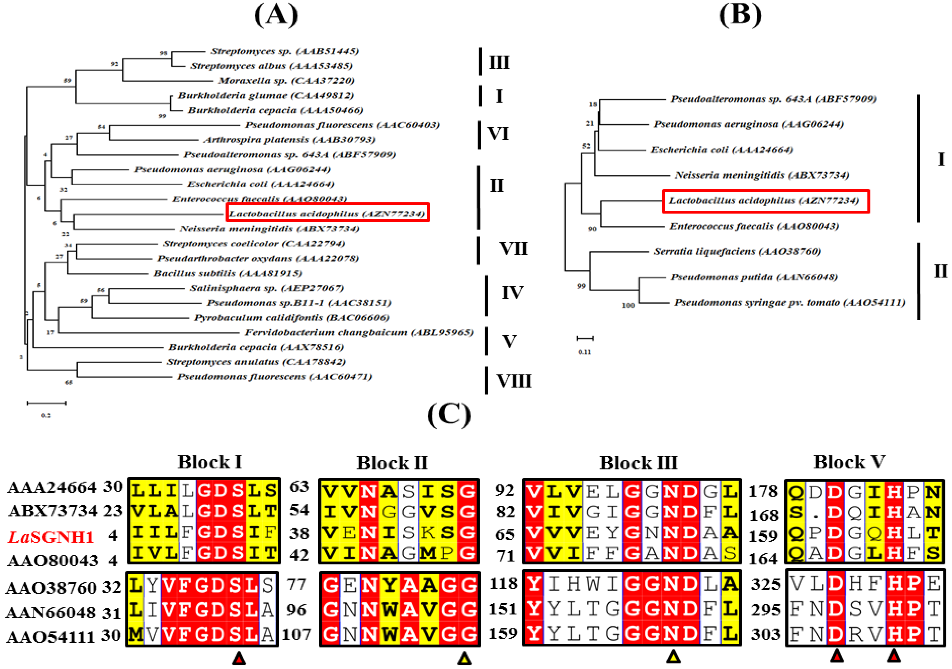
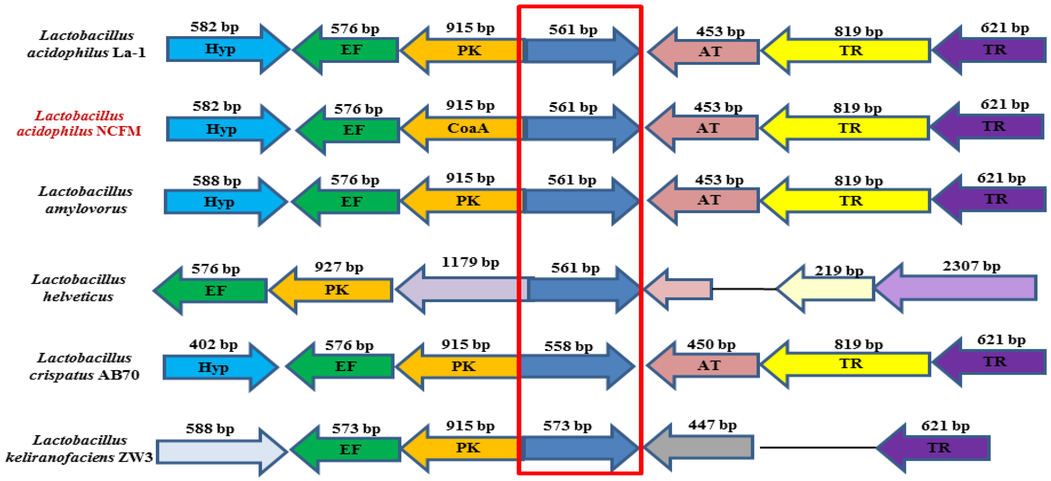
2.1. Bioinformatic Analysis of LaSGNH1

In the chromosome of L. acidophilus, a gene encoding a novel SGNH family esterase (LaSGNH1, locus tag: AZN77234, 561 bp) was identified using in silico bioinformatic analysis. Sequence analysis revealed that LaSGNH1 had a molecular mass of ~21 kDa with a single polypeptide chain of 188 amino acids, with a pI of 5.93. For phylogenetic tree analysis, eight bacterial lipases/esterases families (I–VIII) were investigated (Figure 1A). LaSGNH1 was shown to be a member of family II lipases/esterases, which is further subdivided into clade I and clade II subfamilies [25]. More specifically, as shown in Figure 1B, LaSGNH1 was clustered in the clade I subfamily with a lipase/acylhydrolase from Enterococcus faecalis (AAO80043, 30.4% sequence identity).
As shown in Figure 1C, four blocks (I, II, III, and V) are highly conserved of LaSGNH1 in clade I and II of family II lipases/esterases. The catalytic Ser10 is shown to be located in a GDS motif in block I, while a DXXH motif was localized in block V. Conserved residues in block II and III were shown to be involved in the formation of an oxyanion hole [8,25].
Genomic cluster analysis revealed that LaSGNH1 has homologues in other Lactobacillus species, including Lactobacillus amylovorus, which implies the invariant and important roles of these enzymes in Lactobacillus species (Figure 2). To date, there are no reports on these proteins, and their functional properties are largely unknown. The molar percentage (30.7%) of four hydrophobic amino acid residues (Alanine (Ala), Valine (Val), Isoleucine (Ile), and Leucine (Leu)) in LaSGNH1, which was shown to be important for protein stability [26], is comparable to that of an SGNH hydrolase (LI22) from Listeria innocua [18] or a SGNH hydrolase (Est24) from Sinorhizobium meliloti [19].

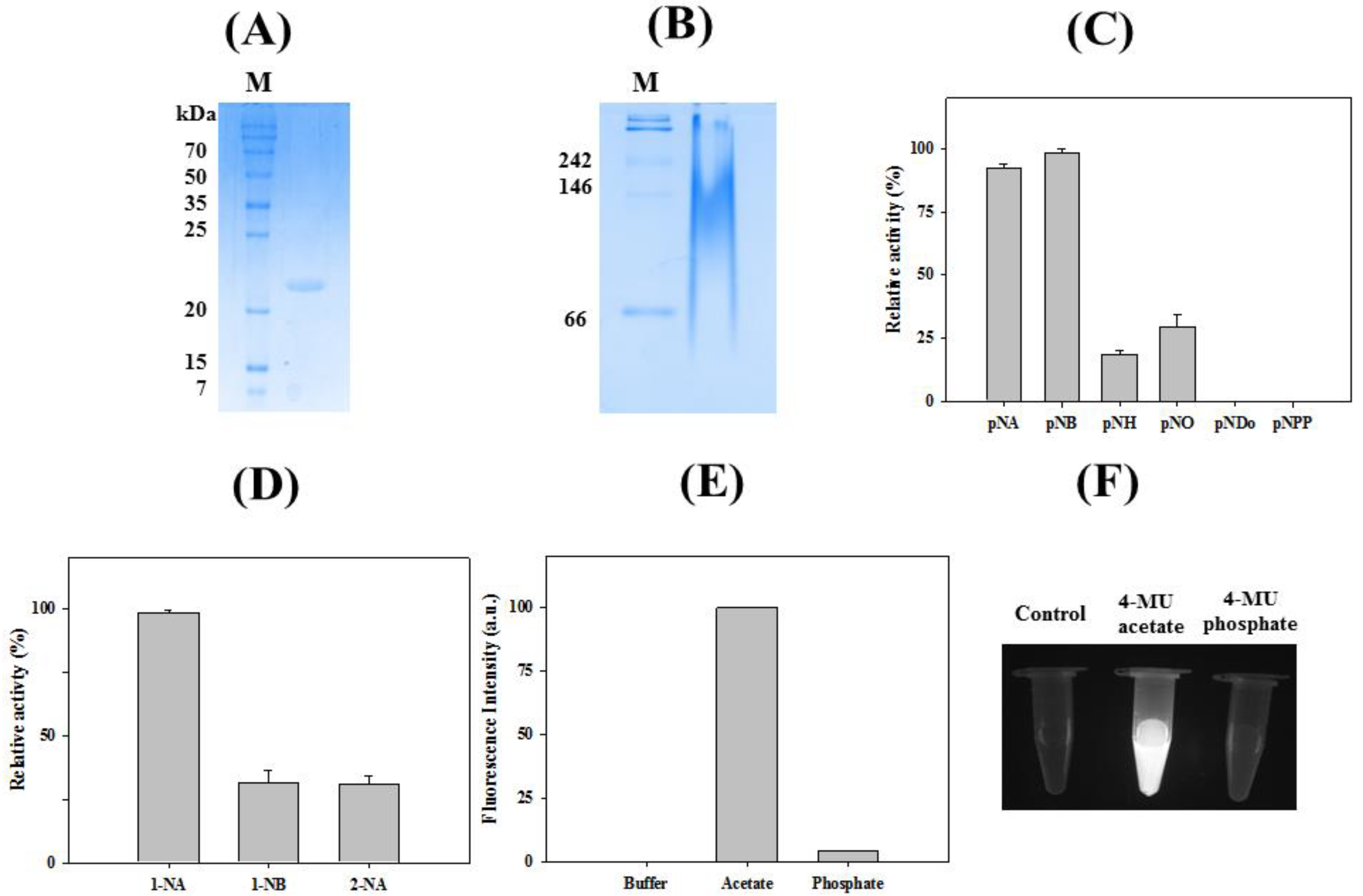
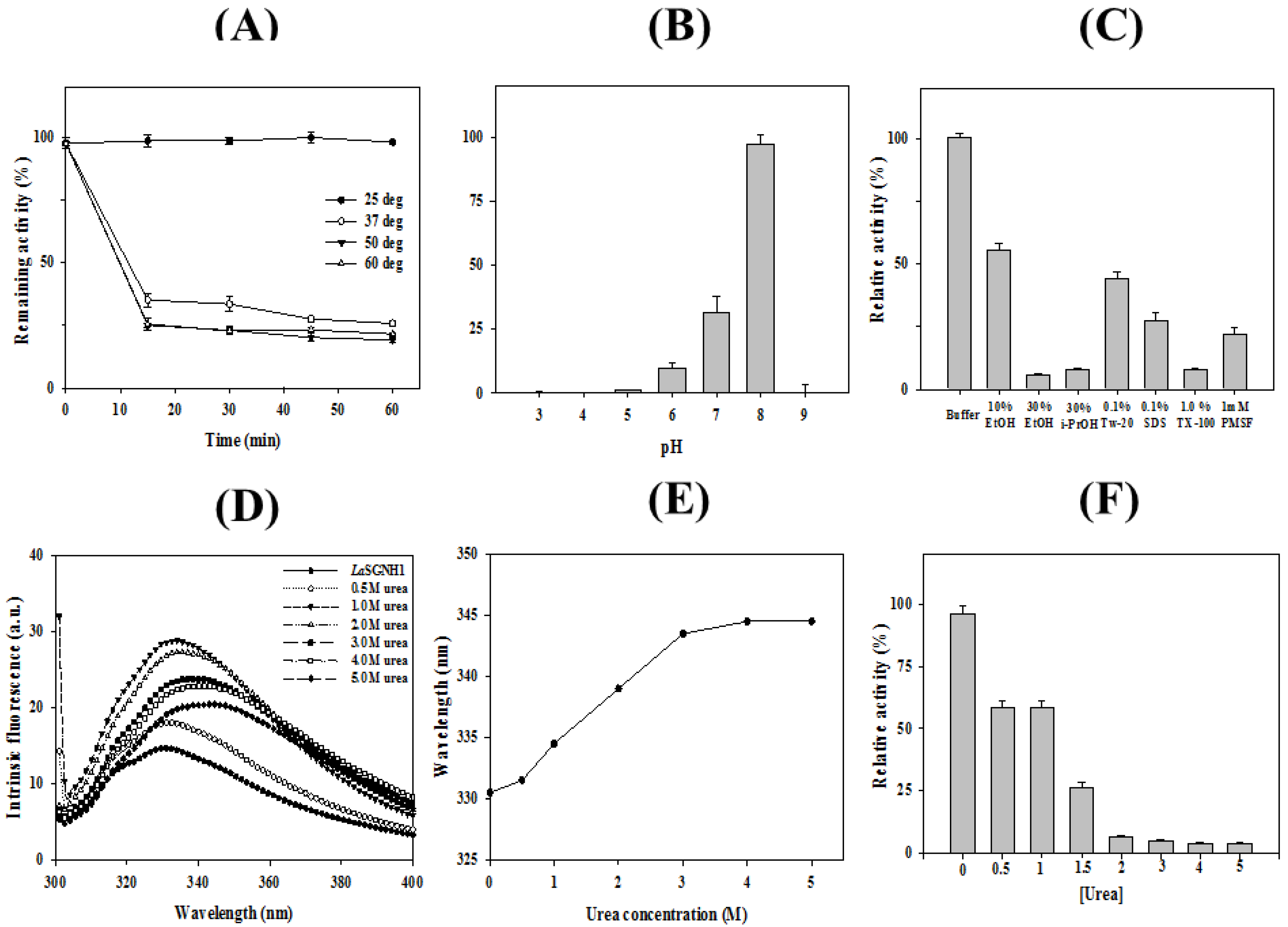
2.2. Characterizations of LaSGNH1

Recombinant LaSGNH1 protein was purified by an immobilized Ni2+-affinity column to near homogeneity (Figure 3A). The molecular mass of LaSGNH1 is similar to those of other SGNH family esterases, such as a thermostable and alkaline GDSL-motif esterase from Bacillus sp. K91 [16] or Lip2 from Monascus purpureus M7 [27]. However, it is smaller than the mass of a cold-adapted 36 kDa GDSL family esterase from Photobacterium sp. J15 [28]. In native polyacrylamide gel electrophoresis (PAGE), LaSGNH1 showed a diffuse pattern (Figure 3B). The hydrolytic activity of LaSGNH1 was analyzed using p-nitrophenyl esters of different chain lengths. As shown in Figure 3C, LaSGNH1 showed high activities for short-chain substrates, such as p-nitrophenyl acetate (p-NA) and p-nitrophenyl butyrate (p-NB) (Figure 3C). However, very little activity was observed for p-nitrophenyl decanoate (p-ND) or p-nitrophenyl phosphate (p-NPP). This strong preference for short-chain p-nitrophenyl esters was also observed for other SGNH family members, such as an SGNH hydrolase from Listeria innocua 11262 [20] or SGNH hydrolases from Sinorhizobium meliloti [19,29]. When naphthyl esters were used as substrates, the highest activity was observed with 1-naphthyl acetate (1-NA) (Figure 3D). LaSGNH1 showed regioselectivity, exhibiting only 25% activity toward 2-naphthyl acetate (2-NA) compared to 1-NA. Similar substrate specificity was observed in other members of the SGNH esterase family [23,24,28]. As shown in Figure 3E,F, strong fluorescence was observed for 4-methylumbelliferone (4-MU) acetate and LaSGNH1, but not for 4-MU phosphate and LaSGNH1.
Thermostability of LaSGNH1 was investigated over a temperature range from 25 to 60 °C (Figure 4A). Enzyme activity of LaSGNH1 did not change significantly after 1-h of incubation at 25 °C. However, LaSGNH1 showed only ~40% of initial activity after 15 min of incubation at 37 °C. Similarly, cinnamoyl esterases from Lactobacilli and Bifidobacteria showed an optimum temperature of 20–30 °C [30]. However, LaSGNH1 showed lower thermostability compared to other SGNH family esterases, such as an SGNH-type esterase (LpSGNH1) from Lactobacillus plantarum WCFS1 [12], a 7-aminocephalosporanic acid deacetylase [15], an alkaline SGNH hydrolase (Est19) from Bacillus sp. K91 [16], an SGNH hydrolase (LI22) from Listeria innocua [19], and an oligomeric SGNH-arylesterase from Sinorhizobium meliloti [20]. In addition, other esterases from L. acidophilus showed higher thermostability compared to LaSGNH1. For example, an acetylesterase (LaAcE) from L. acidophilus was shown to be stable at 40 °C for 1-h [31]. Moreover, no detectable activity loss of a feruloyl esterase from L. acidophilus was observed after a 2-h incubation at 37 °C [32].
In addition, LaSGNH1 displayed its maximal activity at pH 8.0, whereas ~30% of this maximal activity was observed at pH 7.0 (Figure 4B). This optimum pH is similar to other SGNH family esterases, such as an esterase gene (Tlip) from Thauera sp. [14] or an SGNH hydrolase (LI22) from Listeria innocua [19]. Furthermore, other esterases from L. acidophilus showed the optimum pH of 7.0–8.0 such as a cinnamoyl esterase [30] or LaAcE [31].
As shown in Figure 4C, LaSGNH1 retained ~65% of its initial activity in the presence of 10% ethanol and ~40% of its activity in the presence of 0.1% Tween 20. In contrast, the addition of 1.0% Triton X-100 resulted in less than 10% of its original activity. In the presence of 30% ethanol, LaSGNH1 retained only 10% of its initial activity (Figure 4C). The chemical stability of LaSGNH1 against urea was investigated by monitoring the intrinsic fluorescence changes. In the native state, LaSGNH1 exhibited a λmax at 330 nm, indicating that the tryptophan residues of LaSGNH1 were located in the hydrophobic interior (Figure 4D,E). However, a red shift of λmax to 344 nm was observed with a noteworthy increase of fluorescence intensity at 5 M urea. In contrast, the addition of 2.0 M urea resulted in almost complete loss of LaSGNH1 activity (Figure 4F).

2.3. Homology Modeling and Substrate Analysis of LaSGNH1

A structural model of LaSGNH1 was constructed based on the crystal structure of lipase/acylhydrolase from Enterococcus faecalis (PDB I.D.: 1YZF). The putative catalytic triad of Ser10, Asp161, and His164 are positioned close to the outer solvent available surfaces (Figure 5A). Three amino acids, Gly45, Gly70, and Asn72, were identified to control the entrance of substrates toward the catalytic triad via noncovalent interactions (Figure 5B). These resides are also highly conserved in SGNH family esterases (see also Figure 1C). In molecular docking analysis, Asn72, Tyr118, and Gln163 were shown to stabilize the p-nitrophenol ring (Figure 5C,D). In addition, the backbone nitrogen of Gly163 is involved in the stabilization of an oxyanion hole.
The hydrolytic properties of LaSGNH1 towards a wide range of substrates were studied using a colorimetric assay [33,34]. The ability of LaSGNH1 to hydrolyze tertiary alcohol esters (TAEs) was investigated using tert-butyl acetate, α-terpinyl acetate, and linalyl acetate. As shown in Figure 6A, LaSGNH1 was able to effectively hydrolyze tert-butyl acetate, but not α-terpinyl acetate nor linalyl acetate. Additionally, significant hydrolytic activity of LaSGNH1 was only detected for glyceryl tributyrate, which was indicated by the yellow color of the solution (Figure 6B). In addition, strong hydrolytic activity of LaSGNH1 for glucose pentaacetate was observed, although very little activity was observed in the presence of cellulose acetate or N-acetylglucosamine (Figure 6C). The preference of LaSGNH1 for small-size substrates could be explained by the restricted dimensions of the substrate-binding pocket [35].

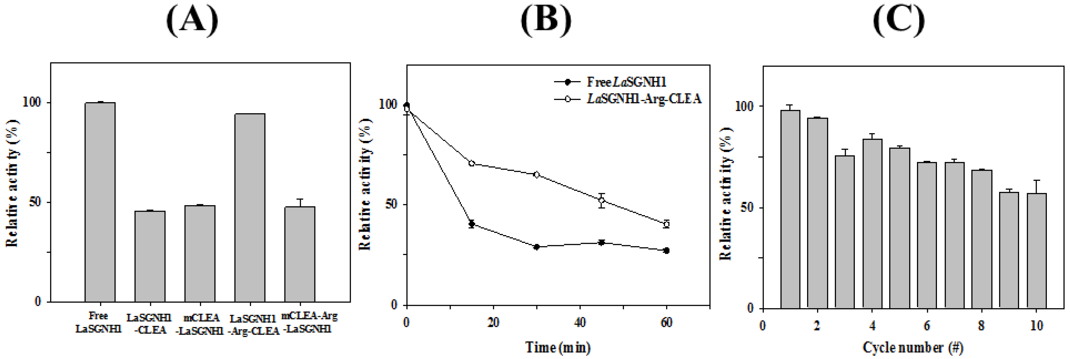
2.4. Immobilization of LaSGNH1

Enzyme immobilization, which could provide low cost, fast recovery, and high product yields, is widely used in industrial applications [36,37]. In previous reports, immobilized SGNH family esterases were shown to have better thermal stability, chemical stability, and recycling ability than free enzymes [12,18,19,29,35]. Specifically, cross-linked enzyme aggregates of LpSGNH1 displayed higher recycling ability and thermal stability than soluble LpSGNH1 [12]. In addition, enhanced thermal and chemical stability as well as good durability were observed in the crosslinked forms of LI22 [18] and Est24 [19]. Based on these studies, we immobilized LaSGNH1 via chemical crosslinking. First, LaSGNH1-crosslinked enzyme aggregates (CLEA) were prepared by precipitating LaSGNH1 with ammonium sulfate and glutaraldehyde. In addition, arginine (Arg) was also included in the preparation of LaSGNH1-Arg-CLEA, which was shown to be effective for the stability of immobilized enzymes [31,38]. Similarly, LaSGNH1 was co-precipitated with magnetite Fe3O4 nanoparticles, and crosslinked using glutaraldehyde to obtain magnetic LaSGNH1-CLEA (mCLEA-LaSGNH1). Enzyme immobilization using magnetite Fe3O4 nanoparticles could be used for fast separation [39]. Among these four different immobilization approaches (LaSGNH1-CLEA, LaSGNH1-Arg-CLEA, mCLEA-LaSGNH1, and mCLEA-Arg-LaSGNH1), LaSGNH1-Arg-CLEA showed the highest immobilization efficiency, which was comparable to that of free LaSGNH1 (Figure 7A).
Next, the thermal stability of LaSGNH1-Arg-CLEA was investigated for 1-h of incubation at 37 °C. As shown in Figure 7B, immobilized LaSGNH1-Arg-CLEA retained ~70% of its original activity after 30 min, while the free LaSGNH1 showed only 31% of its activity. Furthermore, the reusability of LaSGNH1-Arg-CLEA was studied over 10 cycles. After each cycle, the LaSGNH1-Arg-CLEAs were separated by centrifugation and washed for the next cycle. As shown in Figure 7C, LaSGNH1-Arg-CLEA showed good recycling ability, retaining about 60% of the original activity even after the 10th cycle. Therefore, LaSGNH1-Arg-CLEA showed good immobilization efficiency, enhanced thermal stability, and high reusability, which could be exploited to facilitate the applications of LaSGNH1.

3. Materials and Methods

3.1. Reagents

DNA-modifying enzymes were obtained from New England BioLabs (Ipswich, MA, USA). DNA purification kits were obtained from Qiagen Korea (Daejon, Korea), and protein purification columns were purchased from GE Healthcare (Seoul, Korea). All other reagents were of analytical grade and were purchased from Sigma-Aldrich Korea (Yongin, Korea).

3.2. Bioinformatic Analysis

The primary sequences of LaSGNH1 and related proteins were retrieved from the NCBI database. Multiple sequence alignments and sequence comparisons were carried out using Clustal Omega [40] and ESPript [41]. A phylogenetic tree was constructed by MEGA v. 7.0 using the neighbor-joining method with 2000 iterations [42]. A structural model of LaSGNH1 was constructed based on the crystal structure of lipase/acylhydrolase from Enterococcus faecalis (PDB I.D.: 1YZF) using the SWISS-MODEL server. Molecular docking analysis was performed using flexible side chain methods and AutoDock Vina [43].

3.3. Cloning and Purification

L. acidophilus NCFM (KCTC 3145; Korean Collection for Type Cultures) were cultured in MRS medium (BD Difco, NJ, USA) and chromosomal DNA was purified using a DNeasy Tissue and Blood Kit (Qiagen, USA). The open reading frame of the LaSGNH1 gene was amplified by polymerase chain reaction (PCR), and the PCR product was cloned into pQE-30 plasmid using BamHI and XhoI. After DNA sequencing, the recombinant plasmid (pET-LaSGNH1) was transformed into Escherichia coli cells for protein expression of LaSGNH1. E. coli cells were grown until the optical density (OD600nm) reached 0.6–0.8. After 1 mM isopropyl-β-D-1-thiogalactoside induction for 4 h at 37 °C, cells were centrifuged and resuspended in lysis buffer (20 mM Tris-HCl pH 8.0, 50 mM NaCl, 1 mM EDTA). After keeping on ice for 15 min, the cellular membrane was disrupted using a microtip (1-s pulse, 3-s pause, and 51% amplitude) in a Q500 sonicator (Terra Universal, Fullerton, CA, USA). After sonication, the supernatants were loaded onto a HisTrap HP column using an AKTA Prime Plus (GE healthcare, Chicago, IL, USA). The recombinant LaSGNH1 protein was eluted with an imidazole gradient from 50 to 300 mM. After a washing process, the pooled fractions were desalted with a lysis buffer. Protein concentration was determined using a Biorad Protein assay kit (Bio-rad Laboratories, Chicago, IL, USA) and purified LaSGNH1 was stored at −20 °C.

3.4. Biochemical Characterization of LaSGNH1

Substrate specificities of LaSGNH1 were investigated using p-nitrophenyl (p-NP) esters and naphthyl esters. The amounts of released p-nitrophenol were measured at 405 nm using p-nitrophenyl acetate (p-NA), p-nitrophenyl butyrate (p-NB), p-nitrophenyl hexanoate (p-NH), p-nitrophenyl octanoate (p-NO), p-nitrophenyl decanoate (p-ND), and p-nitrophenyl phosphate (p-NPP) [44,45]. For naphthyl esters, the formation of naphthol was monitored at 310 nm. The standard assay solution included 50 µM substrate in 20 mM Tris-HCl (pH 8.0) with 1 µg of LaSGNH1, and the assay ran for 10 min at 25 °C. All spectroscopic analyses were carried out using an Epoch 2 Microplate Spectrophotometer (BioTek, Winooski, VT, USA). Hydrolysis of 4-MU acetate or phosphate was also measured using a Jasco FP-8200 spectrofluorometer (Jasco, Japan) or an Eppendorf tube containing LaSGNH1 in a UV illumination box.
The thermostability and pH stability of LaSGNH1 were investigated at different temperatures ranging from 25 to 60 °C and across a pH range of 3.0 to 10.0. Effects of chemicals (10% ethanol, 30% ethanol, 30% iso-propanol, 0.1% Tween 20, 0.1% SDS, 1.0% Triton X-100, 1 Mm PMSF, and urea (from 0 to 5 M)) on the activity of LaSGNH1 were investigated after 1-h incubation using p-nitrophenyl butyrate (p-NB) as a substrate, and the enzyme activity of LaSGNH1 in buffer alone was defined as 100%. For intrinsic fluorescence spectra, the emission spectra from 300 to 400 nm were measured after excitation at 295 nm. All spectra were measured with a scan speed of 500 nm·min−1 and a 2 nm bandwidth using a Jasco FP-8200 spectrofluorometer.
For pH-indicator-based colorimetric assays, 1 µg of LaSGNH1 was added to a phenol-red-containing substrate solution. The substrates included lipids (glyceryl tributyrate, glyceryl trioleate, olive oil, and fish oil), tertiary alcohol esters (tertiary butyl acetate, α-terpinyl acetate, and linalyl acetate), and acetylated carbohydrates (glucose pentaacetate, cellulose acetate, and N-acetyl-glucosamine) [33,45].

3.5. Immobilization of LaSGNH1

For the preparation of cross-linked enzyme aggregates (CLEA), 0.5 mg·mL−1 of LaSGNH1 was co-precipitated with 70% ammonium sulfate with glutaraldehyde, incubated overnight, and centrifuged. The pellet (LaSGNH1-CLEA) was resuspended and washed extensively until no significant enzyme activity was detected in the supernatant. Addition of Arg and Fe3O4 magnetic nanoparticles for the preparation of LaSGNH1-Arg-CLEA, mCLEA-LaSGNH1, and mCLEA-Arg-LaSGNH1 was carried out as described previously [31,45]. For thermal stability, LaSGNH1-Arg-CLEA and free LaSGNH1 were incubated at 37 °C for 1-h. For the reusability experiments, LaSGNH1-Arg-CLEA was reused after extensive washing in subsequent cycles.

4. Conclusions

Although SGNH family esterases have attracted interest due to their potential applications, there remains little information about this family from lactic acid bacteria. Here, a novel SGNH family esterase (LaSGNH1) from L. acidophilus NCFM was identified, characterized, and immobilized. The novel properties of LaSGNH1 could make it a promising candidate for the food, cosmetics, pharmaceutical, and biofuel industries. In addition, this study could help us to better understand the SGNH family esterases, although the physiological role of LaSGNH1 has not yet been revealed. Further studies on LaSGNH1, including mutagenesis of key residues, structural determination, and formation of the enzyme–substrate complex, will be necessary to further our understanding of this LaSGNH1 enzyme.

Author Contributions

Conceptualization, L.T.H.L.L., W.Y., and T.D.K.; Methodology, L.T.H.L.L., W.Y., S.J., and T.D.K.; Validation, K.K.K., and T.D.K.; Investigation, L.T.H.L.L., W.Y., S.J., K.K.K., and T.D.K.; Writing—Original Draft Preparation, L.T.H.L.L., W.Y., and T.D.K.; Funding Acquisition, T.D.K. All authors have read and agreed to the published version of the manuscript.

Funding

This work was supported by a grant from the National Research Foundation of Korea funded by the Korean Government (NRF-2018R1D1A1B07044447).

Conflicts of Interest

The authors declare no conflicts of interest. The funders had no role in the design of the study; in the collection, analyses, or interpretation of data; in the writing of the manuscript, or in the decision to publish the results.

References

  1. Sarmah, N.; Revathi, D.; Sheelu, G.; Yamuna Rani, K.; Sridhar, S.; Mehtab, V.; Sumana, C. Recent advances on sources and industrial applications of lipases. Biotechnol. Prog. 2018, 34, 5–28. [Google Scholar] [CrossRef] [PubMed]
  2. Yang, J.; Liu, Y.; Liang, X.; Yang, Y.; Li, Q. Enantio-, regio-, and chemoselective lipase-catalyzed polymer synthesis. Macromol. Biosci. 2018, 18, e1800131. [Google Scholar] [CrossRef] [PubMed]
  3. Chen, Y.; Black, D.S.; Reilly, P.J. Carboxylic ester hydrolases: Classification and database derived from their primary, secondary, and tertiary structures. Protein Sci. 2016, 25, 1942–1953. [Google Scholar] [CrossRef] [PubMed] [Green Version]
  4. Jochen, H.; Hesseler, M.; Stiba, K.; Padhi, S.K.; Kazlauskas, R.J.; Bronscheuer, U.T. Protein engineering of α/β-hydrolase fold enzymes. Chembiochem 2011, 12, 1508–1517. [Google Scholar] [CrossRef] [PubMed]
  5. Khan, F.I.; Lan, D.; Durrani, R.; Huan, W.; Zhao, Z.; Wang, Y. The lid domain in lipases: Structural and functional determinant of enzymatic properties. Front. Bioeng. Biotechnol. 2017, 5, 16. [Google Scholar] [CrossRef] [PubMed] [Green Version]
  6. Chen, H.; Meng, X.; Xu, X.; Liu, W.; Li, S. The molecular basis for lipase stereoselectivity. Appl. Microbiol. Biotechnol. 2018, 102, 3487–3495. [Google Scholar] [CrossRef]
  7. De Miranda, A.S.; Miranda, L.S.; de Souza, R.O. Lipases: Valuable catalysts for dynamic kinetic resolutions. Biotechnol. Adv. 2015, 33, 372–393. [Google Scholar] [CrossRef]
  8. Akoh, C.C.; Lee, G.C.; Liaw, Y.C.; Huang, T.H.; Shaw, J.F. GDSL family of serine esterases/lipases. Prog. Lipid Res. 2004, 43, 534–552. [Google Scholar] [CrossRef]
  9. Leščić Ašler, I.; Ivić, N.; Kovačić, F.; Schell, S.; Knorr, J.; Krauss, U.; Wilhelm, S.; Kojić-Prodić, B.; Jaeger, K.E. Probing enzyme promiscuity of SGNH hydrolases. Chembiochem 2010, 11, 2158–2167. [Google Scholar] [CrossRef]
  10. Wilhelm, S.; Rosenau, F.; Kolmar, H.; Jaeger, K.E. Autotransporters with GDSL passenger domains: Molecular physiology and biotechnological applications. Chembiochem 2011, 12, 1476–1485. [Google Scholar] [CrossRef]
  11. Soni, S.; Sathe, S.S.; Odaneth, A.A.; Lali, A.M.; Chandrayan, S.K. SGNH hydrolase-type esterase domain containing Cbes-AcXE2: A novel and thermostable acetyl xylan esterase from Caldicellulosiruptor bescii. Extremophiles 2017, 21, 687–697. [Google Scholar] [CrossRef] [PubMed]
  12. Kim, Y.; Ryu, B.H.; Kim, J.; Yoo, W.; An, D.R.; Kim, B.Y.; Kwon, S.; Lee, S.; Wang, Y.; Kim, K.K.; et al. Characterization of a novel SGNH-type esterase from Lactobacillus plantarum. Int. J. Biol. Macromol. 2017, 96, 560–568. [Google Scholar] [CrossRef] [PubMed]
  13. Shahinyan, G.; Margaryan, A.; Panosyan, H.; Trchounian, A. Identification and sequence analyses of novel lipase encoding novel thermophillic bacilli isolated from Armenian geothermal springs. BMC Microbiol. 2017, 17, 103. [Google Scholar] [CrossRef] [PubMed] [Green Version]
  14. Yu, N.; Yang, J.C.; Yin, G.T.; Li, R.S.; Zou, W.T.; He, C. Identification and characterization of a novel esterase from Thauera sp. Biotechnol. Appl. Biochem. 2018, 65, 748–755. [Google Scholar] [CrossRef] [PubMed]
  15. Ding, J.M.; Yu, T.T.; Han, N.Y.; Yu, J.L.; Li, J.J.; Yang, Y.J.; Tang, X.H.; Xu, B.; Zhou, J.P.; Tang, H.Z.; et al. Identification and characterization of a new 7-Aminocephalosporanic acid deacetylase from Thermophilic Bacterium Alicyclobacillus tengchongensis. J. Bacteriol. 2015, 198, 311–320. [Google Scholar] [CrossRef] [Green Version]
  16. Ding, J.; Zhu, H.; Ye, Y.; Li, J.; Han, N.; Wu, Q.; Huang, Z.; Meng, Z. A thermostable and alkaline GDSL-motif esterase from Bacillus sp. K91: Crystallization and X-ray crystallographic analysis. Acta Crystallogr. Sect. F Struct. Biol. Commun. 2018, 74, 117–121. [Google Scholar] [CrossRef] [Green Version]
  17. Privé, F.; Kaderbhai, N.N.; Girdwood, S.; Worgan, H.J.; Pinloche, E.; Scollan, N.D.; Huws, S.A.; Newbold, C.J. Identification and characterization of three novel lipases belonging to families II and V from Anaerovibrio lipolyticus 5ST. PLoS ONE 2013, 8, e69076. [Google Scholar] [CrossRef]
  18. Kim, S.; Bae, S.Y.; Kim, S.J.; Ngo, T.D.; Kim, K.K.; Kim, T.D. Characterization, amyloid formation, and immobilization of a novel SGNH hydrolase from Listeria innocua 11262. Int. J. Biol. Macromol. 2012, 50, 103–111. [Google Scholar] [CrossRef]
  19. Bae, S.Y.; Ryu, B.H.; Jang, E.; Kim, S.; Kim, T.D. Characterization and immobilization of a novel SGNH hydrolase (Est24) from Sinorhizobium meliloti. Appl. Microbiol. Biotechnol. 2013, 97, 1637–1647. [Google Scholar] [CrossRef]
  20. Brown, L.; Pingitore, E.V.; Mozzi, F.; Saavedra, L.; Villegas, J.M.; Hebert, E.M. Lactic acid bacteria as cell factories for the generation of bioactive peptides. Protein Pept. Lett. 2017, 24, 146–155. [Google Scholar] [CrossRef]
  21. Hatti-Kaul, R.; Chen, L.; Dishisha, T.; Enshasy, H.E. Lactic acid bacteria: From starter cultures to producers of chemicals. FEMS Microbiol. Lett. 2018, 365, fny213. [Google Scholar] [CrossRef] [PubMed] [Green Version]
  22. Stefanovic, E.; Fitzgerald, G.; McAuliffe, O. Advances in the genomics and metabolomics of dairy lactobacilli: A review. Food Microbiol. 2017, 61, 33–49. [Google Scholar] [CrossRef] [PubMed]
  23. Heeney, D.D.; Gareau, M.G.; Marco, M.L. Intestinal Lactobacillus in health and disease, a driver or just along for the ride? Curr. Opin. Biotechnol. 2018, 49, 140–147. [Google Scholar] [CrossRef] [PubMed] [Green Version]
  24. Lili, Z.; Junyan, W.; Hongfei, Z.; Baoqing, Z.; Bolin, Z. Detoxification of cancerogenic compounds by lactic acid bacteria strains. Crit. Rev. Food Sci. Nutr. 2018, 58, 2727–2742. [Google Scholar] [CrossRef]
  25. Dong, X.; Yi, H.; Han, C.T.; Nou, I.S.; Hur, Y. GDSL esterase/lipase genes in Brassica rapa L.: Genome-wide identification and expression analysis. Mol. Genet. Genomics 2016, 291, 531–542. [Google Scholar] [CrossRef]
  26. Mozhaev, V.V.; Martinek, K. Structure-stability relationships in proteins: New approaches to stabilizing enzymes. Enzym. Microb. Technol. 1984, 6, 50–59. [Google Scholar] [CrossRef]
  27. Kang, L.J.; Meng, Z.T.; Hu, C.; Zhang, Y.; Guo, H.L.; Li, Q.; Li, M. Screening, purification, and characterization of a novel organic solvent-tolerant esterase, Lip2, from Monascus purpureus strain M7. Extremophiles 2017, 21, 345–355. [Google Scholar] [CrossRef]
  28. Shakiba, M.H.; Ali, M.S.; Rahman, R.N.; Salleh, A.B.; Leow, T.C. Cloning, expression and characterization of a novel cold-adapted GDSL family esterase from Photobacterium sp. strain J15. Extremophiles 2016, 20, 44–55. [Google Scholar] [CrossRef]
  29. Hwang, H.; Kim, S.; Yoon, S.; Ryu, Y.; Lee, S.Y.; Kim, T.D. Characterization of a novel oligomeric SGNH-arylesterase from Sinorhizobium meliloti 1021. Int. J. Biol. Macromol. 2010, 46, 145–152. [Google Scholar] [CrossRef]
  30. Fritsch, C.; Jänsch, A.; Ehrmann, M.A.; Toelstede, S.; Vogel, R.F. Characterization of cinnamoyl esterases from different Lactobacilli and Bifidobacteria. Curr. Microbiol. 2017, 74, 247–256. [Google Scholar] [CrossRef]
  31. Wang, Y.; Ryu, B.H.; Yoo, W.; Lee, C.W.; Kim, K.K.; Lee, J.H.; Kim, T.D. Identification, characterization, immobilization, and mutational analysis of a novel acetylesterase with industrial potential (LaAcE) from Lactobacillus acidophilus. Biochim. Biophys. Acta Gen. Subj. 2018, 1862, 197–210. [Google Scholar] [CrossRef] [PubMed]
  32. Wang, X.; Geng, X.; Egashira, Y.; Sanada, H. Purification and characterization of a feruloyl esterase from the intestinal bacterium Lactobacillus acidophilus. Appl. Environ. Microbiol. 2004, 70, 2367–2372. [Google Scholar] [CrossRef] [PubMed] [Green Version]
  33. Lee, C.W.; Kwon, S.; Park, S.H.; Kim, B.Y.; Yoo, W.; Ryu, B.H.; Kim, H.W.; Shin, S.C.; Kim, S.; Park, H.; et al. Crystal structure and functional characterization of an esterase (EaEST) from Exiguobacterium antarcticum. PLoS ONE 2017, 12, e0169540. [Google Scholar] [CrossRef] [PubMed]
  34. Oh, C.; Ryu, B.H.; Yoo, W.; Nguyen, D.D.; Kim, T.; Ha, S.C.; Kim, T.D.; Kim, K.K. Identification and Crystallization of Penicillin-Binding Protein/β-Lactamase Homolog (Rp46) from Ruegeria Pomeroyi. Crystals 2017, 7, 6. [Google Scholar]
  35. Kim, K.; Ryu, B.H.; Kim, S.S.; An, D.R.; Ngo, T.D.; Pandian, R.; Kim, K.K.; Kim, T.D. Structural and biochemical characterization of a carbohydrate acetylesterase from Sinorhizobium melioti 1021. FEBS Lett. 2015, 589, 117–122. [Google Scholar] [CrossRef] [Green Version]
  36. Prasad, S.; Roy, I. Converting Enzymes into Tools of Industrial Importance. Recent Pat. Biotechnol. 2018, 12, 33–56. [Google Scholar] [CrossRef]
  37. Hoarau, M.; Badieyan, S.; Marsh, E.N.G. Immobilized enzymes: understanding enzyme—Surface interactions at the molecular level. Org. Biomol. Chem. 2017, 15, 9539–9551. [Google Scholar] [CrossRef]
  38. Mukherjee, J.; Majumder, A.B.; Gupta, M.N. Adding an appropriate amino acid during crosslinking results in more stable crosslinked enzyme aggregates. Anal. Biochem. 2016, 507, 27–32. [Google Scholar] [CrossRef]
  39. Cui, J.; Cui, L.; Jia, S.; Su, Z.; Zhang, S. Hybrid cross-linked lipase aggregates with magnetic nanoparticles: A robust and recyclable biocatalysis for the epoxidation of oleic acid. J. Agric. Food Chem. 2016, 64, 7179–7187. [Google Scholar] [CrossRef]
  40. Sievers, F.; Higgins, D.G. Clustal Omega for making accurate alignments of many protein sequences. Protein Sci. 2018, 27, 135–145. [Google Scholar] [CrossRef] [Green Version]
  41. Gouet, P.; Robert, X.; Courcelle, E. ESPript/ENDscript: Extracting and rendering sequence and 3D information from atomic structures of proteins. Nucleic Acids Res. 2003, 31, 3320–3323. [Google Scholar] [CrossRef] [PubMed]
  42. Kumar, S.; Stecher, G.; Tamura, K. MEGA7: Molecular evolutionary genetics analysis version 7.0 for bigger datasets. Mol. Biol. Evol. 2016, 33, 1870–1874. [Google Scholar] [CrossRef] [PubMed] [Green Version]
  43. Seeliger, D.; de Groot, B.L. Ligand docking and binding site analysis with PyMOL and Autodock/Vina. J. Comput. Aided Mol. Des. 2010, 24, 417–422. [Google Scholar] [CrossRef] [PubMed] [Green Version]
  44. Oh, C.; Ryu, B.H.; Yoo, W.; Nguyen, D.D.; Kim, T.; Ha, S.; Kim, T.D.; Kim, K.K. Identification and crystallographic analysis of a new carbohydrate acetylesterase (SmAcE1) from Sinorhizobium meliloti. Crystals 2018, 8, 12. [Google Scholar] [CrossRef] [Green Version]
  45. Park, S.H.; Yoo, W.; Lee, C.W.; Jeong, C.S.; Shin, S.C.; Kim, H.W.; Park, H.; Kim, K.K.; Kim, T.D.; Lee, J.H. Crystal structure and functional characterization of a cold-active acetyl xylan esterase (PbAcE) from psychrophilic soil microbe Paenibacillus sp. PLoS ONE 2018, 13, e0206260. [Google Scholar] [CrossRef] [PubMed]
Figure 1. Phylogenetic tree and sequence analysis of LaSGNH1. (A) Bacterial lipases/esterases family I–VIII, and (B) clade I and II of family II are shown. A red box in each panel indicates the location of LaSGNH1. The phylogenetic trees were constructed with MEGA v. 7.0 using the neighbor-joining method, and all sequences were retrieved from the NCBI database. (C) Sequence alignments of four conserved blocks (Block I, II, III, and V) are shown, and highly conserved residues are highlighted in red. Sequences are aligned with Clustal Omega and ESPript. Highly conserved catalytic triad, glycine, and asparagine are shown as red or yellow triangles. Four sequences of the clade I subfamily are shown in the upper region, while three sequences of the clade II subfamily are shown in the bottom region. Highly important amino acids for catalysis are shown as red and yellow triangles.
Ijms 21 00091 g001
Figure 2. Gene cluster analysis of LaSGNH1. Similar gene clusters were found in Lactobacillus species including in Lactobacillus acidophilus La-1, L. acidophilus NCFM, Lactobacillus amylovorus, Lactobacillus helveticus, Lactobacillus crispatus AB70, and Lactobacillus kefiranofaciens ZW3. EF: elongation factor, PK: type I pantothenate kinase, AT: acetyltransferase, TR: amino acid ABC transporter. Homologous proteins of LaSGNH1 are shown in the red box.
Ijms 21 00091 g002
Figure 3. Characterization of LaSGNH1. (A) Sodium dodecyl (lauryl) sulfate-polyacrylamide gel electrophoresis (SDS-PAGE) analysis of purified LaSGNH1. (B) Native-polyacrylamide gel electrophoresis (PAGE) analysis. (C) Substrate specificity of LaSGNH1 using p-nitrophenyl (p-NP) esters. The hydrolase activities are shown relative to the activity toward p-NB. (D) Regioselectivity of LaSGNH1 was studied using 1-naphthyl acetate (1-NA), 1-naphthyl butyrate (1-NB), and 2-naphthyl acetate (2-NA). The hydrolase activities are shown relative to the activity toward 1-NA. (E,F) Detection of fluorescence due to the formation of 4-methylumbelliferone (4-MU) by LaSGNH1. All experiments were performed at least in triplicate.
Ijms 21 00091 g003
Figure 4. Stability of LaSGNH1. (A) Thermal stability of LaSGNH1. The residual activity of LaSGNH1 was measured during incubation for 1-h. (B) The pH stability of LaSGNH1 was studied at a pH from 3 to 10. (C) Chemical stability of LaSGNH1 was studied against various chemicals. (D,E) Urea-induced unfolding of LaSGNH1. Fluorescence was monitored after 1-h of incubation in from 1 to 5 M urea. A red-shift of λmax from 330 to 344 nm was detected. (F) Activity of LaSGNH1 in the different concentrations of urea. All experiments were performed at least in triplicate.
Ijms 21 00091 g004
Figure 5. Homology modeling of LaSGNH1. (A) Ribbon representation of LaSGNH1. The substrate binding pocket is also shown in the square and important residues for catalysis are shown as sticks. (B) Electrostatic potential diagram of substrate-binding regions of LaSGNH1. (C) Modeling of p-nitrophenyl acetate (pNA, cyan) in the substrate-binding pocket of LaSGNH1. The amino acid residues interacting with pNA are shown as sticks (green). (D) LigPlot analysis of p-nitrophenyl acetate in the substrate-binding pocket of LaSGNH1.
Ijms 21 00091 g005
Figure 6. Hydrolysis of various substrates by LaSGNH1. A pH shift assay was performed for (A) tertiary alcohol esters (TAEs), including tert-butyl acetate, α-terpinyl acetate, and linalyl acetate, (B) glyceryl esters, including glyceryl tributyrate (GTB) and glyceryl trioleate (GTO), and oils, including olive oil (O.O.) and fish oil (F.O.), and (C) acetylated carbohydrates, including glucose pentaacetate, cellulose acetate, and N-acetyl-glucosamine. The hydrolysis reaction changed the color of the solution from red to yellow.
Ijms 21 00091 g006
Figure 7. Immobilization of LaSGNH1. (A) Immobilization efficiency of free LaSGNH1, LaSGNH1- crosslinked enzyme aggregates (CLEA), mCLEA-LaSGNH1, LaSGNH1-Arg-CLEA, and mCLEA-Arg-LaSGNH1. (B) Thermal stability of free LaSGNH1 and LaSGNH1-Arg-CLEA. (C) Reusability of LaSGNH1-Arg-CLEA. The reaction was repeated for 10 cycles after each washing step. All assays were performed at least in triplicate.
Ijms 21 00091 g007

