Article

Structural Features of Carnivorous Plant (Genlisea, Utricularia) Tubers as Abiotic Stress Resistance Organs

by
Bartosz J. Płachno
1,*,
Saura R. Silva
2,
Piotr Świątek
3,
Kingsley W. Dixon
4,
Krzystof Lustofin
1,
Guilherme C. Seber
2 and
Vitor F. O. Miranda
2
1
Department of Plant Cytology and Embryology, Institute of Botany, Faculty of Biology, Jagiellonian University in Kraków, Gronostajowa 9 St. 30-387 Cracow, Poland
2
Laboratory of Plant Systematics, School of Agricultural and Veterinarian Sciences, São Paulo State University (Unesp), Jaboticabal, CEP 14884-900 SP, Brazil
3
Faculty of Natural Sciences, Institute of Biology, Biotechnology and Environmental Protection, University of Silesia in Katowice, Jagiellońska 28, 40-032 Katowice, Poland
4
School of Molecular and Life Sciences, Curtin University, Kent Street, Bentley, Perth, WA 6102, Australia
*
Author to whom correspondence should be addressed.
Int. J. Mol. Sci. 2020, 21(14), 5143; https://doi.org/10.3390/ijms21145143
Submission received: 28 June 2020 / Revised: 16 July 2020 / Accepted: 18 July 2020 / Published: 21 July 2020

Abstract

:
Carnivorous plants from the Lentibulariaceae form a variety of standard and novel vegetative organs and survive unfavorable environmental conditions. Within Genlisea, only G. tuberosa, from the Brazilian Cerrado, formed tubers, while Utricularia menziesii is the only member of the genus to form seasonally dormant tubers. We aimed to examine and compare the tuber structure of two taxonomically and phylogenetically divergent terrestrial carnivorous plants: Genlisea tuberosa and Utricularia menziesii. Additionally, we analyzed tubers of U. mannii. We constructed phylogenetic trees using chloroplast genes matK/trnK and rbcL and used studied characters for ancestral state reconstruction. All examined species contained mainly starch as histologically observable reserves. The ancestral state reconstruction showed that specialized organs such as turions evolved once and tubers at least 12 times from stolons in Lentibulariaceae. Different from other clades, tubers probably evolved from thick stolons for sect. Orchidioides and both structures are primarily water storage structures. In contrast to species from section Orchidioides, G. tuberosa, U. menziesii and U. mannii form starchy tubers. In G. tuberosa and U. menziesii, underground tubers provide a perennating bud bank that protects the species in their fire-prone and seasonally desiccating environments.

Graphical Abstract

1. Introduction

The Lentibulariaceae are carnivorous plants that produce three types of traps: flypaper (Pinguicula), eel (Genlisea), and suction (Utricularia) (e.g., [1,2,3,4]). Genlisea and Utricularia exhibit a novel plant architecture (e.g., [5,6,7,8]) linked to their growth in wet habitats. However, some are exposed seasonally to abiotic stresses such as drought, high temperatures or frost. To survive unfavorable ecological conditions, members of the Lentibulariaceae form various vegetative organs such as: hibernaculae, dormant rosettes, subterranean bulb-like rosettes, turions, thick stolons, stem and tubers.
Hibernacula occur in temperate species like Pinguicula vulgaris, P. grandiflora and P. alpina. The hibernaculum consists of an abbreviated stem-bearing scales and leaf primordia. Reserves (starch) are stored mainly in swollen hibernaculum scales (P. vulgaris and P. grandiflora) or in the stout perennial roots (P. alpina) [9]. Dormant rosettes and subterranean bulb-like rosettes occur in the heterophyllous Pinguicula species of Mexico and Central America. Leaves in these rosettes are non-carnivorous have a reduced lamina and possess succulent characteristics (e.g., [10]).
In the case of Genlisea, the stress resistance organs might be: scapes, thick stems, leaves and tubers. Large, thick and succulent photosynthetic scapes occur in Genlisea uncinata and G. oligophylla [11,12]. According to Fleischmann [12], Genlisea metallica survives seasonal aridity by a perennating short, succulent vertical underground stem. Rivadavia [11] noted that Genlisea aurea may form the stem up to approximately 1.5 cm in length by 4 mm in diameter and this thickened structure may also provide drought protection. Thick, succulent leaves occur in G. uncinata (Fleischmann, 2012). However, among the members of Genlisea, G. tuberosa is unique in forming tubers ([11] as found in G. pygmaea; [12,13]). According to Rivadavia [11] each plant produces a single tuber. These stolon-derived tubers arise in the early stages of seedling development [12], however there are no data on the anatomy and morphological characters in Genlisea tubers.
In Utricularia, stress resistance organs include turions, thick stolons and tubers [14]. The ten species of aquatic Utricularia from sect. Utricularia form turions in response to unfavorable ecological conditions, usually at the beginning of autumn [14,15]. The Utricularia turion is an overwintering organ, which is formed by the condensation of short, modified leaves in the shoot apex. In comparison to other tissues, the turion has highly reduced metabolism and contains starch as the primary reserve material and protein (e.g., [14,15,16,17,18,19]). According to Taylor [14], tubers in Utricularia occur mostly in the epiphytic and lithophytic species from sect. Orchidioides, Phyllaria, Chelidon and this organ is important for surviving the dry season. Thick stolons also occur in some Utricularia species (U. reniformis, U. humboldtii, U. nelumbifolia, U. cornigera) from former sect. Iperua (now included in sect. Orchidioides) and provide drought resistance [14,20,21]. Thick stolons occur in some species from sect. Pleiochasia with the root tubers of Utricularia menziesii being unique [14,22]. This terrestrial species survives the dry season by senescing to quiescent over-summering tubers [14,23]. Tubers or tuber-like structures have been recorded in aquatic species U. benjaminiana, U. reflexa, U. inflata, and U. radiata [14,20]. However, in these aquatic species sect. Utricularia tubers perhaps may function as vegetative propagules similar to the turions. According to Rice [20], there are two types of Utricularia tubers: water storage and carbohydrate-rich tubers. Rice [20] outlined that Utricularia menziesii possessed carbohydrate-rich tubers, however, he did not provide histochemical evidence and also no literature sources for this information. Pate and Dixon [22] studied mineral and carbohydrate content of U. menziesii tubers and highlighted that these act as the primary over-summering phase for the species though no histological analysis was provided. For annual species of Genlisea and Utricularia, the primary drought tolerance mechanism is the production of a soil seed bank [24]. Besides the drought tolerance, the small seed size associated with large testa cells with a foveolate surface of Genlisea and Utricularia species permits aggregation of air bubbles and thereby facilitating the floatability and the dispersion through the water channels produced by seasonal rains in the Cerrado [25].
Genlisea tuberosa and Utricularia menziesii were selected for this study as they occur in special fire-prone environments. Both are exposed to high irradiance and air temperatures during the dry season and aestivate using underground tubers. These species occur in the old climatically-buffered infertile landscapes (OCBILs, [26]); Genlisea tuberosa in campos rupestres (rupestrian grasslands) (Figure 1A–C) and Utricularia menziesii in kwongkan of south-western Australia. U. menziesii belongs to sect. Pleiochasia, within U. subgen. Polypompholyx [27,28], an early-branching lineage which has unexpected features such as specialization for bird pollination [29]. Therefore, compared tuber structures from the divergent lineages in Lentibulariaceae (Genlisea genus and Utricularia subgen. Polypompholyx sect. Pleiochasia) with tuberous structures of species (subgen. Utricularia sect. Orchidioides, [21]) that are thought to be primarily for storage of water [21,30,31]. Additionally, we chose tubers of Utricularia mannii (sect. Chelidon) as Taylor [14] noted that this species has many common features (including tuber formation) with species from sect. Orchidioides (subgen. Utricularia). However, according to the phylogeny based on trnK/matK and trnL-F DNA sequences [32] U. mannii is nested within subgen. Bivalvaria (sensu [33]). This species, in contrast to the terrestrial Genlisea tuberosa and Utricularia menziesii, is an epiphyte found on Mount Cameroon, Cameroon, Africa. Thus, our study aims to examine and compare the tuber structures from the divergent lineages in Lentibulariaceae, represented by Genlisea tuberosa, Utricularia menziesii and U. mannii, to more fully understand the relationship between anatomical features as related to phylogenetic links and perennation mode.

2. Results

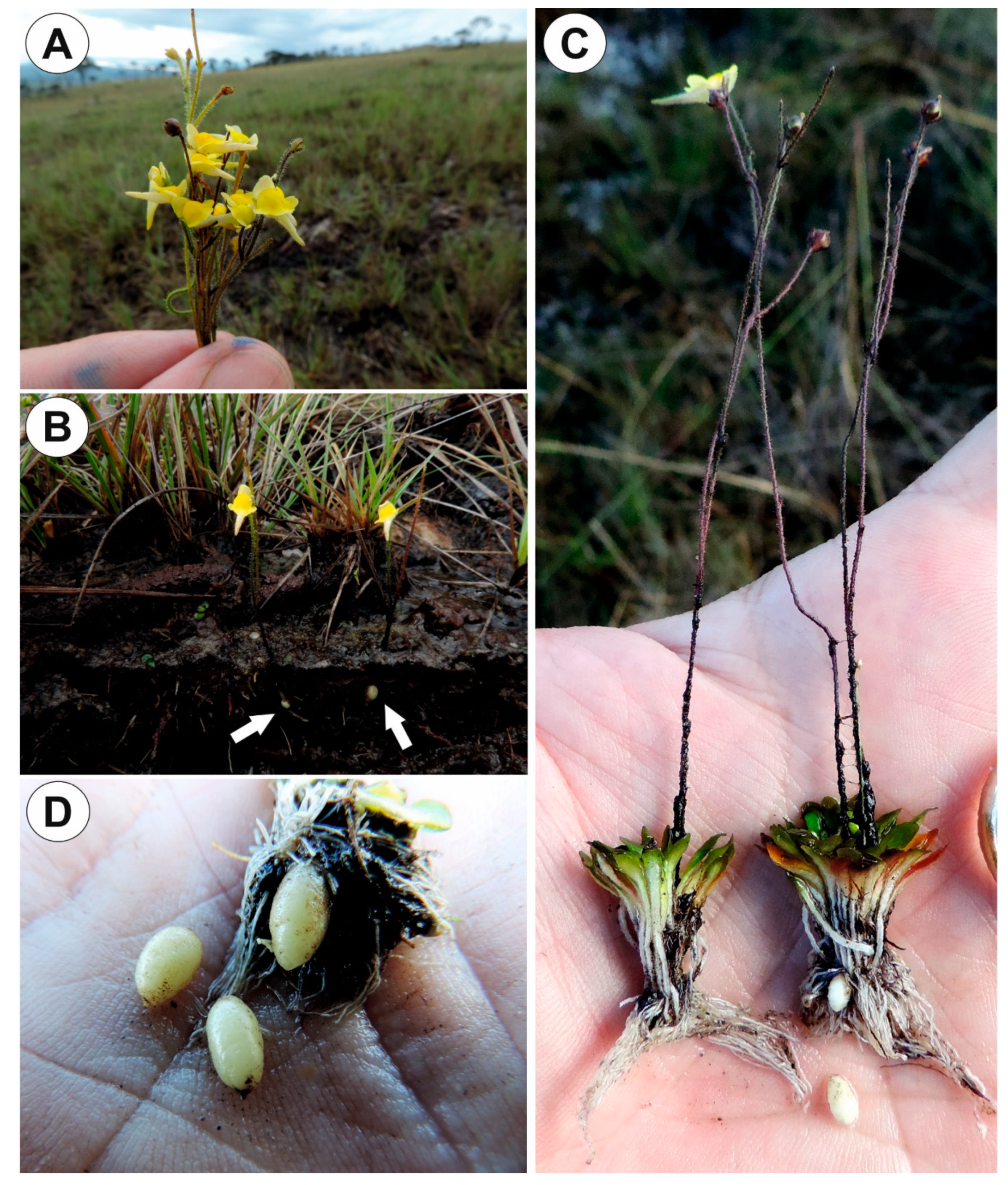
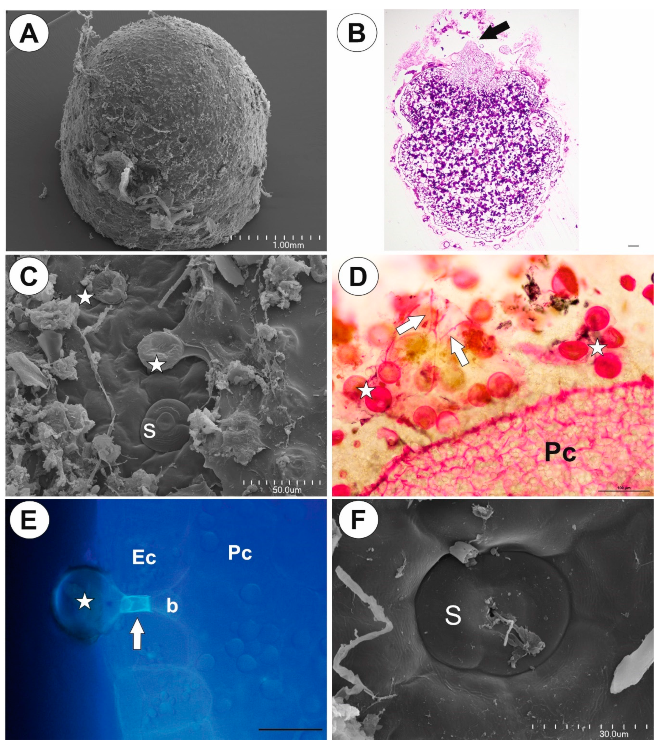
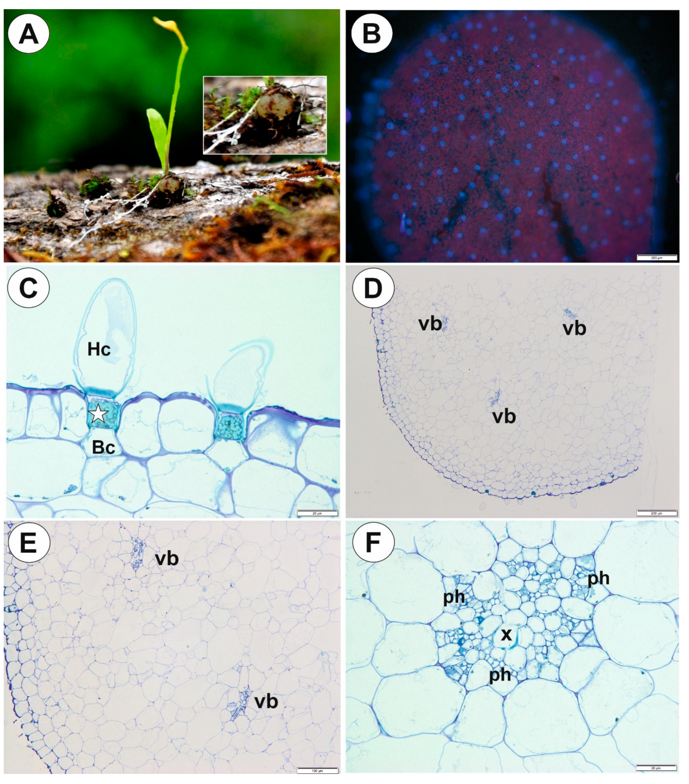
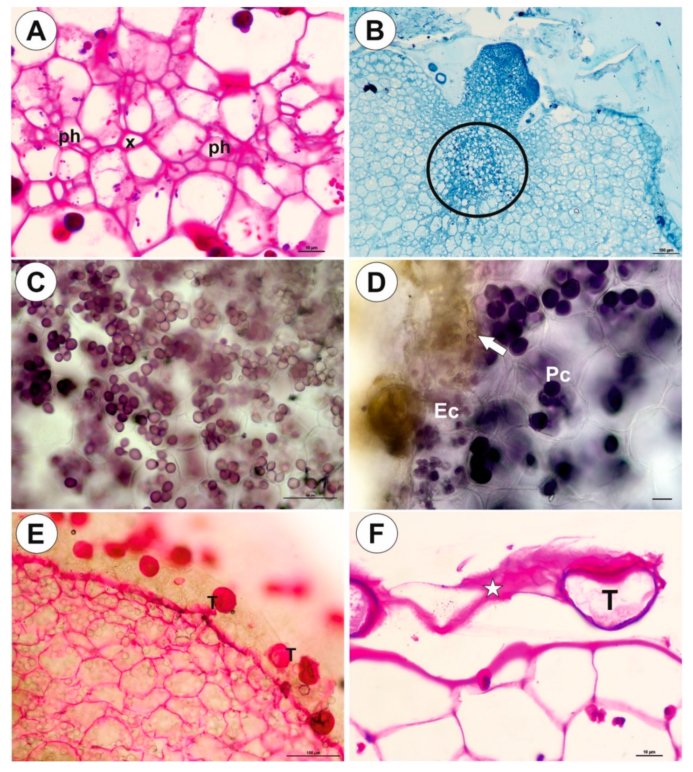
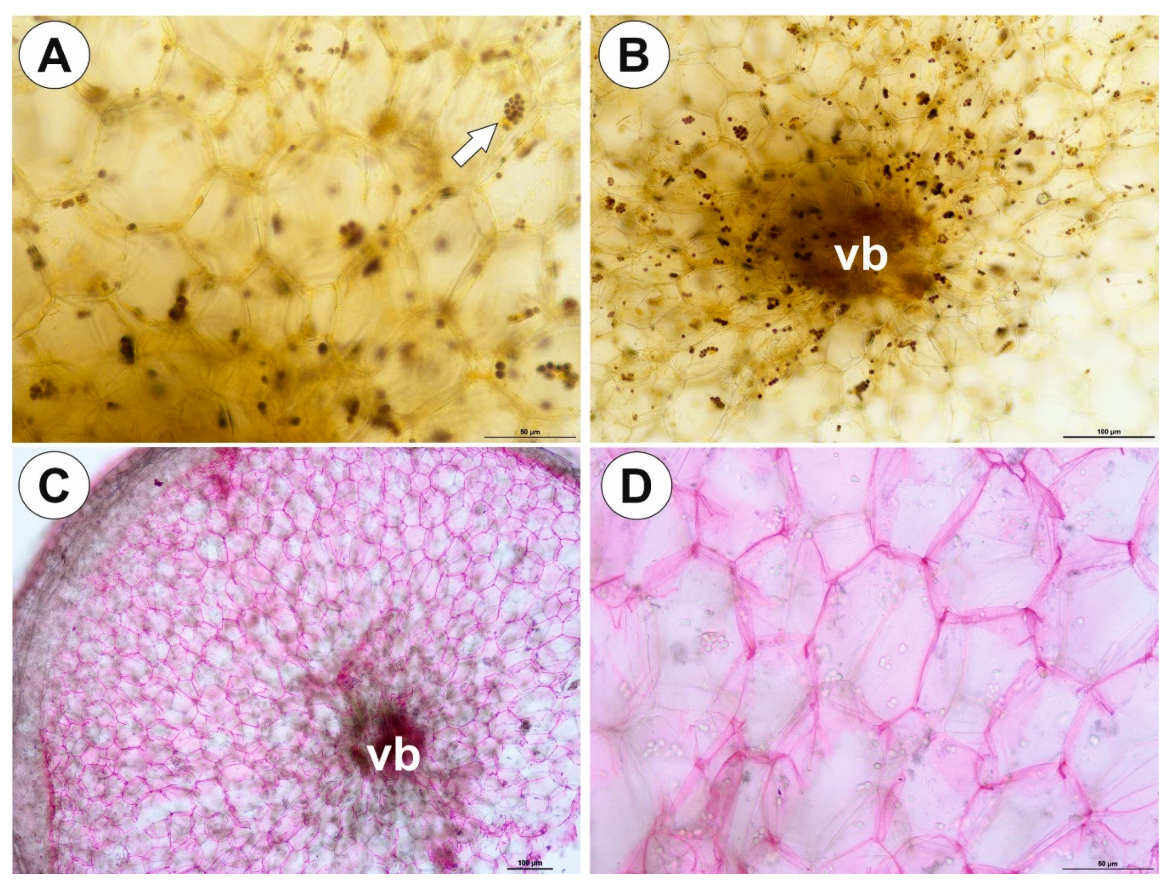
2.1. Genlisea tuberosa Rivadavia, Gonella & A. Fleischm

Up to three tubers occur per plant. The tubers were white-yellow in color, ovoid-shaped (Figure 1A–C). The tuber is attached to the plant by a stalk (Figure 2A). At the tuber pole, where there was stalk, the developing primordia of new organs were observed in the bud (Figure 2B). The tuber surface was covered by mucilage, debris and fungal hyphae (Figure 2C,D). Small epidermal trichomes occurred, each consisting of one basal cell, one short, pedestal cell and a head cell (Figure 2C–E). The lateral wall of the pedestal cell was impregnated by cutin (Figure 2E). The head cell was globular shaped. These trichomes produced mucilage. Stomata were present on the tuber surface (Figure 2C,F). Both open and closed stomata were observed (Figure 2F). As shown in the transverse sections, the tuber was round (Figure 3A,B). The parenchymatous cortex was well developed. Vascular bundles (about 10) formed a ring, with centrally located additional vascular bundles (Figure 3B–D) that showed evidence of branching within the tuber. Within these vascular bundles, there were two groups of phloem cells near the xylem. The xylem is composed of one or two tracheary elements with evident vessels. The vasculature associated with the central part of pith comprised a vessel surrounded by radially elongated parenchymatous cells (Figure 3D). In the pith, there were intercellular spaces. Parenchyma cells of both cortex and pith were highly vacuolated and rich in large starch grains (SGs) (Figure 3E). Smaller SGs also occurred in the epidermal cells (Figure 3F, Table 1). Small SGs were in parenchyma cells of vascular bundles (Figure 4A). SGs, in parenchyma cells of both cortex and pith, were simple (Figure 4C), but in epidermal cells, mixed configurations comprising compound and simple grains co-occurred in the same cell (Figure 4D). Protein storage vacuoles were recorded in the pith near the place where the new bud was formed (Figure 4B). Ruthenium red stained cell walls of parenchyma (Figure 4E), but also head cells of trichomes (Figure 2D and Figure 4E), indicating pectins and mucilage. The PAS (periodic acid-Schiff) reaction revealed that material secreted by trichomes was most likely a polysaccharide (Figure 4F).

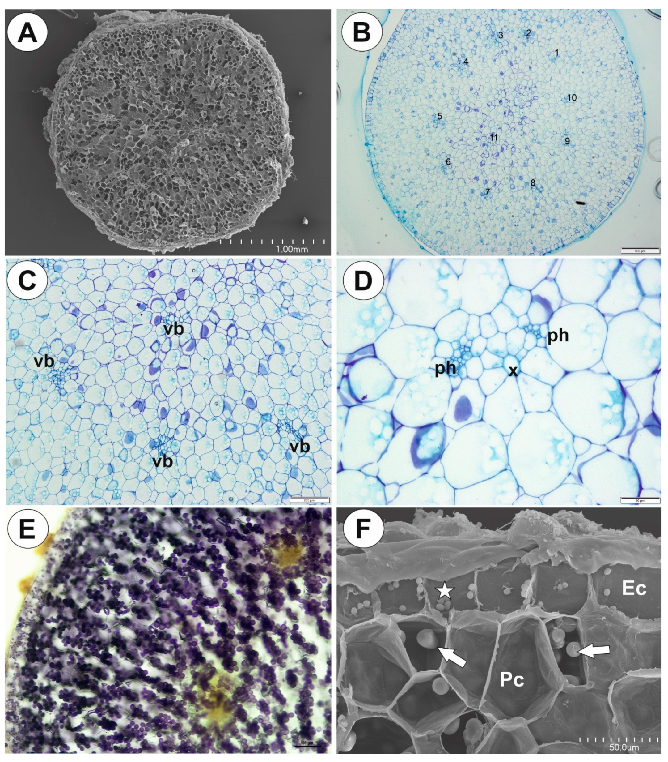
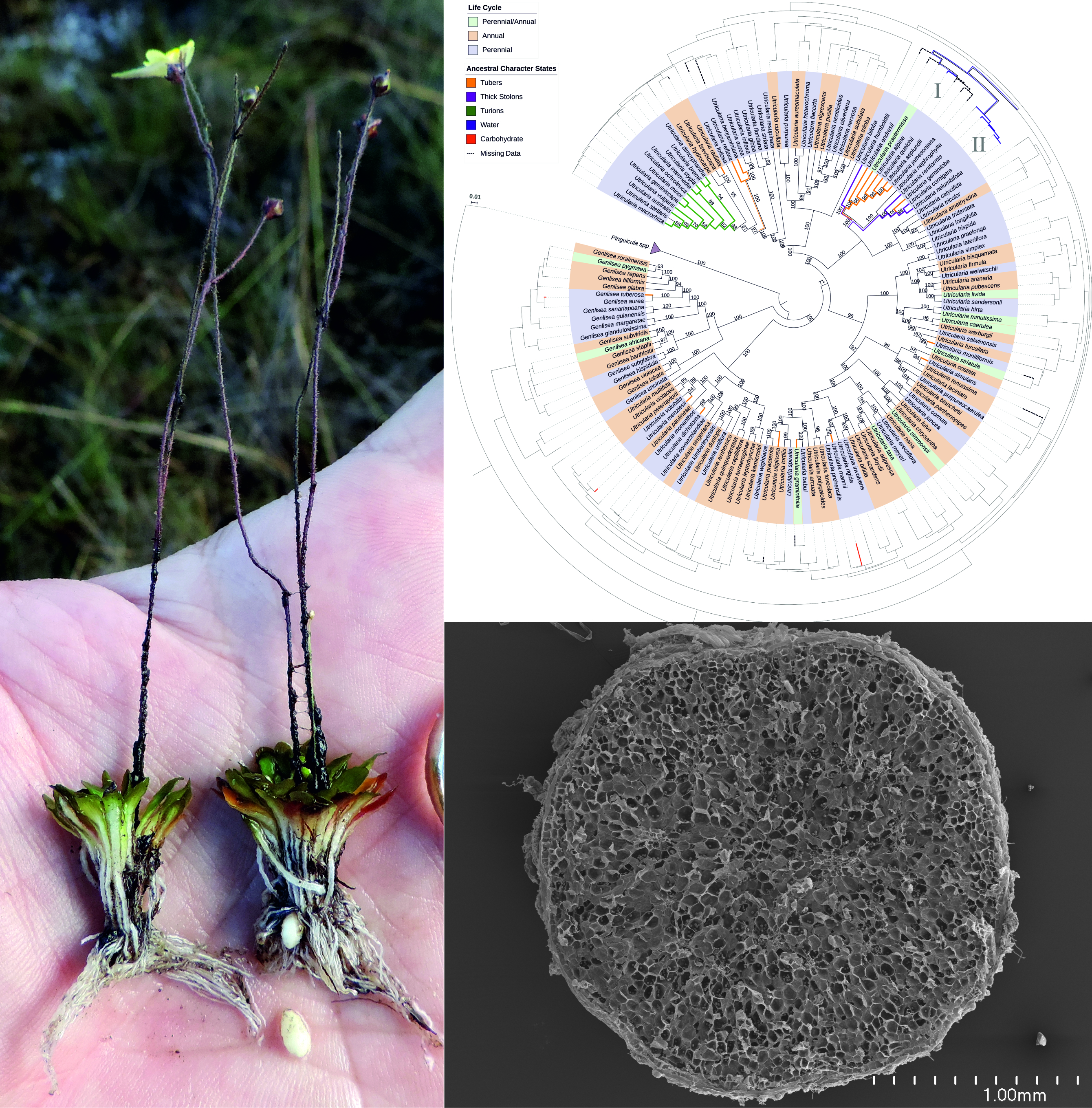
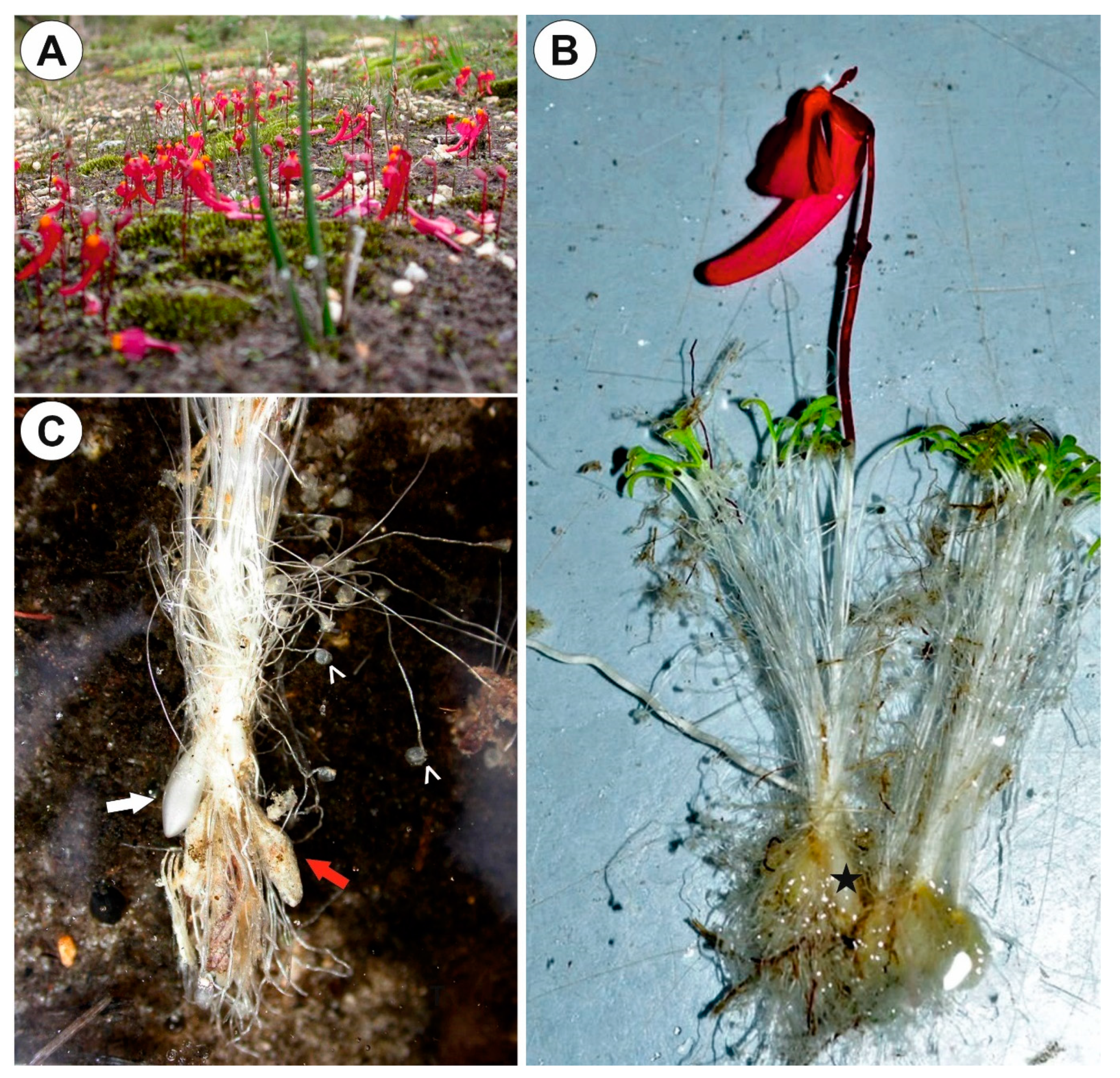
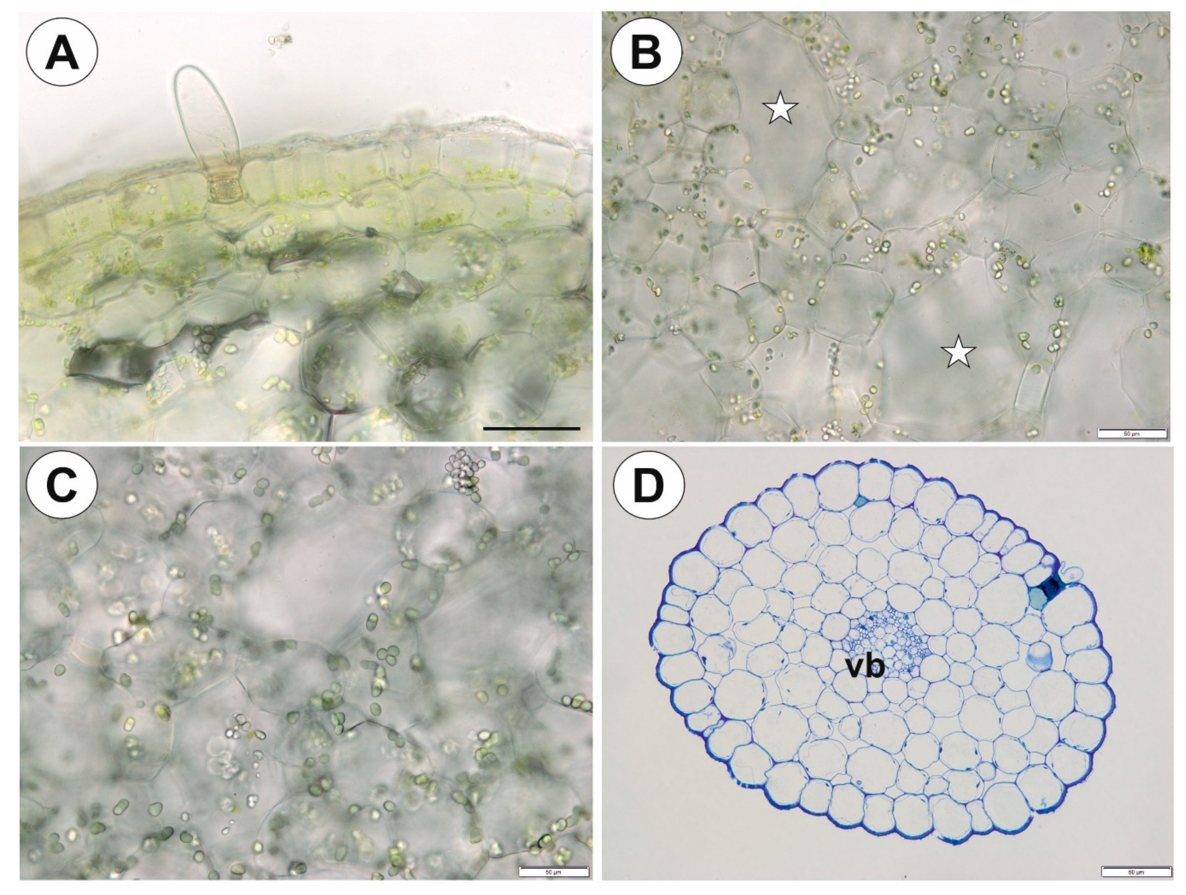
2.2. Utricularia menziesii R.Br.

The tubers were white, turgid, ovoid (Figure 5B,C) attached to a highly abbreviated stem. Old tubers (from the previous season) were flattened and comprised no obvious intact storage cells (Figure 5C and Figure 6A). The tuber was attached to the plant with a stalk, at the other tuber pole no buds or other organs were found. The tuber surface was covered by mucilage, debris and fungal hyphae, nevertheless, no trichomes were recorded (Figure 6B). As shown in the transverse sections, the tuber was round (Figure 6C,D) with a well-developed parenchymatous cortex. There was no clear border between the cortex and pith. Vascular tissues: phloem and xylem were located centrally in the organ (Figure 6C,D). Xylem with one tracheary element and a vessel element surrounded by phloem (Figure 6E,F). Although the xylem and phloem elements were distant from each other, this structure may be treated as a single vascular bundle. Parenchyma cells were highly vacuolated and contained small SGs (Figure 7A,B, Table 1). There were uniform simple and compound SGs (Figure 7A). More SGs occurred in parenchyma which surrounded the vascular tissues (Figure 7B). Mucilage in parenchyma cells was absent, and ruthenium red staining only occurred in the cell walls (Figure 7C,D).

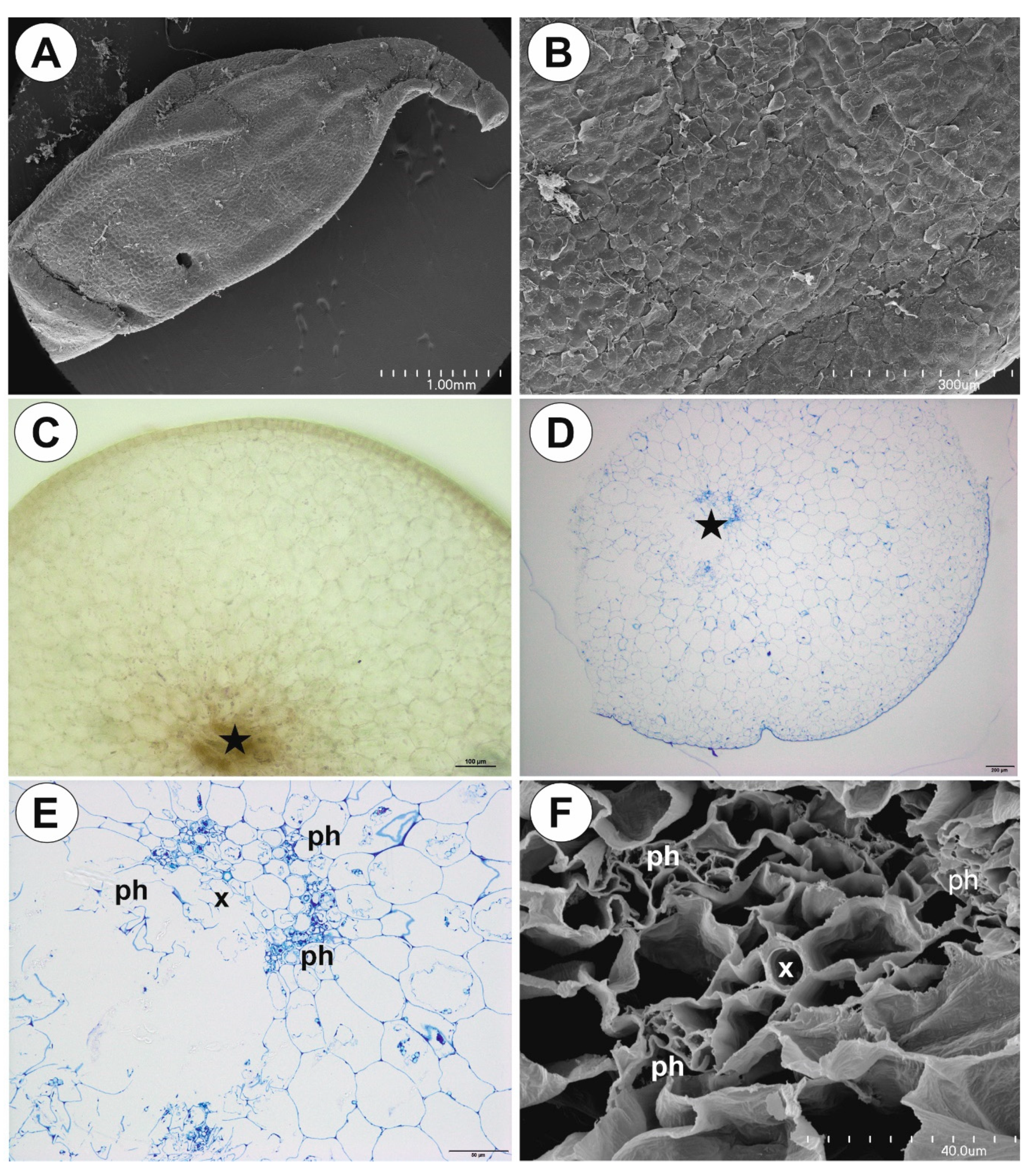
2.3. Utricularia mannii Oliv.

The tubers were green in color, obovoid to globose (Figure 8A). Epidermal trichomes were numerous (Figure 8B), each consisting of one basal cell, one short, pedestal cell and a head cell (Figure 8C). The lateral wall of the pedestal cell was impregnated by cutin (Figure 8B,C). The head cell was strongly elongated (Figure 8C). As shown in the transverse sections, the epidermis and parenchymatous cortex surrounded a large central cylinder (Figure 8D). Vascular bundles formed a ring, with centrally located additional vascular bundles that were larger than the perimeter bundles (Figure 8E,F). Xylem elements were surrounded by phloem (Figure 8F). Chloroplasts occurred in epidermal cells and parenchyma (Figure 9A). Intercellular spaces were well developed and were present both in the cortex and pith (Figure 9B). Parenchyma cells were highly vacuolated and contained small SGs (Figure 9C, Table 1). There were uniform simple SGs and also compound. The stolon had a similar vascular anatomy to the tuber (Figure 9D) though restricted to just one vascular bundle (Figure 9D).

2.4. Phylogenetic Analyses

The concatenated matrix comprehends an alignment of 212 DNA sequences of different species of Lentibulariaceae: 20 of Genlisea, 126 of Utricularia, and 66 sequences of Pinguicula used as the outgroup. The analyses were performed in 4012 characters of which 1755 were parsimony-informative.
The ancestral character tracing (Figure 10, inner tree) shows that stolons have changed to tubers once in Genlisea and that tubers are highly homoplasious in Utricularia, and occurred at least 11 times in the U. radiata, U. reflexa, U. benjaminiana, sect. Orchidioides, U. moniliformis, U. simulans, U. mannii, U. graminifolia, U. uliginosa, U. dichotoma and U. menziesii groups. However, for the U. reflexa-U. benjaminiana-U.aurea-U. inflexa clade, tubers possibly emerged from an ancestor with a stoloniferous habit or that was already bearing tubers. But, for sect. Orchidioides, the two most parsimonious hypotheses for tubers appearance are: (1) that thick stolons and tubers originated from simple (thin) stolons (Figure 10, as indicated by I: thin stolon -> thick stolon [one step] or thick stolon -> tuber [one step]), or (2) that thick stolons originated from simple (thin) stolons with the thickened stolons originating the tubers (Figure 10, indicated by II, in two steps: thin stolon -> thick stolon -> tuber).
Tubers with carbohydrate content evolved at least five times independently in the Lentibulariaceae: in Genlisea tuberosa, Utricularia menziesii, U. mannii lineages (Figure 10, outer tree), U. brachiata and U. inflata. In addition, water storage is found as a synapomorphy, thus only derived once with thick stolons and tubers in sect. Orchidioides.
In addition, ancestral character tracing (Figure 10) shows that turions have appeared as a novel adaptation at least once in Utricularia, within the species of sect Utricularia from temperate climates.

3. Discussion

Darwin [30] examined tubers of Utricularia alpina (as Utricularia montana) and concluded that this species accumulates water that enable the plant to survive seasonal drought. This was later confirmed by Adlassnig [35] and Rodrigues et al. [21]. In addition, other species from sect. Orchidioides produce water-storing tubers or thick stolons [21]. Compton [36] studied tubers of U. brachiata (sect. Phyllaria) and found tuber parenchyma contained starch grains (SG). He concluded the water-storing function is a secondary with the primary function being carbohydrate storage. We found that Genlisea tuberosa, Utricularia menziesii and U. mannii form carbohydrate-rich tubers. SGs exhibited different morphologies and sizes depending on the species. The largest SGs occurred in parenchyma cells of Genlisea tuberosa tubers. The highest number of SGs was recorded in Utricularia menziesii. Accumulation of SGs was recorded also in other Lentibulariaceae perennating organs such as Pinguicula hibernacula ([9]; Płachno unp.) and Utricularia turions (e.g., [19]). Carbohydrate-rich tubers were recorded in the Lentibulariaceae in Genlisea tuberosa and four species of Utricularia that are not closely related (subgen. Polypompholyx: sect. Pleiochasia U. menziesii, subgen. Bivalvaria: sect. Phyllaria U. brachiata, sect. Chelidon U. mannii and subgenus Utricularia: sect. Utricularia U. inflata). Thus, these starch-rich tubers evolved at least five times independently in the Lentibulariaceae lineages. Nonetheless, further investigations are required across storage structures in the family, for example in another two species in sect. Phyllaria showing 1–2-mm-thick tubers, probably also serving as starch-rich vegetative propagules as found in U. christopheri and U. forrestii [14].
Regardless of whether the tubers accumulate starch grains or water, the tubers have well-developed parenchyma, which perform a storage function. Epidermal trichomes produce mucilage that may additionally protect the tuber surface. Mucilage may also interact with microflora and fungi though the function of such co-associating microorganisms is unclear.
Tubers exhibit different vascularization; however, in all species, phloem and xylem elements are evident. The morphological and anatomical tuber characters enable these species to occupy otherwise hostile ecological niches through perennation structures that survive periods of drought. However, tuber vascularization is most likely more related to evolutionary history of species than to organ specialization.
In seasonally dry environments such as the Brazilian Cerrado and kwongkan of south-western Australia the ability to sequester nutrients, moisture and energy in underground organs represents a key strategy for survival [37]. Carbohydrates are stored in various types of organs: tubers, corms, bulbs, rhizomes, rhizophores, tuberous roots, lignotubers, and xylopodia. Starch is the most abundant storage material, with combinations of insoluble and soluble carbohydrates occurring in underground storage organs [37,38,39].
In Genlisea tuberosa and Utricularia menziesii, the subterranean tubers act as a key structure and bud repository able to withstand seasonal drought, summer temperatures and the periodic passage of fire (soil is good heat insulator; see [40]). Several fire adaptations are found in endemic Cerrado flora, including functionally herbaceous or woody geoxylic suffrutices (the ‘underground trees’, see [41]) with enlarged underground xylopodia, lignotubers, thick corky bark, thick shoots, leaves congested at shoot tips and also specialized flowering and fruiting phenologies [42]. However, there is little known of the morphological and anatomical bases to ecological adaptation in tuberous structures (see [43]).
Utricularia menziesii has adopted a growth and development phenology that is similar to that found in most other herbaceous perennial geophytes that occur in the same habitat [22]. Dry season (summer) dormancy occurs from November to May, with sprouting occurring in concert with the onset of cooler and wetter winter conditions. Within a month of sprouting, tuber primordia are evident and these extend and enlarge over the growing season as the parent tubers shrivel and wither. The tuber lacks protective oversummering structures found in many other herbaceous perennial geophytes in the area where U. menziesii grows. For example, common geophytic Drosera accumulate around the tuber paper-like tissues from spent tubers that act to enclose the tuber and function to enhance water retention [22]. Though Utricularia menziesii grows in wet sandy swamps to moss aprons on granite rocks it is surprising that no other Utricularia in the southwest Australian region, a noted hotspot for carnivorous plants, have developed any form of vegetative perennation.
Tubers from sect. Orchidioides and secondary tubers of Utricularia mannii are tuberized (enlarged) stolons [14,21,44], which can continue apical growth to form the tuberous structures. In U. alpina, tubers may have also lateral branches—stolons bearing traps (see illustration in Darwin [30]). In contrast to that, tubers of Genlisea tuberosa and U. menziesii have reduced apical growth and no lateral additional organs. According to Taylor [14], U. mannii grows as an epiphyte on mossy tree trunks; however, this species may grow on rocks as a lithophyte (Figure 8A; Ľuboš Majeský personal observations). Thus, tubers of this species may also have a water-storing function. In addition, Compton [36] proposed both starch and water storage for tubers of U. brachiata. Therefore, the division into tubers which storage only water or only carbohydrates represents an artificial separation.
However, there are some similarities and differences in tuberous structures in the Lentibulariaceae. Epidermal trichomes occur in tubers of Genlisea tuberosa, U. mannii and tubers and thick stolons of species Utricularia from sect. Orchidioides [21]. Darwin [30] did not observe intercellular spaces in tubers of Utricularia alpine however in this study they were found in tubers of U. mannii. Well-developed lacunae were observed in Utricularia nelumbifolia stolons [21]. In the study species, there was no clear partitioning between cortex and pith, but this border is clearly seen in tubers and thick stolons of species Utricularia sect. Orchidioides [21]. There are also clear differences in vascularization in tubers of Genlisea tuberosa, Utricularia menziesii and U. mannii where phloem and xylem formed vascular bundles, but in the tubers and thick stolons of species Utricularia from sect. Orchidioides, [21] the xylem and phloem elements are separated. Vascular bundles with phloem groups flanking the xylem were observed in stolons of Utricularia dichotoma [6]. In tubers of Genlisea tuberosa and U. mannii, vascular bundles are branching in contrast to the tuber of Utricularia menziesii, with single vascular bundle in the tuber. With single, centrally located vascular bundles, tubers of Utricularia menziesii resembles the classical anatomy of root tubers (a ring of collateral bundles is typical for stem tubers in eudicots); however, Utricularia species do not produce roots [14]. Compton [36] also noted single vascular bundles in tubers of U. brachiata. This type of tuber originates from a stolon with one vascular bundle. Such stolons are known for species from section Pleiochasia, to which U. menziesii belongs [6].
For Utricularia mannii, the tubers are very similar to the tubers of species Utricularia from sect. Orchidioides, particularly the occurrence of chlorophyll and glandular trichomes. However, they differ in starch and vascular tissue development.
The phylogenetic hypothesis (Figure 10) are in general congruent to previously published studies [12,27,33,45,46,47,48]. Indeed, we used accessible public databases (mostly from Westermeier [46] and Silva [45]), and performed the analyses using a supermatrix approach with two concatenated chloroplast regions. One is a phylogenetically informative marker for Utricularia and Genlisea species, the matK gene with trnK intron, while the other is the rbcL gene, which is more conserved for these genera. Therefore, the polymorphic nature of the matK/trnK region and the conservation of rbcL [49,50] could provide greater resolution. Considering the two main clades with perennating organs, and ignoring the situation in which these organs are autapomorphic (Genlisea tuberosa, Utricularia menziesii, U. dichotoma, U. uliginosa, U. graminifolia, U. mannii, U. simulans, U. moniliformis and U. radiata), thick stolons and tubers for sect. Orchidioides (Figure 10, I and II) and turions for sect. Utricularia are present in lineages with relatively short branches in comparison with other groups (see the outer tree, Figure 10), even regarding the several factors that could reflect in mutation rates, as body size, population dynamics, and lifestyle among others [51,52]. Taking into account that we applied plastidial DNA sequences, the generation time effect and life history may also affect the rates of molecular evolution in flowering plants (e.g., [53]). This could explain this pattern for lineages of sect. Orchidioides and sect. Utricularia.

4. Materials and Methods

Material of Genlisea tuberosa Rivadavia, Gonella & A.Fleischm. was collected in the Serra da Canastra region, southern Minas Gerais State (Southeastern Brazil), in campos rupestres (rupestrian grasslands) of the Cerrado (collecting permits was ICMBio/MMA/SISBIO #74307-1). Utricularia menziesii R.Br. was collected from the Alison Baird Reserve (Yule Brook) in Western Australia, a privately owned and managed nature reserve. Utricularia mannii Oliv. (from Mount Cameroon, Cameroon, Africa) was cultivated in the Department of Plant Cytology and Embryology, Jagiellonian University in Kraków. Tubers were fixed as below for anatomical and histochemical studies.
The tubers were examined using light microscopy (LM) and scanning electron microscopy (SEM) as follows. Material was fixed in a mixture of 2.5% or 5% glutaraldehyde with 2.5% formaldehyde in a 0.05 M cacodylate buffer (Sigma-Aldrich, Sigma-Aldrich LLB, Poznan, Poland; pH 7.2) overnight or for several days, washed three times in a 0.1 M sodium cacodylate buffer and post-fixed in a 1% osmium tetroxide solution at room temperature for 1.5 h. Later material was treated as previously [54]. The semi-thin sections (0.9–1.0 µm thick) prepared for the LM were stained with aqueous methylene blue/azure II (MB/AII) for 1–2 min [55] and examined using an Olympus BX60, as well as Nikon Eclipse E400 light microscope for the general histology. The periodic acid-Schiff (PAS) reaction for the LM (semi-thin sections) was also used to reveal the presence of insoluble polysaccharides.
Materials were also embedded in Technovit 7100 (Kulzer, Germany) for further histological analysis. This material was fixed (as above), washed three times in a 0.1 M sodium cacodylate buffer, dehydrated in a graded ethanol series for 15 min at each concentration and kept overnight in absolute ethanol. Later, the samples were infiltrated for 1 h each in 3:1, 1:1 and 1:3 (v/v) mixtures of absolute ethanol and Technovit and then stored for 12 h in pure Technovit. The resin was polymerised by adding a hardener. Materials were also sectioned to 5 μm thick using a rotary microtome, stained with 0.1% toluidine blue O (TBO) and mounted in DPX (Sigma-Aldrich, Sigma-Aldrich LLB, Poznan, Poland). The selected Technovit sections were stained with naphthol blue black (NBB) for total protein staining or the periodic acid-Schiff (PAS) reaction was performed for starch visualization.
In order to identify the main classes of the chemical compounds that are present in the tubers, histochemical procedures with the fixed tubers using Lugol’s solution were performed to detect the starch grains and proteins [56]. 0.1% ruthenium red was used for pectin and mucilage detection [57,58].
Tubers were cut using a razor blade and observed under UV light using an Olympus BX60, as well as Nikon Eclipse E400 light microscope to determine: cell walls impregnated with cutin and autofluorescence of chlorophyll.
For the SEM, tubers were fixed (as above) and later dehydrated and critical point dried using CO2. They were then sputter-coated with gold and examined at an accelerating voltage of 20 kV using a Hitachi S-4700 scanning electron microscope (Hitachi, Tokyo, Japan), which is housed in the Institute of Geological Sciences, Jagiellonian University in Kraków, Poland.
We measured starch grain diameter for each species (Table 1) as follows. For each species, one randomly chosen tuber section was selected. The number of starch grain measurements per cell type of a particular species was 100. Each variable was tested using the Shapiro–Wilk W-test for normality. The homogeneity of variance was estimated with Levene’s test. Statistical differences in starch grains diameter in each species were assessed using one-way ANOVA, followed by Tukey’s post-hoc comparison test. Statistical analyses were performed on the raw data using Statistica 13 software (StatSoft Inc., Oklahoma, USA). Data from measurements of starch grain diameter were expressed in µm as mean ± SD. Data were considered statistically significant at *** p < 0.001. We measured also number of starch grains per 100 µm2 (Table 2). For each species one randomly chosen tuber section was selected.
To reconstruct the phylogenetic hypothesis, we aligned sequences of chloroplast regions matK/trnK and rbcL genes from GenBank Nucleotide database (Table S1) using MAFFT version 7 [59] with default parameters and generated a supermatrix with FASConCAT-G version 1.04 [60]. With the resulting supermatrix we calculated a Maximum Likelihood phylogenetic trees with IQ-TREE version 1.6.12 [61] using TVM+F+I+G4 model parameters chosen according with AIC criterion [62] using ModelFinder [63] implemented in IQ-TREE. Clade support was evaluated using ultrafast bootstrap [64] with 1000 replicates. Gaps were treated as missing data. The ancestral state reconstruction was performed using Maximum Parsimony criteria from a character matrix developed according to data from the present study and previous publications [12,14,20,21,34] to map the absence or presence of turions, tubers, thick stolons and the type of storage material into the molecular phylogeny using Mesquite version 3.61 [65]. The final tree was edited using Interactive Tree of Life (iTol) version 5.5.1 [66].

5. Conclusions

In contrast to species from section Orchidioides, which produce tubers storing mainly water, Genlisea tuberosa, Utricularia menziesii and U. mannii form starch-rich tubers. Such tubers evolved independently at least five times in the family Lentibulariaceae. In Genlisea tuberosa and Utricularia menziesii, underground tubers are a perennating structure that are key strategy to survive in summer fire-prone environments. In contrast to examined species here, in tubers of species from section Orchidioides the xylem and phloem elements are separated from each other which supports molecular studies from previous studies that this species is not related to species from Orchidioides. In contrast to most Utricularia and Genlisea lineages, where tubers evolved from regular stolons, sect. Orchidioides probably evolved through a continuum of transformation going from stolons, thick stolons to tubers. The occurrence of stomata on the underground tubers of G. tuberosa is unusual and point to a stem-based origin for the tuber tissue. Both tubers of G. tuberosa, and U. mannii had epidermal trichomes, which produce mucilage for protection. In G. tuberosa and U. mannii, vascular bundles formed a ring, but there was a centrally located additional vascular bundle. However, in U. menziesii, the single vascular bundle was located centrally in the tuber.

Supplementary Materials

Supplementary Materials can be found at https://www.mdpi.com/1422-0067/21/14/5143/s1. Table S1. Data used for the phylogenetic analyses. “-” denotes missing data. Pinguicula species were 20 used as outgroup.

Author Contributions

B.J.P. designed the project; B.J.P., V.F.O.M., G.C.S. collected plants and cultivated the plants; B.J.P., P.Ś., K.L., S.R.S. performed analysis and data visualization; B.J.P., S.R.S., prepared the original manuscript draft; P.Ś., K.W.D., V.F.O.M. were involved in the experimental ideas and critically revised the manuscript. All authors have read and agreed to the published version of the manuscript.

Funding

This research was partially supported financially to B.J.P. by the Ministry of Science and Higher Education of Poland as part of the statutory activities of the Department of Plant Cytology and Embryology, Institute of Botany, Faculty of Biology, Jagiellonian University in Kraków (N18/DBS/000002), and Institute of Biology, Biotechnology and Environmental Protection, Faculty of Natural Sciences, University of Silesia in Katowice. The scientific stay of BJP in Brazil was financed by the Jagiellonian University within the project Integrate JU-Comprehensive Development Programme of the Jagiellonian University as part of the Knowledge Education Development 2014–2020 Operational Programme co-financed by the European Social Fund of the European Union (POWR.03.05.00-00Z309/17-00). We thank Conselho Nacional de Desenvolvimento Científico e Tecnológico (CNPq) for the fellowship of V.F.O.M. (Bolsa de Produtividade - Proc. # 312908/2018-0).

Acknowledgments

Sincere thanks are due to Ľuboš Majeský (Palacký University in Olomouc) and André J. Arruda for permission to use of their photos. Sincere thanks are due to Hans Lambers for permission to use his photos and kind help both in case colleting of material and discussion about manuscript. We cordially thank Rolf Rutishauser ISEB-UZH (Zurich, Switzerland) for critically reading the manuscript and for all valuable suggestions.

Conflicts of Interest

The authors declare no conflicts of interest.

References

  1. Król, E.; Płachno, B.J.; Adamec, L.; Stolarz, M.; Dziubińska, H.; Trebacz, K. Quite a few reasons for calling carnivores “the most wonderful plants in the world”. Ann. Bot. 2012, 109, 47–64. [Google Scholar] [CrossRef] [PubMed] [Green Version]
  2. Poppinga, S.; Weisskopf, C.; Westermeier, A.S.; Masselter, T.; Speck, T. Fastest predators in plant kingdom: Functional morphology and biomechanics of suction traps found in the largest genus of carnivorous plants. AoB Plants 2015, 8, plv140. [Google Scholar] [CrossRef] [PubMed] [Green Version]
  3. Płachno, B.J.; Muravnik, L.E. Functional anatomy of carnivorous traps. In Carnivorous Plants: Physiology, Ecology, and Evolution; Elisson, A.M., Adamec, L., Eds.; Oxford University Press: Oxford, UK, 2018; pp. 167–179. [Google Scholar]
  4. Płachno, B.J.; Świątek, P.; Miranda, V.F.O.; Stolarczyk, P. The structure and occurrence of a velum in Utricularia traps (Lentibulariaceae). Front. Plant Sci. 2019, 10, 302. [Google Scholar] [CrossRef]
  5. Rutishauser, R. Evolution of unusual morphologies in Lentibulariaceae (bladderworts and allies) and Podostemaceae (river-weeds): A pictorial report at the interface of developmental biology and morphological diversification. Ann. Bot. 2016, 117, 811–832. [Google Scholar] [CrossRef] [PubMed] [Green Version]
  6. Reut, M.S.; Płachno, B.J. Unusual developmental morphology and anatomy of vegetative organs in Utricularia dichotoma—Leaf, shoot and root dynamics. Protoplasma 2020, 371–390. [Google Scholar] [CrossRef] [PubMed] [Green Version]
  7. Whitewoods, C.D.; Gonçalves, B.; Cheng, J.; Cui, M.; Kennaway, R.; Lee, K.; Bushell, C.; Yu, M.; Piao, C.; Coen, E. Evolution of carnivorous traps from planar leaves through simple shifts in gene expression. Science 2020, 367, 91–96. [Google Scholar] [CrossRef] [PubMed]
  8. Płachno, B.J.; Adamec, L.; Świątek, P.; Kapusta, M.; Miranda, V.F.O. Life in the Current: Anatomy and Morphology of Utricularia neottioides. Int. J. Mol. Sci. 2020, 21, 4474. [Google Scholar] [CrossRef]
  9. Heslop-Harrison, Y. Pinguicula, L. J. Ecol. 2004, 92, 1071–1118. [Google Scholar] [CrossRef]
  10. Fleischmann, A.; Roccia, A. Systematics and evolution of Lentibulariaceae: I. Pinguicula, 1st ed.; Ellison, A.M., Adamec, L., Eds.; Oxford University Press: Oxford, UK, 2018; Volume 1, ISBN 9780198779841. [Google Scholar]
  11. Rivadavia, F. A Genlisea myth is confirmed. Carniv. Plant Newletter 2007, 36, 122–125. [Google Scholar]
  12. Fleischmann, A. Monograph of the Genus Genlisea; Redfern Natural History Productions: Dorset, UK, 2012; ISBN 9781908787002. [Google Scholar]
  13. Rivadavia, F.; Gonella, P.M.; Fleischmann, A. A New and Tuberous Species of Genlisea (Lentibulariaceae) from the Campos Rupestres of Brazil. Syst. Bot. 2013, 38, 464–470. [Google Scholar] [CrossRef]
  14. Taylor, P. The Genus Utricularia—A Taxonomic Monograph.; The Royal Botanic Gardens: London, UK, 1989; p. 724. [Google Scholar]
  15. Adamec, L. Ecophysiology of aquatic carnivorous plants. In Carnivorous Plants: Physiology, Ecology, and Evolution; Elisson, A.M., Adamec, L., Eds.; Oxford University Press: Oxford, UK, 2018; Volume 1. [Google Scholar]
  16. Adamec, L. Turion overwintering of aquatic carnivorous plants. Carniv. Plant Newletter 1999, 28, 19–24. [Google Scholar]
  17. Adamec, L. Respiration of turions and winter apices in aquatic carnivorous plants. Biologia (Bratisl). 2008, 63, 515–520. [Google Scholar] [CrossRef] [Green Version]
  18. Adamec, L. Ecophysiological characteristics of turions of aquatic plants: A review. Aquat. Bot. 2018, 148, 64–77. [Google Scholar] [CrossRef]
  19. Płachno, B.J.; Adamec, L.; Kozieradzka-Kiszkurno, M.; Światek, P.; Kamińska, I. Cytochemical and ultrastructural aspects of aquatic carnivorous plant turions. Protoplasma 2014, 251, 1449–1454. [Google Scholar] [CrossRef] [Green Version]
  20. Rice, B. Tuberous organs in Utricularia, and new observations of sub-tuberous stolons on Utricularia radiata Small. Carniv. Plant Newsl. 2011, 40, 88–91. [Google Scholar]
  21. Rodrigues, F.G.; Marulanda, N.F.; Silva, S.R.; Płachno, B.J.; Adamec, L.; Miranda, V.F.O. Phylogeny of the “orchid-like” bladderworts (gen. Utricularia sect. Orchidioides and Iperua: Lentibulariaceae) with remarks on the stolon-tuber system. Ann. Bot. 2017, 120, 709–723. [Google Scholar]
  22. Pate, J.S.; Dixon, K.W. Tuberous, Cormous and Bulbous Plants: Biology of an Adaptive Strategy in Western Australia; University of Western Australia Press: Nedlands, Australia, 1982. [Google Scholar]
  23. Lowrie, A. Carnivorous Plants of Australia: Magnum Opus; Robinson, A., Ed.; Redfern Natural History Productions: Dorset, UK, 2013; Volume 1. [Google Scholar]
  24. Cross, A.T.; Davis, A.R.; Fleischmann, A.; Horner, J.D.; Jürgens, A.; Merritt, D.J.; Murza, G.L.; Turner, S.R. Reproductive biology and pollinator-prey conflicts. In Carnivorous Plants: Physiology, Ecology, and Evolution; Elisson, A.M., Adamec, L., Eds.; Oxford University Press: Oxford, UK, 2018; Volume 1. [Google Scholar]
  25. Menezes, C.G.; Gasparino, E.C.; Baleeiro, P.C.; Miranda, V.F.O. Seed morphology of bladderworts: A survey on Utricularia sect. Foliosa and sect. Psyllosperma (Lentibulariaceae) with taxonomic implications. Phytotaxa 2014, 167, 173. [Google Scholar] [CrossRef] [Green Version]
  26. Silveira, F.A.O.; Negreiros, D.; Barbosa, N.P.U.; Buisson, E.; Carmo, F.F.; Carstensen, D.W.; Conceição, A.A.; Cornelissen, T.G.; Echternacht, L.; Fernandes, G.W.; et al. Ecology and evolution of plant diversity in the endangered campo rupestre: A neglected conservation priority. Plant Soil 2016, 403, 129–152. [Google Scholar] [CrossRef] [Green Version]
  27. Jobson, R.W.; Baleeiro, P.C.; Reut, M.S. Molecular phylogeny of subgenus Polypompholyx (Utricularia; Lentibulariaceae) based on three plastid markers: Diversification and proposal for a new section. Aust. Syst. Bot. 2017, 30, 259–278. [Google Scholar] [CrossRef]
  28. Jobson, R.W.; Baleeiro, P.C.; Guisande, C. Systematics and evolution of Lentibulariaceae: III. Utricularia. In Carnivorous Plants: Physiology, Ecology, and Evolution; Elisson, A.M., Adamec, L., Eds.; Oxford University Press: Oxford, UK, 2018; pp. 89–104. [Google Scholar]
  29. Płachno, B.J.; Stpiczyńska, M.; Świątek, P.; Lambers, H.; Miranda, V.F.O.; Nge, F.J.; Stolarczyk, P.; Cawthray, G.R. Floral micromorphology of the bird-pollinated carnivorous plant species Utricularia menziesii R.Br. (Lentibulariaceae). Ann. Bot. 2018, 123, 213–220. [Google Scholar] [CrossRef] [Green Version]
  30. Darwin, C. Insectivorous Plants; Murray, J., Ed.; London, 1875. Available online: http://darwin-online.org.uk/ (accessed on 5 May 2020).
  31. Adlassnig, W.; Peroutka, M.; Eder, G.; Pois, W.; Lichtscheidl, I.K. Ecophysiological observations on Drosophyllum lusitanicum. Ecol. Res. 2005, 21, 255–262. [Google Scholar] [CrossRef]
  32. Westermeier, A.S.; Fleischmann, A.; Müller, K.; Schäferhoff, B.; Rubach, C.; Speck, T.; Poppinga, S. Trap diversity and character evolution in carnivorous Lentibulariaceae). Sci. Rep. 2017, 7, 1–24. [Google Scholar] [CrossRef] [PubMed]
  33. Müller, K.; Borsch, T. Phylogenetics of Utricularia (Lentibulariaceae) and molecular evolution of the trnK intron in a lineage with high substitutional rates. Plant Syst. Evol. 2005, 250, 39–67. [Google Scholar] [CrossRef]
  34. Fleischmann, A.; Schäferhoff, B.; Heubl, G.; Rivadavia, F.; Barthlott, W.; Müller, K.F. Phylogenetics and character evolution in the carnivorous plant genus Genlisea A. St.-Hil. (Lentibulariaceae). Mol. Phylogenet. Evol. 2010, 56, 768–783. [Google Scholar] [CrossRef] [PubMed]
  35. Adlassnig, W.; Peroutka, M.; Lambers, H.; Lichtscheidl, I.K. The roots of carnivorous plants. Plant Soil 2005, 274, 127–140. [Google Scholar] [CrossRef]
  36. Compton, R.H. The morphology and anatomy of Utricularia brachiata Oliver. New Phytol. 1909, 8, 117–130. [Google Scholar] [CrossRef]
  37. Oliveira, R.S.; Abrahão, A.; Pereira, C.; Teodoro, G.S.; Brum, M.; Alcantara, S.; Lambers, H. Ecophysiology of campos rupestres plants. In Ecology and Conservation of Mountaintop Grasslands in Brazil; Fernandes, G.W., Ed.; Springer International Publishing AG: Cham, Switzerland, 2016; pp. 227–272. [Google Scholar]
  38. de Joaquim, E.O.; de Figueiredo-Ribeiro, R.C.L.; Hayashi, A.H.; de Carvalho, M.A.M. Inulin contents and tissue distribution in underground storage organs of Asteraceae species from the Brazilian rocky fields. Botany 2014, 92, 827–836. [Google Scholar] [CrossRef]
  39. Carvalho, M.A.M.; Asega, A.F.; Figueiredo-Ribeiro, R.C.L. Fructans in Asteraceae from Brazilian cerrado. In Recent Advances In Fructooligosaccharides Research; Shiomi, N., Benkeblia, N., Onodera, S., Eds.; Research Signpost: Kerala, India, 2007; pp. 69–91. [Google Scholar]
  40. Auld, T.D.; Bradstock, R.A. Soil temperatures after the passage of a fire: Do they influence the germination of buried seeds? Aust. J. Ecol. 1996, 21, 106–109. [Google Scholar] [CrossRef]
  41. Meerts, P. Geoxylic suffrutices of African savannas: Short but remarkably similar to trees. J. Trop. Ecol. 2017, 33, 295–298. [Google Scholar] [CrossRef] [Green Version]
  42. Simon, M.F.; Grether, R.; De Queiroz, L.P.; Skemae, C.; Pennington, R.T.; Hughes, C.E. Recent assembly of the Cerrado, a neotropical plant diversity hotspot, by in situ evolution of adaptations to fire. Proc. Natl. Acad. Sci. USA 2009, 106, 20359–20364. [Google Scholar] [CrossRef] [Green Version]
  43. Pausas, J.G.; Lamont, B.B.; Paula, S.; Appezzato-da-Glória, B.; Fidelis, A. Unearthing belowground bud banks in fire-prone ecosystems. New Phytol. 2018, 217, 1435–1448. [Google Scholar] [CrossRef] [PubMed] [Green Version]
  44. Juniper, B.E.; Robins, R.J.; Joel, D.M. The Carnivorous Plants; Academic Press: London, UK, 1989; p. 353. [Google Scholar]
  45. Silva, S.R.; Gibson, R.; Adamec, L.; Domínguez, Y.; Miranda, V.F.O. Molecular phylogeny of bladderworts: A wide approach of Utricularia (Lentibulariaceae) species relationships based on six plastidial and nuclear DNA sequences. Mol. Phylogenet. Evol. 2018, 118, 244–264. [Google Scholar] [CrossRef] [PubMed] [Green Version]
  46. Westermeier, A.S.; Fleischmann, A.; Müller, K.; Schäferhoff, B.; Rubach, C.; Speck, T.; Poppinga, S. Trap diversity and character evolution in carnivorous bladderworts (Utricularia, Lentibulariaceae). Sci. Rep. 2017, 7, 12052. [Google Scholar] [CrossRef]
  47. Reut, M.S.; Jobson, R.W. A phylogenetic study of subgenus Polypompholyx: A parallel radiation of Utricularia (Lentibulariaceae) throughout Australasia. Aust. Syst. Bot. 2010, 23, 152–161. [Google Scholar] [CrossRef]
  48. Jobson, R.W.; Playford, J.; Cameron, K.M.; Albert, V.A. Molecular Phylogenetics of Lentibulariaceae Inferred from Plastid rps16 Intron and trnL-F DNA Sequences: Implications for Character Evolution and Biogeography. Syst. Bot. 2003, 28, 157–171. [Google Scholar]
  49. Silva, S.R.; Pinheiro, D.G.; Penha, H.A.; Płachno, B.J.; Michael, T.P.; Meer, E.J.; Miranda, V.F.O.; Varani, A.M. Intraspecific Variation within the Utricularia amethystina Species Morphotypes Based on Chloroplast Genomes. Int. J. Mol. Sci. 2019, 20, 6130. [Google Scholar] [CrossRef] [Green Version]
  50. Silva, S.R.; Michael, T.P.; Meer, E.J.; Pinheiro, D.G.; Varani, A.M.; Miranda, V.F.O. Comparative genomic analysis of Genlisea (corkscrew plants—Lentibulariaceae) chloroplast genomes reveals an increasing loss of the ndh genes. PLoS ONE 2018, 13, e0190321. [Google Scholar] [CrossRef] [Green Version]
  51. Bromham, L. Why do species vary in their rate of molecular evolution? Biol. Lett. 2009, 5, 401–404. [Google Scholar] [CrossRef] [Green Version]
  52. Lehtonen, J.; Lanfear, R. Generation time, life history and the substitution rate of neutral mutations. Biol. Lett. 2014, 10, 20140801. [Google Scholar] [CrossRef] [PubMed] [Green Version]
  53. Smith, S.A.; Donoghue, M.J. Rates of molecular evolution are linked to life history in flowering plants. Science 2008, 322, 86–89. [Google Scholar] [CrossRef] [PubMed] [Green Version]
  54. Płachno, B.J.; Stpiczyńska, M.; Davies, K.L.; Świątek, P.; de Miranda, V.F.O. Floral ultrastructure of two Brazilian aquatic-epiphytic bladderworts: Utricularia cornigera Studnička and U. nelumbifolia Gardner (Lentibulariaceae). Protoplasma 2017, 254, 353–366. [Google Scholar] [CrossRef] [PubMed] [Green Version]
  55. Humphrey, C.D.; Pittman, F.E. A Simple Methylene Blue-Azure Ii-Basic Fuchsin Stain for Epoxy-Embedded Tissue Sections. Stain Technol. 1974, 49, 9–14. [Google Scholar] [CrossRef] [PubMed]
  56. Johansen, D.A. Plant Microtechnique; McGraw-Hil Book Company Inc.: New York, NY, USA, 1940. [Google Scholar]
  57. Ruzin, S.E. Plant Microtechnique and Microscopy; Oxford University Press: New York, NY, USA, 1999. [Google Scholar]
  58. Kuzdowicz, A. Mikrotechnika roślinna; Panstw. Wyd. Roln. I Leśne: Warszawa, Polska, 1951. [Google Scholar]
  59. Kazutaka Katoh; John Rozewicki; Kazunori D Yamada MAFFT online service: Multiple sequence alignment, interactive sequence choice and visualization. Brief. Bioinform. 2019, 20, 1160–1166. [CrossRef] [PubMed] [Green Version]
  60. Kück, P.; Longo, G.C. FASconCAT-G: Extensive functions for multiple sequence alignment preparations concerning phylogenetic studies. Front. Zool. 2014, 11, 81. [Google Scholar] [CrossRef] [PubMed] [Green Version]
  61. Nguyen, L.-T.; Schmidt, H.A.; von Haeseler, A.; Minh, B.Q. IQ-TREE: A Fast and Effective Stochastic Algorithm for Estimating Maximum-Likelihood Phylogenies. Mol. Biol. Evol. 2014, 32, 268–274. [Google Scholar] [CrossRef] [PubMed]
  62. Akaike, H. Information theory and an extension of the maximum likelihood principle. Int. Symp. Inf. Theory 1973, 267–281. [Google Scholar]
  63. Kalyaanamoorthy, S.; Minh, B.Q.; Wong, T.K.F.; von Haeseler, A.; Jermiin, L.S. ModelFinder: Fast model selection for accurate phylogenetic estimates. Nat. Methods 2017, 14, 587–589. [Google Scholar] [CrossRef] [Green Version]
  64. Hoang, D.T.; Chernomor, O.; von Haeseler, A.; Minh, B.Q.; Vinh, L.S. UFBoot2: Improving the Ultrafast Bootstrap Approximation. Mol. Biol. Evol. 2017, 35, 518–522. [Google Scholar] [CrossRef]
  65. Maddison, W.P.; Maddison, D.R. Mesquite: A modular system for evolutionary analysis. Version 3.61. Available online: http://mesquiteproject.org/ (accessed on 5 May 2020).
  66. Letunic, I.; Bork, P. Interactive Tree Of Life v2: Online annotation and display of phylogenetic trees made easy. Nucleic Acids Res. 2011, 39, W475–W478. [Google Scholar] [CrossRef]
Figure 1. Genlisea tuberosa in its natural habitat, the Serra da Canastra, southern Minas Gerais State (south-eastern Brazil), in the rocky fields of the Cerrado. (A). Inflorescences of Genlisea tuberosa in the Serra da Canastra. (B). Exposed traps and tubers (arrows). (C). Two excavated plants. (D). Tubers.
Ijms 21 05143 g001
Figure 2. Structure of Genlisea tuberosa tubers. (A). Scanning electron micrograph (SEM) of a tuber; scale bar = 1 mm. (B). Longitudinal section through the tuber, note bud (arrow), PAS (periodic acid-Schiff) reaction; scale bar = 100 µm. (C). SEM micromorphology of the tuber surface: glandular trichomes (star), stoma (S); scale bar = 50 µm. (D). A tuber section stained with ruthenium red: glandular trichomes (star), fungal hyphae (arrows), parenchyma (Pc); scale bar = 100 µm. (E). Structure of epidermal glandular trichome, note fluorescence of the pedestal cell that is heavily impregnated with cutin: basal cell (b), pedestal cell (arrow), head cell (star), epidermal cell (Ec), parenchyma cell (Pc); scale bar = 60 µm. (F). SEM, stoma from tuber surface; scale bar = 30 µm.
Ijms 21 05143 g002
Figure 3. Anatomy and histochemistry of Genlisea tuberosa tubers. (A,B). General tuber anatomy. Note the numerous vascular bundles (numbers), A. SEM; scale bar = 1mm and 500 µm. (C,D). Vascular bundles (vb): phloem (ph), xylem vessel (x); scale bar = 200 µm and 50 µm. E. A part of tuber section after Lugol’s solution treatment, note numerous starch grains fill the cells; scale bar = 100 µm. (F). SEM, of the outer part of the tuber, note differences in sizes between starch grains in parenchyma cells (arrows, Pc) and epidermal cells (star, EC); scale 50 µm.
Ijms 21 05143 g003
Figure 4. Histochemistry of Genlisea tuberosa tubers. (A). Vascular bundle, PAS reaction, note small starch grains: phloem (ph), xylem vessel (x); scale bar = 10 µm. (B). Tuber section stained with naphthol blue black for protein, note accumulation of protein storage vacuoles accumulation in cells (circle) near bud; scale bar = 100 µm. (C). Simple starch grains in parenchyma cells, Lugol’s solution treatment; scale bar = 100 µm. (D). Starch grains in parenchyma cells (Pc) and composed starch grains (arrow) in epidermal cells (Ec); scale bar = 20 µm. (E). Tuber section treated with ruthenium red: note positive reaction in cell walls of parenchyma cells and head cells of glandular trichomes (T)); scale bar = 100 µm. (F). Positive result of the PAS reaction of material (star) secreted by trichomes (T); scale bar = 10 µm.
Ijms 21 05143 g004
Figure 5. Utricularia menziesii in natural habitat, the Alison Baird Reserve (Yule Brook) in Western Australia. (A). Habitat of Utricularia menziesii, (photo Hans Lambers). (B). Excavated plants; tuber (star), author of photo André J. Arruda. (C). A part of excavated plant with traps (arrowhead) and tubers: young tuber (white arrow), old tuber (red arrows) (photo Hans Lambers).
Ijms 21 05143 g005
Figure 6. Tuber structure of Utricularia menziesii. (A). SEM, morphology of tuber; scale bar = 1mm. (B). SEM, micromorphology of tuber surface; scale bar = 300 µm. (C,D). General tuber anatomy. Note the vascular tissues (black star); scale bar = 100 µm and scale bar = 200 µm. (E,F). Structure of vascular tissues: phloem (ph), xylem (x); (F). SEM; scale bar = 50 µm and scale bar = 40 µm.
Ijms 21 05143 g006
Figure 7. Histochemistry of Utricularia menziesii tubers. (A,B). Tuber section after Lugol’s solution treatment: compound starch grain (arrow), vascular bundle (vb); scale bar = 50 µm and scale bar = 100 µm. (C,D). Tuber sections treated with ruthenium red: note positive reaction in case cell walls of parenchyma cells; vascular bundle (vb); scale bar = 100 µm and scale bar = 50 µm.
Ijms 21 05143 g007
Figure 8. Tuber structure of Utricularia mannii. (A). Utricularia mannii habitat, Cameroon, Africa; (photo Dr. Ľuboš Majeský), insert – tuber. (B). Surface of tuber covered by numerous glandular trichomes, note bright blue fluorescence of trichome pedestal cells and red fluorescence of chlorophyll – chloroplast in epidermal cells and parenchyma cells; scale bar = 10 µm. (C). Structure of epidermal glandular trichome: basal cell (Bc), pedestal cell (star), head cell (Hc); scale bar = 20 µm. (D,E). General tuber anatomy. Note the vascular bundles (vb); scale bar = 200 µm and scale bar = 100 µm. (F). Structure of vascular bundle: phloem (ph), xylem vessel (x); scale bar = 20 µm.
Ijms 21 05143 g008
Figure 9. Tuber and stolon structure of Utricularia mannii. (A). Chloroplasts in epidermal cells and parenchyma; scale bar = 50 µm. (B,C). Starch grains in the parenchyma cells, note well developed intercellular spaces; scale bar = 50 µm. (D). Stolon anatomy, vascular bundle (vb); scale bar = 50 µm.
Ijms 21 05143 g009
Figure 10. Phylogenetic trees were reconstructed with the Maximum Likelihood method. The inner tree and outer tree indicate the ancestral character states reconstructed according to parsimony criteria. The inner tree shows the possible ancestral character states of tubers, thick stolons and turions (orange, purple and green branch color, respectively). Branch numbers correspond to ultrafast bootstrap values. The outer tree shows the possible ancestral character states of the tuber and thick stolons content: carbohydrate in red; water in blue; dashed lines correspond to unknown data. The names of species are colored according to life cycle: purple for perennial, orange for annual and green for annual and/or perennial species according to Taylor [14] and Fleischmann [34]. I and II denote potential points of tuber appearance. Pinguicula species were used as the outgroup.
Ijms 21 05143 g010
Table 1. Starch grain diameter in Genlisea tuberosa, Utricularia mannii and U. menziesii.
SpeciesCell TypeDiameter of Starch Grains (mean ± SD)
Genlisea tuberosaParenchyma17.34 ± 3.07 µm
Epidermis7.57 ± 1.53 µm
Utricularia manniiParenchyma7.01 ± 1.46 µm
Utricularia menziesiiParenchyma3.76 ± 0.70 µm
Table 2. Number of starch grains per 100 µm2 in Genlisea tuberosa, Utricularia mannii and U. menziesii.
SpeciesNumber of Starch Grains Per 100 µm2 (mean ± SD)
Genlisea tuberosa22.6 ± 3.59
Utricularia mannii32.0 ± 5.83
Utricularia menziesii36.4 ± 5.85

