

Is Melanoma Progression Affected by Thyroid Diseases?
 , , , , , , and
Abstract
:1. Introduction
2. Thyroid Diseases
3. Melanoma
4. Epidemiological Evidence for Thyroid Disease–Melanoma Association
4.1. Benign Thyroid Diseases and Melanoma
4.2. Malignant Thyroid Diseases and Melanoma
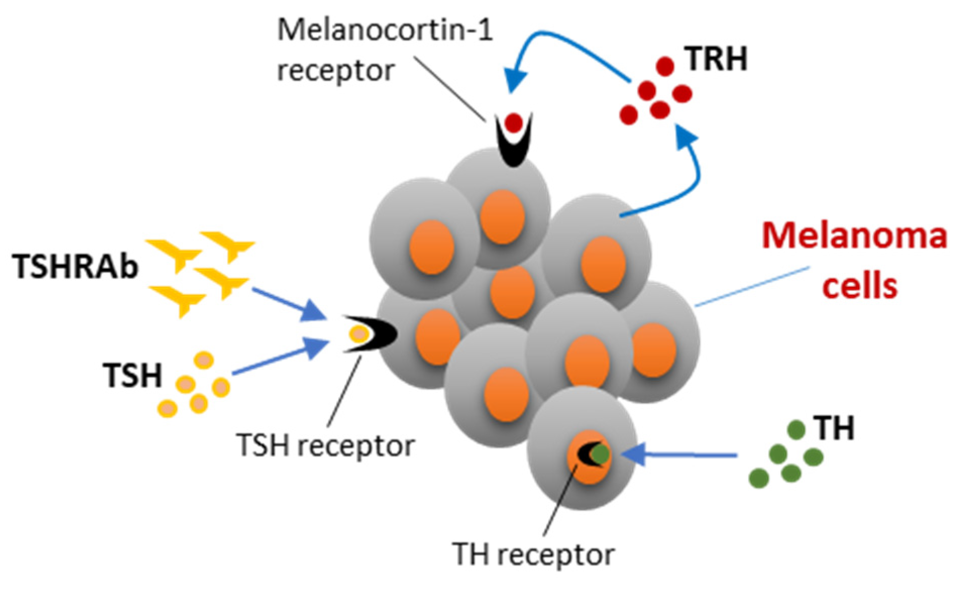
5. Hormonal Players of the Hypothalamic–Pituitary–Thyroid Axis and Melanoma
5.1. Thyrotropin-Releasing Hormone and Melanoma
5.2. Thyroid-Stimulating Hormone and Melanoma
5.3. Thyroid Hormones and Melanoma
5.3.1. A Brief Overview of Molecular Mechanisms Underlying Thyroid Hormones Action
5.3.2. Thyroid Hormones’ Effects on Melanoma Development
6. Conclusions
Author Contributions
Funding
Institutional Review Board Statement
Informed Consent Statement
Conflicts of Interest
References
- Prinzi, N.; Sorrenti, S.; Baldini, E.; DE Vito, C.; Tuccilli, C.; Catania, A.; Coccaro, C.; Bianchini, M.; Nesca, A.; Grani, G.; et al. Association of Thyroid Diseases with Primary Extra-Thyroidal Malignancies in Women: Results of a Cross-Sectional Study of 6,386 Patients. PLoS ONE 2015, 10, e0122958. [Google Scholar] [CrossRef] [PubMed]
- Prinzi, N.; Baldini, E.; Sorrenti, S.; DE Vito, C.; Tuccilli, C.; Catania, A.; Carbotta, S.; Mocini, R.; Coccaro, C.; Nesca, A.; et al. Prevalence of breast cancer in thyroid diseases: Results of a cross-sectional study of 3,921 patients. Breast Cancer Res. Treat. 2014, 144, 683–688. [Google Scholar] [CrossRef] [PubMed]
- Hardefeldt, P.J.; Eslick, G.D.; Edirimanne, S. Benign thyroid disease is associated with breast cancer: A meta-analysis. Breast Cancer Res. Treat. 2012, 133, 1169–1177. [Google Scholar] [CrossRef] [PubMed]
- Chen, Y.-K.; Lin, C.-L.; Chang, Y.-J.; Cheng, F.T.-F.; Peng, C.-L.; Sung, F.-C.; Cheng, Y.-H.; Kao, C.-H. Cancer Risk in Patients with Graves’ Disease: A Nationwide Cohort Study. Thyroid 2013, 23, 879–884. [Google Scholar] [CrossRef]
- Chen, Y.-K.; Lin, C.-L.; Cheng, F.T.-F.; Sung, F.-C.; Kao, C.-H. Cancer risk in patients with Hashimoto’s thyroiditis: A nationwide cohort study. Br. J. Cancer 2013, 109, 2496–2501. [Google Scholar] [CrossRef] [PubMed]
- Bhatti, P.; Veiga, L.H.S.; Ronckers, C.M.; Sigurdson, A.J.; Stovall, M.; Smith, S.A.; Weathers, R.; Leisenring, W.; Mertens, A.C.; Hammond, S.; et al. Risk of Second Primary Thyroid Cancer after Radiotherapy for a Childhood Cancer in a Large Cohort Study: An Update from the Childhood Cancer Survivor Study. Radiat. Res. 2010, 174, 741–752. [Google Scholar] [CrossRef]
- Trinh, L.; Crawford, A.; Hussein, M.; Zerfaoui, M.; Toraih, E.; Randolph, G.; Kandil, E. Deciphering the Risk of Developing Second Primary Thyroid Cancer Following a Primary Malignancy—Who Is at the Greatest Risk? Cancers 2021, 13, 1402. [Google Scholar] [CrossRef]
- Zhang, Y.; Liang, J.; Li, H.; Cong, H.; Lin, Y. Risk of second primary breast cancer after radioactive iodine treatment in thyroid cancer: A systematic review and meta-analysis. Nucl. Med. Commun. 2016, 37, 110–115. [Google Scholar] [CrossRef]
- Mahmood, S.; Vu, K.; Tai, P.; Joseph, K.; Koul, R.; Dubey, A.; Yu, E. Radiation-induced second malignancies. Anticancer Res. 2015, 35, 2431–2434. [Google Scholar]
- Shu, X.; Ji, J.; Li, X.; Sundquist, J.; Hemminki, K.; Sundquist, K. Cancer risk in patients hospitalised for Graves’ disease: A population-based cohort study in Sweden. Br. J. Cancer 2010, 102, 1397–1399. [Google Scholar] [CrossRef]
- Chen, S.; Wu, F.; Hai, R.; You, Q.; Xie, L.; Shu, L.; Zhou, X. Thyroid disease is associated with an increased risk of breast cancer: A systematic review and meta-analysis. Gland Surg. 2021, 10, 336–346. [Google Scholar] [CrossRef] [PubMed]
- Pan, X.-F.; Ma, Y.-J.; Tang, Y.; Yu, M.-M.; Wang, H.; Fan, Y.-R. Breast cancer populations may have an increased prevalence of thyroglobulin antibody and thyroid peroxidase antibody: A systematic review and meta-analysis. Breast Cancer 2020, 27, 828–836. [Google Scholar] [CrossRef] [PubMed]
- Ellerhorst, J.; Cooksley, C.; Broemeling, L.; Johnson, M.; Grimm, E. High prevalence of hypothyroidism among patients with cutaneous melanoma. Oncol. Rep. 2003, 10, 1317–1320. [Google Scholar] [CrossRef]
- Shah, M.; Orengo, I.F.; Rosen, T. High prevalence of hypothyroidism in male patients with cutaneous melanoma. Dermatol. Online J. 2006, 12, 1. [Google Scholar] [CrossRef] [PubMed]
- Lazzara, D.R.; Zarkhin, S.G.; Rubenstein, S.N.; Glick, B.P. Melanoma and Thyroid Carcinoma: Our Current Understanding. J. Clin. Aesthetic Dermatol. 2019, 12, 39–41. [Google Scholar]
- Vanderpump, M.P.J. The epidemiology of thyroid disease. Br. Med Bull. 2011, 99, 39–51. [Google Scholar] [CrossRef] [PubMed]
- Paschke, R. Molecular pathogenesis of nodular goiter. Langenbecks Arch. Surg. 2011, 396, 1127–1136. [Google Scholar] [CrossRef]
- Haugen, B.R.; Alexander, E.K.; Bible, K.C.; Doherty, G.M.; Mandel, S.J.; Nikiforov, Y.E.; Pacini, F.; Randolph, G.W.; Sawka, A.M.; Schlumberger, M.; et al. 2015 American Thyroid Association Management Guidelines for Adult Patients with Thyroid Nodules and Differentiated Thyroid Cancer: The American Thyroid Association Guidelines Task Force on Thyroid Nodules and Differentiated Thyroid Cancer. Thyroid 2016, 26, 1–133. [Google Scholar] [CrossRef]
- Baldini, E.; Sorrenti, S.; Tartaglia, F.; Catania, A.; Palmieri, A.; Pironi, D.; Filippini, A.; Ulisse, S. New perspectives in the diagnosis of thyroid follicular lesions. Int. J. Surg. 2017, 41, S7–S12. [Google Scholar] [CrossRef]
- Nikiforov, Y.E.; Biddinger, P.W.; Thompson, L.D.R. Diagnostic Pathology and Molecular Genetics of the Thyroid; Wolters Kluwer—Lippincott Williams & Wilkins: Philadelphia, PA, USA, 2009. [Google Scholar]
- Ferrari, S.M.; Fallahi, P.; Ruffilli, I.; Elia, G.; Ragusa, F.; Paparo, S.R.; Ulisse, S.; Baldini, E.; Giannini, R.; Miccoli, P.; et al. Molecular testing in the diagnosis of differentiated thyroid carcinomas. Gland Surg. 2018, 7, S19–S29. [Google Scholar] [CrossRef]
- Trimboli, P.; Ulisse, S.; Graziano, F.; Marzullo, A.; Ruggieri, M.; Calvanese, A.; Piccirilli, F.; Cavaliere, R.; Fumarola, A.; D’Armiento, M. Trend in Thyroid Carcinoma Size, Age at Diagnosis, and Histology in a Retrospective Study of 500 Cases Diagnosed Over 20 Years. Thyroid 2006, 16, 1151–1155. [Google Scholar] [CrossRef] [PubMed]
- Trimboli, P.; Ulisse, S.; Dal, M.; Solari, F.; Fumarola, A.; Ruggieri, M.; De Antoni, E.; Catania, A.; Sorrenti, S.; Nardi, F.; et al. Analysis of clinical, ultrasound and colour flow-Doppler characteristics in predicting malignancy in follicular thyroid neoplasms. Clin. Endocrinol. 2008, 69, 342–344. [Google Scholar] [CrossRef] [PubMed]
- Papale, F.; Cafiero, G.; Grimaldi, A.; Marino, G.; Rosso, F.; Mian, C.; Barollo, S.; Pennelli, G.; Sorrenti, S.; De Antoni, E.; et al. Galectin-3 expression in thyroid fine needle cytology (t-FNAC) uncertain cases: Validation of molecular markers and technology innovation. J. Cell. Physiol. 2013, 228, 968–974. [Google Scholar] [CrossRef] [PubMed]
- Sorrenti, S.; Trimboli, P.; Catania, A.; Ulisse, S.; De Antoni, E.; D’Armiento, M. Comparison of Malignancy Rate in Thyroid Nodules with Cytology of Indeterminate Follicular or Indeterminate Hürthle Cell Neoplasm. Thyroid 2009, 19, 355–360. [Google Scholar] [CrossRef]
- Ulisse, S.; Bosco, D.; Nardi, F.; Nesca, A.; D’Armiento, E.; Guglielmino, V.; DE Vito, C.; Sorrenti, S.; Pironi, D.; Tartaglia, F.; et al. Thyroid Imaging Reporting and Data System Score Combined with the New Italian Classification for Thyroid Cytology Improves the Clinical Management of Indeterminate Nodules. Int. J. Endocrinol. 2017, 2017, 9692304. [Google Scholar] [CrossRef]
- Ohori, N.P. Molecular testing and thyroid nodule management in North America. Gland Surg. 2020, 9, 1628–1638. [Google Scholar] [CrossRef]
- Fresilli, D.; David, E.; Pacini, P.; Del Gaudio, G.; Dolcetti, V.; Lucarelli, G.; Di Leo, N.; Bellini, M.; D’Andrea, V.; Sorrenti, S.; et al. Thyroid Nodule Characterization: How to Assess the Malignancy Risk. Update of the Literature. Diagnostics 2021, 11, 1374. [Google Scholar] [CrossRef]
- Sorrenti, S.; Dolcetti, V.; Fresilli, D.; Del Gaudio, G.; Pacini, P.; Huang, P.; Camponovo, C.; Leoncini, A.; D’Andrea, V.; Pironi, D.; et al. The Role of CEUS in the Evaluation of Thyroid Cancer: From Diagnosis to Local Staging. J. Clin. Med. 2021, 10, 4559. [Google Scholar] [CrossRef]
- Medas, F.; Canu, G.L.; Cappellacci, F.; Boi, F.; Lai, M.L.; Erdas, E.; Calò, P.G. Predictive Factors of Lymph Node Metastasis in Patients with Papillary Microcarcinoma of the Thyroid: Retrospective Analysis on 293 Cases. Front. Endocrinol. 2020, 11, 551. [Google Scholar] [CrossRef]
- Wang, Y.; Liyanarachchi, S.; Miller, K.E.; Nieminen, T.T.; Comiskey, D.F., Jr.; Li, W.; Brock, P.; Symer, D.E.; Akagi, K.; DeLap, K.E.; et al. Identification of Rare Variants Predisposing to Thyroid Cancer. Thyroid. 2019, 29, 946–955. [Google Scholar] [CrossRef]
- Yang, S.P.; Ngeow, J. Familial non-medullary thyroid cancer: Unraveling the genetic maze. Endocr.-Relat. Cancer 2016, 23, R577–R595. [Google Scholar] [CrossRef] [PubMed]
- Moses, W.; Weng, J.; Kebebew, E. Prevalence, Clinicopathologic Features, and Somatic Genetic Mutation Profile in Familial Versus Sporadic Nonmedullary Thyroid Cancer. Thyroid 2011, 21, 367–371. [Google Scholar] [CrossRef] [PubMed] [Green Version]
- Cancer Genome Atlas Research Network. Integrated Genomic Characterization of Papillary Thyroid Carcinoma. Cell 2014, 159, 676–690. [Google Scholar] [CrossRef] [PubMed]
- Yoo, S.-K.; Lee, S.; Kim, S.-J.; Jee, H.-G.; Kim, B.-A.; Cho, H.; Song, Y.S.; Cho, S.W.; Won, J.-K.; Shin, J.-Y.; et al. Comprehensive Analysis of the Transcriptional and Mutational Landscape of Follicular and Papillary Thyroid Cancers. PLoS Genet. 2016, 12, e1006239. [Google Scholar] [CrossRef]
- Soares, P.; Lima, J.; Preto, A.; Castro, P.; Vinagre, J.; Celestino, R.; Couto, J.P.; Prazeres, H.; Eloy, C.; Maximo, V.; et al. Genetic Alterations in Poorly Differentiated and Undifferentiated Thyroid Carcinomas. Curr. Genom. 2011, 12, 609–617. [Google Scholar] [CrossRef]
- Baldini, E.; Tuccilli, C.; Pironi, D.; Catania, A.; Tartaglia, F.; Di Matteo, F.M.; Palumbo, P.; Arcieri, S.; Mascagni, D.; Palazzini, G.; et al. Expression and Clinical Utility of Transcription Factors Involved in Epithelial–Mesenchymal Transition during Thyroid Cancer Progression. J. Clin. Med. 2021, 10, 4076. [Google Scholar] [CrossRef]
- Ulisse, S.; Baldini, E.; Lauro, A.; Pironi, D.; Tripodi, D.; Lori, E.; Ferent, I.C.; Amabile, M.I.; Catania, A.; Di Matteo, F.M.; et al. Papillary Thyroid Cancer Prognosis: An Evolving Field. Cancers 2021, 13, 5567. [Google Scholar] [CrossRef]
- Sorrenti, S.; Carbotta, G.; Di Matteo, F.M.; Catania, A.; Pironi, D.; Tartaglia, F.; Tarroni, D.; Gagliardi, F.; Tripodi, D.; Watanabe, M.; et al. Evaluation of Clinicopathological and Molecular Parameters on Disease Recurrence of Papillary Thyroid Cancer Patient: A Retrospective Observational Study. Cancers 2020, 12, 3637. [Google Scholar] [CrossRef]
- Ulisse, S.; Baldini, E.; Sorrenti, S.; Barollo, S.; Gnessi, L.; Catania, A.; Pellizzo, M.R.; Nardi, F.; Mian, C.; De Antoni, E.; et al. High Expression of the Urokinase Plasminogen Activator and Its Cognate Receptor Associates with Advanced Stages and Reduced Disease-Free Interval in Papillary Thyroid Carcinoma. J. Clin. Endocrinol. Metab. 2011, 96, 504–508. [Google Scholar] [CrossRef]
- Ulisse, S.; Baldini, E.; Sorrenti, S.; Barollo, S.; Prinzi, N.; Catania, A.; Nesca, A.; Gnessi, L.; Pelizzo, M.R.; Mian, C.; et al. In papillary thyroid carcinoma BRAFV600E is associated with increased expression of the urokinase plasminogen activator and its cognate receptor, but not with disease-free interval. Clin. Endocrinol. 2012, 77, 780–786. [Google Scholar] [CrossRef]
- Baldini, E.; Tuccilli, C.; Prinzi, N.; Sorrenti, S.; Falvo, L.; DE Vito, C.; Catania, A.; Tartaglia, F.; Mocini, R.; Coccaro, C.; et al. Deregulated Expression of Aurora Kinases Is Not a Prognostic Biomarker in Papillary Thyroid Cancer Patients. PLoS ONE 2015, 10, e0121514. [Google Scholar] [CrossRef] [PubMed]
- Medas, F.; Erdas, E.; Canu, G.L.; Longheu, A.; Pisano, G.; Tuveri, M.; Calò, P.G. Does hyperthyroidism worsen prognosis of thyroid carcinoma? A retrospective analysis on 2820 consecutive thyroidectomies. J. Otolaryngol.-Head Neck Surg. 2018, 47, 6. [Google Scholar] [CrossRef] [PubMed] [Green Version]
- Ferrari, S.M.; Bocci, G.; Di Desidero, T.; Elia, G.; Ruffilli, I.; Ragusa, F.; Orlandi, P.; Paparo, S.R.; Patrizio, A.; Piaggi, S.; et al. Lenvatinib exhibits antineoplastic activity in anaplastic thyroid cancer in vitro and in vivo. Oncol. Rep. 2018, 39, 2225–2234. [Google Scholar] [CrossRef] [PubMed]
- Ferrari, S.M.; Bocci, G.; Di Desidero, T.; Ruffilli, I.; Elia, G.; Ragusa, F.; Fioravanti, A.; Orlandi, P.; Paparo, S.R.; Patrizio, A.; et al. Vandetanib has antineoplastic activity in anaplastic thyroid cancer, in vitro and in vivo. Oncol. Rep. 2018, 39, 2306–2314. [Google Scholar] [CrossRef] [PubMed]
- Macerola, E.; Poma, A.M.; Vignali, P.; Proietti, A.; Ugolini, C.; Torregrossa, L.; Basolo, A.; Elisei, R.; Santini, F.; Basolo, F. Predictive Biomarkers in Thyroid Cancer. Front. Oncol. 2022, 12, 901004. [Google Scholar] [CrossRef]
- Baldini, E.; Tuccilli, C.; Prinzi, N.; Sorrenti, S.; Antonelli, A.; Gnessi, L.; Morrone, S.; Moretti, C.; Bononi, M.; Arlot-Bonnemains, Y.; et al. Effects of selective inhibitors of Aurora kinases on anaplastic thyroid carcinoma cell lines. Endocr.-Relat. Cancer 2014, 21, 797–811. [Google Scholar] [CrossRef]
- Ragusa, F.; Ferrari, S.M.; Elia, G.; Paparo, S.R.; Balestri, E.; Botrini, C.; Patrizio, A.; Mazzi, V.; Guglielmi, G.; Foddis, R.; et al. Combination Strategies Involving Immune Checkpoint Inhibitors and Tyrosine Kinase or BRAF Inhibitors in Aggressive Thyroid Cancer. Int. J. Mol. Sci. 2022, 23, 5731. [Google Scholar] [CrossRef]
- Ulisse, S.; Tuccilli, C.; Sorrenti, S.; Antonelli, A.; Fallahi, P.; D’Armiento, E.; Catania, A.; Tartaglia, F.; Amabile, M.I.; Giacomelli, L.; et al. PD-1 Ligand Expression in Epithelial Thyroid Cancers: Potential Clinical Implications. Int. J. Mol. Sci. 2019, 20, 1405. [Google Scholar] [CrossRef]
- Tuccilli, C.; Baldini, E.; Sorrenti, S.; Catania, A.; Antonelli, A.; Fallahi, P.; Tartaglia, F.; Barollo, S.; Mian, C.; Palmieri, A.; et al. CTLA-4 and PD-1 Ligand Gene Expression in Epithelial Thyroid Cancers. Int. J. Endocrinol. 2018, 2018, 174295. [Google Scholar] [CrossRef]
- Baldini, E.; Presutti, D.; Favoriti, P.; Santini, S.; Papoff, G.; Tuccilli, C.; Carletti, R.; Di Gioia, C.; Lori, E.; Ferent, I.C.; et al. In Vitro and In Vivo Effects of the Urokinase Plasminogen Activator Inhibitor WX-340 on Anaplastic Thyroid Cancer Cell Lines. Int. J. Mol. Sci. 2022, 23, 3724. [Google Scholar] [CrossRef]
- Fallahi, P.; Ferrari, S.M.; Santini, F.; Corrado, A.; Materazzi, G.; Ulisse, S.; Miccoli, P.; Antonelli, A. Sorafenib and Thyroid Cancer. BioDrugs 2013, 27, 615–628. [Google Scholar] [CrossRef] [PubMed]
- Wu, X.; Hammer, J.A. Melanosome transfer: It is best to give and receive. Curr. Opin. Cell Biol. 2014, 29, 1–7. [Google Scholar] [CrossRef] [PubMed] [Green Version]
- Dimitriou, F.; Krattinger, R.; Ramelyte, E.; Barysch, M.J.; Micaletto, S.; Dummer, R.; Goldinger, S.M. The World of Melanoma: Epidemiologic, Genetic, and Anatomic Differences of Melanoma Across the Globe. Curr. Oncol. Rep. 2018, 20, 87. [Google Scholar] [CrossRef]
- Andrews, M.C.; Oba, J.; Wu, C.-J.; Zhu, H.; Karpinets, T.; Creasy, C.A.; Forget, M.-A.; Yu, X.; Song, X.; Mao, X.; et al. Multi-modal molecular programs regulate melanoma cell state. Nat. Commun. 2022, 13, 4000. [Google Scholar] [CrossRef] [PubMed]
- Schadendorf, D.; van Akkooi, A.C.J.; Berking, C.; Griewank, K.G.; Gutzmer, R.; Hauschild, A.; Stang, A.; Roesch, A.; Ugurel, S. Melanoma. Lancet 2018, 392, 971–984. [Google Scholar] [CrossRef]
- Siegel, R.L.; Miller, K.D.; Fuchs, H.E.; Jemal, A. Cancer Statistics, 2021. CA Cancer J. Clin. 2021, 71, 7–33. [Google Scholar] [CrossRef]
- Arnaut, J.R.M.B.; Guimarães, I.D.S.; dos Santos, A.C.E.; Silva, F.D.M.L.D.; Machado, J.R.; de Melo, A.C. Molecular landscape of Hereditary Melanoma. Crit. Rev. Oncol. Hematol. 2021, 164, 103425. [Google Scholar] [CrossRef]
- Elder, D.E.; Barnhill, R.I.; Bastian, B.C.; Cook, M.G.; de la Fouchardière, A.; Gerami, P.; Lazar, A.J.; Massi, D.; Migm, M.C., Jr.; Nagore, E.; et al. Melanocytic tumour classification and the pathway concept of melanoma pathogenesis. In WHO Classification of Skin Tumours, 4th ed.; Elder, D.E., Massi, D., Scolyer, R.A., Willemze, R., Eds.; International Agency for Research on Cancer (IARC): Lyon, France, 2018; pp. 66–71. [Google Scholar]
- Elder, D.E.; Bastian, B.C.; Cree, I.A.; Massi, D.; Scolyer, R.A. The 2018 World Health Organization Classification of Cutaneous, Mucosal, and Uveal Melanoma: Detailed Analysis of 9 Distinct Subtypes Defined by Their Evolutionary Pathway. Arch. Pathol. Lab. Med. 2020, 144, 500–522. [Google Scholar] [CrossRef]
- Yeh, I.; Bastian, B. Melanoma pathology: New approaches and classification*. Br. J. Dermatol. 2021, 185, 282–293. [Google Scholar] [CrossRef]
- Ferrara, G.; Argenziano, G. The WHO 2018 Classification of Cutaneous Melanocytic Neoplasms: Suggestions from Routine Practice. Front. Oncol. 2021, 11, 675296. [Google Scholar] [CrossRef]
- Garbe, C.; Amaral, T.; Peris, K.; Hauschild, A.; Arenberger, P.; Bastholt, L.; Bataille, V.; del Marmol, V.; Dréno, B.; Fargnoli, M.C.; et al. European consensus-based interdisciplinary guideline for melanoma. Part 1: Diagnostics—Update 2019. Eur. J. Cancer 2020, 126, 141–158. [Google Scholar] [CrossRef]
- Balch, C.M.; Gershenwald, J.E.; Soong, S.-J.; Thompson, J.F.; Atkins, M.B.; Byrd, D.R.; Buzaid, A.C.; Cochran, A.J.; Coit, D.G.; Ding, S.; et al. Final Version of 2009 AJCC Melanoma Staging and Classification. J. Clin. Oncol. 2009, 27, 6199–6206. [Google Scholar] [CrossRef] [PubMed] [Green Version]
- Cianfarani, F.; Mastroeni, S.; Odorisio, T.; Passarelli, F.; Cattani, C.; Mannooranparampil, T.J.; Fortes, C.; Failla, C.M. Expression of vascular endothelial growth factor-C in primary cutaneous melanoma predicts sentinel lymph node positivity. J. Cutan. Pathol. 2012, 39, 826–834. [Google Scholar] [CrossRef] [PubMed]
- Garbe, C.; Amaral, T.; Peris, K.; Hauschild, A.; Arenberger, P.; Bastholt, L.; Bataille, V.; del Marmol, V.; Dréno, B.; Fargnoli, M.C.; et al. European consensus-based interdisciplinary guideline for melanoma. Part 2: Treatment—Update 2019. Eur. J. Cancer 2020, 126, 159–177. [Google Scholar] [CrossRef] [PubMed]
- Eggermont, A.M.M.; Chiarion-Sileni, V.; Grob, J.-J.; Dummer, R.; Wolchok, J.D.; Schmidt, H.; Hamid, O.; Robert, C.; Ascierto, P.A.; Richards, J.M.; et al. Adjuvant ipilimumab versus placebo after complete resection of high-risk stage III melanoma (EORTC 18071): A randomised, double-blind, phase 3 trial. Lancet Oncol. 2015, 16, 522–530. [Google Scholar] [CrossRef]
- Weber, J.; Mandalà, M.; Del Vecchio, M.; Gogas, H.J.; Arance, A.M.; Cowey, C.L.; Dalle, S.; Schenker, M.; Chiarion-Sileni, V.; Marquez-Rodas, I.; et al. Adjuvant Nivolumab versus Ipilimumab in Resected Stage III or IV Melanoma. N. Engl. J. Med. 2017, 377, 1824–1835. [Google Scholar] [CrossRef]
- Eggermont, A.M.M.; Blank, C.U.; Mandalà, M.; Long, G.V.; Atkinson, V.; Dalle, S.; Haydon, A.; Lichinitser, M.; Khattak, A.; Carlino, M.S.; et al. Adjuvant Pembrolizumab versus Placebo in Resected Stage III Melanoma. N. Engl. J. Med. 2018, 378, 1789–1801. [Google Scholar] [CrossRef]
- Eggermont, A.M.M.; Blank, C.U.; Mandalà, M.; Long, G.V.; Atkinson, V.G.; Dalle, S.; Haydon, A.M.; Meshcheryakov, A.; Khattak, A.; Carlino, M.S.; et al. Adjuvant pembrolizumab versus placebo in resected stage III melanoma (EORTC 1325-MG/KEYNOTE-054): Distant metastasis-free survival results from a double-blind, randomised, controlled, phase 3 trial. Lancet Oncol. 2021, 22, 643–654. [Google Scholar] [CrossRef]
- Long, G.V.; Hauschild, A.; Santinami, M.; Atkinson, V.; Mandalà, M.; Sileni, V.C.; Larkin, J.; Nyakas, M.; Dutriaux, C.; Haydon, A.; et al. Adjuvant Dabrafenib plus Trametinib in Stage IIIBRAF-Mutated Melanoma. N. Engl. J. Med. 2017, 377, 1813–1823. [Google Scholar] [CrossRef]
- Eggermont, A.M.; Chiarion-Sileni, V.; Grob, J.-J.; Dummer, R.; Wolchok, J.D.; Schmidt, H.; Hamid, O.; Robert, C.; Ascierto, P.A.; Richards, J.M.; et al. Prolonged Survival in Stage III Melanoma with Ipilimumab Adjuvant Therapy. N. Engl. J. Med. 2016, 375, 1845–1855. [Google Scholar] [CrossRef]
- Ellerhorst, J.A.; Cooksley, C.D.; Grimm, E.A. Autoimmunity and hypothyroidism in patients with uveal melanoma. Melanoma Res. 2001, 11, 633–637. [Google Scholar] [CrossRef]
- Ferrari, S.M.; Fallahi, P.; Galetta, F.; Citi, E.; Benvenga, S.; Antonelli, A. Thyroid disorders induced by checkpoint inhibitors. Rev. Endocr. Metab. Disord. 2018, 19, 325–333. [Google Scholar] [CrossRef]
- Ferrari, S.M.; Fallahi, P.; Elia, G.; Ragusa, F.; Ruffilli, I.; Patrizio, A.; Galdiero, M.R.; Baldini, E.; Ulisse, S.; Marone, G.; et al. Autoimmune Endocrine Dysfunctions Associated with Cancer Immunotherapies. Int. J. Mol. Sci. 2019, 20, 2560. [Google Scholar] [CrossRef]
- Killock, D. ICI for resected stage IV melanoma. Nat. Rev. Clin. Oncol. 2020, 17, 450. [Google Scholar] [CrossRef]
- Herrscher, H.; Robert, C. Immune checkpoint inhibitors in melanoma in the metastatic, neoadjuvant, and adjuvant setting. Curr. Opin. Oncol. 2020, 32, 106–113. [Google Scholar] [CrossRef]
- Dawidowska, A.; Jagodzinska-Mucha, P.; Koseła-Paterczyk, H.; Jaczewska, S.; Sobczuk, P.; Chelstowska, M.; Kowalska, M.; Badziak-Sterczewska, H.; Poleszczuk, J.; Rutkowski, P.; et al. Immune-Related Thyroid Adverse Events Predict Response to PD-1 Blockade in Patients with Melanoma. Cancers 2022, 14, 1248. [Google Scholar] [CrossRef]
- Bertrand, A.; Kostine, M.; Barnetche, T.; Truchetet, M.-E.; Schaeverbeke, T. Immune related adverse events associated with anti-CTLA-4 antibodies: Systematic review and meta-analysis. BMC Med. 2015, 13, 211. [Google Scholar] [CrossRef]
- Larkin, J.; Chiarion-Sileni, V.; Gonzalez, R.; Grob, J.-J.; Cowey, C.L.; Lao, C.D.; Schadendorf, D.; Dummer, R.; Smylie, M.; Rutkowski, P.; et al. Combined Nivolumab and Ipilimumab or Monotherapy in Untreated Melanoma. N. Engl. J. Med. 2015, 373, 23–34. [Google Scholar] [CrossRef]
- Almutairi, A.R.; McBride, A.; Slack, M.; Erstad, B.L.; Abraham, I. Potential Immune-Related Adverse Events Associated with Monotherapy and Combination Therapy of Ipilimumab, Nivolumab, and Pembrolizumab for Advanced Melanoma: A Systematic Review and Meta-Analysis. Front. Oncol. 2020, 10, 91. [Google Scholar] [CrossRef]
- Rogado, J.; Sánchez-Torres, J.; Romero-Laorden, N.; Ballesteros, A.; Pacheco-Barcia, V.; Ramos-Leví, A.; Arranz, R.; Lorenzo, A.; Gullón, P.; Donnay, O.; et al. Immune-related adverse events predict the therapeutic efficacy of anti–PD-1 antibodies in cancer patients. Eur. J. Cancer 2019, 109, 21–27. [Google Scholar] [CrossRef]
- Bilen, M.A.; Patel, A.; Hess, K.R.; Munoz, J.; Busaidy, N.L.; Wheler, J.J.; Janku, F.; Falchook, G.S.; Hong, D.S.; Meric-Bernstam, F.; et al. Association between new-onset hypothyroidism and clinical response in patients treated with tyrosine kinase inhibitor therapy in phase I clinical trials. Cancer Chemother. Pharmacol. 2016, 78, 167–171. [Google Scholar] [CrossRef] [PubMed]
- Oakley, G.M.; Curtin, K.; Layfield, L.; Jarboe, E.; Buchmann, L.O.; Hunt, J.P. Increased Melanoma Risk in Individuals with Papillary Thyroid Carcinoma. JAMA Otolaryngol. Head Neck Surg. 2014, 140, 423–427. [Google Scholar] [CrossRef] [PubMed] [Green Version]
- Kim, Y.J.; Lee, M.; Kim, E.H.; Jung, I.; Lee, C.S. Coexisting and Second Primary Cancers in Patients with Uveal Melanoma: A 10-Year Nationwide Database Analysis. J. Clin. Med. 2021, 10, 4744. [Google Scholar] [CrossRef] [PubMed]
- Schonfeld, S.J.; Morton, L.M.; de González, A.B.; Curtis, R.E.; Kitahara, C.M. Risk of second primary papillary thyroid cancer among adult cancer survivors in the United States, 2000–2015. Cancer Epidemiol. 2019, 64, 101664. [Google Scholar] [CrossRef]
- Kim, C.Y.; Lee, S.H.; Oh, C.W. Cutaneous Malignant Melanoma Associated with Papillary Thyroid Cancer. Ann. Dermatol. 2010, 22, 370–372. [Google Scholar] [CrossRef]
- Kolaii, S.S.T.J.; Dehghanian, A.R.; Jeddi, M. Concomitant uveal melanoma and papillary thyroid carcinoma: A case report. J. Med Case Rep. 2022, 16, 29. [Google Scholar] [CrossRef]
- Zerfaoui, M.; Dokunmu, T.; Toraih, E.; Rezk, B.; Elmageed, Z.A.; Kandil, E. New Insights into the Link between Melanoma and Thyroid Cancer: Role of Nucleocytoplasmic Trafficking. Cells 2021, 10, 367. [Google Scholar] [CrossRef]
- Papanikolaou, M.; Chohan, T.; Millington, G.W.M. Malignant melanoma, papillary thyroid carcinoma and Erdheim–Chester disease, associated with both BRAF V600E and mosaic Turner syndrome. Clin. Exp. Dermatol. 2020, 45, 512–514. [Google Scholar] [CrossRef]
- Tavares, C.; Melo, M.; Cameselle-Teijeiro, J.M.; Soares, P.; Sobrinho-Simões, M. ENDOCRINE TUMOURS: Genetic predictors of thyroid cancer outcome. Eur. J. Endocrinol. 2016, 174, R117–R126. [Google Scholar] [CrossRef]
- Cicenas, J.; Tamosaitis, L.; Kvederaviciute, K.; Tarvydas, R.; Staniute, G.; Kalyan, K.; Meskinyte-Kausiliene, E.; Stankevicius, V.; Valius, M. KRAS, NRAS and BRAF mutations in colorectal cancer and melanoma. Med Oncol. 2017, 34, 26. [Google Scholar] [CrossRef]
- Greenspan, F.S. The thyroid gland. In Basic & Clinical Endocrinology, 7th ed.; Greenspan, F.S., Gardner, D.G., Eds.; Lange Medical Books/McGraw-Hill: New York, NY, USA, 2004; pp. 215–294. [Google Scholar]
- Tan, M.-H.; Mester, J.L.; Ngeow, J.; Rybicki, L.A.; Orloff, M.S.; Eng, C. Lifetime Cancer Risks in Individuals with Germline PTEN Mutations. Clin. Cancer Res. 2012, 18, 400–407. [Google Scholar] [CrossRef]
- Robles- Espinoza, C.D.; Harland, M.; Ramsay, A.J.; Aoude, L.G.; Quesada, V.; Ding, Z.; Pooley, K.A.; Pritchard, A.L.; Tiffen, J.C.; Petljak, M.; et al. POT1 loss-of-function variants predispose to familial melanoma. Nat. Genet. 2014, 46, 478–481. [Google Scholar] [CrossRef] [Green Version]
- Potrony, M.; Puig-Butille, J.; Ribera-Sola, M.; Iyer, V.; Robles-Espinoza, C.; Aguilera, P.; Carrera, C.; Malvehy, J.; Badenas, C.; Landi, M.; et al. POT 1 germline mutations but not TERT promoter mutations are implicated in melanoma susceptibility in a large cohort of Spanish melanoma families. Br. J. Dermatol. 2018, 181, 105–113. [Google Scholar] [CrossRef]
- Wong, K.; Robles-Espinoza, C.D.; Rodriguez, D.; Rudat, S.S.; Puig, S.; Potrony, M.; Wong, C.C.; Hewinson, J.; Aguilera, P.; Puig-Butille, J.A.; et al. Association of the POT1 Germline Missense Variant p.I78T With Familial Melanoma. JAMA Dermatol. 2019, 155, 604–609. [Google Scholar] [CrossRef]
- Srivastava, A.; Miao, B.; Skopelitou, D.; Kumar, V.; Kumar, A.; Paramasivam, N.; Bonora, E.; Hemminki, K.; Försti, A.; Bandapalli, O.R. A Germline Mutation in the POT1 Gene Is a Candidate for Familial Non-Medullary Thyroid Cancer. Cancers 2020, 12, 1441. [Google Scholar] [CrossRef]
- Orois, A.; Badenas, C.; Reverter, J.L.; López, V.; Potrony, M.; Mora, M.; Halperin, I.; Oriola, J. Lack of Mutations in POT1 Gene in Selected Families with Familial Non-Medullary Thyroid Cancer. Horm. Cancer 2020, 11, 111–116. [Google Scholar] [CrossRef]
- McDonnell, K.J.; Gallanis, G.T.; Heller, K.A.; Melas, M.; Idos, G.E.; Culver, J.O.; Martin, S.-E.; Peng, D.H.; Gruber, S.B. A novel BAP1 mutation is associated with melanocytic neoplasms and thyroid cancer. Cancer Genet. 2015, 209, 75–81. [Google Scholar] [CrossRef]
- Wilber, J.F.; Spinella, P. Identification of Immunoreactive Thyrotropin-Releasing Hormone in Human Neoplasia. J. Clin. Endocrinol. Metab. 1984, 59, 432–435. [Google Scholar] [CrossRef]
- Slominski, A.; Pisarchik, A.; Wortsman, J.; Kohn, L.; Ain, K.B.; Venkataraman, G.M.; Chung, J.H.; Giuliani, C.; Thornton, M.; Slugocki, G.; et al. Expression of Hypothalamic–Pituitary–Thyroid Axis RelatedGenes in the Human Skin. J. Investig. Dermatol. 2002, 119, 1449–1455. [Google Scholar] [CrossRef]
- Ellerhorst, J.A.; Naderi, A.A.; Johnson, M.K.; Pelletier, P.; Prieto, V.G.; Diwan, A.H.; Johnson, M.M.; Gunn, D.C.; Yekell, S.; Grimm, E.A. Expression of Thyrotropin-Releasing Hormone by Human Melanoma and Nevi. Clin. Cancer Res. 2004, 10, 5531–5536. [Google Scholar] [CrossRef]
- Schiöth, H.B.; Prusis, P.; Muceniece, R.; Mutulis, F.; Mutule, I.; Wikberg, J.E. Thyrotropin releasing hormone (TRH) selectively binds and activates the melanocortin 1 receptor. Peptides 1999, 20, 395–400. [Google Scholar] [CrossRef]
- Gáspár, E.; Nguyen-Thi, K.T.; Hardenbicker, C.; Tiede, S.; Plate, C.; Bodó, E.; Knuever, J.; Funk, W.; Bíró, T.; Paus, R. Thyrotropin-Releasing Hormone Selectively Stimulates Human Hair Follicle Pigmentation. J. Investig. Dermatol. 2011, 131, 2368–2377. [Google Scholar] [CrossRef] [Green Version]
- Chu, Y.-D.; Yeh, C.-T. The Molecular Function and Clinical Role of Thyroid Stimulating Hormone Receptor in Cancer Cells. Cells 2020, 9, 1730. [Google Scholar] [CrossRef]
- Cianfarani, F.; Baldini, E.; Cavalli, A.; Marchioni, E.; Lembo, L.; Teson, M.; Persechino, S.; Zambruno, G.; Ulisse, S.; Odorisio, T.; et al. TSH Receptor and Thyroid-Specific Gene Expression in Human Skin. J. Investig. Dermatol. 2010, 130, 93–101. [Google Scholar] [CrossRef]
- Bodó, E.; Kromminga, A.; Bíró, T.; Borbíró, I.; Gáspár, E.; Zmijewski, M.A.; van Beek, N.; Langbein, L.; Slominski, A.T.; Paus, R. Human Female Hair Follicles Are a Direct, Nonclassical Target for Thyroid-Stimulating Hormone. J. Investig. Dermatol. 2009, 129, 1126–1139. [Google Scholar] [CrossRef]
- Ellerhorst, J.A.; Sendi-Naderi, A.; Johnson, M.K.; Cooke, C.P.; Dang, S.M.; Diwan, A.H. Human melanoma cells express functional receptors for thyroid-stimulating hormone. Endocr.-Relat. Cancer 2006, 13, 1269–1277. [Google Scholar] [CrossRef]
- Ursu, H.I. Functional TSH Receptors, Malignant Melanomas and Subclinical Hypothyroidism. Eur. Thyroid J. 2012, 1, 208. [Google Scholar] [CrossRef]
- Cheng, S.-Y.; Leonard, J.L.; Davis, P.J. Molecular Aspects of Thyroid Hormone Actions. Endocr. Rev. 2010, 31, 139–170. [Google Scholar] [CrossRef]
- Davis, P.J.; Goglia, F.; Leonard, J.L. Nongenomic actions of thyroid hormone. Nat. Rev. Endocrinol. 2015, 12, 111–121. [Google Scholar] [CrossRef]
- Visser, T.J. Thyroid Hormone Transporters and Resistance. Endocr. Dev. 2013, 24, 1–10. [Google Scholar] [CrossRef]
- Chin, W.W. Nuclear thyroid hormone receptors. In Nuclear Hormone Receptor; Parker, M.G., Ed.; Academic Press: London, UK, 1991; pp. 79–102. [Google Scholar]
- Ortiga-Carvalho, T.M.; Sidhaye, A.R.; Wondisford, F.E. Thyroid hormone receptors and resistance to thyroid hormone disorders. Nat. Rev. Endocrinol. 2014, 10, 582–591. [Google Scholar] [CrossRef]
- Lazar, M.A.; Chin, W.W. Nuclear thyroid hormone receptors. J. Clin. Investig. 1990, 86, 1777–1782. [Google Scholar] [CrossRef] [Green Version]
- Sap, J.; Munoz, A.; Damm, K.; Goldberg, Y.; Ghysdael, J.; Leutz, A.; Beug, H.; Vennström, B. The c-erb-A protein is a high-affinity receptor for thyroid hormone. Nature 1986, 324, 635–640. [Google Scholar] [CrossRef]
- Weinberger, C.; Thompson, C.C.; Ong, E.S.; Lebo, R.; Gruol, D.J.; Evans, R.M. The c-erb-A gene encodes a thyroid hormone receptor. Nature 1986, 324, 641–646. [Google Scholar] [CrossRef]
- Chassande, O.; Fraichard, A.; Gauthier, K.; Flamant, F.; Legrand, C.; Savatier, P.; Laudet, V.; Samarut, J. Identification of Transcripts Initiated from an Internal Promoter in the c-erbAα Locus That Encode Inhibitors of Retinoic Acid Receptor-α and Triiodothyronine Receptor Activities. Mol. Endocrinol. 1997, 11, 1278–1290. [Google Scholar] [CrossRef]
- Gauthier, K.; Chassande, O.; Plateroti, M.; Roux, J.-P.; Legrand, C.; Pain, B.; Rousset, B.; Weiss, R.; Trouillas, J.; Samarut, J. Different functions for the thyroid hormone receptors TRalpha and TRbeta in the control of thyroid hormone production and post-natal development. EMBO J. 1999, 18, 623–631. [Google Scholar] [CrossRef]
- Lin, H.; Chin, Y.; Yang, Y.S.H.; Lai, H.; Whang-Peng, J.; Liu, L.F.; Tang, H.; Davis, P.J. Thyroid Hormone, Cancer, and Apoptosis. Compr. Physiol. 2011, 6, 1221–1237. [Google Scholar] [CrossRef]
- Lin, H.-Y.; Sun, M.; Tang, H.-Y.; Lin, C.; Luidens, M.K.; Mousa, S.A.; Incerpi, S.; Drusano, G.L.; Davis, F.B.; Davis, P.J. l-Thyroxine vs. 3,5,3′-triiodo-l-thyronine and cell proliferation: Activation of mitogen-activated protein kinase and phosphatidylinositol 3-kinase. Am. J. Physiol. Cell Physiol. 2009, 296, C980–C991. [Google Scholar] [CrossRef]
- McMenamin, S.K.; Bain, E.J.; McCann, A.E.; Patterson, L.B.; Eom, D.S.; Waller, Z.P.; Hamill, J.C.; Kuhlman, J.A.; Eisen, J.S.; Parichy, D.M. Thyroid hormone–dependent adult pigment cell lineage and pattern in zebrafish. Science 2014, 345, 1358–1361. [Google Scholar] [CrossRef]
- Guillot, R.; Muriach, B.; Rocha, A.; Rotllant, J.; Kelsh, R.; Cerdá-Reverter, J.M. Thyroid Hormones Regulate Zebrafish Melanogenesis in a Gender-Specific Manner. PLoS ONE 2016, 11, e0166152. [Google Scholar] [CrossRef]
- Di Cicco, E.; Moran, C.; Visser, W.E.; Nappi, A.; Schoenmakers, E.; Todd, P.; Lyons, M.G.; Dattani, M.; Ambrosio, R.; Parisi, S.; et al. Germ Line Mutations in the Thyroid Hormone Receptor Alpha Gene Predispose to Cutaneous Tags and Melanocytic Nevi. Thyroid 2021, 31, 1114–1126. [Google Scholar] [CrossRef] [PubMed]
- Wändell, P.; Carlsson, A.C.; Li, X.; Sundquist, J.; Sundquist, K. Levothyroxine treatment is associated with an increased relative risk of overall and organ specific incident cancers—A cohort study of the Swedish population. Cancer Epidemiol. 2020, 66, 101707. [Google Scholar] [CrossRef] [PubMed]
- Gupta, S.; Artomov, M.; Goggins, W.B.; Daly, M.J.; Tsao, H. Gender Disparity and Mutation Burden in Metastatic Melanoma. JNCI J. Natl. Cancer Inst. 2015, 107, djv221. [Google Scholar] [CrossRef] [PubMed]
- Janick, M.E.; Belkot, K.; Przybylo, M. Is oestrogen an important player in melanoma progression? Contemp. Oncol. 2014, 18, 302–306. [Google Scholar] [CrossRef] [PubMed]
- Di Donato, M.; Migliaccio, A.; Castoria, G. Targeting ERβ to fight melanoma: A new valid approach? J. Transl. Med. 2022, 20, 156. [Google Scholar] [CrossRef]
- Bechmann, N.; Calsina, B.; Richter, S.; Pietzsch, J. Therapeutic Potential of Nitric Oxide—Releasing Selective Estrogen Receptor Modulators in Malignant Melanoma. J. Investig. Dermatol. 2022, 142, 2217–2227. [Google Scholar] [CrossRef]
- Joosse, A.; Collette, S.; Suciu, S.; Nijsten, T.; Patel, P.M.; Keilholz, U.; Eggermont, A.M.; Coebergh, J.W.W.; de Vries, E. Sex Is an Independent Prognostic Indicator for Survival and Relapse/Progression-Free Survival in Metastasized Stage III to IV Melanoma: A Pooled Analysis of Five European Organisation for Research and Treatment of Cancer Randomized Controlled Trials. J. Clin. Oncol. 2013, 31, 2337–2346. [Google Scholar] [CrossRef]
- Morgese, F.; Sampaolesi, C.; Torniai, M.; Conti, A.; Ranallo, N.; Giacchetti, A.; Serresi, S.; Onofri, A.; Burattini, M.; Ricotti, G.; et al. Gender Differences and Outcomes in Melanoma Patients. Oncol. Ther. 2020, 8, 103–114. [Google Scholar] [CrossRef]
- Siegel, R.L.; Miller, K.D.; Jemal, A. Cancer statistics, 2019. CA Cancer J. Clin. 2019, 69, 7–34. [Google Scholar] [CrossRef]
- Joosse, A.; de Vries, E.; Eckel, R.; Nijsten, T.; Eggermont, A.M.; Hölzel, D.; Coebergh, J.W.W.; Engel, J.; Munich Melanoma Group. Gender Differences in Melanoma Survival: Female Patients Have a Decreased Risk of Metastasis. J. Investig. Dermatol. 2011, 131, 719–726. [Google Scholar] [CrossRef]
- Mervic, L.; Leiter, U.; Meier, F.; Eigentler, T.; Forschner, A.; Metzler, G.; Bartenjev, I.; Büttner, P.; Garbe, C. Sex differences in survival of cutaneous melanoma are age dependent: An analysis of 7338 patients. Melanoma Res. 2011, 21, 244–252. [Google Scholar] [CrossRef] [PubMed]
- Maggiolini, M.; Picard, D. The unfolding stories of GPR30, a new membrane-bound estrogen receptor. J. Endocrinol. 2010, 204, 105–114. [Google Scholar] [CrossRef] [PubMed] [Green Version]
- Warner, M.; Huang, B.; Gustafsson, J.-A. Estrogen Receptor β as a Pharmaceutical Target. Trends Pharmacol. Sci. 2017, 38, 92–99. [Google Scholar] [CrossRef] [PubMed]
- Ulisse, S.; Tata, J.R. Thyroid hormone and glucocorticoid independently regulate the expression of estrogen receptor in male Xenopus liver cells. Mol. Cell. Endocrinol. 1994, 105, 45–53. [Google Scholar] [CrossRef]
- Alarid, E.T.; Preisler-Mashek, M.T.; Solodin, N.M. Thyroid Hormone Is an Inhibitor of Estrogen-Induced Degradation of Estrogen Receptor-α Protein: Estrogen-Dependent Proteolysis Is Not Essential for Receptor Transactivation Function in the Pituitary. Endocrinology 2003, 144, 3469–3476. [Google Scholar] [CrossRef]
- Glass, C.K.; Holloway, J.M. Regulation of gene expression by the thyroid hormone receptor. Biochim. Biophys. Acta 1990, 1032, 157–176. [Google Scholar] [CrossRef]
- Tang, H.-Y.; Lin, H.-Y.; Zhang, S.; Davis, F.B.; Davis, P.J. Thyroid Hormone Causes Mitogen-Activated Protein Kinase-Dependent Phosphorylation of the Nuclear Estrogen Receptor. Endocrinology 2004, 145, 3265–3272. [Google Scholar] [CrossRef]
- Hercbergs, A.; Mousa, S.A.; Leinung, M.; Lin, H.-Y.; Davis, P.J. Thyroid Hormone in the Clinic and Breast Cancer. Horm. Cancer 2018, 9, 139–143. [Google Scholar] [CrossRef]
- Hammes, S.R.; Davis, P.J. Overlapping nongenomic and genomic actions of thyroid hormone and steroids. Best Pr. Res. Clin. Endocrinol. Metab. 2015, 29, 581–593. [Google Scholar] [CrossRef] [Green Version]


| Melanoma Classification | |
|---|---|
| Melanoma arising in sun-exposed skin. | 1. Low-CSD melanoma/superficial spreading melanoma |
| 2. High-CSD melanoma/lentigo maligna melanoma | |
| 3. Desmoplastic melanoma | |
| Melanoma arising at sun-shielded sites or without known etiological association with UV radiation exposure. | 4. Malignant Spitz tumor (Spitz melanoma) |
| 5. Acral melanoma | |
| 6. Mucosal melanoma | |
| 7. Melanoma arising in congenital naevus | |
| 8. Melanoma arising in blue naevus | |
| 9. Uveal melanoma | |
Publisher’s Note: MDPI stays neutral with regard to jurisdictional claims in published maps and institutional affiliations. |
© 2022 by the authors. Licensee MDPI, Basel, Switzerland. This article is an open access article distributed under the terms and conditions of the Creative Commons Attribution (CC BY) license (https://creativecommons.org/licenses/by/4.0/).
Share and Cite
Ulisse, S.; Baldini, E.; Pironi, D.; Gagliardi, F.; Tripodi, D.; Lauro, A.; Carbotta, S.; Tarroni, D.; D’Armiento, M.; Morrone, A.; et al. Is Melanoma Progression Affected by Thyroid Diseases? Int. J. Mol. Sci. 2022, 23, 10036. https://doi.org/10.3390/ijms231710036
Ulisse S, Baldini E, Pironi D, Gagliardi F, Tripodi D, Lauro A, Carbotta S, Tarroni D, D’Armiento M, Morrone A, et al. Is Melanoma Progression Affected by Thyroid Diseases? International Journal of Molecular Sciences. 2022; 23(17):10036. https://doi.org/10.3390/ijms231710036
Chicago/Turabian StyleUlisse, Salvatore, Enke Baldini, Daniele Pironi, Federica Gagliardi, Domenico Tripodi, Augusto Lauro, Sabino Carbotta, Danilo Tarroni, Matteo D’Armiento, Aldo Morrone, and et al. 2022. "Is Melanoma Progression Affected by Thyroid Diseases?" International Journal of Molecular Sciences 23, no. 17: 10036. https://doi.org/10.3390/ijms231710036