Article

SWATH Mass Spectrometry-Based CSF Proteome Profile of GBA-Linked Parkinson’s Disease Patients

1
Clinical Department of Neurology, University Medical Center Göttingen and the German Center for Neurodegenerative Diseases (DZNE), Robert-Koch-Straße 40, 37075 Göttingen, Germany
2
Biomedical Engineering and Sciences Department, School of Mechanical and Manufacturing Engineering (SMME), National University of Sciences and Technology (NUST), Bolan Road, H-12, Islamabad 44000, Pakistan
3
Institute of Neuropathology, University Medical Center Hamburg-Eppendorf, Martinistraße 52, 20246 Hamburg, Germany
4
Department of Neurodegenerative Diseases, Hertie-Institute for Clinical Brain Research, University of Tübingen, Hoppe-Seyler-Str. 3, 72076 Tübingen, Germany
5
German Center for Neurodegenerative Disease (DZNE), 72076 Tübingen, Germany
*
Author to whom correspondence should be addressed.
These authors contributed equally to this work.
Int. J. Mol. Sci. 2022, 23(22), 14166; https://doi.org/10.3390/ijms232214166
Submission received: 5 September 2022 / Revised: 7 November 2022 / Accepted: 11 November 2022 / Published: 16 November 2022

Abstract

:
β-glucocerebrosidase (GBA)-associated mutations are a significant risk factor for Parkinson’s disease (PD) that aggravate the disease pathology by upregulating the deposition of α-Synuclein (α-Syn). The resultant clinical profile varies for PD patients without GBA mutations. The current study aimed to identify the proteomic targets involved in the pathogenic pathways leading to the differential clinical presentation of GBA-associated PD. CSF samples (n = 32) were obtained from PD patients with GBA mutations (n = 22), PD patients without GBA mutations (n = 7), and healthy controls that were carriers of GBA mutations (n = 3). All samples were subjected to in-gel tryptic digestion followed by the construction of the spectral library and quantitative SWATH-based analysis. CSF α-Syn levels were reduced in both PDIdiopathic and PDGBA cases. Our SWATH-based mass spectrometric analysis detected 363 proteins involved in immune response, stress response, and cell signaling in various groups. Intergroup analysis showed that 52 proteins were significantly up- or downregulated in various groups. Of these 52 targets, 20 proteins were significantly altered in PDGBA cases only while 2 showed different levels in PDIdiopathic patients. Our results show that the levels of several pathologically relevant proteins, including Contactin-1, Selenium-binding protein 1, Adhesion G Protein-Coupled Receptor, and Apolipoprotein E are significantly different among the sporadic and genetic variants of PD and hint at aggravated synaptic damage, oxidative stress, neuronal loss, and aggregation of α-Syn in PDGBA cases.

1. Introduction

Parkinson’s disease (PD) is the second most prevalent neurodegenerative disorder and affects around 1% of the population above the age of 60 [1]. Characterized by the intraneuronal accumulation of α-Synuclein (α-Syn) and extensive death of dopaminergic neurons in the substantia nigra, PD results in debilitating motor symptoms [2,3]. The patients present resting tremor, rigidity, bradykinesia, and postural instability along with multiple non-motor symptoms such as hyposmia, REM sleep behavior disorder, mood swings, and cognitive dysfunction [4,5]. Decades of research have identified ageing, environmental toxicants (e.g., pesticides and herbicides), and genetic predisposition as the major risk factors for this disorder [6,7,8].
Although genome-wide association studies have identified >80 risk loci for PD, mutations in β-glucocerebrosidase (GBA) are amongst the most significant risk factors [9,10]. Mutations in the GBA gene were initially discovered in association with Gaucher’s disease whereby a decrease in the activity of GBA leads to abnormalities in the metabolism of glucosylceramide and multisystem damage [11]. Over the last decade, an increasing amount of evidence has correlated the occurrence of PD with various GBA mutations. Although around 300 mutations have been reported for the GBA gene, not all play an equal role in the pathogenesis of PD [12]. L444P, N370S, and E326K mutations are most common among PD patients and, interestingly, the occurrence of E326K exclusively results in PD and has no role in Gaucher’s disease [13]. Furthermore, these PD-associated mutations show differences in PD risk, biological profiles, and clinical trajectories based on mutation severity.
GBA mutations have various effects on the pathophysiology of PD [14,15]. Loss-of-function mutations in the GBA gene have been hypothesized to increase the accumulation of α-Syn, possibly initiating and aggravating the neurodegenerative process in PD [16]. PD patients with GBA mutations, compared with those without mutations, have been reported to present a characteristic biological and clinical phenotype with reference to aggregation of α-Syn, impaired lipid metabolism, cognitive dysfunction, disease progression, and survival time, indicating a definite effect of GBA mutations on the prognosis of PD [16,17,18,19,20,21,22]. Although the role of the GBA gene has been established in PD, we lack proteome-wide studies in human subjects that may allow us to pinpoint the factors that are being affected by these mutations and establish an understanding of the disorder.
Cerebrospinal fluid (CSF) is being extensively used to understand the pathophysiology of neurodegenerative disorders and hunt for potential biomarkers. In contrast to other biological fluids, CSF has the advantage of providing a direct window into disease-associated changes in the nervous system. Previous studies have reported the relative levels of key pathogenic proteins in CSF from PD patients with and without GBA mutation to highlight the differences in the pathophysiology [20]. However, the lack of proteome-wide studies has limited our understanding of the CSF signature of PD proteome to very few targets. With the recent advances in mass spectrometry (MS), it is now possible to accurately identify and quantify proteins in biofluids. Approaches such as sequential window acquisition of all theoretical spectra (SWATH) provide an ideal platform to analyze complex protein samples reproducibly and predict potential biomarkers confidently [23].
In the current study, we used the advantages of SWATH to analyze and quantify globe-wide proteome alterations in CSF samples from PD patients with and without GBA mutations and identify the differentially regulated pathological targets to decipher the role this mutation in PD.

2. Results

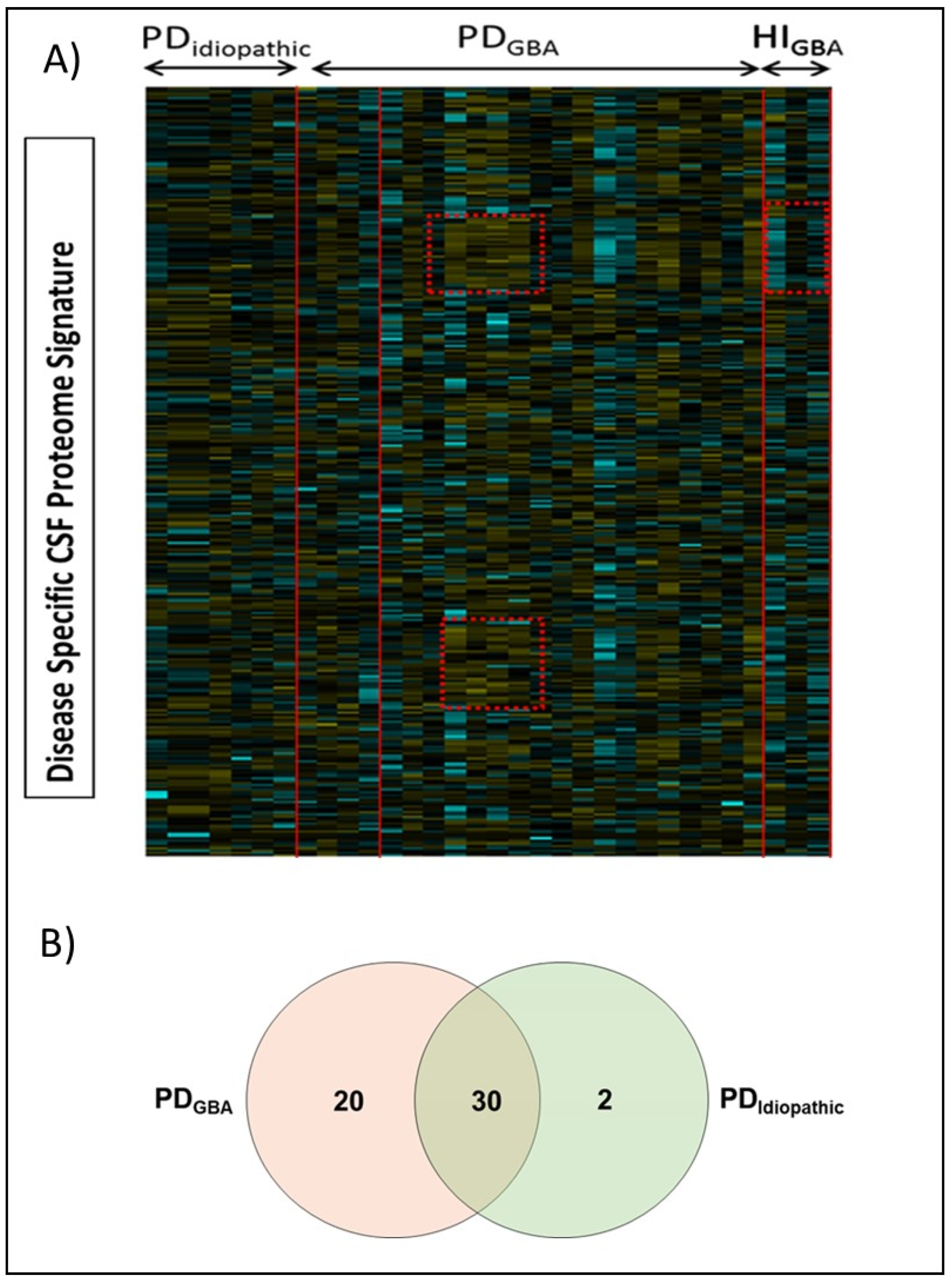
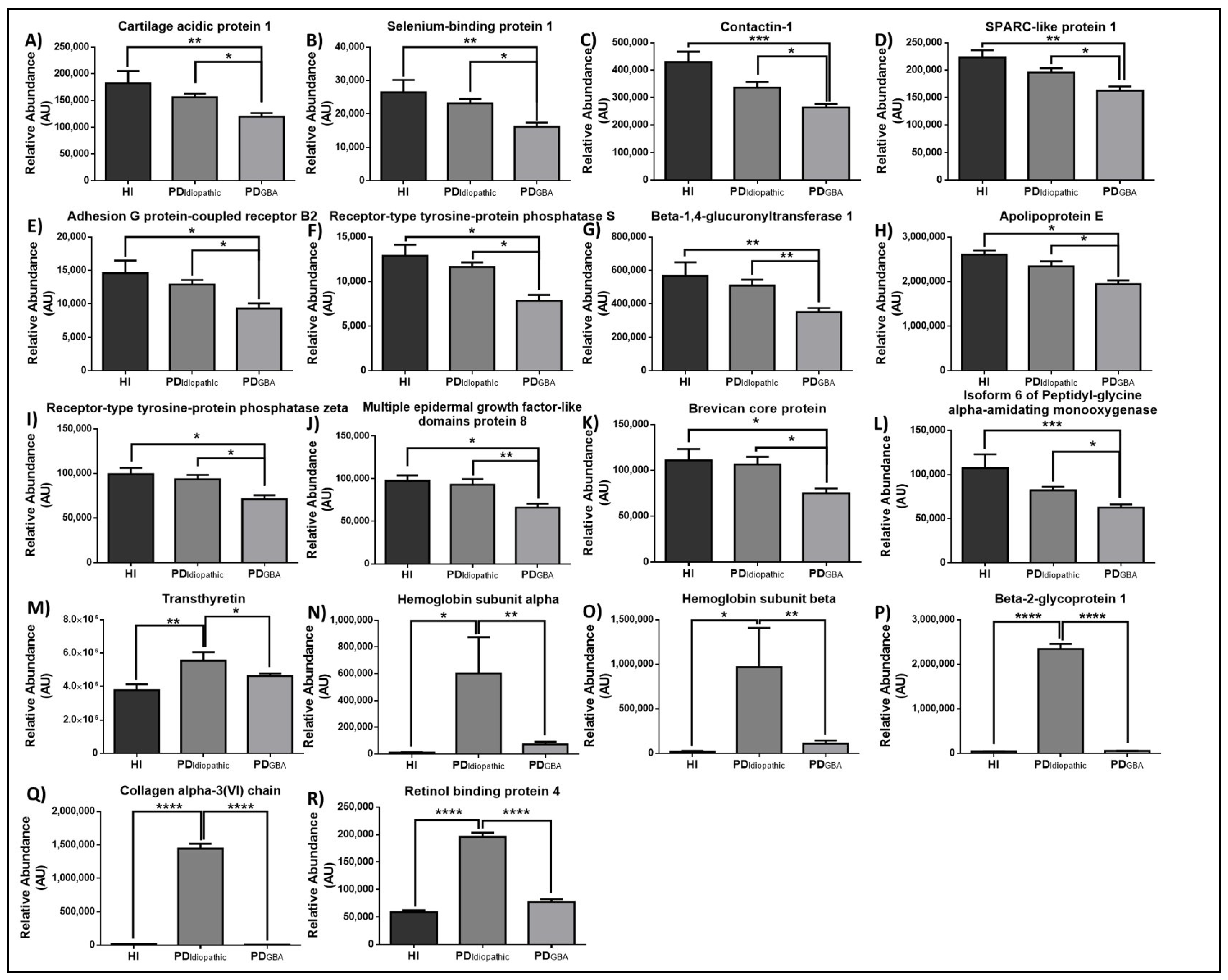
2.1. GBA-Mutation-Linked Proteomic Alterations in PD Patients

We created a spectral library with a traditional data-dependent acquisition (DDA) mass spectrometry method. It contained approximately 255 distinct protein groups and 2854 peptides, and 81,037 spectral counts were identified with greater than 99% confidence and passed the false discovery rate (FDR) from fit analysis using a critical FDR of 1%. However, at a global 1% FDR, 290 distinct protein groups, 3915 peptides, and 101,293 spectral counts were identified with greater than 99% confidence by using ProteinPilotTM software (Supplementary File S1). Following the generation of the spectral library, the identification and quantification of GBA-linked altered proteins and an intergroup analysis was conducted to identify the differentially regulated targets (Supplementary File S2). Overall, we were able to identify altered proteins from the generated spectral libraries and could establish clear differences in amount of the same proteins in response to GBA mutation with high confidence (Figure 1A). After removing the common contaminants, 52 differentially regulated proteins were obtained. Of these 52 targets, 20 proteins were significantly altered in PDGBA cases only while 2 showed different levels in PDIdiopathic patients. Additionally, subtype-specific analysis showed the number of uniquely regulated proteins, in comparison to HI in PD cases with and without GBA mutations (Figure 1B).

2.2. Pathway Analysis and Functional Characterization

A comprehensive pathway and functional analysis, using GO in conjunction with Panther, Uniprot, and ProteinPilot software, allowed us to annotate CSF proteins to various subgroups based on their functions. Most of the identified proteins were found to play a role in immune response, stress response, and response to an external and biotic stimulus. The components of the Wnt signaling pathway, plasminogen activating cascades, and Cadherin signaling pathways were most abundantly present in the CSF. In accordance with these findings, most of the proteins could be assigned to the class of defense proteins or were enzymes/enzyme modulators. Moreover, most of the proteins detected in CSF were of extracellular origin. Supplementary Figure S1 shows a detailed overview of the functional subclasses and roles of identified proteins along with the number of proteins specific to each subgroup.

2.3. Proteomic Alterations in Response to GBA-Mutation in PD Patients

We compared the profiles of CSF proteins of all groups to identify differentially regulated proteins that may play a role in disease pathology and were able to identify 52 significantly altered proteins for various groups. In PDGBA, 20 proteins were found to be significantly altered in comparison to HI, but not PDIdiopathic cases (Supplementary Figure S2). In comparison to the other groups, PDidiopathic cases presented two unique proteins, all of which were significantly upregulated (Supplementary Figure S3). The former category contained mainly secreted and membrane-bound proteins involved in neuronal structure (Neurotrimin, Neural cell adhesion molecule, Neural cell adhesion molecule 1, Fibronectin, Receptor-type tyrosine-protein phosphatase delta, Seizure 6-like protein, Fibulin 1, Fibulin 5, and Reticulon-4 receptor), neurotransmission (Isoform 2 of Amyloid-like protein 1), protein folding/ degradation (Clusterin, Plasma protease C1 inhibitor, Antithrombin -III), transport (Alpha-1-acid glycoprotein 2), immunity (Complement C6, Prostaglandin-H2), and metabolism (Galectin-3-binding protein, B4GALT1, Sulfhydryl oxidase 1, Alpha-mannosidase 2×). The two targets in the latter category were involved in metabolism (Beta hexoaminidase) and antioxidation (extracellular superoxide dismutase).
The remaining targets were significantly altered in both variants of PD. Within this category, 12 proteins showed a significant reduction in comparison to HI in both PDIdiopathic and PDGBA cases (Supplementary Figure S4). This category involved secreted, cytoplasmic, and membrane-bound neuronal structure (Contactin-2, Cell surface glycoprotein MUC 18, Gelsolin, Neural cell adhesion molecule 2, Neural cell adhesion molecule L1), neurotransmission (Ephrin type-A receptor 4), protein folding/degradation (Procollagen C-endopeptidase enhancer 1, Alpha-2-macroglobulin), and metabolism (Aspartate aminotransferase, Pyruvate kinase, L-lactate dehydrogenase, Transcobalamin-2).
Significant and clinically relevant differences were evident in PDIdiopathic and PDGBA cases among 18 cytoplasmic and secreted targets involved in neuronal structure (Contactin 1, Receptor-type tyrosine-protein phosphatase S, Receptor-type tyrosine-protein phosphatase zeta, Brevican core protein, Collagen alpha-3(VI) chain), signaling (Cartilage acidic protein 1, SPARC-like protein 1, Adhesion G Protein-Coupled Receptor B2), transport (Apolipoprotein E, Hemoglobin subunit alpha, Hemoglobin subunit alpha, Transthyretin, Beta-2-glycoprotein 1, Retinol binding protein 4), immunity (Multiple epidermal growth factor-like domains protein and 8), and metabolism (Selenium-binding protein 1, Beta-1,4-glucuronyltransferase 1, Isoform 6 of Peptidyl-glycine alpha-amidating monooxygenase) (Figure 2).

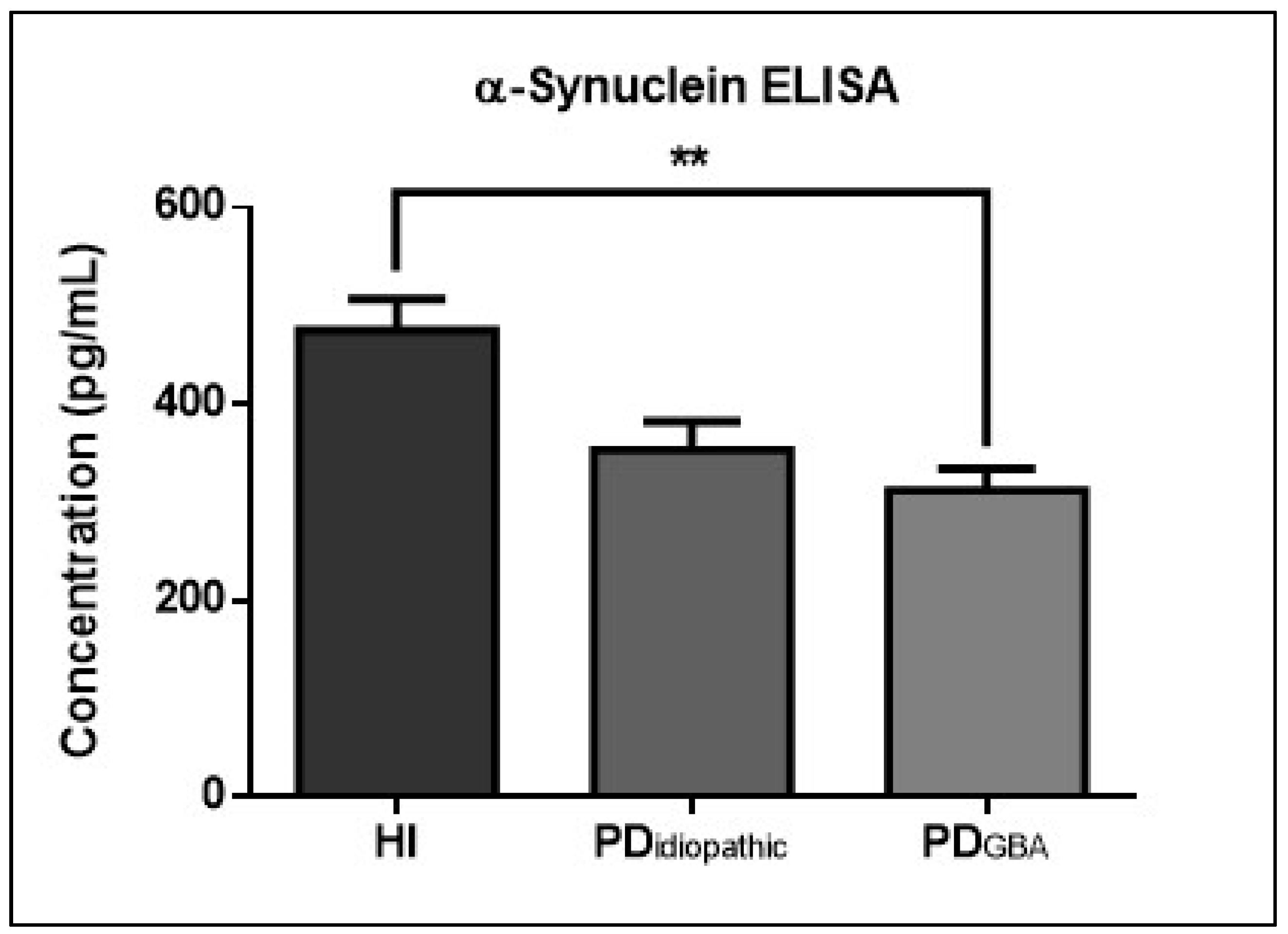
2.4. CSF α-Syn Profiles in PD Patients with GBA Mutations

ELISA-based quantification detected GBA genotype-associated alterations in CSF α-Syn levels in PD patients (Figure 3). Healthy individuals with GBA mutations presented higher levels of α-Syn in comparison to PD patients from both clinical variants; however, this trend was statistically significant in PDGBA cases only. The mean level of α-Syn was slightly higher in PDIdiopathic cases in comparison PDGBA cases.

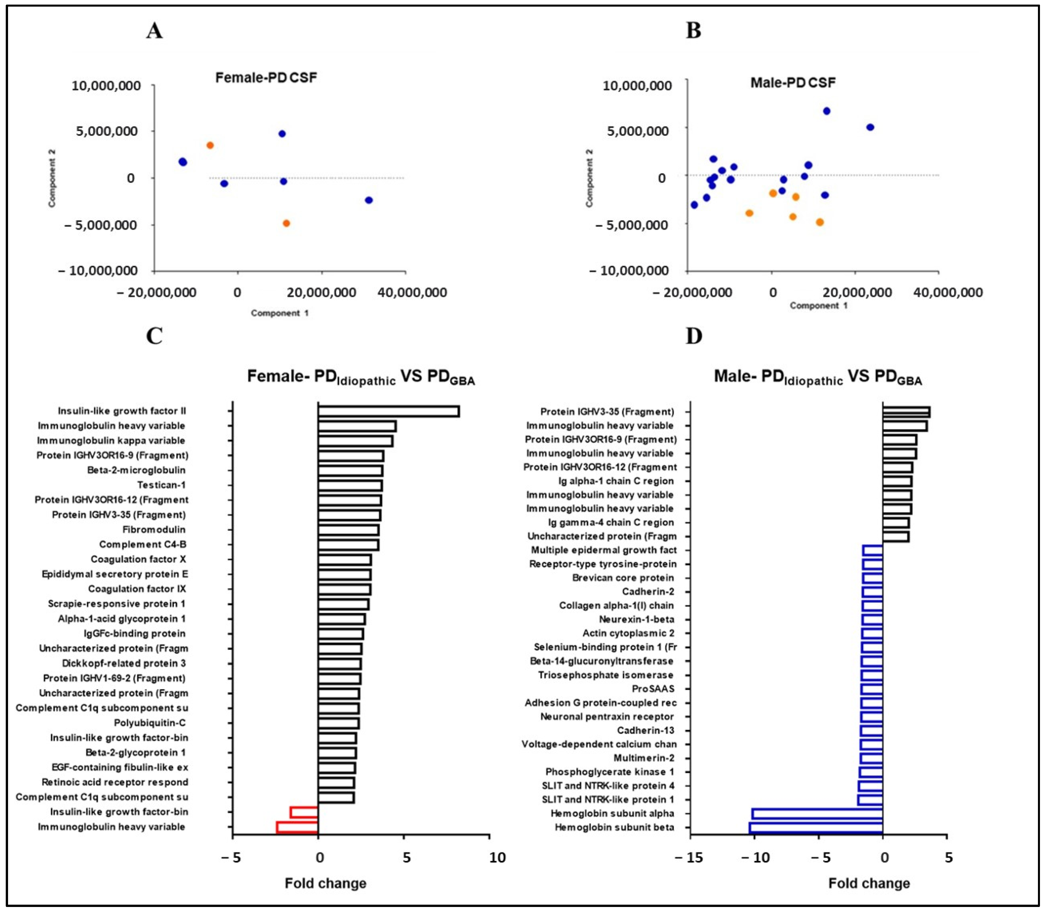
2.5. Gender-Specific Proteome Alterations in PD Patients with GBA Mutations

To elucidate the effects of gender on the CSF proteome of PD patients, we segregated our PD cohort into male and female cases. The PCA plot visualizes the effects of GBA mutation on male and female PD patients. Although no obvious clusters can be observed for female cases, possibly due to the low sample number, male samples present clear clustering for samples with and without mutations (Figure 4A,B). We further analyzed the targets in a gender-specific manner to determine the proteins that were accounting for the variation among male and female clusters (Figure 4C,D). Modifications specific to males showed two-fold or more change in 31 proteins while those specific to females depicted changes in 29 targets. However, the latter group had two cases of PD cases without GBA mutations so they need to be interpreted with caution.

3. Discussion

GBA mutations have been previously reported to influence the clinical outcome of PD [24]. Our study analyzed the impact of GBA mutations on the CSF proteome in PD patients with the aim of identifying disease triggers and potential biomarkers. For the quantitative assessment of the CSF proteome, we opted for a SWATH MS-based approach that is based on targeted peptide identification using a reference library. It combines the benefits of traditional discovery-based and targeted proteomics resulting in a high-throughput, reproducible, and accurate analysis of proteins in biological samples [25]. In the first study of its kind, we compared the CSF proteome of PD patients with and without GBA mutations to healthy controls. Owing to the dynamic and heterogeneous nature of the CSF proteome, it was not surprising that identified proteins belonged to many different functional classes and biological pathways. However, most of our targets were extracellular proteins involved in immune response, neurotransmission, and various signaling pathways. Due to a greater incidence of PD in men, we also targeted the gender-based differences within the proteome of the samples in our cohort [26].
Although all the differentially expressed proteins and their functional annotation provided valuable insights into the pathophysiology of PD, we focused on proteins that were uniquely regulated in certain groups with the aim of identifying possible causes of phenotypic diversity in sporadic and genetic PD. In the first subset, proteomic changes associated with PDIdiopathic cases were targeted. These patients exhibited a significant alteration in the levels of superoxide dismutase whose activity has been previously reported to be reduced in CSF [27]. Similarly, the increased levels of β-hexosaminidase have also been associated with the duration and severity of PD [28]. These proteins, along with the other amyloidogenic (Transthyretin), structural (Collagen), and metabolic (Retinol binding protein 4) proteins hint at the alterations associated with sporadic PD only.
The second subset of altered proteins involved the targets that show significant alterations in PDGBA patients only. This group involved immune (Complement C6, Prostaglandin-H2), transport (Clusterin), signaling (Galectin-3-binding protein, Receptor-type tyrosine-protein phosphatase delta, Alpha-1-acid glycoprotein 2), metabolic (B4GALT1, Plasma protease C1 inhibitor, Sulfhydryl oxidase 1, Antithrombin-III, Isoform 2 of Amyloid-like protein 1), and structural (Neurotrimin, Fibronectin, Seizure 6-like protein, Neural cell adhesion molecule, Neural cell adhesion molecule 1, Reticulon-4 receptor, Fibulin 1) proteins. Interestingly, the trends in this group mimicked the trends observed in α- Syn ELISA hinting at their direct involvement in the pathology of PD. Prostaglandins have been previously reported to modulate the unfolding of amyloids generated in PD and their reduced levels may cause increased aggregation of α-Syn [29]. Reduced levels of proteins involved in cell adhesion have been associated with aberrations in the structure of neurites and altering the ratio of excitatory neurons in comparison with inhibitory neurons, thereby contributing towards neurological disorders [30]. Fibronectin, also downregulated in all PD cases, has been known to exert neuroprotective effects in PD [31].
Proteins that were significantly altered in both PDIdiopathic and PDGBA cases allowed the identification of shared pathological mechanisms. Reduced levels of Gelsolin in both clinical variants can be attributed to its involvement in the aggregation of α-Syn and colocalization with the Lewy bodies [32]. The Neural cell adhesion molecule is also downregulated in all PD cases and is involved in the trafficking and internalization of dopamine [33]. Polymorphisms in Alpha-2-macroglobulin are a risk factor for PD and its CSF levels are also significantly downregulated in all the PD cases targeted in the current study [34].
The most promising targets were identified in the last subset where PDGBA cases differed significantly from PDIdiopathic and HI cases. Reduction in the CSF levels of Contactin-1, a target identified in this subset, has been reported previously and has been associated with synaptic degradation caused by the accumulation of Lewy bodies [35]. In the current study, its levels are significantly lower in PDGBA cases in comparison to PDIdiopathic samples indicating increased synaptic damage. Selenium-binding protein 1 may contribute towards aggravated pathology in PDGBA cases by modulating oxidative stress [36]. The reduced level of Adhesion G Protein-Coupled Receptor has been correlated with dopaminergic cell death and unveils another mechanism which may lead to aggravated pathology in PDGBA patients [37]. Decreased levels of Apolipoprotein E in CSF can be explained by increased aggregation of α-Syn in the brain as the former is known to increase the aggregation of the latter which may influence its release into the CSF [38]. The amount of Hemoglobin, another target identified in this subset, has been known to correlate with the severity of PD [39]. Interestingly, several targets identified in this subset showed a greater fold change in samples from males (known to have a greater incidence of PD) further validating their contribution to the disease pathology.
To establish the impact of GBA mutation on α-Syn accumulation, one of the key pathological markers of PD, we compared the levels of α-Syn in CSF. In accordance with the previously published data, our cohort also showed lowered α-Syn levels in CSF from PD patients, possibly in response to its increased aggregation in the brain and decreased elimination into CSF [40,41]. Previous studies have postulated that different GBA mutations may have a different impact on the phenotype of PD and these mutations may further reduce the levels of α-Syn in CSF [42,43,44]. However, this difference was not as evident in our cohort, possibly due to the smaller size of the PDIdiopathic group. The mean level of α-Syn was slightly higher in PDIdiopathic cases in comparison to PDGBA cases, but the difference was not significant.
Although GBA mutations have been known to increase the incidence of PD and contribute towards its clinical presentation (published previously for the current cohort [45,46]), the exact mechanism of their pathogenicity was not understood. We addressed this problem using proteomic approaches and could highlight some major differences that occur in PD patients in the response to GBA mutations. Our results show that the levels of several pathologically relevant proteins, including Contactin-1, Selenium-binding protein 1, Adhesion G Protein-Coupled Receptor, and Apolipoprotein E are significantly different among the sporadic and genetic variants of PD and hint at aggravated synaptic damage, oxidative stress, neuronal loss, and aggregation of α-Syn in PDGBA cases. However, the limited number of samples, especially those of controls (n = 3), and the dynamic nature of the CSF proteome demand the replication of the results in a larger cohort. Similarly, the gender-based differences also need to be interpreted cautiously due to a smaller number of female patients and a considerable difference in group size. A comprehensive analysis, that includes sequenced GBA mutations in larger cohorts, might be needed to establish the relative pathogenicity of specific polymorphisms on the GBA gene in PD in addition to their effects on the age at onset in different genders and their response to various medications. Nevertheless, the outcomes of this study provide important candidates for further validation as diagnostic, prognostic, and therapeutic markers.

4. Materials and Methods

4.1. Participants and Ethical Approval

A total of 32 participants (Table 1) were included in this study. A detailed neurological assessment was conducted and the patients who fulfilled Movement Disorder Society Clinical Diagnostic Criteria for PD were included in the study. Genetic assessment of GBA mutations (N370S, L444P, and E326K) was conducted through genotyping and restriction digestion using slight modifications in previously established criteria [45,46]. The control cases underwent clinical examination to exclude any neurological disorders that may affect the study. All participants provided informed consent prior to the collection of CSF samples and the study was approved by the ethical committee of the University of Tübingen (702/2013BO1).

4.2. Sample Collection, Preparation and In-Gel Tryptic Digestion

CSF samples were collected through a lumbar puncture, centrifuged at 2000× g for 10 min at 4 °C and stored at −80 °C until further analysis. Samples (50 µL) were concentrated using SpeedVac (sc100, American laboratory trading, Groton, CT, USA) and resuspended in 40 µL of lysis buffer (7M Urea, 2M thiourea and 4% CHAPS, protease, and phosphatase inhibitor cocktail). They were loaded onto a 4–12% NuPAGE Novex Bis-Tris Minigels (Invitrogen, Carlsbad, CA, USA) and allowed to run to a length of 1.5 cm. The gels were subjected to Coomassie staining and protein bands were excised.
In-gel tryptic digestion was performed as previously described [47]. Briefly, the protein bands were diced, reduced, and alkylated followed by overnight digestion with trypsin. The solution containing Tryptic peptides was dried in a SpeedVac and stored at −20 °C until further analysis.

4.3. Quantitative SWATH Analysis

For the generation of spectral library, equal amounts of proteins from each sample were pooled to a final concentration of 80 µg and separated into eight fractions using reversed-phase spin columns (Pierce High pH Reversed-Phase Peptide Fractionation Kit, Thermo Fisher Scientific, Waltham, MA, USA). Each fraction was divided into two technical replicates prior to analysis. Digested peptides were dissolved in a loading buffer (2% acetonitrile, 0.1% formic acid in water) to a final concentration of 0.3 µg/µL. For each analysis, 1.5 µg of the analyte was enriched on a pre-column (0.18 mm ID × 20 mm, Symmetry C18-AQ 5 µm, Waters, Milford, MA, USA) and separated on an analytical RP-C18 column (0.075 mm ID × 250 mm, HSS T3, 1.8 µm, Waters). A Nanoflow chromatography system (Eksigent nanoLC425) hyphenated to a hybrid triple quadrupole-TOF mass spectrometer (TripleTOF 5600+) equipped with a Nanospray III ion source (Ionspray Voltage 2400 V, Interface Heater Temperature 150 °C, Sheath Gas Setting 12) and controlled by Analyst TF 1.7.1 software build 1163 (all AB Sciex, Redwood City, CA, USA) was used for analysis.
For the analysis of samples, MS/MS data were acquired using 65 variable size windows across the 400–1050 m/z range. PeakView Software version 2.1 build 11,041 (AB Sciex, Redwood City, CA, USA) and SWATH quantitation microApp version 2.0 build 2003 were used for generating spectral library and extracting SWATH peaks.

4.4. Protein Annotation and Functional Characterization

ProteinPilot Software version 5.0 build 4769 (AB Sciex, Redwood City, CA, USA) was used for protein identification. MS/MS spectra from the combined qualitative analyses were searched against the UniProtKB Homo sapiens reference proteome (revision 02-2017, 92,928 entries) augmented with a set of 51 known common laboratory contaminants to identify 363 proteins at a False Discovery Rate (FDR) of 1%. Functional characterization was also achieved through UniProtKB Homo sapiens reference proteome and Panther classification system [48].

4.5. ELISA

The commercially available ELISA kit (Biolegend, San Diego, CA, USA, catalogue # 844101) was used for the analysis of CSF levels of α-Syn according to the manufacturer’s protocol.

4.6. Statistical Analysis

Data visualization and statistical analysis were performed using R. Studio (version 1.1.383) and ClustVis [49]. ANOVA in combination with Tukey Multiple comparisons test were used to identify the differentially regulated targets and p-value < 0.05 was considered significant.

Supplementary Materials

The following supporting information can be downloaded at: https://www.mdpi.com/article/10.3390/ijms232214166/s1.

Author Contributions

S.Z., A.N., T.G., I.Z.: Study design, experimentation, data analysis and manuscript compilation. N.Y., M.S. (Mohsin Shafiq) and, M.S. (Matthias Schmitz): Data analysis, experimentation. I.W., K.B.: Characterization of samples and genotyping. All authors have read and agreed to the published version of the manuscript.

Funding

This research received no external funding.

Institutional Review Board Statement

The study was approved by the ethical committee of the University of Tübingen (702/2013BO1).

Informed Consent Statement

All participants provided informed consent prior to the collection of CSF samples.

Data Availability Statement

Raw data is included as Supplementary Materials. All raw MS files can be obtained from the corresponding author upon request.

Conflicts of Interest

The authors declare no conflict of interest.

References

  1. Tysnes, O.B.; Storstein, A. Epidemiology of Parkinson’s disease. J. Neural Transm. Suppl. 2017, 124, 901–905. [Google Scholar] [CrossRef]
  2. Parkinson, J. An essay on the shaking palsy. J. Neuropsychiatry Clin. Neurosci. 2002, 14, 223–236. [Google Scholar] [CrossRef] [PubMed]
  3. Baba, M.; Nakajo, S.; Tu, P.H.; Tomita, T.; Nakaya, K.; Lee, V.M.; Trojanowski, J.Q.; Iwatsubo, T. Aggregation of alpha-synuclein in Lewy bodies of sporadic Parkinson’s disease and dementia with Lewy bodies. Am. J. Pathol. 1998, 152, 879. [Google Scholar] [PubMed]
  4. Jankovic, J. Parkinson’s disease: Clinical features and diagnosis. J. Neurol. Neurosurg. Psychiatry 2008, 79, 368–376. [Google Scholar] [CrossRef] [PubMed] [Green Version]
  5. Frank, C.; Pari, G.; Rossiter, J.P. Approach to diagnosis of Parkinson disease. Can. Fam. Physician 2006, 52, 862–868. [Google Scholar]
  6. McCormack, A.L.; Thiruchelvam, M.; Manning-Bog, A.B.; Thiffaulta, C.; Langstona, J.W.; Cory-Slechta, D.A.; Di Monte, D.A. Environmental risk factors and Parkinson’s disease: Selective degeneration of nigral dopaminergic neurons caused by the herbicide paraquat. Neurobiol. Dis. 2002, 10, 119–127. [Google Scholar] [CrossRef] [Green Version]
  7. Satake, W.; Nakabayashi, Y.; Mizuta, I.; Hirota, Y.; Ito, C.; Kubo, M.; Kawaguchi, T.; Tsunoda, T.; Watanabe, M.; Takeda, A.; et al. Genome-wide association study identifies common variants at four loci as genetic risk factors for Parkinson’s disease. Nat. Genet. 2009, 41, 1303–1307. [Google Scholar] [CrossRef]
  8. Collier, T.J.; Kanaan, N.M.; Kordower, J.H. Aging and Parkinson’s disease: Different sides of the same coin? Mov. Disord. 2017, 32, 983–990. [Google Scholar] [CrossRef] [Green Version]
  9. Goker-Alpan, O.; Schiffmann, R.; LaMarca, M.E.; Nussbaum, R.L.; McInerney-Leo, A.; Sidransky, E. Parkinsonism among Gaucher disease carriers. J. Med. Genet. 2004, 41, 937–940. [Google Scholar] [CrossRef] [Green Version]
  10. Klein, C.; Westenberger, A. Genetics of Parkinson’s disease. Cold Spring Harb. Perspect. Med. 2012, 2. [Google Scholar] [CrossRef] [Green Version]
  11. Grabowski, G.A.; Dinur, T.; Osiecki, K.M.; Kruse, J.R.; Legler, G.; Gatt, S. Gaucher disease types 1, 2, and 3: Differential mutations of the acid beta-glucosidase active site identified with conduritol B epoxide derivatives and sphingosine. Am. J. Hum. Genet. 1985, 37, 499. [Google Scholar] [PubMed]
  12. Hruska, K.S.; LaMarca, M.E.; Scott, C.R.; Sidransky, E. Gaucher disease: Mutation and polymorphism spectrum in the glucocerebrosidase gene (GBA). Hum. Mutat. 2008, 29, 567–583. [Google Scholar] [CrossRef] [PubMed]
  13. O’Regan, G.; deSouza, R.M.; Balestrino, R.; Schapira, A.H. Glucocerebrosidase mutations in Parkinson disease. J. Parkinsons. Dis. 2017, 7, 411–422. [Google Scholar] [CrossRef] [PubMed] [Green Version]
  14. Neumann, J.; Bras, J.; Deas, E.; O’Sullivan, S.S.; Parkkinen, L.; Lachmann, R.H.; Li, A.; Holton, J.; Guerreiro, R.; Paudel, R.; et al. Glucocerebrosidase mutations in clinical and pathologically proven Parkinson’s disease. Brain 2009, 132, 1783–1794. [Google Scholar] [CrossRef] [Green Version]
  15. Gan-Or, Z.; Amshalom, I.; Kilarski, L.L.; Bar-Shira, A.; Gana-Weisz, M.; Mirelman, A.; Marder, K.; Bressman, S.; Giladi, N.; Orr-Urtreger, A. Differential effects of severe vs mild GBA mutations on Parkinson disease. Neurology 2015, 84, 880–887. [Google Scholar] [CrossRef] [Green Version]
  16. Du, T.-T.; Wang, L.; Duan, C.-L.; Lu, L.-L.; Zhang, J.-L.; Gao, G.; Qiu, X.-B.; Wang, X.-M.; Yang, H. GBA deficiency promotes SNCA/α-synuclein accumulation through autophagic inhibition by inactivated PPP2A. Autophagy 2015, 11, 1803–1820. [Google Scholar] [CrossRef] [Green Version]
  17. Chahine, L.M.; Qiang, J.; Ashbridge, E.; Minger, J.; Yearout, D.; Horn, S.; Colcher, A.; Hurtig, H.I.; Lee, V.M.-Y.; Van Deerlin, V.M.; et al. Clinical and biochemical differences in patients having Parkinson disease with vs without GBA mutations. JAMA Neurol. 2013, 70, 852–858. [Google Scholar] [CrossRef] [Green Version]
  18. Mata, I.F.; Leverenz, J.B.; Weintraub, D.; Trojanowski, J.Q.; Chen-Plotkin, A.; Van Deerlin, V.M.; Ritz, B.; Rausch, R.; Do, S.A.F.; Wood-Siverio, C.; et al. GBA Variants are associated with a distinct pattern of cognitive deficits in Parkinson’s disease. Mov. Disord. 2016, 31, 95–102. [Google Scholar] [CrossRef] [Green Version]
  19. Davis, M.Y.; Johnson, C.O.; Leverenz, J.B.; Weintraub, D.; Trojanowski, J.Q.; Chen-Plotkin, A.; Van Deerlin, V.M.; Quinn, J.F.; Chung, K.A.; Peterson-Hiller, A.L.; et al. Association of GBA mutations and the E326K polymorphism with motor and cognitive progression in Parkinson disease. JAMA Neurol. 2016, 73, 1217–1224. [Google Scholar] [CrossRef] [Green Version]
  20. Brockmann, K.; Srulijes, K.; Pflederer, S.; Hauser, A.; Schulte, C.; Maetzler, W.; Gasser, T.; Berg, D. GBA-associated Parkinson’s disease: Reduced survival and more rapid progression in a prospective longitudinal study. Move. Disord. 2015, 30, 407–411. [Google Scholar] [CrossRef]
  21. Guedes, L.C.; Chan, R.B.; Gomes, M.A.; Conceição, V.A.; Machado, R.B.; Soares, T.; Xu, Y.; Gaspar, P.; Carriço, J.A.; Alcalay, R.N.; et al. Serum lipid alterations in GBA-associated Parkinson’s disease. Park. Relat. Disord. 2017, 44, 58–65. [Google Scholar] [CrossRef] [PubMed]
  22. Adler, C.H.; Beach, T.G.; Shill, H.A.; Caviness, J.N.; Driver-Dunckley, E.; Sabbagh, M.N.; Patel, A.; Sue, L.I.; Serrano, G.; Jacobson, S.A.; et al. GBA mutations in Parkinson disease: Earlier death but similar neuropathological features. Eur. J. Neurol. 2017, 24, 1363–1368. [Google Scholar] [CrossRef] [PubMed]
  23. Gillet, L.C.; Navarro, P.; Tate, S.; Röst, H.; Selevsek, N.; Reiter, L.; Bonner, R.; Aebersold, R. Targeted data extraction of the MS/MS spectra generated by data-independent acquisition: A new concept for consistent and accurate proteome analysis. Mol. Cell Proteom. 2012, 11, O111.016717. [Google Scholar] [CrossRef] [PubMed] [Green Version]
  24. Winder-Rhodes, S.E.; Evans, J.R.; Ban, M.; Mason, S.L.; Williams-Gray, C.; Foltynie, T.; Duran, R.; Mencacci, N.E.; Sawcer, S.J.; Barker, R.A. Glucocerebrosidase mutations influence the natural history of Parkinson’s disease in a community-based incident cohort. Brain 2013, 136, 392–399. [Google Scholar] [CrossRef] [PubMed]
  25. Anjo, S.I.; Santa, C.; Manadas, B. Short GeLC-SWATH: A fast and reliable quantitative approach for proteomic screenings. Proteomics 2015, 15, 757–762. [Google Scholar] [CrossRef]
  26. Miller, I.N.; Cronin-Golomb, A. Gender differences in Parkinson’s disease: Clinical characteristics and cognition. Mov. Disord. 2010, 25, 2695–2703. [Google Scholar] [CrossRef] [Green Version]
  27. De Deyn, P.P.; Hiramatsu, M.; Borggreve, F.; Goeman, J.; Dʼhooge, R.; Saerens, J.; Mori, A. Superoxide dismutase activity in cerebrospinal fluid of patients with dementia and some other neurological disorders. Alzheimer Dis. Assoc. Disord. 1998, 12, 26–32. [Google Scholar] [CrossRef]
  28. Kim, H.J.; Jeon, B.; Song, J.; Lee, W.W.; Park, H.; Shin, C. Leukocyte glucocerebrosidase and β-hexosaminidase activity in sporadic and genetic Parkinson disease. Park. Relat. Disord. 2016, 23, 99–101. [Google Scholar] [CrossRef]
  29. Famitafreshi, H.; Karimian, M. Prostaglandins as the agents that modulate the course of brain disorders. Degener. Neurol. Neuromuscul. Dis. 2020, 10, 1–13. [Google Scholar] [CrossRef]
  30. Singh, K.; Jayaram, M.; Kaare, M.; Leidmaa, E.; Jagomäe, T.; Heinla, I.; Hickey, M.; Kaasik, A.; Schäfer, M.K.; Innos, J.; et al. Neural cell adhesion molecule Negr1 deficiency in mouse results in structural brain endophenotypes and behavioural deviations related to psychiatric disorders. Sci. Rep. 2019, 9, 5457. [Google Scholar] [CrossRef] [Green Version]
  31. Wang, J.; Yin, L.; Chen, Z. Neuroprotective role of fibronectin in neuron-glial extrasynaptic transmission. Neural Regen. Res. 2013, 8, 376. [Google Scholar] [CrossRef] [PubMed]
  32. Welander, H.; Bontha, S.V.; Näsström, T.; Karlsson, M.; Nikolajeff, F.; Danzer, K.; Kostka, M.; Kalimo, H.; Lannfelt, L.; Ingelsson, M.; et al. Gelsolin co-occurs with Lewy bodies in vivo and accelerates α-synuclein aggregation in vitro. Biochem. Biophys. Res. Commun. 2011, 412, 32–38. [Google Scholar] [CrossRef] [PubMed]
  33. Xiao, M.F.; Xu, J.C.; Tereshchenko, Y.; Novak, D.; Schachner, M.; Kleene, R. Neural cell adhesion molecule modulates dopaminergic signaling and behavior by regulating dopamine D2 receptor internalization. J. Neurosci. 2009, 29, 14752–14763. [Google Scholar] [CrossRef] [Green Version]
  34. Guo, X.; Tang, P.; Li, X.; Chong, L.; Zhang, X.; Li, R.; Xingzhi, G.; Peng, T.; Xiaoqing, L.; Li, C.; et al. Association between two α-2-macroglobulin gene polymorphisms and Parkinson’s disease: A meta-analysis. Int. J. Neurosci. 2016, 126, 193–198. [Google Scholar] [CrossRef] [PubMed]
  35. Chatterjee, M.; van Steenoven, I.; Huisman, E.; Oosterveld, L.; Berendse, H.; Van Der Flier, W.M.; Del Campo, M.; Lemstra, A.W.; Van De Berg, W.D.J.; Teunissen, C.E. Contactin-1 is reduced in cerebrospinal fluid of parkinson’s disease patients and is present within lewy bodies. Biomolecules 2020, 10, 1177. [Google Scholar] [CrossRef] [PubMed]
  36. Ellwanger, J.H.; Franke, S.I.; Bordin, D.L.; Prá, D.; Henriques, J.A. Biological functions of selenium and its potential influence on Parkinson’s disease. An. Acad. Bras. Cienc. 2016, 88, 1655–1674. [Google Scholar] [CrossRef] [PubMed] [Green Version]
  37. Choi, J.S.; Bae, W.Y.; Nam, S.; Jeong, J.-W. New targets for Parkinson’s disease: Adhesion G protein-coupled receptor B1 is downregulated by AMP-activated protein kinase activation. OMICS J. Integr. Biol. 2018, 22, 493–501. [Google Scholar] [CrossRef] [PubMed]
  38. Fyfe, I. APOE* ε4 promotes synucleinopathy. Nat. Rev. Neurol. 2020, 16, 185. [Google Scholar] [CrossRef]
  39. Shohimardonov, S.H.; Daminova, K. Examining level of haemoglobin in patients with Parkinson’s disease and the role of haemoglobin level in the disease course. Mov. Disord. 2019, 34, 1075. [Google Scholar]
  40. Gao, L.; Tang, H.; Nie, K.; Wang, L.; Zhao, J.; Gan, R.; Huang, J.; Zhu, R.; Feng, S.; Duan, Z.; et al. Cerebrospinal fluid alpha-synuclein as a biomarker for Parkinson’s disease diagnosis: A systematic review and meta-analysis. Int. J. Neurosci. 2015, 125, 645–654. [Google Scholar] [CrossRef]
  41. Stav, A.L.; Aarsland, D.; Johansen, K.K.; Hessen, E.; Auning, E.; Fladby, T. Amyloid-β and α-synuclein cerebrospinal fluid biomarkers and cognition in early Parkinson’s disease. Park. Relat. Disord. 2015, 21, 758–764. [Google Scholar] [CrossRef] [PubMed]
  42. Thaler, A.; Bregman, N.; Gurevich, T.; Shiner, T.; Dror, Y.; Zmira, O.; Gan-Or, Z.; Bar-Shira, A.; Gana-Weisz, M.; Orr-Urtreger, A.; et al. Parkinson’s disease phenotype is influenced by the severity of the mutations in the GBA gene. Park. Relat. Disord. 2018, 55, 45–49. [Google Scholar] [CrossRef] [PubMed]
  43. Lerche, S.; Wurster, I.; Roeben, B.; Zimmermann, M.; Riebenbauer, B.; Deuschle, C.; Hauser, A.; Schulte, C.; Berg, D.; Maetzler, W.; et al. Parkinson’s disease: Glucocerebrosidase 1 mutation severity is associated with CSF alpha-synuclein profiles. Mov. Disord. 2020, 35, 495–499. [Google Scholar] [CrossRef] [PubMed]
  44. Brockmann, K.; Quadalti, C.; Lerche, S.; Rossi, M.; Wurster, I.; Baiardi, S.; Roeben, B.; Mammana, A.; Zimmermann, M.; Hauser, A.-K.; et al. Association between CSF alpha-synuclein seeding activity and genetic status in Parkinson’s disease and dementia with Lewy bodies. Acta Neuropathol. Commun. 2021, 9, 175. [Google Scholar] [CrossRef] [PubMed]
  45. Brockmann, K.; Schulte, C.; Deuschle, C.; Hauser, A.-K.; Heger, T.; Gasser, T.; Maetzler, W.; Berg, D. Neurodegenerative CSF markers in genetic and sporadic PD: Classification and prediction in a longitudinal study. Park. Relat. Disord. 2015, 21, 1427–1434. [Google Scholar] [CrossRef] [PubMed]
  46. Brockmann, K.; Srulijes, K.; Hauser, A.K.; Schulte, C.; Csoti, I.; Gasser, T.; Berg, D. GBA-associated PD presents with nonmotor characteristics. Neurology 2011, 77, 276–280. [Google Scholar] [CrossRef]
  47. Schmidt, C.; Urlaub, H. iTRAQ-labeling of in-gel digested proteins for relative quantification. In Proteomics; Humana Press: Totowa, NJ, USA, 2009; pp. 207–226. [Google Scholar] [CrossRef] [Green Version]
  48. Thomas, P.D.; Campbell, M.J.; Kejariwal, A.; Mi, H.; Karlak, B.; Daverman, R.; Diemer, K.; Muruganujan, A.; Narechania, A. PANTHER: A library of protein families and subfamilies indexed by function. Genome Res. 2003, 13, 2129–2141. [Google Scholar] [CrossRef] [Green Version]
  49. Metsalu, T.; Vilo, J. ClustVis: A web tool for visualizing clustering of multivariate data using Principal Component Analysis and heatmap. Nucleic Acids Res. 2015, 43, 566–570. [Google Scholar] [CrossRef]
Figure 1. Disease-specific alterations in CSF proteome. (A) The heat map shows the relative levels of identified proteins across all analyzed samples. The red lines mark clustering in response to GBA genotype in PD cases. Both rows and columns were clustered using correlation distance and average linkage and these clusters are represented by red lines (B) Venn diagram presenting the number and distribution of differentially regulated proteins of each group (in comparison to controls).
Ijms 23 14166 g001
Figure 2. Relative CSF concentrations of the proteins differentially regulated in PDIdiopathic vs PDGBA cases. The graphs depict the relative amount of (A) Cartilage acidic protein 1, (B) Selenium-binding protein 1, (C) Contactin 1, (D) SPARC-like protein 1, (E) Adhesion G Protein-Coupled Receptor B2, (F) Receptor-type tyrosine-protein phosphatase S, (G) Beta-1,4-glucuronyltransferase 1, (H) Apolipoprotein E, (I) Receptor-type tyrosine-protein phosphatase zeta, (J) Multiple epidermal growth factor-like domains protein 8, (K) Brevican core protein, (L) Isoform 6 of Peptidyl-glycine alpha-amidating monooxygenase, (M) Transthyretin, (N) Hemoglobin subunit alpha, (O) Hemoglobin subunit alpha, (P) Beta-2-glycoprotein 1, (Q) Collagen alpha-3(VI) chain and (R) Retinol binding protein 4 in HI, PDIdiopathic, and PDGBA cases. One-way ANOVA, followed by Tukey’s multiple comparisons test, was used for statistical analysis. Error bars represent SEM. (* p ≤ 0.05; ** p ≤ 0.01; *** p ≤ 0.001; **** p ≤ 0.0001).
Ijms 23 14166 g002
Figure 3. Correlation between α-Syn levels in CSF of PD patients with and without GBA mutations. The graph depicts the levels of α-Syn (pg/mL) in CSF from HI, PDidiopathic, and PDGBA cases. One-way ANOVA, followed by Tukey’s multiple comparisons test, was used for statistical analysis. Error bars represent SEM. (** p ≤ 0.01).
Ijms 23 14166 g003
Figure 4. Effect of gender on CSF proteome in PD patients: Principal Component Analysis shows clusters for (A) female and (B) male cases with (blue circles) and without GBA mutation (orange circles). The column bar graph depicts the targets that show two-fold or higher fold change in PDIdiopathic (C) female and (D) male cases in comparison to the patients carrying a GBA mutation. The red and blue columns represent the downregulated proteins in female and male patients respectively.
Ijms 23 14166 g004
Table 1. List of samples used in the study. A total of 32 CSF samples were used for the study including 22 PD cases with GBA mutation (PDGBA), 7 PD cases without mutations (PDIdiopathic), and 3 healthy controls with GBA mutation (HIGBA).
No.Patient IDAgeGenderAge at Onset
PDIdiopathic (Mean age 63.4 ± 7.78 years)
1PDIdiopathic-166Male63
2PDIdiopathic-274Male62
3PDIdiopathic-362Female53
4PDIdiopathic-453Male42
5PDIdiopathic-560Male51
6PDIdiopathic-674Female67
7PDIdiopathic-755Male50
PDGBA (Mean age 60.5 ± 9.97 years)
8PDGBA-176Female47
9PDGBA-258Male50
10PDGBA-365Female55
11PDGBA-461Male45
12PDGBA-544Male28
13PDGBA-660Male49
14PDGBA-759Male 52
15PDGBA-851Male41
16PDGBA-950Male44
17PDGBA-1075Male70
18PDGBA-1141Male39
19PDGBA-1267Female56
20PDGBA-1373Female65
21PDGBA-1463Male58
22PDGBA-1558Female51
23PDGBA-1670Male60
24PDGBA-1767Male 63
25PDGBA-1842Male40
26PDGBA-1964Female51
27PDGBA-2059Male51
28PDGBA-2168Male56
29PDGBA-2262Male52
HIGBA (Mean age 52.6 ± 13.76 years)
30HIGBA-172Female-
31HIGBA-245Male-
32HIGBA-341Female-
Publisher’s Note: MDPI stays neutral with regard to jurisdictional claims in published maps and institutional affiliations.

