Review

Interactions of Gram-Positive Bacterial Membrane Vesicles and Hosts: Updates and Future Directions

by
Giuseppe Sangiorgio
*,
Emanuele Nicitra
,
Dalida Bivona
,
Carmelo Bonomo
,
Paolo Bonacci
,
Maria Santagati
,
Nicolò Musso
,
Dafne Bongiorno
* and
Stefania Stefani
Department of Biomedical and Biotechnological Sciences (BIOMETEC), University of Catania, 95125 Catania, Italy
*
Authors to whom correspondence should be addressed.
Int. J. Mol. Sci. 2024, 25(5), 2904; https://doi.org/10.3390/ijms25052904
Submission received: 24 January 2024 / Revised: 26 February 2024 / Accepted: 29 February 2024 / Published: 1 March 2024
(This article belongs to the Special Issue Host-Pathogen Interaction 5.0)

Abstract

:
Extracellular vesicles (EVs) are lipid bilayers derived from cell membranes, released by both eukaryotic cells and bacteria into the extracellular environment. During production, EVs carry proteins, nucleic acids, and various compounds, which are then released. While Gram-positive bacteria were traditionally thought incapable of producing EVs due to their thick peptidoglycan cell walls, recent studies on membrane vesicles (MVs) in Gram-positive bacteria have revealed their significant role in bacterial physiology and disease progression. This review explores the current understanding of MVs in Gram-positive bacteria, including the characterization of their content and functions, as well as their interactions with host and bacterial cells. It offers a fresh perspective to enhance our comprehension of Gram-positive bacterial EVs.

1. Introduction

The fundamental process of secreting cellular components across the plasma membrane is a universal occurrence in all life forms, facilitating interactions between organisms and their surroundings. This mechanism is achieved through the release of vesicles—spherical, nanosized structures derived from the lipid membranes of the cell surface [1,2,3]. Recently, there has been increased attention on extracellular vesicles (EVs) [4,5,6]. Originally discovered in eukaryotes, EVs from the plasma membrane carry proteins, nucleic acids, and lipids, categorized mainly into exosomes, microvesicles, and apoptotic bodies by size [7,8]. Subsequently, EVs were identified in prokaryotes, including bacteria [1,9]. Bacteria are classified into Gram-negative or Gram-positive based on membrane structure, with Gram-negative bacteria having two membrane layers separated by the periplasm. In contrast, Gram-positive bacteria possess a distinct membrane structure consisting of one membrane and a thicker layer of peptidoglycan. In Gram-negative bacteria, EVs are termed outer membrane vesicles (OMVs), while in Gram-positive bacteria, they are referred to as membrane vesicles (MVs) [10,11]. Both OMVs and MVs constitute subclasses of microbial EVs. Extensive research has been conducted on OMVs to elucidate their functions [12,13,14]. Questions about the relatively low production of vesicles in Gram-positive bacteria were prevalent in the past. However, over the last decade, substantial research has been dedicated to understanding the role of Gram-positive MVs [15]. Observations have indicated the presence of MVs in both pathogenic and nonpathogenic Gram-positive bacteria across diverse growth conditions and environments, suggesting the universal and widespread nature of MV secretion [16,17,18,19]. Depending on the packaged cargo, EVs have been implicated in pathogenesis, antibiotic resistance, stress response, intercellular competition, and nucleic acid transfer. The increase in studies on Gram-positive bacteria, such as Staphylococcus aureus, Bacillus anthracis, and Streptococcus mutans, has revealed that MVs in Gram-positive bacteria exhibit a 20- to 400-nm bilayer spherical structure [20,21,22]. This review explores the current understanding of the characterization of the content and functions of MVs in Gram-positive bacteria, as well as their interactions with host and bacterial cells.

2. Vesiculogenesis

The formation of membrane vesicles is well-documented in archaeal, Gram-negative, and mammalian cells, yet understanding MV biogenesis in Gram-positive bacteria, with their peptidoglycan-rich structure, has been challenging [1,23]. Various models for OMV biogenesis have been proposed, with genetic and biochemical analyses shedding light on the process [24,25]. Despite the relatively recent exploration of MVs in Gram-positive bacteria, with fewer studies compared to their Gram-negative counterparts, only a limited number of investigations have identified genetic factors responsible for vesicle formation [23,26]. Two hypotheses have been proposed to elucidate the mechanism of MVs crossing the barrier of the peptidoglycan. One suggestion has posited that MVs may be propelled through pores in the cell wall by turgor pressure, driven by budding from the cell membrane. Alternatively, it has been proposed that the peptidoglycan may undergo localized degradation, either due to enzymes associated with EVs or released alongside them [1]. MV release appears to initiate with the budding of the cytoplasmic membrane, with a hypotonic environment being pivotal for vesiculogenesis [27] (Figure 1). Lipidomic studies have revealed similarities between MVs and cytoplasmic membranes. However, variations in fatty acids and phospholipid content suggest that vesicle budding may occur in specific lipid-enriched membrane domains [22,28]. Recently, nanopods or nanotubes, filamentous structures facilitating cell-to-cell transfer and associated with EVs, have gained attention in bacterial research [29]. These structures, resembling eukaryotic ‘tunneling nanotubes’, were initially observed in hyperthermophilic bacteria and later identified in various bacteria, including Firmicutes, Myxobacteria, and Proteobacteria [30,31,32,33]. Nanotubes can bridge neighboring cells, promoting communication and facilitating molecular exchange [30]. Notably, some nanotubes contain a calcineurin-like protein, YmdB, essential for their formation and intercellular molecular exchange in B. subtilis [33].
In Staphylococcus aureus, a link between MV formation and the production of Phenol-Soluble Modulins (PSMs) has been established, enhancing membrane fluidity [27,34]. Following their production, MVs actively contribute to the weakening of peptidoglycan cross-links, with S. aureus utilizing autolysin to create pores for MV release [23]. Several Gram-positive species, including Group A Streptococci and Bacillus subtilis, exhibit the up-regulation of genes like spo0A and spf, and the transcription factor σB, contributing to MV production [9,24]. Rath et al. demonstrated that Mycobacterium tuberculosis promotes MV formation by acting on virR gene expression, strongly stimulating the immune system [35]. Environmental factors, including surfactin and serum albumin, disrupt Gram-positive bacterial MVs, indicating host–bacteria interactions [9,36]. The release of MVs involves the facilitation of cell wall pores after their packaging at the cytoplasmic level. Holes in the peptidoglycan layer may be associated with prophage-encoded endolysin, compromising cell structural integrity and causing cytoplasmic content to be released as MVs [37]. Prophages, transmitted from the parental strain to its daughter cells under favorable environmental conditions, can induce the expression of lytic genes during stressful situations. This promotes the assembly of new phages that lyse the bacterial host cell, concurrently leading to the dumping of EVs into the extracellular space [19]. Proteomic analyses of MVs have detected penicillin-binding proteins (PBPs) and autolysins, suggesting that cell wall modification plays a pivotal role in vesicle release. Antibiotic treatment, specifically with β-lactam antibiotics, reduces peptidoglycan cross-linking, thereby increasing MV release [38]. While Gram-positive bacteria may share mechanisms with their Gram-negative counterparts, involving explosive cell lysis and prophage-derived endolysins under stress conditions [19,37], the specifics of MV release at division septa, as observed in Gram-negative bacteria, remain unproven for Gram-positive bacteria [39]. Membrane fluidity and peptidoglycan cross-linking have emerged as critical determinants of vesicle release in Gram-positive bacteria, providing insights into the dynamic processes underlying MV biogenesis in these organisms.

3. Composition of Membrane Vesicles

Despite decades of observation of OMVs in Gram-negative organisms, the precise mechanism behind their production remains incompletely understood. The enrichment or depletion of EV content compared to the cell suggests a regulated process. Gram-positive EVs display a diverse composition encompassing fatty acids, phospholipids, cytoplasmic and membrane-associated proteins, virulence factors, lipid acids, peptidoglycans, DNA, and sRNA. Omics technologies facilitate assigning specific roles to vesicles based on their content [39,40]. This sorting mechanism reflects differences in EV protein and nucleic acid compositions [41]. The presence of a protein core in EVs suggests a conserved sorting mechanism guiding protein packaging [42]. The sorting of proteins, lipids, and nucleic acids is lipoprotein-dependent, using molecular charge to determine EV composition. Evidence suggests molecules intercept the curvature caused by EV blubbing, guiding their direction and insertion into EVs.

3.1. Protein Cargo

Proteomic analysis has demonstrated variation in protein content among bacteria within the same genus [43,44]. The complex protein arrangement within vesicles includes membrane-associated and lumen-protected proteins [45]. Clinical strains of S. aureus exhibit diverse cytotoxic profiles, yet a conserved core composition includes surface proteins, transporter proteins, and PBPs [20]. Comparable findings occur in other Gram-positive bacteria like M. tuberculosis, Streptococcus pneumoniae, and Bacillus anthracis [9,22,46,47]. MVs have a bilayer structure with a central lumen, leading to intricate protein organization [48]. Moreover, protein distribution in MVs reflects their origin: the majority of membrane-associated proteins in MVs come from the cytoplasmic membrane, while those in the lumen are cytoplasmic proteins packaged during vesiculogenesis [41]. Some studies have focused on the fact that the MV proteome can include not only proteins from the MV lumen and membrane but also proteins associated with the MV surface [18]. Functional analysis has revealed a prevalence of metabolism-associated proteins in MVs [22,49]. The role of these non-virulence-associated proteins in MVs remains unclear [45,46]. Proteomic analyses of Gram-positive bacterial MVs highlight lipoprotein enrichment [9,49]. Lipoproteins, being Toll-like receptor 2 (TLR2) ligands, play a pivotal role in host immune responses to bacterial infection [50,51]. The presence of lipoproteins significantly affects the host immune response to MVs, influencing various aspects of bacterial growth, immune activation, and virulence [52,53].

3.2. Genetic Cargo

Previous studies have shown that genetic materials were present in MVs isolated from Gram-positive bacteria [54,55]. With the continuous development of vesicle studies, it has been assessed that DNA of either chromosomal, plasmid, or phage origin and RNA (including mRNA, rRNA, sRNA, and tRNA) are carried through MVs [56,57]. These vesicles protect genetic material cargo from the degradation process through several molecules able to counteract extracellular nucleases [58,59]. A recent study on Streptomyces coelicolor showed that the DNA content carried by its MVs represents the entire chromosome of the bacterium [60]. Numerous studies have reported the association of DNA with bacterial MVs produced by Firmicutes. In Ruminococcus spp. strain YE71, the DNA in MVs was associated with short chromosomal fragments resistant to digestion, indicating differences in restriction/modification patterns compared to those in chromosomal DNA [61].
The majority of RNA in bacterial EVs tends to be relatively short and resistant to RNase treatment. For instance, miRNAs in Streptococcus sanguinis MVs are protected from degradation, ensuring safe transport to host cells [58]. In Group A Streptococcus EVs, Resch et al. observed the presence of many mRNA species, some of which were specifically enriched [24]. This suggests that EVs might induce the production of new proteins in recipient cells. These findings have significant implications for lateral gene transfer, potentially contributing to the spread of antibiotic resistance and virulence genes, as well as broader implications for bacterial evolution. Nevertheless, the specific mechanisms underlying the involvement of DNA and RNA remain unclear, warranting further investigation.

3.3. Virulence Factor Cargo

The exploration of cargo in pathogenic Gram-positive bacterial MVs has revealed the inclusion or association of virulence factors, suggesting a pivotal role in pathological conditions. Some of the MV protein cargo identified in Firmicutes encompasses enzymes engaged in peptidoglycan degradation, antibiotic degradation, and virulence factors (e.g., anthrolysin, anthrax toxin components, coagulases, hemolysins, and lipases). Wang et al. reported that macrophages activate their NLRP3 inflammasome when exposed to EV release containing pore-forming toxins and lipoproteins [62]. The polyketide toxin mycolactone, an important virulence factor of M. ulcerans, has been found in purified vesicles extracted from the abundant extracellular matrix that the bacteria use to infect humans, causing Buruli ulcer [63]. Similarly, MVs detected and purified from Staphylococcus aureus strains have been shown to contain biologically active toxins such as α- and γ-hemolysins, which disrupt eukaryotic cell membranes by pore formation, as well as exhibit superantigens (SEQ, SSaA1, and SSaA2) that can elicit proinflammatory mediators and cytotoxicity [20,23,64]. Analyses on Listeria monocytogenes MVs have revealed enrichment in proteins crucial for survival and virulence, including the hemolysin listeriolysin O [65]. MVs generated by Group A Streptococcus unveiled a set of 195 proteins in the EV proteome, including both distinctive proteins and those selectively enriched within the EVs [24].

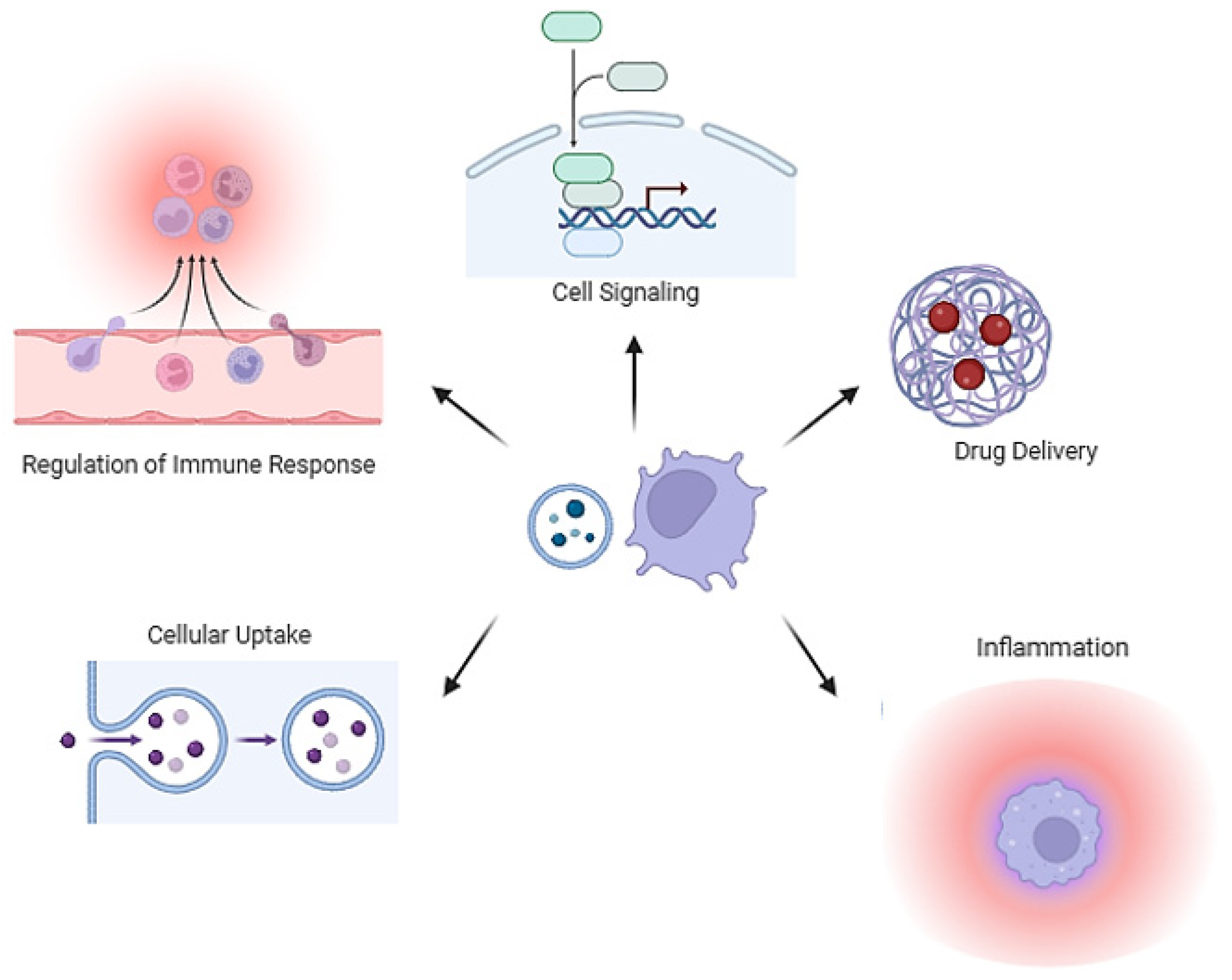
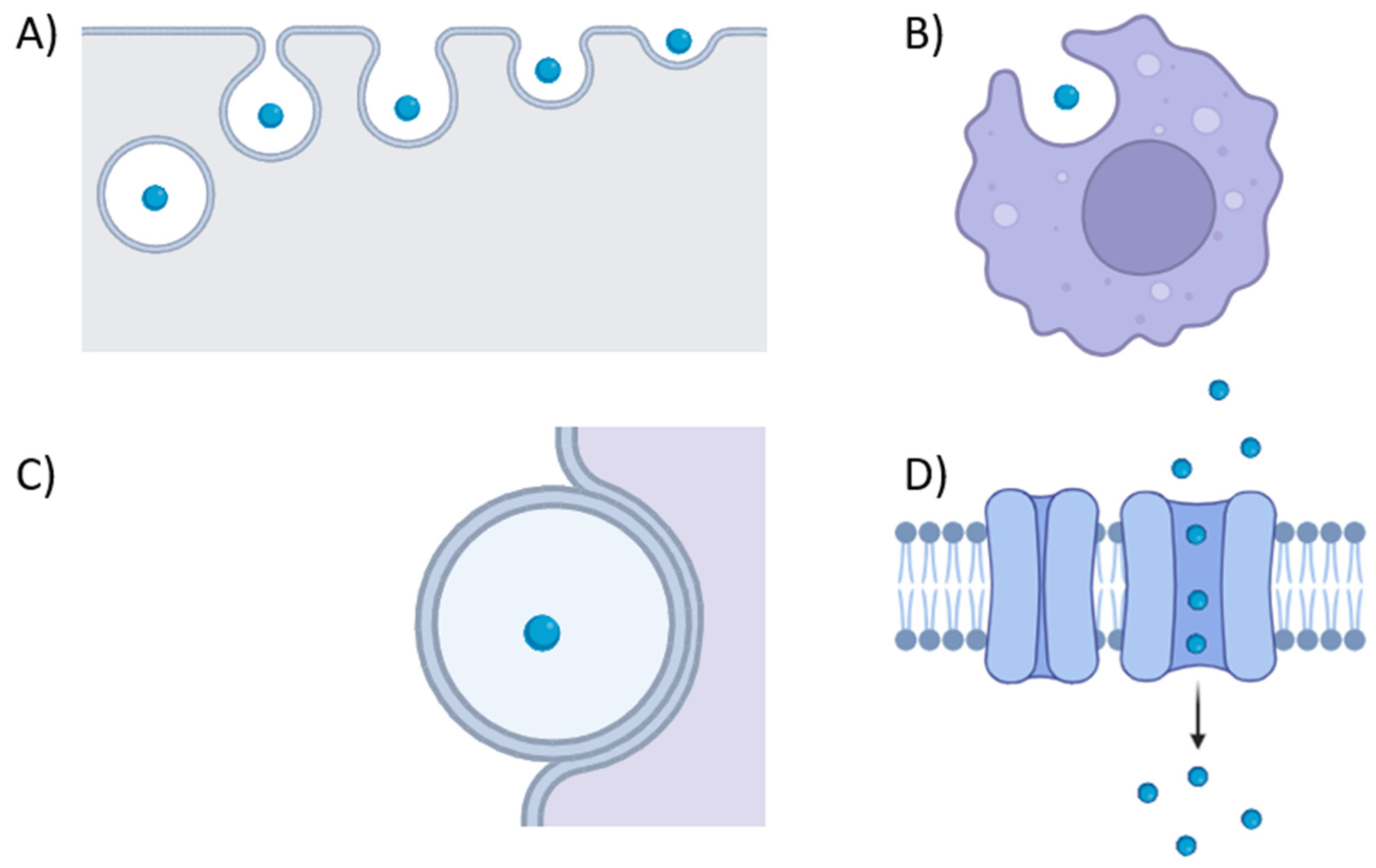
4. Membrane Vesicle in Host–Pathogen Interactions

The role of vesicles in host–bacteria interactions is significantly determined by the cargo packaged during vesiculogenesis. The inclusion of toxins, siderophores, immune evasion proteins, adhesins, and antibiotic resistance proteins strongly suggests MVs’ involvement in virulence (Figure 2). Pathogenic bacteria can use MVs as a mechanism to deliver virulence factors to eukaryotic host cells. For instance, S. aureus MVs harbor superantigens, including T-cell-activating enterotoxin SeQ, lipase, immune evasion proteins (e.g., protein A and SbI), toxins such as PSMs, and bicomponent pore-forming toxins like alpha-toxin, LukSF-PV, and LukAB. MV-associated alpha-toxin from S. aureus significantly impacts certain diseases like atopic dermatitis more than its soluble forms [66]. Both forms induce keratinocyte death, but only MV-associated alpha-toxin provokes keratinocyte necrosis and eosinophilic infiltration specific to atopic dermatitis in mice [66]. Additionally, they may carry β-lactamase, which degrades β-lactam antibiotics, and staphopain A (a papain-like cysteine protease), contributing to extracellular matrix degradation and promoting tissue invasion [42,49,67]. Similarly, EVs from L. monocytogenes encapsulate the pore-forming toxin listeriolysin O, aiding the bacterium in escaping host vacuoles [25]. Notably, the cytosolic pore-forming toxin pneumolysin in S. pneumoniae lacks export signal sequences and is exclusively released into host cells through MV secretion, underscoring the pivotal role of MVs in S. pneumoniae virulence [47]. MVs enriched with extracellular-matrix-degrading enzymes from Group B streptococcus (GBS) play a role in disrupting physical barriers and causing host cell death. In mice, GBS-MV treatment led to collagen fragmentation, immune cell infiltration, and membrane integrity loss, resulting in preterm birth [28]. The heightened effectiveness of MV-associated toxins may be explained by their encapsulation within MVs. This allows toxins to be delivered at concentrated levels, avoiding dilution over a distance and providing protection from immune system clearance, including antibodies and protease activity. The differential impact of associated and soluble forms of toxins, whether MV-associated or soluble, is likely attributed to their distinct delivery mechanisms. The entry of membrane vesicles into eukaryotic host cells is a finely tuned process, involving various mechanisms tailored to the vesicle type recipient cell (Figure 3). The treatment of THP-1 cells or monocyte-derived macrophages with dynamin-dependent endocytosis prevents S. aureus MV internalization, hindering pore-forming toxin delivery [62]. Methyl-β-cyclodextrins abolished the internalization of EV-associated protein A from S. aureus inside human laryngeal carcinoma cells, potentially demonstrating that EVs fuse with cholesterol-rich domains of host cell membranes [64]. In P. acnes, clathrin-dependent endocytosis is the major route for MV internalization in human epidermal keratinocyte cells, with the specific receptor remaining unidentified [68]. Additional entry modes are probable, considering diverse routes described for OMVs, influenced by vesicle size and infected cell type [69].

5. Membrane Vesicles in Inter-Bacterial Interactions

Vesicle-mediated interactions among bacteria have been extensively explored in Gram-negative bacteria [70], particularly focusing on resistance gene transfer, bactericidal activities, and long-distance signaling within bacterial populations. Initially believed to be limited by thicker cell walls [1], MV-mediated processes in Gram-positive bacteria have gained attention, challenging prior assumptions. Well-known horizontal gene transfer (HGT) mechanisms, such as transformation, transduction, and conjugation, coexist with the proposed concept of “vesiduction”, which employs EVs in prokaryotes for the transfer of genes related to antibiotic resistance, virulence, and metabolic factors [55,71]. Recent studies, particularly in clinically significant species like Staphylococcus aureus [49] and Enterococcus spp. [72], have demonstrated the presence of MV-mediated mechanisms in Gram-positive bacteria. For instance, Lee et al. revealed the transfer of β-lactam antibiotic resistance from methicillin-resistant S. aureus ST541 to susceptible E. coli RC85 through MVs [73]. Contrary to previous beliefs, intracellular and extracellular DNA has been identified in Gram-positive bacteria, including Streptococcus mutans and Clostridium perfringens [21,56], challenging assumptions about MV-mediated genetic material transfer [74]. Despite uncertainties regarding determinant composition, MVs represent a potential strategy to mitigate the dissemination of multidrug-resistant (MDR) bacteria. Streptomyces has been found to frequently produce antimicrobial vesicles containing diverse compounds such as actinomycins, anthracyclines, candicidin, and actinorhodin, which exhibit diverse antibacterial and antifungal activity [75]. These antimicrobial-containing vesicles achieve direct delivery of their cargo to other microbes via membrane fusion, suggesting an alternative and broad delivery system for antimicrobial specialized metabolites with significant implications for interbacterial communication and new clinical strategies against antibiotic resistance. Additionally, vesicular inter-bacterial interactions extend to bactericidal activities, with Gram-positive vesicles commonly housing cell-wall-degrading enzymes [76]. MVs labeled with the lipophilic probe R18 from B. subtilis exhibit fusion capabilities with other B. subtilis cells, showcasing their broad interaction potential [77]. Lactobacillus acidophilus MVs seamlessly merge with membranes of Lactobacillus delbrueckii and E. coli, accompanied by the growth inhibition of target cells due to the presence of bacteriocins [78]. Further potential roles of MVs in inter-bacterial interactions could be represented by long-distance signaling within bacterial populations [79,80], as showed in research on Gram-negative bacteria. Specifically, studies on Pseudomonas aeruginosa have shown the use of its extracellular vesicles to transport PQS, a quorum sensing molecule. These vesicular PQS molecules can then directly interact with other bacteria through LPS [81]. The delivery of quorum sensing molecules embedded in MVs could specifically activate the recipient cells, leading to heterogenous gene activation within a bacterial population [81]. However, this aspect remains underexplored in the literature for Gram-positive bacteria; future studies have the potential to elucidate not only the transfer of resistance determinants but also the fundamental knowledge about the ways microbial communities interacts.

6. Immune Regulation of MVs

The growing recognition of the role of MVs in host immune regulation is attributed to their content, which encompasses various immune-related molecules readily accepted by host cells [82]. These molecules, including lipoproteins and toxins, stimulate both innate and adaptive immunity through pattern recognition receptors (PRRs) [83]. MVs from Gram-positive bacteria, whether pathogenic or nonpathogenic, contribute to innate immunity involving key molecules like macrophages, dendritic cells, and Toll-like TLR2 [56,64,84]. The evolutionary costs of microbial MV immunogenicity must be balanced against the benefits to the microorganism from EV emission [85]. Additionally, MVs released by infected cells carry microbial molecules, potentially influencing the immune response indirectly [86]. Gram-positive bacterial EVs induce innate immune responses; for instance, MVs from Clostridium perfringens trigger the release of IL-6 through the TLR2 signaling pathway [56]. Staphylococcus aureus expresses co-stimulatory molecules via the TLR2 pathway, leading to the production of inflammatory proteins like tumor necrosis factor, IL-6, and IL-12, and releases EVs containing DNA, RNA, and peptidoglycans. These EVs are recognized by PRRs such as TLR7, TLR8, TLR9, and NOD2, ultimately undergoing autophagosomal degradation [64,87,88]. MVs from Streptococcus suis activate the nuclear factor kappa B signaling pathway, inducing the secretion of pro-inflammatory cytokines [84]. Filifactor alocis EVs elevate the secretion of various molecules in Thp-1 cells and oral keratinocyte cell lines [89]. Nonpathogenic bacteria, like Lactobacillus sakei subsp. sakei NBRC15893, show increased immunoglobulin A production through TLR2 signaling [90]. The cell wall components of Gram-positive bacteria, rich in TLR2 ligands, suggest that MVs may collaboratively exert innate immunomodulatory effects via TLR2 [91,92,93,94]. Lipoproteins within MVs play a role in immunomodulatory responses across various Gram-positive bacteria [90,95]. The TLR2-dependent pathway is crucial for the proinflammatory effect of EVs, with other pathways likely contributing. In the case of Gram-negative bacteria, various pathways leading to proinflammatory cytokine production, including TLR-4 and TLR-8 activation, the recognition of OMV-associated LPS and RNA, and NOD1 interaction with peptidoglycan, have been described [69,96]. Pseudomonas aeruginosa OMVs carrying sRNA52320 modulate the immune response by downregulating genes in the LPS-stimulated MAPK signaling pathway [97], suggesting potential interkingdom communication facilitated by RNA molecules in bacterial vesicles. It is probable that some of these mechanisms are shared and additional unique activation pathways exist for Gram-positive EVs. Additionally, anti-inflammatory potential has been predominantly observed in nonpathogenic species like Lactobacillus paracasei, demonstrating an in vivo impact against colitis [98]. EVs from other Lactobacilli spp. also dampen proinflammatory responses [99]. Surprisingly, EVs from the pathogenic bacterium M. tuberculosis exhibit anti-inflammatory activity by inhibiting CD4 T cell activation during macrophage infection [100]. Furthermore, MVs play a crucial role in adaptive immunity, with MVs from Streptococcus pneumoniae inducing specific antibodies [44], and S. aureus stimulating Th1, Th17, and Th2 cells, along with IgG antibody responses [87], suggesting the effective vaccination efficacy of MVs in Gram-positive bacteria.

7. Clinical Applications

Exploring bacterial MVs highlights their potential positive contributions to disease diagnosis and treatment. Current clinical applications of the utilization of MVs from Gram-positive bacteria have emerged as a promising therapy in several fields. Firstly, the immunity and stability of MVs highlight their potential as vaccine candidates. This is corroborated by evidence for major clinical Gram-positive pathogens. Choi et al. proposed how active immunization through S. aureus MVs induces an adaptive immunity of antibody and T cell responses. In particular, data showed that S. aureus MVs effectively protect against lethality and pneumonia induced by S. aureus infection, mainly via IFN-γ-producing Th1 cellular response rather than B-cell-mediated antibody response in mice [87]. Findings on Streptococcus pneumoniae revealed that extracellular MVs derived from S. pneumoniae BAA-255 effectively protected mice without causing significant side effects. Furthermore, the study identified specific immunogenic proteins present in these MVs. The results indicated that MVs, being more immunogenic than an equivalent bacterial cell extract, hold significant promise as potential vaccine antigens [101]. Moreover, Prados-Rosales et al. showed how MVs are highly immunogenic without adjuvants and elicit immune responses comparable to those achieved with BCG in protection against Mycobacterium tuberculosis [102]. To date, only one vaccine has been developed using OMVs for Neisseria meningitidis serogroup B: the MeNZB OMV vaccine. This vaccine was designed to provide broader protection against multiple strains of meningococcal B, reflecting advancements in vaccine technology to enhance its effectiveness [103]. However, further research should be carried out to pave the way for proper newly updated immunization therapies in Gram-positive bacteria.
Another clinically impactful theme is cancer therapy and the anti-tumor effects of bacterial MVs. Currently, the emphasis in cancer treatment development lies on targeted drugs; however, alternative approaches to identify and stimulate tumor immunity are also under investigation. Considering the immunomodulatory properties of MVs, they can be engineered to express cancer-specific epitopes or carry small noncoding RNAs, as previously explored for Gram-negative bacteria. Hence, OMVs have demonstrated the ability to induce a sustained antitumor immune response, effectively inhibiting tumor growth in various models [104]. For example, it has been shown that basic fibroblast growth factor (BFGF)-OMVs, used as vaccines, can successfully induce the body to produce persistent anti-BFGF autoantibodies to inhibit tumor growth and metastasis [105]. For Gram-positive bacteria, most of the evidence comes from several studies on probiotic strains belonging to the Lactobacillus genus, illustrating the existing approaches to regulate human gut microbial ecology, aiming at alternative methods to reduce the damage and improve the effectiveness of cancer therapy [106]. Notably, Lactobacillus rhamnosus GG, a commonly used probiotic supplement, generates MVs with cytotoxic effects on hepatic cancer cells. This is achieved, in part, through the downregulation of the bcl-2 and bax genes in cancer cells [107]. Furthermore, MVs from Lacticaseibacillus paracasei PC-H1 can inhibit colorectal cancer cell growth both in vivo and in vitro, inducing apoptosis through the PDK1/AKT/Bcl-2 signaling pathway [108]. Gram-positive bacterial MVs may also play a role in various pathologies, including dermatological and viral diseases. Streptococcus epidermidis MVs show potential therapeutic value for psoriasis [109], while Lactobacillus druckerii MVs may have applications for hyperplastic scars [110]. In conclusion, Palomino et al. demonstrated that MVs from vaginally isolated Lactobacillus inhibited the adhesion and entry of the HIV-1 virus into human T cells [111] (Figure 4).

8. Future Directions

Engineered Bacterial Vesicles (BEVs) have emerged as versatile tools with promising applications across various domains, offering a glimpse into the future of medical and biotechnological advancements [112]. One of the most innovative applications lies in vaccine development, where BEVs could revolutionize the current approach to infectious diseases, providing a platform for the rapid development of effective vaccines [103,113,114,115]. Indeed, MVs can be engineered to carry specific antigens from pathogens, mimicking their natural presentation and potentially eliciting a more robust and targeted immune response [116].
In addition, BEVs hold great potential for drug delivery since they can be loaded with therapeutic agents such as antibiotics, antivirals, or immunomodulators, offering a controlled and targeted release system. This approach maximizes the efficacy of treatments, enhancing the stability of encapsulated cargoes [117] and at the same time minimizing side effects by directly delivering drugs to the site of infection [118]. Moreover, loaded BEVs preserve their immunogenicity, thereby simultaneously treating and preventing the infection. This combined effect may be helpful to counteract the spread of MDR bacteria. Since BEVs can contain a variety of bacterial biomolecules such as DNA, RNA, lipids, proteins, and metabolites, there is increasing evidence that they have significant potential as diagnostic biomarkers enabling the detection of early signs of infection and specifically identify the bacterial etiological agent [119]. The applications of BEVs extend beyond infectious diseases, opening new avenues for treating a wide range of diseases such as cancer or osteoporosis. In this context, they can be engineered to carry specific cargo, such as therapeutic proteins or small interfering RNA (siRNA) in order to modulate cellular processes for therapeutic purposes [120,121].
Moreover, there has been growing interest in investigating the impact of BEVs on Neurodegenerative Diseases (NDs) [122,123]. In recent years, research has highlighted the potential of BEVs to play a substantial role in the treatment and therapeutics of NDs [124]. Recent studies have suggested that these vehicles exhibit immuno-modulatory and neuroprotective properties, offering potential benefits in addressing NDs. Additionally, it has been noted that BEVs can effectively cross the Blood-Brain Barrier (BBB) and target specific regions of the brain, making them an appealing drug delivery system for ND treatment [124]. Despite their immense potential, engineered BEVs come with inherent challenges and limitations. Long-term studies are necessary to assess their safety and toxicity, especially as they progress toward clinical applications [125]. The standardization of production processes, scalability, and storage stability are critical obstacles that must be addressed to facilitate widespread use [126]. Due to their isolation from growth medium, batch-to-batch differences may occur, challenging current isolation methods to remove non-encapsulated bacterial components and discriminate between empty or filled vesicles [117].

9. Conclusions

There is a pressing need for innovative approaches to combat bacterial infections caused by Gram-positive bacteria. As far as we know, this study provides the first comprehensive review on how Gram-positive bacteria naturally interact with the host through MVs. Ongoing advancements in bacterial MV extraction technology have revealed the ability of Gram-positive bacteria to produce MVs, intricately linked to their virulence, immune capabilities, and pathogenic factors. While MVs assist bacteria in resisting host defenses, eliciting immune responses, and transferring drug-resistance genes, the molecular mechanisms remain unclear. MVs resist external environmental stresses, like antibiotic killing, suggesting a therapeutic strategy against antibiotic resistance. In addition, the immune response triggered by MVs, along with their adaptability for engineering, highlights their potential for various clinical applications. Probiotic MVs from Gram-positive bacteria hold promise for oral administration or incorporation into pharmaceutical supplements due to their regulatory and anti-inflammatory benefits. The prospect of revolutionizing vaccine development, drug delivery, diagnostics, and therapeutics is substantial, making BEVs a groundbreaking frontier in medicine and biotechnology. Nevertheless, practical implementation on a large scale requires addressing current limitations, ensuring safety, and developing robust production and regulatory frameworks. As research progresses, engineered MVs and their role in host–pathogen interactions hold the promise of transforming the healthcare and disease management landscape.

Author Contributions

Conceptualization, G.S. and D.B. (Dafne Bongiorno); methodology, G.S.; writing—original draft preparation, G.S., C.B., E.N., D.B. (Dalida Bivona), P.B. and N.M.; writing—review and editing, G.S. and D.B. (Dafne Bongiorno); visualization, G.S. and D.B. (Dafne Bongiorno); supervision, M.S., D.B. (Dafne Bongiorno), and S.S. All authors have read and agreed to the published version of the manuscript.

Funding

This research received no external funding.

Informed Consent Statement

Not applicable.

Conflicts of Interest

The authors declare no conflicts of interest.

Abbreviations

EV: extracellular vesicle; OMVs, outer membrane vesicles; MVs, membrane vesicles; PSM, Phenol-Soluble Modulins; PBPs, penicillin-binding proteins; TLR2, Toll-like receptor 2; GBS, Group B streptococcus; HGT, horizontal gene transfer; MDR, multidrug-resistant, PRRs, pattern recognition receptors; BCG, Bacillus Calmette-Guéri; BFGF, basic fibroblast growth factor; BEVs, Engineered Bacterial Vesicles; siRNA, small interfering RNA; NDs, Neurodegenerative Diseases; BBB, Blood-Brain Barrier.

References

  1. Brown, L.; Wolf, J.M.; Prados-Rosales, R.; Casadevall, A. Through the wall: Extracellular vesicles in Gram-positive bacteria, mycobacteria and fungi. Nat. Rev. Microbiol. 2015, 13, 620–630. [Google Scholar] [CrossRef]
  2. Deatherage, B.L.; Cookson, B.T. Membrane vesicle release in bacteria, eukaryotes, and archaea: A conserved yet underappreciated aspect of microbial life. Infect. Immun. 2012, 80, 1948–1957. [Google Scholar] [CrossRef]
  3. György, B.; Szabó, T.G.; Pásztói, M.; Pál, Z.; Misják, P.; Aradi, B.; László, V.; Pállinger, É.; Pap, E.; Kittel, Á.; et al. Membrane vesicles, current state-of-the-art: Emerging role of extracellular vesicles. Cell. Mol. Life Sci. 2011, 68, 2667–2688. [Google Scholar] [CrossRef]
  4. Clemmens, H.; Lambert, D.W. Extracellular vesicles: Translational challenges and opportunities. Biochem. Soc. Trans. 2018, 46, 1073–1082. [Google Scholar] [CrossRef]
  5. van Niel, G.; D’Angelo, G.; Raposo, G. Shedding light on the cell biology of extracellular vesicles. Nat. Rev. Mol. Cell Biol. 2018, 19, 213–228. [Google Scholar] [CrossRef]
  6. EL Andaloussi, S.; Mäger, I.; Breakefield, X.O.; Wood, M.J.A. Extracellular vesicles: Biology and emerging therapeutic opportunities. Nat. Rev. Drug Discov. 2013, 12, 347–357. [Google Scholar] [CrossRef]
  7. Popa, S.J.; Stewart, S.E. Socially Distanced Intercellular Communication: Mechanisms for Extracellular Vesicle Cargo Delivery. Subcell Biochem. 2021, 97, 179–209. [Google Scholar] [CrossRef]
  8. Raposo, G.; Stoorvogel, W. Extracellular vesicles: Exosomes, microvesicles, and friends. J. Cell Biol. 2013, 200, 373–383. [Google Scholar] [CrossRef]
  9. Brown, L.; Kessler, A.; Cabezas-Sanchez, P.; Luque-Garcia, J.L.; Casadevall, A. Extracellular vesicles produced by the Gram-positive bacterium Bacillus subtilis are disrupted by the lipopeptide surfactin. Mol. Microbiol. 2014, 93, 183–198. [Google Scholar] [CrossRef]
  10. Avila-Calderón, E.D.; Araiza-Villanueva, M.G.; Cancino-Diaz, J.C.; López-Villegas, E.O.; Sriranganathan, N.; Boyle, S.M.; Contreras-Rodríguez, A. Roles of bacterial membrane vesicles. Arch. Microbiol. 2015, 197, 1–10. [Google Scholar] [CrossRef]
  11. Sartorio, M.G.; Pardue, E.J.; Feldman, M.F.; Haurat, M.F. Bacterial Outer Membrane Vesicles: From Discovery to Applications. Annu. Rev. Microbiol. 2021, 75, 609–630. [Google Scholar] [CrossRef]
  12. Balhuizen, M.D.; van Dijk, A.; Jansen, J.W.A.; van de Lest, C.H.A.; Veldhuizen, E.J.A.; Haagsman, H.P. Outer Membrane Vesicles Protect Gram-Negative Bacteria against Host Defense Peptides. mSphere 2021, 6, e0052321. [Google Scholar] [CrossRef] [PubMed]
  13. Jan, A.T. Outer Membrane Vesicles (OMVs) of Gram-negative Bacteria: A Perspective Update. Front. Microbiol. 2017, 8, 1053. [Google Scholar] [CrossRef] [PubMed]
  14. Tan, K.; Li, R.; Huang, X.; Liu, Q. Outer Membrane Vesicles: Current Status and Future Direction of These Novel Vaccine Adjuvants. Front. Microbiol. 2018, 9, 783. [Google Scholar] [CrossRef]
  15. Toyofuku, M.; Nomura, N.; Eberl, L. Types and origins of bacterial membrane vesicles. Nat. Rev. Microbiol. 2019, 17, 13–24. [Google Scholar] [CrossRef]
  16. da Barreira, D.S.; Laurent, J.; Lourenço, J.; Ducassou, J.N.; Couté, Y.; Guzzo, J.; Rieu, A. Membrane vesicles released by Lacticaseibacillus casei BL23 inhibit the biofilm formation of Salmonella Enteritidis. Sci. Rep. 2023, 13, 1163. [Google Scholar] [CrossRef]
  17. da Silva Barreira, D.; Lapaquette, P.; Ducassou, J.N.; Couté, Y.; Guzzo, J.; Rieu, A. Spontaneous Prophage Induction Contributes to the Production of Membrane Vesicles by the Gram-Positive Bacterium Lacticaseibacillus casei BL23. mBio 2022, 13, e0237522. [Google Scholar] [CrossRef]
  18. Faddetta, T.; Renzone, G.; Vassallo, A.; Rimini, E.; Nasillo, G.; Buscarino, G.; Agnello, S.; Licciardi, M.; Botta, L.; Scaloni, A.; et al. Streptomyces coelicolor Vesicles: Many Molecules To Be Delivered. Appl. Environ. Microbiol. 2022, 88, e0188121. [Google Scholar] [CrossRef]
  19. Andreoni, F.; Toyofuku, M.; Menzi, C.; Kalawong, R.; Mairpady Shambat, S.; François, P.; Zinkernagel, A.S.; Eberl, L. Antibiotics Stimulate Formation of Vesicles in Staphylococcus aureus in both Phage-Dependent and -Independent Fashions and via Different Routes. Antimicrob. Agents Chemother. 2019, 63, e01439-18. [Google Scholar] [CrossRef]
  20. Jeon, H.; Oh, M.H.; Jun, S.H.; Kim, S.I.; Choi, C.W.; Kwon, H.I.; Na, S.H.; Kim, Y.J.; Nicholas, A.; Selasi, G.N.; et al. Variation among Staphylococcus aureus membrane vesicle proteomes affects cytotoxicity of host cells. Microb. Pathog. 2016, 93, 185–193. [Google Scholar] [CrossRef]
  21. Liao, S.; Klein, M.I.; Heim, K.P.; Fan, Y.; Bitoun, J.P.; Ahn, S.-J.; Burne, R.A.; Koo, H.; Brady, L.J.; Wen, Z.T. Streptococcus mutans extracellular DNA is upregulated during growth in biofilms, actively released via membrane vesicles, and influenced by components of the protein secretion machinery. J. Bacteriol. 2014, 196, 2355–2366. [Google Scholar] [CrossRef] [PubMed]
  22. Rivera, J.; Cordero, R.J.B.; Nakouzi, A.S.; Frases, S.; Nicola, A.; Casadevall, A. Bacillus anthracis produces membrane-derived vesicles containing biologically active toxins. Proc. Natl. Acad. Sci. USA 2010, 107, 19002–19007. [Google Scholar] [CrossRef] [PubMed]
  23. Wang, X.; Thompson, C.D.; Weidenmaier, C.; Lee, J.C. Release of Staphylococcus aureus extracellular vesicles and their application as a vaccine platform. Nat. Commun. 2018, 9, 1379. [Google Scholar] [CrossRef] [PubMed]
  24. Resch, U.; Tsatsaronis, J.A.; Le Rhun, A.; Stübiger, G.; Rohde, M.; Kasvandik, S.; Holzmeister, S.; Tinnefeld, P.; Wai, S.N.; Charpentier, E. A Two-Component Regulatory System Impacts Extracellular Membrane-Derived Vesicle Production in Group A Streptococcus. mBio 2016, 7, e00207-16. [Google Scholar] [CrossRef] [PubMed]
  25. Coelho, C.; Brown, L.C.; Maryam, M.; Vij, R.; Smith, D.F.; Burnet, M.C.; Kyle, J.E.; Heyman, H.M.; Ramirez, J.; Prados-Rosales, R.; et al. Listeria monocytogenes virulence factors, including listeriolysin O, are secreted in biologically active extracellular vesicles. J. Biol. Chem. 2019, 294, 1202–1217. [Google Scholar] [CrossRef]
  26. White, D.W.; Elliott, S.R.; Odean, E.; Bemis, L.T.; Tischler, A.D. Mycobacterium tuberculosis Pst/SenX3-RegX3 Regulates Membrane Vesicle Production Independently of ESX-5 Activity. mBio 2018, 9, e00778-18. [Google Scholar] [CrossRef]
  27. Schlatterer, K.; Beck, C.; Hanzelmann, D.; Lebtig, M.; Fehrenbacher, B.; Schaller, M.; Ebner, P.; Nega, M.; Otto, M.; Kretschmer, D.; et al. The Mechanism behind Bacterial Lipoprotein Release: Phenol-Soluble Modulins Mediate Toll-Like Receptor 2 Activation via Extracellular Vesicle Release from Staphylococcus aureus. mBio 2018, 9, e01851-18. [Google Scholar] [CrossRef]
  28. Surve, M.V.; Anil, A.; Kamath, K.G.; Bhutda, S.; Sthanam, L.K.; Pradhan, A.; Srivastava, R.; Basu, B.; Dutta, S.; Sen, S.; et al. Membrane Vesicles of Group B Streptococcus Disrupt Feto-Maternal Barrier Leading to Preterm Birth. PLoS Pathog. 2016, 12, e1005816. [Google Scholar] [CrossRef]
  29. Baidya, A.K.; Bhattacharya, S.; Dubey, G.P.; Mamou, G.; Ben-Yehuda, S. Bacterial nanotubes: A conduit for intercellular molecular trade. Curr. Opin. Microbiol. 2018, 42, 1–6. [Google Scholar] [CrossRef]
  30. Baidya, A.K.; Bhattacharya, S.; Dubey, G.P.; Mamou, G.; Ben-Yehuda, S. Architecture and Characteristics of Bacterial Nanotubes. Dev. Cell 2016, 36, 453–461. [Google Scholar] [CrossRef]
  31. Wei, X.; Vassallo, C.N.; Pathak, D.T.; Wall, D. Myxobacteria produce outer membrane-enclosed tubes in unstructured environments. J. Bacteriol. 2014, 196, 1807–1814. [Google Scholar] [CrossRef]
  32. Pande, S.; Shitut, S.; Freund, L.; Westermann, M.; Bertels, F.; Colesie, C.; Bischofs, I.B.; Kost, C. Metabolic cross-feeding via intercellular nanotubes among bacteria. Nat. Commun. 2015, 6, 6238. [Google Scholar] [CrossRef]
  33. Dubey, G.P.; Ben-Yehuda, S. Intercellular nanotubes mediate bacterial communication. Cell 2011, 144, 590–600. [Google Scholar] [CrossRef] [PubMed]
  34. Wang, G.; Zhao, G.; Chao, X.; Xie, L.; Wang, H. The Characteristic of Virulence, Biofilm and Antibiotic Resistance of Klebsiella pneumoniae. Int. J. Environ. Res. Public Health 2020, 17, 6278. [Google Scholar] [CrossRef] [PubMed]
  35. Rath, P.; Huang, C.; Wang, T.; Wang, T.; Li, H.; Prados-Rosales, R.; Elemento, O.; Casadevall, A.; Nathan, C.F. Genetic regulation of vesiculogenesis and immunomodulation in Mycobacterium tuberculosis. Proc. Natl. Acad. Sci. USA 2013, 110, E4790–E4797. [Google Scholar] [CrossRef] [PubMed]
  36. Wolf, J.M.; Rivera, J.; Casadevall, A. Serum albumin disrupts Cryptococcus neoformans and Bacillus anthracis extracellular vesicles. Cell Microbiol. 2012, 14, 762–773. [Google Scholar] [CrossRef] [PubMed]
  37. Toyofuku, M.; Cárcamo-Oyarce, G.; Yamamoto, T.; Eisenstein, F.; Hsiao, C.-C.; Kurosawa, M.; Gademann, K.; Pilhofer, M.; Nomura, N.; Eberl, L. Prophage-triggered membrane vesicle formation through peptidoglycan damage in Bacillus subtilis. Nat. Commun. 2017, 8, 481. [Google Scholar] [CrossRef] [PubMed]
  38. Biagini, M.; Garibaldi, M.; Aprea, S.; Pezzicoli, A.; Doro, F.; Becherelli, M.; Taddei, A.R.; Tani, C.; Tavarini, S.; Mora, M.; et al. The Human Pathogen Streptococcus pyogenes Releases Lipoproteins as Lipoprotein-rich Membrane Vesicles. Mol. Cell. Proteom. 2015, 14, 2138–2149. [Google Scholar] [CrossRef]
  39. Yu, Y.-J.; Wang, X.-H.; Fan, G.-C. Versatile effects of bacterium-released membrane vesicles on mammalian cells and infectious/inflammatory diseases. Acta Pharmacol. Sin. 2018, 39, 514–533. [Google Scholar] [CrossRef]
  40. Gan, Y.; Zhao, G.; Wang, Z.; Zhang, X.; Wu, M.X.; Lu, M. Bacterial Membrane Vesicles: Physiological Roles, Infection Immunology, and Applications. Adv. Sci. 2023, 10, e2301357. [Google Scholar] [CrossRef]
  41. Briaud, P.; Carroll, R.K. Extracellular Vesicle Biogenesis and Functions in Gram-Positive Bacteria. Infect. Immun. 2020, 88, e00433-20. [Google Scholar] [CrossRef]
  42. Tartaglia, N.R.; Nicolas, A.; de Rezende Rodovalho, V.; da Luz, B.S.R.; Briard-Bion, V.; Krupova, Z.; Thierry, A.; Coste, F.; Burel, A.; Martin, P.; et al. Extracellular vesicles produced by human and animal Staphylococcus aureus strains share a highly conserved core proteome. Sci. Rep. 2020, 10, 8467. [Google Scholar] [CrossRef]
  43. Mehanny, M.; Kroniger, T.; Koch, M.; Hoppstädter, J.; Becher, D.; Kiemer, A.K.; Lehr, C.; Fuhrmann, G. Yields and Immunomodulatory Effects of Pneumococcal Membrane Vesicles Differ with the Bacterial Growth Phase. Adv. Healthc. Mater. 2022, 11, e2101151. [Google Scholar] [CrossRef]
  44. Olaya-Abril, A.; Prados-Rosales, R.; McConnell, M.J.; Martín-Peña, R.; González-Reyes, J.A.; Jiménez-Munguía, I.; Gómez-Gascón, L.; Fernández, J.; Luque-García, J.L.; García-Lidón, C.; et al. Characterization of protective extracellular membrane-derived vesicles produced by Streptococcus pneumoniae. J. Proteom. 2014, 106, 46–60. [Google Scholar] [CrossRef]
  45. Tartaglia, N.R.; Breyne, K.; Meyer, E.; Cauty, C.; Jardin, J.; Chrétien, D.; Dupont, A.; Demeyere, K.; Berkova, N.; Azevedo, V.; et al. Staphylococcus aureus Extracellular Vesicles Elicit an Immunostimulatory Response in vivo on the Murine Mammary Gland. Front. Cell. Infect. Microbiol. 2018, 8, 277. [Google Scholar] [CrossRef]
  46. Lee, J.; Choi, D.; Lee, J.S.; Kim, D.; Go, G.; Park, S.; Kim, S.H.; Shin, J.H.; Chang, C.L.; Gho, Y.S. Proteomic analysis of extracellular vesicles derived from Mycobacterium tuberculosis. Proteomics 2015, 15, 3331–3337. [Google Scholar] [CrossRef]
  47. Codemo, M.; Muschiol, S.; Iovino, F.; Nannapaneni, P.; Plant, L.; Wai, S.N.; Henriques-Normark, B. Immunomodulatory Effects of Pneumococcal Extracellular Vesicles on Cellular and Humoral Host Defenses. mBio 2018, 9, e00559-18. [Google Scholar] [CrossRef]
  48. Cao, Y.; Lin, H. Characterization and function of membrane vesicles in Gram-positive bacteria. Appl. Microbiol. Biotechnol. 2021, 105, 1795–1801. [Google Scholar] [CrossRef]
  49. Lee, E.Y.; Choi, D.Y.; Kim, D.K.; Kim, J.W.; Park, J.O.; Kim, S.; Kim, S.H.; Desiderio, D.M.; Kim, Y.K.; Kim, K.P.; et al. Gram-positive bacteria produce membrane vesicles: Proteomics-based characterization of Staphylococcus aureus-derived membrane vesicles. Proteomics 2009, 9, 5425–5436. [Google Scholar] [CrossRef]
  50. Zähringer, U.; Lindner, B.; Inamura, S.; Heine, H.; Alexander, C. TLR2-promiscuous or specific? A critical re-evaluation of a receptor expressing apparent broad specificity. Immunobiology 2008, 213, 205–224. [Google Scholar] [CrossRef]
  51. Tomlinson, G.; Chimalapati, S.; Pollard, T.; Lapp, T.; Cohen, J.; Camberlein, E.; Stafford, S.; Periselneris, J.; Aldridge, C.; Vollmer, W.; et al. TLR-mediated inflammatory responses to Streptococcus pneumoniae are highly dependent on surface expression of bacterial lipoproteins. J. Immunol. 2014, 193, 3736–3745. [Google Scholar] [CrossRef]
  52. Kopparapu, P.K.; Deshmukh, M.; Hu, Z.; Mohammad, M.; Maugeri, M.; Götz, F.; Valadi, H.; Jin, T. Lipoproteins Are Responsible for the Pro-Inflammatory Property of Staphylococcus aureus Extracellular Vesicles. Int. J. Mol. Sci. 2021, 22, 7099. [Google Scholar] [CrossRef] [PubMed]
  53. Bhatnagar, S.; Shinagawa, K.; Castellino, F.J.; Schorey, J.S. Exosomes released from macrophages infected with intracellular pathogens stimulate a proinflammatory response in vitro and in vivo. Blood 2007, 110, 3234–3244. [Google Scholar] [CrossRef] [PubMed]
  54. Diallo, I.; Ho, J.; Lambert, M.; Benmoussa, A.; Husseini, Z.; Lalaouna, D.; Massé, E.; Provost, P. A tRNA-derived fragment present in E. coli OMVs regulates host cell gene expression and proliferation. PLoS Pathog. 2022, 18, e1010827. [Google Scholar] [CrossRef] [PubMed]
  55. Domingues, S.; Nielsen, K.M. Membrane vesicles and horizontal gene transfer in prokaryotes. Curr. Opin. Microbiol. 2017, 38, 16–21. [Google Scholar] [CrossRef]
  56. Jiang, Y.; Kong, Q.; Roland, K.L.; Curtiss, R. Membrane vesicles of Clostridium perfringens type A strains induce innate and adaptive immunity. Int. J. Med. Microbiol. 2014, 304, 431–443. [Google Scholar] [CrossRef]
  57. Rubio, A.P.D.; Martínez, J.H.; Casillas, D.C.M.; Leskow, F.C.; Piuri, M.; Pérez, O.E. Lactobacillus casei BL23 Produces Microvesicles Carrying Proteins That Have Been Associated with Its Probiotic Effect. Front. Microbiol. 2017, 8, 1783. [Google Scholar] [CrossRef]
  58. Choi, J.-W.; Kwon, T.-Y.; Hong, S.-H.; Lee, H.-J. Isolation and Characterization of a microRNA-size Secretable Small RNA in Streptococcus sanguinis. Cell. Biochem. Biophys. 2018, 76, 293–301. [Google Scholar] [CrossRef]
  59. Théry, C. Exosomes: Secreted vesicles and intercellular communications. F1000 Biol. Rep. 2011, 3, 15. [Google Scholar] [CrossRef]
  60. Faddetta, T.; Vassallo, A.; Del Duca, S.; Gallo, G.; Fani, R.; Puglia, A.M. Unravelling the DNA sequences carried by Streptomyces coelicolor membrane vesicles. Sci. Rep. 2022, 12, 16651. [Google Scholar] [CrossRef]
  61. Klieve, A.V.; Yokoyama, M.T.; Forster, R.J.; Ouwerkerk, D.; Bain, P.A.; Mawhinney, E.L. Naturally occurring DNA transfer system associated with membrane vesicles in cellulolytic Ruminococcus spp. of ruminal origin. Appl. Environ. Microbiol. 2005, 71, 4248–4253. [Google Scholar] [CrossRef] [PubMed]
  62. Wang, X.; Eagen, W.J.; Lee, J.C. Orchestration of human macrophage NLRP3 inflammasome activation by Staphylococcus aureus extracellular vesicles. Proc. Natl. Acad. Sci. USA 2020, 117, 3174–3184. [Google Scholar] [CrossRef] [PubMed]
  63. Marsollier, L.; Brodin, P.; Jackson, M.; Korduláková, J.; Tafelmeyer, P.; Carbonnelle, E.; Aubry, J.; Milon, G.; Legras, P.; André, J.-P.S.; et al. Impact of Mycobacterium ulcerans biofilm on transmissibility to ecological niches and Buruli ulcer pathogenesis. PLoS Pathog. 2007, 3, e62. [Google Scholar] [CrossRef] [PubMed]
  64. Gurung, M.; Moon, D.C.; Choi, C.W.; Lee, J.H.; Bae, Y.C.; Kim, J.; Lee, Y.C.; Seol, S.Y.; Cho, D.T.; Kim, S.I.; et al. Staphylococcus aureus produces membrane-derived vesicles that induce host cell death. PLoS ONE 2011, 6, e27958. [Google Scholar] [CrossRef] [PubMed]
  65. Vdovikova, S.; Luhr, M.; Szalai, P.; Skalman, L.N.; Francis, M.K.; Lundmark, R.; Engedal, N.; Johansson, J.; Wai, S.N. A Novel Role of Listeria monocytogenes Membrane Vesicles in Inhibition of Autophagy and Cell Death. Front. Cell. Infect. Microbiol. 2017, 7, 154. [Google Scholar] [CrossRef]
  66. Hong, S.-W.; Choi, E.-B.; Min, T.-K.; Kim, J.-H.; Kim, M.-H.; Jeon, S.G.; Lee, B.-J.; Gho, Y.S.; Jee, Y.-K.; Pyun, B.-Y.; et al. An important role of α-hemolysin in extracellular vesicles on the development of atopic dermatitis induced by Staphylococcus aureus. PLoS ONE 2014, 9, e100499. [Google Scholar] [CrossRef]
  67. Askarian, F.; Lapek, J.D., Jr.; Dongre, M.; Tsai, C.-M.; Kumaraswamy, M.; Kousha, A.; Valderrama, J.A.; Ludviksen, J.A.; Cavanagh, J.P.; Uchiyama, S.; et al. Staphylococcus aureus Membrane-Derived Vesicles Promote Bacterial Virulence and Confer Protective Immunity in Murine Infection Models. Front. Microbiol. 2018, 9, 262. [Google Scholar] [CrossRef]
  68. Choi, E.-J.; Lee, H.G.; Bae, I.-H.; Kim, W.; Park, J.; Lee, T.R.; Cho, E.-G. Propionibacterium acnes-Derived Extracellular Vesicles Promote Acne-Like Phenotypes in Human Epidermis. J. Investig. Dermatol. 2018, 138, 1371–1379. [Google Scholar] [CrossRef]
  69. Kaparakis-Liaskos, M.; Ferrero, R.L. Immune modulation by bacterial outer membrane vesicles. Nat. Rev. Immunol. 2015, 15, 375–387. [Google Scholar] [CrossRef]
  70. Rumbo, C.; Fernández-Moreira, E.; Merino, M.; Poza, M.; Mendez, J.A.; Soares, N.C.; Mosquera, A.; Chaves, F.; Bou, G. Horizontal transfer of the OXA-24 carbapenemase gene via outer membrane vesicles: A new mechanism of dissemination of carbapenem resistance genes in Acinetobacter baumannii. Antimicrob. Agents Chemother. 2011, 55, 3084–3090. [Google Scholar] [CrossRef] [PubMed]
  71. Soler, N.; Forterre, P. Vesiduction: The fourth way of HGT. Environ. Microbiol. 2020, 22, 2457–2460. [Google Scholar] [CrossRef]
  72. Wagner, T.; Joshi, B.; Janice, J.; Askarian, F.; Škalko-Basnet, N.; Hagestad, O.; Mekhlif, A.; Wai, S.; Hegstad, K.; Johannessen, M. Enterococcus faecium produces membrane vesicles containing virulence factors and antimicrobial resistance related proteins. J. Proteom. 2018, 187, 28–38. [Google Scholar] [CrossRef]
  73. Lee, A.R.; Bin Park, S.; Kim, S.W.; Jung, J.W.; Chun, J.H.; Kim, J.; Kim, Y.R.; Lazarte, J.M.S.; Bin Jang, H.; Thompson, K.D.; et al. Membrane vesicles from antibiotic-resistant Staphylococcus aureus transfer antibiotic-resistance to antibiotic-susceptible Escherichia coli. J. Appl. Microbiol. 2022, 132, 2746–2759. [Google Scholar] [CrossRef]
  74. Dorward, D.W.; Garon, C.F. DNA Is Packaged within Membrane-Derived Vesicles of Gram-Negative but Not Gram-Positive Bacteria. Appl. Environ. Microbiol. 1990, 56, 1960–1962. [Google Scholar] [CrossRef] [PubMed]
  75. Meyer, K.J.; Nodwell, J.R. Streptomyces extracellular vesicles are a broad and permissive antimicrobial packaging and delivery system. J. Bacteriol. 2024, e0032523. [Google Scholar] [CrossRef] [PubMed]
  76. Liu, Y.; Defourny, K.A.Y.; Smid, E.J.; Abee, T. Gram-Positive Bacterial Extracellular Vesicles and Their Impact on Health and Disease. Front. Microbiol. 2018, 9, 1502. [Google Scholar] [CrossRef] [PubMed]
  77. Kim, Y.; Edwards, N.; Fenselau, C. Extracellular vesicle proteomes reflect developmental phases of Bacillus subtilis. Clin. Proteom. 2016, 13, 6. [Google Scholar] [CrossRef]
  78. Dean, S.N.; Rimmer, M.A.; Turner, K.B.; Phillips, D.A.; Caruana, J.C.; Hervey, W.J.; Leary, D.H.; Walper, S.A. Lactobacillus acidophilus Membrane Vesicles as a Vehicle of Bacteriocin Delivery. Front. Microbiol. 2020, 11, 710. [Google Scholar] [CrossRef]
  79. Daubin, V.; Szöllősi, G.J. Horizontal Gene Transfer and the History of Life. Cold Spring Harb. Perspect. Biol. 2016, 8, a018036. [Google Scholar] [CrossRef] [PubMed]
  80. Mukherjee, S.; Bassler, B.L. Bacterial quorum sensing in complex and dynamically changing environments. Nat. Rev. Microbiol. 2019, 17, 371–382. [Google Scholar] [CrossRef]
  81. Mashburn, L.M.; Whiteley, M. Membrane vesicles traffic signals and facilitate group activities in a prokaryote. Nature 2005, 437, 422–425. [Google Scholar] [CrossRef] [PubMed]
  82. Buzas, E.I. The roles of extracellular vesicles in the immune system. Nat. Rev. Immunol. 2023, 23, 236–250. [Google Scholar] [CrossRef] [PubMed]
  83. Johnston, E.L.; Heras, B.; Kufer, T.A.; Kaparakis-Liaskos, M. Detection of Bacterial Membrane Vesicles by NOD-Like Receptors. Int. J. Mol. Sci. 2021, 22, 1005. [Google Scholar] [CrossRef] [PubMed]
  84. Haas, B.; Grenier, D. Isolation, Characterization and Biological Properties of Membrane Vesicles Produced by the Swine Pathogen Streptococcus suis. PLoS ONE 2015, 10, e0130528. [Google Scholar] [CrossRef] [PubMed]
  85. McMillan, H.M.; Kuehn, M.J. The extracellular vesicle generation paradox: A bacterial point of view. EMBO J. 2021, 40, e108174. [Google Scholar] [CrossRef] [PubMed]
  86. Schorey, J.S.; Cheng, Y.; Singh, P.P.; Smith, V.L. Exosomes and other extracellular vesicles in host-pathogen interactions. EMBO Rep. 2015, 16, 24–43. [Google Scholar] [CrossRef] [PubMed]
  87. Choi, S.J.; Kim, M.-H.; Jeon, J.; Kim, O.Y.; Choi, Y.; Seo, J.; Hong, S.-W.; Lee, W.-H.; Jeon, S.G.; Gho, Y.S.; et al. Active Immunization with Extracellular Vesicles Derived from Staphylococcus aureus Effectively Protects against Staphylococcal Lung Infections, Mainly via Th1 Cell-Mediated Immunity. PLoS ONE 2015, 10, e0136021. [Google Scholar] [CrossRef] [PubMed]
  88. Bitto, N.J.; Cheng, L.; Johnston, E.L.; Pathirana, R.; Phan, T.K.; Poon, I.K.H.; O’Brien-Simpson, N.M.; Hill, A.F.; Stinear, T.P.; Kaparakis-Liaskos, M. Staphylococcus aureus membrane vesicles contain immunostimulatory DNA, RNA and peptidoglycan that activate innate immune receptors and induce autophagy. J. Extracell. Vesicles 2021, 10, e12080. [Google Scholar] [CrossRef]
  89. Kim, H.Y.; Lim, Y.; An, S.-J.; Choi, B.-K. Characterization and immunostimulatory activity of extracellular vesicles from Filifactor alocis. Mol. Oral Microbiol. 2020, 35, 1–9. [Google Scholar] [CrossRef]
  90. Yamasaki-Yashiki, S.; Miyoshi, Y.; Nakayama, T.; Kunisawa, J.; Katakura, Y. IgA-enhancing effects of membrane vesicles derived from Lactobacillus sakei subsp. sakei NBRC15893. Biosci. Microbiota Food Health 2019, 38, 23–29. [Google Scholar] [CrossRef]
  91. Kaji, R.; Kiyoshima-Shibata, J.; Nagaoka, M.; Nanno, M.; Shida, K. Bacterial teichoic acids reverse predominant IL-12 production induced by certain lactobacillus strains into predominant IL-10 production via TLR2-dependent ERK activation in macrophages. J. Immunol. 2010, 184, 3505–3513. [Google Scholar] [CrossRef]
  92. Matsuguchi, T.; Takagi, A.; Matsuzaki, T.; Nagaoka, M.; Ishikawa, K.; Yokokura, T.; Yoshikai, Y. Lipoteichoic acids from Lactobacillus strains elicit strong tumor necrosis factor alpha-inducing activities in macrophages through Toll-like receptor 2. Clin. Diagn Lab. Immunol. 2003, 10, 259–266. [Google Scholar] [CrossRef]
  93. Shida, K.; Kiyoshima-Shibata, J.; Kaji, R.; Nagaoka, M.; Nanno, M. Peptidoglycan from lactobacilli inhibits interleukin-12 production by macrophages induced by Lactobacillus casei through Toll-like receptor 2-dependent and independent mechanisms. Immunology 2009, 128, e858–e869. [Google Scholar] [CrossRef] [PubMed]
  94. Zeuthen, L.H.; Fink, L.N.; Frøkiaer, H. Toll-like receptor 2 and nucleotide-binding oligomerization domain-2 play divergent roles in the recognition of gut-derived lactobacilli and bifidobacteria in dendritic cells. Immunology 2008, 124, 489–502. [Google Scholar] [CrossRef]
  95. Prados-Rosales, R.; Baena, A.; Martinez, L.R.; Luque-Garcia, J.; Kalscheuer, R.; Veeraraghavan, U.; Camara, C.; Nosanchuk, J.D.; Besra, G.S.; Chen, B.; et al. Mycobacteria release active membrane vesicles that modulate immune responses in a TLR2-dependent manner in mice. J. Clin. Investig. 2011, 121, 1471–1483. [Google Scholar] [CrossRef]
  96. Han, E.-C.; Choi, S.-Y.; Lee, Y.; Park, J.-W.; Hong, S.-H.; Lee, H.-J. Extracellular RNAs in periodontopathogenic outer membrane vesicles promote TNF-α production in human macrophages and cross the blood-brain barrier in mice. FASEB J. 2019, 33, 13412–13422. [Google Scholar] [CrossRef]
  97. Koeppen, K.; Hampton, T.H.; Jarek, M.; Scharfe, M.; Gerber, S.A.; Mielcarz, D.W.; Demers, E.G.; Dolben, E.L.; Hammond, J.H.; Hogan, D.A.; et al. A Novel Mechanism of Host-Pathogen Interaction through sRNA in Bacterial Outer Membrane Vesicles. PLoS Pathog. 2016, 12, e1005672. [Google Scholar] [CrossRef] [PubMed]
  98. Choi, J.H.; Moon, C.M.; Shin, T.-S.; Kim, E.K.; McDowell, A.; Jo, M.-K.; Joo, Y.H.; Kim, S.-E.; Jung, H.-K.; Shim, K.-N.; et al. Lactobacillus paracasei-derived extracellular vesicles attenuate the intestinal inflammatory response by augmenting the endoplasmic reticulum stress pathway. Exp. Mol. Med. 2020, 52, 423–437. [Google Scholar] [CrossRef]
  99. Forsberg, M.M.; Björkander, S.; Pang, Y.; Lundqvist, L.; Ndi, M.; Ott, M.; Escribá, I.B.; Jaeger, M.-C.; Roos, S.; Sverremark-Ekström, E. Extracellular Membrane Vesicles from Lactobacilli Dampen IFN-γ Responses in a Monocyte-Dependent Manner. Sci. Rep. 2019, 9, 17109. [Google Scholar] [CrossRef]
  100. Athman, J.J.; Sande, O.J.; Groft, S.G.; Reba, S.M.; Nagy, N.; Wearsch, P.A.; Richardson, E.T.; Rojas, R.; Boom, W.H.; Shukla, S.; et al. Mycobacterium tuberculosis Membrane Vesicles Inhibit T Cell Activation. J. Immunol. 2017, 198, 2028–2037. [Google Scholar] [CrossRef] [PubMed]
  101. Choi, C.-W.; Park, E.C.; Yun, S.H.; Lee, S.-Y.; Kim, S.I.; Kim, G.-H. Potential Usefulness of Streptococcus pneumoniae Extracellular Membrane Vesicles as Antibacterial Vaccines. J. Immunol. Res. 2017, 2017, 7931982. [Google Scholar] [CrossRef]
  102. Prados-Rosales, R.; Carreño, L.J.; Batista-Gonzalez, A.; Baena, A.; Venkataswamy, M.M.; Xu, J.; Yu, X.; Wallstrom, G.; Magee, D.M.; LaBaer, J.; et al. Mycobacterial membrane vesicles administered systemically in mice induce a protective immune response to surface compartments of Mycobacterium tuberculosis. mBio 2014, 5, e01921-14. [Google Scholar] [CrossRef] [PubMed]
  103. Micoli, F.; MacLennan, C.A. Outer membrane vesicle vaccines. Semin. Immunol. 2020, 50, 101433. [Google Scholar] [CrossRef]
  104. Zhang, Y.; Fang, Z.; Li, R.; Huang, X.; Liu, Q. Design of Outer Membrane Vesicles as Cancer Vaccines: A New Toolkit for Cancer Therapy. Cancers 2019, 11, 1314. [Google Scholar] [CrossRef]
  105. Huang, W.; Shu, C.; Hua, L.; Zhao, Y.; Xie, H.; Qi, J.; Gao, F.; Gao, R.; Chen, Y.; Zhang, Q.; et al. Modified bacterial outer membrane vesicles induce autoantibodies for tumor therapy. Acta Biomater. 2020, 108, 300–312. [Google Scholar] [CrossRef]
  106. Li, W.; Deng, X.; Chen, T. Exploring the Modulatory Effects of Gut Microbiota in Anti-Cancer Therapy. Front. Oncol. 2021, 11, 644454. [Google Scholar] [CrossRef]
  107. Behzadi, E.; Hosseini, H.M.; Imani Fooladi, A.A. The inhibitory impacts of Lactobacillus rhamnosus GG-derived extracellular vesicles on the growth of hepatic cancer cells. Microb. Pathog. 2017, 110, 1–6. [Google Scholar] [CrossRef]
  108. Shi, Y.; Meng, L.; Zhang, C.; Zhang, F.; Fang, Y. Extracellular vesicles of Lacticaseibacillus paracasei PC-H1 induce colorectal cancer cells apoptosis via PDK1/AKT/Bcl-2 signaling pathway. Microbiol. Res. 2021, 255, 126921. [Google Scholar] [CrossRef] [PubMed]
  109. Gómez-Chávez, F.; Cedillo-Peláez, C.; Zapi-Colín, L.A.; Gutiérrez-González, G.; Martínez-Torres, I.; Peralta, H.; Chavez-Galan, L.; Avila-Calderón, E.D.; Contreras-Rodríguez, A.; Bartolo-Aguilar, Y.; et al. The Extracellular Vesicles from the Commensal Staphylococcus Epidermidis ATCC12228 Strain Regulate Skin Inflammation in the Imiquimod-Induced Psoriasis Murine Model. Int. J. Mol. Sci. 2021, 22, 13029. [Google Scholar] [CrossRef]
  110. Han, F.; Wang, K.; Shen, K.; Wang, J.; Han, S.; Hu, D.; Wu, G. Extracellular vesicles from Lactobacillus druckerii inhibit hypertrophic scar fibrosis. J. Nanobiotechnology 2023, 21, 113. [Google Scholar] [CrossRef]
  111. Palomino, R.A.; Vanpouille, C.; Laghi, L.; Parolin, C.; Melikov, K.; Backlund, P.; Vitali, B.; Margolis, L. Extracellular vesicles from symbiotic vaginal lactobacilli inhibit HIV-1 infection of human tissues. Nat. Commun. 2019, 10, 5656. [Google Scholar] [CrossRef]
  112. Pathirana, R.D.; Kaparakis-Liaskos, M. Bacterial membrane vesicles: Biogenesis, immune regulation and pathogenesis: Bacterial membrane vesicles: Biogenesis, immune regulation and pathogenesis. Cell. Microbiol. 2016, 18, 1518–1524. [Google Scholar] [CrossRef]
  113. Van Der Pol, L.; Stork, M.; Van Der Ley, P. Outer membrane vesicles as platform vaccine technology. Biotechnol. J. 2015, 10, 1689–1706. [Google Scholar] [CrossRef]
  114. Haque, S.; Swami, P.; Khan, A.S. Typhi derived vaccines and a proposal for outer membrane vesicles (OMVs) as potential vaccine for typhoid fever. Microb. Pathog. 2021, 158, 105082. [Google Scholar] [CrossRef]
  115. Li, P.; Wang, X.; Sun, X.; Cimino, J.; Guan, Z.; Sun, W. Recombinant Pseudomonas Bionanoparticles Induce Protection against Pneumonic Pseudomonas aeruginosa Infection. Infect. Immun. 2021, 89, e00396-21. [Google Scholar] [CrossRef]
  116. Qiao, L.; Rao, Y.; Zhu, K.; Rao, X.; Zhou, R. Engineered Remolding and Application of Bacterial Membrane Vesicles. Front. Microbiol. 2021, 12, 729369. [Google Scholar] [CrossRef]
  117. Gan, Y.; Li, C.; Peng, X.; Wu, S.; Li, Y.; Tan, J.P.; Yang, Y.Y.; Yuan, P.; Ding, X. Fight bacteria with bacteria: Bacterial membrane vesicles as vaccines and delivery nanocarriers against bacterial infections. Nanomed. Nanotechnol. Biol. Med. 2021, 35, 102398. [Google Scholar] [CrossRef] [PubMed]
  118. Jahromi, L.P.; Fuhrmann, G. Bacterial extracellular vesicles: Understanding biology promotes applications as nanopharmaceuticals. Adv. Drug Deliv. Rev. 2021, 173, 125–140. [Google Scholar] [CrossRef] [PubMed]
  119. Wen, M.; Wang, J.; Ou, Z.; Nie, G.; Chen, Y.; Li, M.; Wu, Z.; Xiong, S.; Zhou, H.; Yang, Z.; et al. Bacterial extracellular vesicles: A position paper by the microbial vesicles task force of the Chinese society for extracellular vesicles. Interdiscip. Med. 2023, 1, e20230017. [Google Scholar] [CrossRef]
  120. Zhao, X.; Zhao, R.; Nie, G. Nanocarriers based on bacterial membrane materials for cancer vaccine delivery. Nat. Protoc. 2022, 17, 2240–2274. [Google Scholar] [CrossRef] [PubMed]
  121. Liu, H.; Zhang, H.; Wang, S.; Cui, J.; Weng, W.; Liu, X.; Tang, H.; Hu, Y.; Li, X.; Zhang, K.; et al. Bone-targeted bioengineered bacterial extracellular vesicles delivering siRNA to ameliorate osteoporosis. Compos. Part B Eng. 2023, 255, 110610. [Google Scholar] [CrossRef]
  122. Cuesta, C.M.; Guerri, C.; Ureña, J.; Pascual, M. Role of Microbiota-Derived Extracellular Vesicles in Gut-Brain Communication. IJMS 2021, 22, 4235. [Google Scholar] [CrossRef] [PubMed]
  123. Díaz-Garrido, N.; Badia, J.; Baldomà, L. Microbiota-derived extracellular vesicles in interkingdom communication in the gut. J. Extracell. Vesicle 2021, 10, e12161. [Google Scholar] [CrossRef]
  124. Iyaswamy, A.; Lu, K.; Guan, X.-J.; Kan, Y.; Su, C.; Liu, J.; Jaganathan, R.; Vasudevan, K.; Paul, J.; Thakur, A.; et al. Impact and Advances in the Role of Bacterial Extracellular Vesicles in Neurodegenerative Disease and Its Therapeutics. Biomedicines 2023, 11, 2056. [Google Scholar] [CrossRef]
  125. Yuan, J.; Yang, J.; Hu, Z.; Yang, Y.; Shang, W.; Hu, Q.; Zheng, Y.; Peng, H.; Zhang, X.; Cai, X.; et al. Safe Staphylococcal Platform for the Development of Multivalent Nanoscale Vesicles against Viral Infections. Nano Lett. 2018, 18, 725–733. [Google Scholar] [CrossRef]
  126. Cao, Z.; Liu, J. Bacteria and bacterial derivatives as drug carriers for cancer therapy. J. Control. Release 2020, 326, 396–407. [Google Scholar] [CrossRef]
Figure 1. Vesiculogenesis of Gram-positive EVs (this figure was created by the authors using Biorender.com, app.biorender.com (accessed on 26 February 2024)).
Ijms 25 02904 g001
Figure 2. Example of possible interactions known between membrane vesicles and eukaryotic cells (this figure was created by the authors using Biorender.com, app.biorender.com (accessed on 26 February 2024)).
Ijms 25 02904 g002
Figure 3. The different mechanisms of entry into host cells of membrane vesicles. EVs are represented as dark gray circles. Endocytosis (A), phagocytosis (B), fusion with membrane (C), and internalization mediated by receptor (D) (this figure was created by the authors using Biorender.com, app.biorender.com (accessed on 26 February 2024)).
Ijms 25 02904 g003
Figure 4. Gram-positive bacterial MVs and clinical applications: novel vaccination approaches (A), viral infections blocking (B), anti-tumor effects and cancer therapy (C), and dermatological diseases (D) (this figure was created by the authors using Biorender.com, app.biorender.com (accessed on 26 February 2024)).
Ijms 25 02904 g004
Disclaimer/Publisher’s Note: The statements, opinions and data contained in all publications are solely those of the individual author(s) and contributor(s) and not of MDPI and/or the editor(s). MDPI and/or the editor(s) disclaim responsibility for any injury to people or property resulting from any ideas, methods, instructions or products referred to in the content.

