Article

Metabolomics of Plasma in XLH Patients with Arterial Hypertension: New Insights into the Underlying Mechanisms

by
Luis Carlos López-Romero
1,*,
José Jesús Broseta
2,
Marta Roca-Marugán
3,
Juan R. Muñoz-Castañeda
4,
Agustín Lahoz
5 and
Julio Hernández-Jaras
6
1
Department of Nephrology, Consorci Hospital General Universitari de València, 46014 Valencia, Spain
2
Department of Nephrology and Renal Transplantation, Hospital Clínic of Barcelona, 08036 Barcelona, Spain
3
Metabolomics Unit, Health Research Institute Hospital La Fe (IIS La Fe), 46026 Valencia, Spain
4
Maimonides Institute for Biomedical Research of Córdoba (IMIBIC), Nephrology Clinical Management Unit, Reina Sofia Hospital/University of Cordoba, 14071 Córdoba, Spain
5
Biomarkers and Precision Medicine Unit, Health Research Institute-Hospital La Fe, 46026 Valencia, Spain
6
Department of Nephrology, Hospital Universitari i Politècnic La Fe, 46026 Valencia, Spain
*
Author to whom correspondence should be addressed.
Int. J. Mol. Sci. 2024, 25(6), 3545; https://doi.org/10.3390/ijms25063545
Submission received: 20 February 2024 / Revised: 14 March 2024 / Accepted: 18 March 2024 / Published: 21 March 2024

Abstract

:
X-linked hypophosphatemia (XLH) is a rare genetic disorder that increases fibroblast growth factor 23 (FGF23). XLH patients have an elevated risk of early-onset hypertension. The precise factors contributing to hypertension in XLH patients have yet to be identified. A multicenter cross-sectional study of adult patients diagnosed with XLH. Metabolomic analysis was performed using ultra-performance liquid chromatography (UPLC) coupled to a high-resolution mass spectrometer. Twenty subjects were included, of which nine (45%) had hypertension. The median age was 44 years. Out of the total, seven (35%) subjects had a family history of hypertension. No statistically significant differences were found between both groups for nephrocalcinosis or hyperparathyroidism. Those with hypertension exhibited significantly higher levels of creatinine (1.08 ± 0.31 mg/dL vs. 0.78 ± 0.19 mg/dL; p = 0.01) and LDL-C (133.33 ± 21.92 mg/dL vs. 107.27 ± 20.12 mg/dL, p = 0.01). A total of 106 metabolites were identified. Acetylcarnitine (p = 0.03), pyruvate p = (0.04), ethanolamine (p = 0.03), and butyric acid (p = 0.001) were significantly different between both groups. This study is the first to examine the metabolomics of hypertension in patients with XLH. We have identified significant changes in specific metabolites that shed new light on the potential mechanisms of hypertension in XLH patients. These findings could lead to new studies identifying associated biomarkers and developing new diagnostic approaches for XLH patients.

1. Introduction

X-linked hypophosphatemia (XLH) is a genetic disorder caused by mutations that inactivate the X-linked phosphate-regulating endopeptidase homolog (PHEX) gene. This condition increases fibroblast growth factor 23 (FGF23), a hormone that regulates the sodium-dependent phosphate cotransporters NPT2a and NPT2c in the renal proximal tubules. As a result, XLH patients experience hypophosphaturia, hypophosphatemia, and reduced synthesis of active vitamin D (1.25(OH)2 vitamin D) [1]. XLH affects 1 in every 20,000 individuals, classifying it as a rare disease according to European criteria [2]. XLH exhibits a broad spectrum of symptoms, spanning from isolated hypophosphatemia to severe manifestations, including growth retardation, rickets, lower extremity deformities, bone and muscle pain, osteoarthritis, enthesopathy, and an elevated risk of bone fractures. The clinical manifestations vary between the pediatric and adult populations and among individuals within the same family [3].
Although controversial, some studies indicate a potential link between XLH patients and an elevated risk of early-onset hypertension, along with a greater prevalence of hypertension compared to the general population. In XLH patients, it has been suggested that hypertension may be associated with the presence of secondary or tertiary hyperparathyroidism, nephrocalcinosis, and/or impaired renal function [4,5]. Furthermore, there is evidence of the influence of FGF23 on renal sodium reabsorption and its effect on vessels and cardiomyocytes, which could potentially contribute to the presence of hypertension and left ventricular hypertrophy in this population [1,6]. In addition, there is evidence in observational studies that suggests that vitamin D level influences blood pressure and the risk of hypertension in patients with chronic kidney disease and normal subjects. However, these data have not been confirmed in randomized controlled trials [7].
Hypertension is a significant public health burden and one of the main cardiovascular risk factors and causes of mortality in the general population [8]. As we know, hypertension is a complex condition characterized by the involvement of multiple physiological pathways and organ systems. Metabolomics is one of the “omics” sciences that quantifies the changes in multiple metabolites at once and provides a more precise and complete image of an organism’s molecular structure and function. Several studies have successfully applied metabolomic techniques to characterize hypertension and suggest possible etiological pathways involving lipid, amino acid, and alcohol metabolism. As metabolites are closely linked to phenotypes, studying metabolomics can be an effective way to better understand the biology of hypertension [9].
Despite all this, it remains to be established whether the factors that could cause hypertension in this disorder are mainly due to XLH, secondary to its complications, the treatment, or other proteins and metabolites involved in phosphate homeostasis. For all this, metabolomics could be helpful to explore the associated metabolic alterations and better characterize the pathophysiology of hypertension in patients with XLH. In this study, we performed a systematic metabolomic analysis in XLH patients with and without hypertension. Our study aimed to explore the metabolomic signatures of hypertension in patients with XLH, identifying metabolites that can provide information on the pathophysiology of this disease and future lines of research.

2. Results

2.1. Characteristics of the Study Participants

The baseline characteristics of twenty adult patients diagnosed with XLH are outlined in Table 1. The study included 9 (45%) XLH patients with high blood pressure and 11 (55%) XLH patients with normal blood pressure. The gender distribution was equal, with 50% men and 50% women. Among the hypertensive group, six (66%) were men, compared to four (40%) in the normotensive group. There were no statistically significant differences between genders.
The median age of all individuals was 44 (33.2–54.7) years. The median weight, height, and body mass index (BMI) were 73 (50.7–85.5) kg, 158 (150–170.5) cm, and 27.5 (22–32.2) Kg/m2, respectively. The diagnosis of XLH was made in childhood for ten subjects (50%), with five in each group. The median age at hypertension diagnosis was 35 years. Out of the total, seven (35%) subjects had a family history of hypertension, three (33.3%) in the hypertensive group and four (40%) in the non-hypertensive group. Nephrocalcinosis was present in three (15%) subjects, two (22.2%) of whom had hypertension, and one (10%) did not. In the group with hypertension, four (44.4%) had hyperparathyroidism compared to three (30%) in the normal-blood-pressure group, resulting in a total of seven (35%) patients. Nonsteroidal anti-inflammatory drugs were taken by four (20%) subjects, of which three (30%) did not have hypertension, and only one (11.1%) did. No statistically significant differences were found between hypertensive and normotensive subjects for all these variables.
Out of the total individuals, the mean SBP was 124.75 ± 17.66 mmHg, 139.77 ± 10.67 in XLH subjects with hypertension, and 112.45 ± 11.50 mmHg in normotensives, showing a statistically significant difference (p = 0.001). Moreover, the mean DBP for the group was 71.5 ± 7.72 mmHg, 75.66 ± 8.93 in the hypertensive group, and 68.09 ± 4.59 mmHg in normotensives (p = 0.04).
Out of the subjects with hypertension, six (66.6%) were taking one antihypertensive drug, two (22.2%) were taking two drugs, and one (11.1%) was taking three drugs. Regarding antihypertensive treatment, seven (77.7%) hypertensive patients were on ACE inhibitors/ARBs, five (55.5%) on calcium channel blockers, one (11.1%) on beta-blockers, and one (11.1%) on diuretics.

2.2. Biochemical Analyses

Compared to XLH subjects with normal blood pressure, those with hypertension exhibited significantly higher levels of creatinine (1.08 ± 0.31 mg/dL vs. 0.78 ± 0.19 mg/dL; p = 0.01) and LDL-C (133.33 ± 21.92 mg/dL vs. 107.27 ± 20.12 mg/dL, p = 0.01) and a lower estimated glomerular filtration rate (79.77 ± 19.75 mL/min/m2 vs. 103.45 ± 20.51 mL/min/m2, p = 0.01). There were no statistically significant differences found between the two groups for uric acid, total cholesterol, HDL-C, triglycerides, hemoglobin, alkaline phosphatase, phosphorus, calcium, intact FGF23, 1.25(OH)2D, iPTH, or TmP/GFR as observed in Table 1.

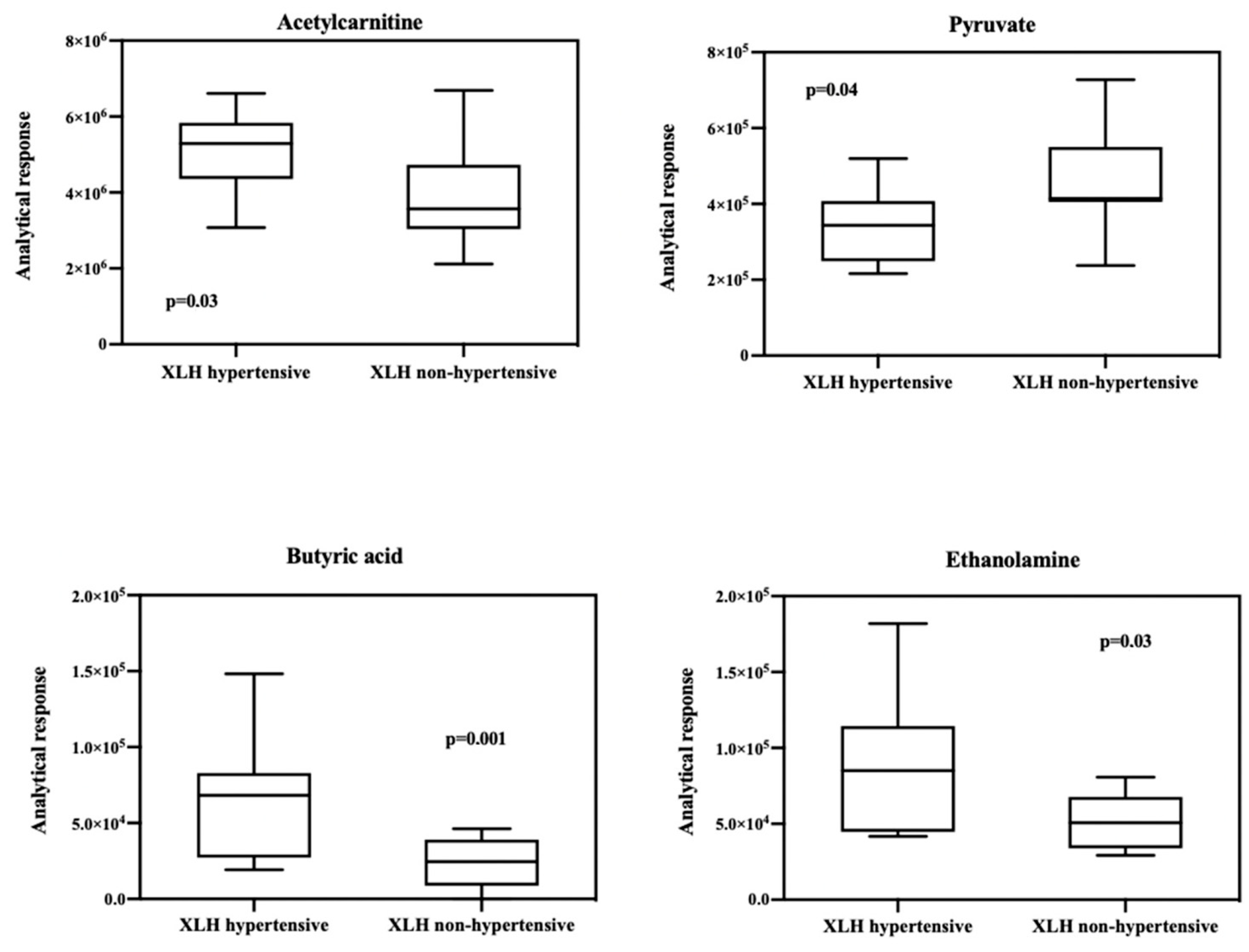
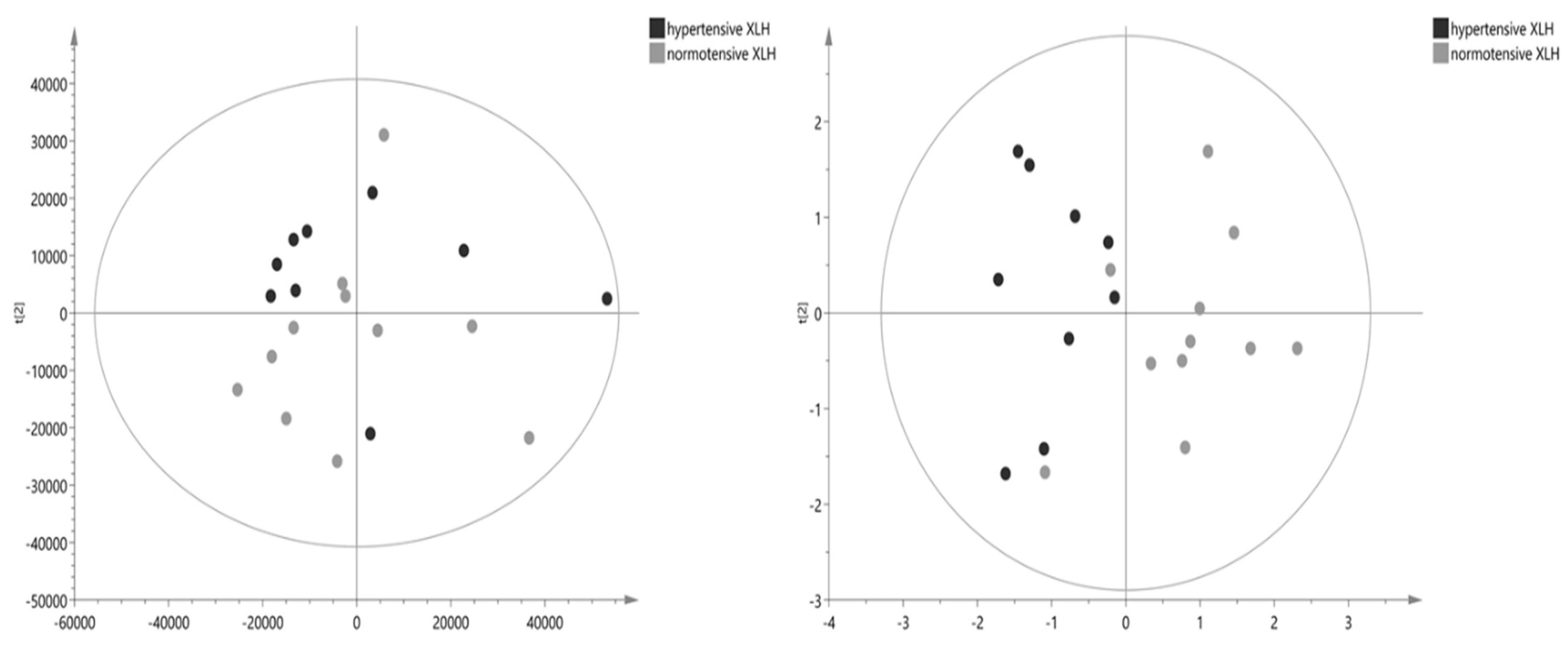
2.3. Metabolomic Results

A comprehensive list of 106 identified metabolites is presented in Table 2. The table provides detailed information on the identified metabolites, including their chemical composition and properties. The Volcano graph showed that acetylcarnitine, pyruvate, ethanolamine, and butyric acid significantly differ between the blood samples of XLH patients with hypertension and those of XLH non-hypertensive patients (Figure 1). Box and whisker plots were used to represent the relative intensities of selected metabolic variables with possible clinical relevance (Figure 2). PCA score plots (Figure 3) were constructed for all metabolites and the discriminative ones, showing that the model discriminates between the two groups. From this model, the most important discriminant variables were selected according to their VIP score (>1), a jack-knife confidence interval that did not include zero, and an FC greater than 1.2. There was no correlation observed between acetylcarnitine, pyruvate, butyric acid, or ethanolamine and the levels of PTH, vitamin D, phosphorus, or FGF23 (Supplementary Materials).

3. Discussion

Hypertension is a chronic condition and a significant risk factor for cardiovascular diseases (CVD). It is estimated that around 20% of adults worldwide are affected by hypertension, of which 90% of cases are classified as “essential” due to an unknown underlying cause [10]. This percentage is higher in XLH patients, who, despite being young, are at a higher risk of morbidity, mortality, and cardiovascular diseases. The prevalence of hypertension in our study comprises 45%, a figure similar to other previously described studies [4]. However, this is controversial, as other cohorts do not find similar results, and the exact pathophysiological mechanisms involved are still unclear.
The development of hypertension is influenced by a combination of genetic and environmental factors, with age, obesity, and physical inactivity being typical risk factors, and is strongly associated with inflammation, oxidative stress, and dyslipidemia [11]. Not in vain, recent studies have shown that alterations in lipid and carbohydrate metabolism often accompany hypertension [12,13], highlighting the connection between hypertension and metabolic disorders. Thus, researchers have increasingly utilized metabolomic profiles to identify specific metabolites that could serve as biomarkers of oxidative stress and inflammation, conditions commonly present in hypertensive patients [14]. With this same idea, our study, employing an untargeted metabolomic technique to identify differences in plasma metabolites between XLH patients with and without hypertension, identified a total of 106 metabolites, among which 4 showed significant clinical and statistical differences.
Firstly, we found higher levels of acetylcarnitine, a metabolite that plays a key role in lipid metabolism, in hypertensive XLH subjects compared to normotensive individuals. Endogenous carnitine biosynthesis occurs in the kidney, liver, and brain and comes from protein lysosomal degradation [15]. The human body produces more than 1200 fatty acids (FAs) using L-carnitine and acylcarnitines [16]. The essential role of L-carnitine and acylcarnitines is to facilitate the transportation of long-chain fatty acids (long-chain fatty acyl-CoA) to the mitochondria for β-oxidation degradation. In contrast, medium and short-chain fatty acids can enter the mitochondria freely through diffusion. This function is crucial to prevent the accumulation of long-chain fatty acids that can cause cellular damage [17].
The analysis of serum acylcarnitine levels serves as a measurable indicator of β-oxidation status, consistently tied to vascular inflammation in various research outcomes [18]. The buildup of acylcarnitine intermediates in extracellular fluid indicates compromised β-oxidation and disrupted mitochondrial metabolism, especially in aging, leading to inefficient fatty acid recycling. On the other hand, it has been suggested that an accumulation of intracellular lipids and acetylcarnitine can occur in hypertensive subjects if the absorption rate of fatty acids exceeds the rate of β-oxidation [19]. Additionally, acylcarnitines constitute some of the most commonly implicated metabolites in cardiovascular diseases [20]. Recent clinical studies have linked acylcarnitines to an elevated risk of atherosclerotic plaque formation, independent of conventional cardiovascular risk factors [21]. Notably, Ramipril was found to significantly reduce the levels of propionylcarnitine, butyrylcarnitine, and isovalerylcarnitine (C3M, C4M, and C5M) in rats among 60 metabolites analyzed that were not affected by treatment with ACE inhibitors [22]. Hence, their role as cardiovascular risk markers in this type of population remains to be studied.
Secondly, we observed lower levels of pyruvate in hypertensive subjects with XLH. There are limited data directly associating pyruvate levels with hypertension, but this correlation was positively made in a study involving restricted participants [19]. Conversely, one study highlighted that decreased alanine and pyruvate levels in people with hypertension may indicate a dynamic balance between aminotransferase reactions in the muscle and the liver [23,24]. While some authors propose that hypertensive patients have an elevated lactate-to-pyruvate (L/P) ratio compared to normotensive individuals [25], this ratio would reflect their response to oxygen and glucose supply variations, suggesting its role as a potential marker for tissue ischemia in anaerobic conditions. The higher L/P ratio might indicate the occurrence of such conditions in hypertensive tissues. Nevertheless, our study did not detect changes in the L/P ratio or a decrease in alanine levels.
Thirdly, elevated levels of ethanolamine were found in hypertensive patients with XLH as compared to the control group. This observation is in line with a study that involved hypertensive pediatric subjects, where ethanolamine proved to be effective in identifying children with hypertension [19]. While only a few studies have focused on the role of this metabolite in arterial hypertension, existing evidence highlights its crucial involvement in the synthesis of phosphatidylethanolamine, glycerophospholipids linked to the formation of atherosclerotic lesions and endothelial damage [26]. Furthermore, its impact on left ventricular function and the development of cardiac fibrosis has been substantiated [27].
Finally, the levels of butyric acid were also significantly higher in the hypertensive XLH population compared to normotensive individuals. Traditionally, cardiovascular research focused mainly on the involvement of long-chain fatty acids. However, increasing evidence indicates a significant impact of molecules produced by the gut microbiota, such as short-chain fatty acids (SCFA), on atherosclerosis and hypertension [28]. Butyric acid (BA) is a significant SCFA that acts as a mediator between the intestinal microbiota and the circulatory system [29]. The role of butyric acid in blood pressure BP was elucidated more than 50 years ago by intravenous administration of tributyrin, a BA prodrug [30]. Another study indicates that an elevation in the concentration of BA in the colon induces a notable hypotensive effect regulated by signals from the afferent vagus nerve of the colon. This bacterial metabolite is believed to exert its hypotensive effect by inhibiting sympathetic activity and inducing a direct vasodilatory effect [31]. In addition, BA suppresses the activation of NF-κB and reduces macrophage migration, thus assuming an anti-inflammatory role and improving atherosclerosis. In light of these findings, it is proposed that SCFAs, particularly butyric acid, could serve as a therapeutic target for atherosclerosis by modulating lipid metabolism and mitigating inflammation [32].
It is important to acknowledge certain limitations of this study. Firstly, the study’s design prevents us from establishing a causal relationship. Secondly, the small sample size necessitates categorizing this study as exploratory and hypothesis-generating until further validation is performed with larger cohorts. It also makes a potentially high type II error. In fact, our study found no correlation between hypertension and well-known related factors with high BP in the general population, such as hyperparathyroidism, nephrocalcinosis, high FGF23 levels, obesity, or the use of nonsteroidal anti-inflammatory drugs, which XLH patients commonly use for pain control. Nonetheless, it did afford us the statistical power required to detect these observed metabolite differences. Thirdly, we did not include unaffected controls with and without hypertension. Fourthly, it is important to consider the effect of medications on blood pressure and metabolism in our analysis. In our study, most patients take minimal medication in addition to active vitamin D and oral phosphorus supplements, but, maybe due to limited statistical power, the study has not yielded significant results on this point. Likewise, the study was conducted under similar conditions, with all patients fasting. The impact of metabolic regulation on flora has not been explored in this manuscript.
Despite the outlined limitations, it is crucial to recognize the substantial strengths present in this study. Our XLH patient cohort is one of the largest published from Europe and the largest among adult XLH patients in Western Europe. Therefore, despite the potential insights gleaned from larger populations, every scientific contribution is significant in this rare disease. The field of omics sciences, such as proteomics, genomics, and metabolomics, is undergoing a surge in medical research, and we are confident that it will eventually provide us with extensive data to understand the pathophysiological mechanisms of diseases, particularly hypertension. Manipulating metabolic pathways and achieving personalized medicine represent the future of medicine.
In conclusion, the changes observed in the metabolite profile suggest that metabolomics can be a useful tool for better understanding the pathogenic mechanisms of hypertension in XLH. This can lead to earlier diagnosis, better treatment options, and the prevention of cardiovascular complications. Our study, which represents the first on hypertension in patients with XLH using a metabolomic approach, identified significant changes in specific metabolites. These findings can help us better understand this disease’s biological and pathological processes and its regulatory mechanisms, which are still unclear.

4. Materials and Methods

4.1. Study Design and Participants

This multicenter cross-sectional study was conducted on a population of adult patients diagnosed with XLH and under follow-up at the Hospital Universitari i Politècnic La Fe and other areas of the Valencian Community. The systematic identification of this cohort was performed using electronic medical records, following a methodology previously published by our group [33]. The population was stratified into two groups based on the presence or absence of hypertension. Demographic, clinical, and laboratory variables, as well as treatments used, were collected. Clinical data and treatments were collected through electronic health records, which were later validated and extended through medical interviews. The study was conducted according to the guidelines of the Declaration of Helsinki and approved by the Institutional Review Board and Ethics Committee of La Fe Health Research Institute (protocol code 2021-016-1, date of approval 27 January 2021). Informed consent was obtained from all subjects involved in the study.

4.2. Measurement of Blood Pressure and Covariables

Body weight and height were measured twice, and the mean values were used to estimate BMI, calculated as weight in kilograms divided by height in square meters. Hypertension was defined as systolic blood pressure (BP) ≥ 130 mm Hg and/or diastolic BP ≥ 80 mm Hg according to the 2017 ACC/AHA guidelines [10]. In all cases of hypertension, the diagnosis had already been made by a primary care physician or another specialist prior to the commencement of our study. BP measurements were obtained during medical visits, with the primary objective of validating the diagnosis and assessing its management status. It was measured following standard protocols and by trained nurses or physicians using validated devices. The participants were in a relaxed, sitting position for 5 min and avoided caffeine, cigarettes, alcohol, and physical activity for 12 h before the clinic visit. At the medical examination, three BP measurements were recorded at 5 min intervals, and the mean BP was calculated based on the last two values. Patients diagnosed with secondary arterial hypertension were excluded. Nephrocalcinosis was defined as diffused calcium deposition within the kidney, identifiable through ultrasonography, whereas renal dysfunction was determined according to KDIGO criteria, eGFR < 60 mL/min/1.73 m2, and/or the presence of structural damage [34]. Hyperparathyroidism (HPT) was diagnosed in cases exhibiting consistently elevated intact parathormone (PTH) levels (>65 pg/mL) for six months or more.

4.3. Blood Sample Collection and Preparation

All patients included in the study were scheduled for a single medical visit at the research unit within the Nephrology Service of Hospital Universitari i Politècnic La Fe. A comprehensive evaluation was carried out during this visit, including blood tests, urine sampling, and kidney ultrasounds. All patients observed a minimum 8 h fasting period before undergoing these assessments. Blood samples were collected using ethylenediaminetetraacetic acid (EDTA) tubes and processed within 30 min to prevent platelet activation, protein degradation, and the decomposition of thermolabile compounds. Intact FGF23 levels were determined using an enzyme-linked immunosorbent assay (ELISA) test kit from Kainos Laboratories, Tokyo, Japan. Total cholesterol, high-density lipoprotein cholesterol (HDL-C), low-density lipoprotein cholesterol (LDL-C), and triglycerides were assayed using enzymatic procedures. The CKD-EPI formula was used to calculate the estimated glomerular filtration rate (eGFR), while the remaining biochemical variables were examined following the established protocols and best-practice guidelines outlined by Universitari I Politècnic La Fe Hospital.
For the metabolomic study, serum samples were promptly centrifuged at room temperature within 15 min of extraction, using the Eppendorf Centrifuge 5702 model at 5000 rpm for 10 min. The resulting serum was then divided into 250 μL aliquots, each labeled with an anonymized numerical code, before being frozen at −80 °C using a New Brunswick Scientific Ultra-Low Temperature Freezer V 410 Premium, ensuring optimal conditions for further analysis.
Upon thawing, 20 µL of serum was combined with 180 µL of cold methanol, an organic solvent that dissociates the protein-bound metabolites. The samples were then shaken at −20 °C for 30 min, inducing protein precipitation, before being centrifuged at 16,000 rpm for 20 min at 4 °C to separate the cellular fraction from the plasma. Then, 90 µL of the upper cleaned was collected from each sample and transferred to an HPLC vial and combined with 10 µL of a standard solution containing internal standards (a mixture of 20 µM caffeine-d9, leucine enkephalin, reserpine, and phenylalanine-d5).
Quality control samples (QCs) were prepared by combining 10 μL from each extract. Blank samples, prepared to replace the extract with ultrapure water, were used to identify artifacts from reagents, the tube, and other materials. Finally, samples, QCs, and blank samples were injected randomly into the chromatographic system. To monitor the stabilities of the instrumental system and the instrumental drift, QC samples were injected into every 7th sample in each sequence, and the blank samples were performed at the end of the sequence.

4.4. Untargeted Metabolomics Based on UPLC-Q-ToF Mass Spectrometry

The metabolomic analysis was performed using Ultra-Performance Liquid Chromatography (UPLC) equipment coupled to a high-resolution mass spectrometer (MS) with Orbitrap UPLC-QExactive Plus detector (UPLC-TOF/MS-Orbitrap, QExactive Plus MS) available at the Analytical Unit of the Instituto de Investigación Sanitaria La Fe (IISLaFe). All reagents and chemicals were purchased from Sigma Aldrich (St. Louis, MO, USA) and Fisher Scientific (CAN, Waltham, MA, USA).
Chromatographic separation was performed using an HILIC UPLC X bridge BEH amide column (150 mm × 2.1 mm, particle size 2.5 uM, Waters, Wexford, Ireland). The column and auto counter were set to 25 °C and 4 °C, respectively, with an injection volume of 5 μL. The total running time of the chromatogram was 25 min at a flow rate of 105 μL/min. Metabolites were eluted with gradients of mobile phase A consisting of H2O and 10 mM ammonium acetate and mobile phase B in acetonitrile (ACN). During the first 2 min, the eluent composition was 90% mobile phase B and 10% mobile phase A; at minute 2, 85% B; from minute 3 to minute 8, 75% B; from minute 8 to minute 10, 70% B; from minute 10 to minute 13, 50% B; from minute 13 to minute 16, 25% B; from minute 16 to minute 22, 0% B; from minute 22 to minute 25, again 90% B reaching the starting conditions. A mass detector (Orbitrap) was used in Full scan in both positive and negative ESI modes with two different events, the first scan event between 70 and 700 Da and a second event in the mass range of 700–1700 Da.
Different measures were taken to ensure the analysis’s quality and reproducibility and mitigate intra-batch variability. The reagent blank was analyzed at the beginning of the sequence to identify sampling artifacts and exclude contaminating signals. Subsequently, a random injection sequence was performed that included the analysis of 5 quality control (QC) samples to condition the column and equipment (these data were not used for data analysis). Sample analyses and quality control were conducted at a 5:1 ratio to monitor and control variability resulting from contamination or changes in the column. Any signal detected with an intensity similar to the quality control and blank samples was eliminated.

4.5. Data Processing and Acquisition

The data obtained during the analysis were converted into mzXML format using the Mass Converter Proteowizard program. Then, they were processed in EI-MAVEN software (Version Version 0.4.1) for alignment, noise filtering, the integration of chromatographic peaks, generation of a peak table, and identification of metabolites using an in-house library of polar compounds previously developed and used at the Analytical Unit [35]. Structures, m/z spectra, and formulas were obtained from the Human Metabolome Database (HMDB, version 4.0).
The configuration parameters of EI-MAVEN for data processing were the following: ionization in automatic mode type ESI, precision q1 and q3 for MRN transitions of 0.5 amu with total filter line. Maximum differences in retention time between spikes in a group were 15 s. The smoothness threshold was 2 λ, and the asymmetry was 0.08 ρ. A weighted average of the m/z values observed in the chromatographic peak was calculated to annotate the chromatographic peaks. Cluster selection criteria for use in the alignment were at least 2 peaks in each cluster with a total limit of 1000 clusters and a clustering window of 20 scans. For peak picking, the minimum intensity was 1000 with a maximum signal-to-noise ratio of 3 and a minimum peak width of 5 scans. The peaks are assigned a clustering score.
We have applied the NetID algorithm to the dataset, creating a peak table containing the exact mass (m/z) and retention time (min). Subsequently, matches were sought with those metabolites in the database. The predefined tolerance was ±10 ppm. Peaks were considered metabolites or artifacts based on the match with the local database formulas. All adducts, fragments, or isotopes of metabolites were considered artifacts.
When comparing with blank samples, background peaks were removed if their intensity was greater than 0.5 times that of the sample. Compounds showing a coefficient of variation (CV) in the analytical response of the quality controls exceeding 30% were excluded.

4.6. Statistical Analysis

Categorical variables were presented as n (%), while continuous variables were presented as median (P25–P75). The distribution of the variables was tested with the Shapiro–Wilk test. Differences between hypertensive and normotensive groups were analyzed using independent samples t-test for normally distributed continuous data, the Mann–Whitney U test for skewed data, and the χ2 test for categorical variables.
The relative abundances of metabolic members of XLH individuals with hypertension were compared with non-hypertensive XLH controls. A univariate statistical analysis was conducted between the case and control groups utilizing a t-test and visualized through a Volcano Plot. This analysis was executed using an in-house script within the R platform (Version 4.3.3). The methodology integrated a Fold Change (FC) approach alongside the significance derived from a paired Student’s t-test for normally distributed variables. For skewed data, the analysis employed the Wilcoxon signed-rank test after confirming normality via the Shapiro–Wilk test.
Molecular characteristics demonstrating the stronger combination of FC and statistical significance are represented in Figure 1. To mitigate the risk of false discoveries, we applied the Benjamini–Hochberg procedure. An exploratory unsupervised Principal Component Analysis (PCA) was performed to extract maximum information and identify behavioral patterns. This involved simplifying data variability by examining their distribution, ultimately grouping them into principal components. Model validity and robustness were assessed using R2(Y) (goodness of fit) and Q2(Y) (goodness of prediction), with a predictive capacity deemed satisfactory when Q2(Y) exceeded 0.5. A PCA score plot accompanies each comparison. For univariate analysis, p < 0.05 was defined to reach statistical significance in a two-tailed Student’s t-test. Metabolic compounds with p < 0.05 and variable importance in the projection (VIP) > 1 were considered statistically different between groups. For statistical analysis, the SPSS system for Windows version 22 and the GraphPad Prism v8 software (San Diego, CA, USA) were used.

Supplementary Materials

The following supporting information can be downloaded at: https://www.mdpi.com/article/10.3390/ijms25063545/s1.

Author Contributions

Conceptualization: L.C.L.-R. and J.J.B.; methodology, L.C.L.-R., M.R.-M., and J.J.B.; software, M.R.-M. and A.L.; validation, L.C.L.-R.; formal analysis, L.C.L.-R., J.J.B. and M.R.-M.; investigation, L.C.L.-R.; resources, J.H.-J.; data curation, J.R.M.-C.; writing—original draft preparation, L.C.L.-R.; writing—review and editing, J.J.B. and M.R.-M.; visualization, L.C.L.-R. and M.R.-M.; supervision, J.J.B.; project administration, J.H.-J.; funding acquisition, J.J.B. and J.H.-J. All authors have read and agreed to the published version of the manuscript.

Funding

Kyowa Kirin International provided financial support for this study but played no role in the design, execution, interpretation, or composition of the study.

Institutional Review Board Statement

The study was authorized by La Fe Health Research Institute Research Ethics Committee (protocol code 2021-016-1, date of approval 27 January 2021) and was conducted in accordance with the ethical principles of the Declaration of Helsinki and data protection regulations (EU) 2016/679 (General Data Protection Regulation) and its subordinate national and regional laws.

Informed Consent Statement

Informed consent was obtained from all subjects involved in the study.

Data Availability Statement

The datasets and materials used and/or analyzed during the current study are available from the corresponding author upon reasonable request.

Acknowledgments

We would like to publicly thank all participating patients and the nursing staff of the nephrology research unit of Health Research Institute Hospital La Fe for their selfless collaboration in this study.

Conflicts of Interest

L.C.L.-R. and J.H.-J. have received lecture fees from Kyowa Kirin Spain, J.J.B. has received lecture fees and advisory fees from Kyowa Kirin Spain and Kyowa Kirin International. The other authors declare no conflicts of interests.

References

  1. Beck-Nielsen, S.S.; Mughal, Z.; Haffner, D.; Nilsson, O.; Levtchenko, E.; Ariceta, G.; de Lucas Collantes, C.; Schnabel, D.; Jandhyala, R.; Mäkitie, O. FGF23 and its role in X-linked hypophosphatemia-related morbidity. Orphanet. J. Rare Dis. 2019, 14, 58. [Google Scholar] [CrossRef]
  2. European Commissions. Rare Diseases. Public Health–Eur Comm 2016. Available online: https://ec.europa.eu/health/non_conmunicable_diaseases/rare_diaseases_en (accessed on 27 March 2019).
  3. González-Lamuño, D.; Lorente Rodríguez, A.; Luis Yanes, M.I.; Marín-Del Barrio, S.; Martínez Díaz-Guerra, G.; Peris, P. Clinical practice recommendations for the diagnosis and treatment of X-linked hypophosphatemia: A consensus based on the ADAPTE method. Med. Clin. 2022, 159, 152.e1–152.e12. [Google Scholar] [CrossRef]
  4. Nakamura, Y.; Takagi, M.; Takeda, R.; Miyai, K.; Hasegawa, Y. Hypertension is a characteristic complication of X-linked hypophosphatemia. Endocr. J. 2017, 64, 283–289. [Google Scholar] [CrossRef] [PubMed]
  5. Alon, U.S.; Monzavi, R.; Lilien, M.; Rasoulpour, M.; Geffner, M.E.; Yadin, O. Hypertension in hypophosphatemic rickets-role of secondary hyperparathyroidism. Pediatr. Nephrol. 2003, 18, 155–158. [Google Scholar] [CrossRef] [PubMed]
  6. Andrukhova, O.; Slavic, S.; Smorodchenko, A.; Zeitz, U.; Shalhoub, V.; Lanske, B.; Pohl, E.E.; Erben, R.G. FGF23 regulates renal sodium handling and blood pressure. EMBO Mol. Med. 2014, 6, 744–759. [Google Scholar] [CrossRef] [PubMed]
  7. McMullan, C.J.; Borgi, L.; Curhan, G.C.; Fisher, N.; Forman, J.P. The effect of vitamin D on renin-angiotensin system activation and blood pressure: A randomized control trial. J. Hypertens. 2017, 35, 822–829. [Google Scholar] [CrossRef] [PubMed]
  8. GBD 2017 Causes of Death Collaborators. Global, regional, and national age-sex-specific mortality for 282 causes of death in 195 countries and territories, 1980–2017: A systematic analysis for the global burden of disease study 2017. Lancet 2018, 392, 1736–1788. [Google Scholar] [CrossRef]
  9. Ameta, K.; Gupta, A.; Kumar, S.; Sethi, R.; Kumar, D.; Mahdi, A.A. Essential hypertension: A filtered serum based metabolomics study. Sci. Rep. 2017, 7, 2153. [Google Scholar] [CrossRef]
  10. Whelton, P.K.; Carey, R.M.; Aronow, W.S.; Casey, D.E., Jr.; Collins, K.J.; Dennison Himmelfarb, C.; DePalma, S.M.; Gidding, S.; Jamerson, K.A.; Jones, D.W.; et al. 2017 ACC/AHA/AAPA/ABC/ACPM/AGS/APhA/ASH/ASPC/NMA/PCNA Guideline for the Prevention, Detection, Evaluation, and Management of High Blood Pressure in Adults: Executive Summary: A Report of the American College of Cardiology/American Heart Association Task Force on Clinical Practice Guidelines. Hypertension 2018, 71, 1269–1324, Erratum in: Hypertension 2018, 71, e136–e139. Erratum in: Hypertension 2018, 72, e33. [Google Scholar] [CrossRef]
  11. Sun, Z. Aging, arterial stiffness, and hypertension. Hypertension 2015, 65, 252–256. [Google Scholar] [CrossRef]
  12. Dietrich, S.; Floegel, A.; Weikert, C.; Prehn, C.; Adamski, J.; Pischon, T.; Boeing, H.; Drogan, D. Identification of serum metabolites associated with incident hypertension in the European prospective investigation into cancer and Nutrition-Potsdam study. Hypertension 2016, 68, 471–477. [Google Scholar] [CrossRef]
  13. Arnett, D.K.; Claas, S.A. Omics of blood pressure and hypertension. Circ. Res. 2018, 122, 1409–1419. [Google Scholar] [CrossRef] [PubMed]
  14. He, W.J.; Li, C.; Mi, X.; Shi, M.; Gu, X.; Bazzano, L.A.; Razavi, A.C.; Nierenberg, J.L.; Dorans, K.; He, H.; et al. An untargeted metabolomics study of blood pressure: Findings from the bogalusa heart study. J. Hypertens. 2020, 38, 1302–1311. [Google Scholar] [CrossRef] [PubMed]
  15. Vaz, F.M.; Wanders, R.J. Carnitine biosynthesis in mammals. Biochem. J. 2002, 361, 417–429. [Google Scholar] [CrossRef] [PubMed]
  16. Li, S.; Gao, D.; Jiang, Y. Function, Detection and Alteration of Acylcarnitine Metabolism in Hepatocellular Carcinoma. Metabolites 2019, 9, 36. [Google Scholar] [CrossRef] [PubMed]
  17. Jones, L.L.; McDonald, D.A.; Borum, P.R. Acylcarnitines: Role in brain. Prog. Lipid Res. 2010, 49, 61–75. [Google Scholar] [CrossRef] [PubMed]
  18. Guasch-Ferré, M.; Zheng, Y.; Ruiz-Canela, M.; Hruby, A.; Martínez-González, M.A.; Clish, C.B.; Corella, D.; Estruch, R.; Ros, E.; Fitó, M.; et al. Plasma Acylcarnitines and Risk of Cardiovascular Disease: Effect of Mediterranean Diet Interventions. Am. J. Clin. Nutr. 2016, 103, 1408–1416. [Google Scholar] [CrossRef] [PubMed]
  19. Sun, J.; Zhao, M.; Yang, L.; Liu, X.; Pacifico, L.; Chiesa, C.; Xi, B. Identification of Potential Metabolic Markers of Hypertension in Chinese Children. Int. J. Hypertens. 2021, 2021, 6691734. [Google Scholar] [CrossRef]
  20. Aitken-Buck, H.M.; Krause, J.; Zeller, T.; Jones, P.P.; Lamberts, R.R. Long-Chain Acylcarnitines and Cardiac Excitation-Contraction Coupling: Links to Arrhythmias. Front. Physiol. 2020, 11, 577856. [Google Scholar] [CrossRef]
  21. Hua, S.; Clish, C.; Scott, J.; Hanna, D.; Haberlen, S.; Shah, S.; Hodis, H.; Landy, A.; Post, W.; Anastos, K.; et al. Abstract P201: Associations of Plasma Acylcarnitines With Incident Carotid Artery Plaque in Individuals With or at Risk of HIV Infection. Circulation 2018, 137, AP201. [Google Scholar] [CrossRef]
  22. Kosacka, J.; Berger, C.; Ceglarek, U.; Hoffmann, A.; Blüher, M.; Klöting, N. Ramipril Reduces Acylcarnitines and Distinctly Increases Angiotensin-Converting Enzyme 2 Expression in Lungs of Rats. Metabolites 2022, 12, 293. [Google Scholar] [CrossRef] [PubMed]
  23. Natali, A.; Santoro, D.; Palombo, C.; Cerri, M.; Ghione, S.; Ferrannini, E. Impaired insulin action on skeletal muscle metabolism in essential hypertension. Hypertension 1991, 17, 170–178. [Google Scholar] [CrossRef] [PubMed]
  24. Newgard, C.B.; An, J.; Bain, J.R.; Muehlbauer, M.J.; Stevens, R.D.; Lien, L.F.; Haqq, A.M.; Shah, S.H.; Arlotto, M.; Slentz, C.A.; et al. A branched-chain amino acid-related metabolic signature that differentiates obese and lean humans and contributes to insulin resistance. Cell Metab. 2009, 9, 311–326. [Google Scholar] [CrossRef] [PubMed]
  25. Fujishima, M.; Nakatomi, Y.; Tamaki, K.; Ishitsuka, T.; Kawasaki, T.; Omae, T. Cerebrospinal fluid lactate and pyruvate concentrations in patients with malignant hypertension. J. Neurol. 1984, 231, 71–74. [Google Scholar] [CrossRef]
  26. Xu, W.; Liu, D.; Lin, D. Comparison of mechanisms of endothelial cell protections between high-density lipo-protein and apolipoprotein A-I mimetic peptide. Front. Pharmacol. 2019, 10, 817. [Google Scholar] [CrossRef] [PubMed]
  27. Yamamoto, K.; Takahashi, Y.; Mano, T.; Sakata, Y.; Nishikawa, N.; Yoshida, J.; Oishi, Y.; Hori, M.; Miwa, T.; Inoue, S.; et al. N-methyl-ethanolamine attenuates cardiac fibrosis and improves diastolic function: Inhibition of phospholipase D as a possible mechanism. Eur. Heart J. 2004, 25, 1221–1229. [Google Scholar] [CrossRef]
  28. Chen, X.F.; Chen, X.; Tang, X. Short-chain fatty acid, acylation and cardiovascular diseases. Clin. Sci. 2020, 134, 657–676. [Google Scholar] [CrossRef]
  29. Huc, T.; Nowinski, A.; Drapala, A.; Konopelski, P.; Ufnal, M. Indole and indoxyl sulfate, gut bacteria metabolites of tryptophan, change arterial blood pressure via peripheral and central mechanisms in rats. Pharmacol. Res. 2018, 130, 172–179. [Google Scholar] [CrossRef]
  30. Wretlind, A. Effect of tributyrin on circulation and respiration. Acta Physiol. Scand. 1957, 40, 59–74. [Google Scholar] [CrossRef]
  31. Onyszkiewicz, M.; Gawrys-Kopczynska, M.; Konopelski, P.; Aleksandrowicz, M.; Sawicka, A.; Koźniewska, E.; Samborowska, E.; Ufnal, M. Butyric acid, a gut bacteria metabolite, lowers arterial blood pressure via colon-vagus nerve signaling and GPR41/43 receptors. Pflug. Arch.–Eur. J. Physiol. 2019, 471, 1441–1453. [Google Scholar] [CrossRef]
  32. Cao, H.; Zhu, Y.; Hu, G.; Zhang, Q.; Zheng, L. Gut microbiome and metabolites, the future direction of diagnosis and treatment of atherosclerosis. Pharmacol. Res. 2023, 187, 106586. [Google Scholar] [CrossRef]
  33. Broseta, J.J. Different approaches to improve cohort identification using electronic health records: X-linked hypophosphatemia as an example. Intractable Rare Dis. Res. 2021, 10, 17–22. [Google Scholar] [CrossRef] [PubMed]
  34. Improving Global Outcomes (KDIGO) CKD Work Group. KDIGO 2012 clinical practice guideline for the evaluation and management of chronic kidney disease. Kidney Int. Suppl. 2013, 3, 19–62. [Google Scholar] [CrossRef]
  35. García-Cañaveras, J.C.; Lancho, O.; Ducker, G.S.; Ghergurovich, J.M.; Xu, X.; da Silva-Diz, V.; Minuzzo, S.; Indraccolo, S.; Kim, H.; Herranz, D.; et al. SHMT inhibition is effective and synergizes with methotrexate in T-cell acute lymphoblastic leukemia. Leukemia 2021, 35, 377–388. [Google Scholar] [CrossRef] [PubMed]
Figure 1. Volcano plot illustrates significant differences in serum metabolites between XLH patients with and without hypertension.
Ijms 25 03545 g001
Figure 2. Box and whisker plot of significantly altered metabolites in the hypertensive group compared with the normotensive group.
Ijms 25 03545 g002
Figure 3. PCA score plot with metabolic variations between hypertensive XLH patients and normotensive XLH patients when all metabolites and only discriminative metabolites are identified.
Ijms 25 03545 g003
Table 1. Demographic and clinical characteristics of the study population.
VariableOverall (n = 20)XLH Hypertensive (n = 9)XLH Non-Hypertensive (n = 11)
Age (years)44 (33.2–54.7)49 (39–57)43 (27–47)
Male sex, n (%)10 (50)6 (66)4 (40)
Weight (kg)73 (50.7–85.5)81.5 (50.2–93.7)71 (50–79.7)
Height (cm)158 (150–170.5)159.5 (152.2–182.7)155.5 (150–163)
BMI (Kg/m2)27.5 (22–32.2)30 (22.5–32.7)25 (22–32)
Diagnosis in childhood, n (%)10 (50)5 (55.5)5 (50)
Office SBP (mmHg)124.75 ± 17.66139.77 ± 10.67112.45 ± 11.50
Office DBP (mmHg)71.5 ± 7.7275.66 ± 8.9368.09 ± 4.59
Familiar history of hypertension n (%)7 (35)3 (33.3)4 (40)
Nephrocalcinosis n (%)3 (15)2 (22.2)1 (10)
Hyperparathyroidism n (%)7 (35)4 (44.4)3 (30)
Non-steroidal anti-inflammatories n (%)4 (20)1 (11.1)3 (30)
Creatinine (mg/dL), mean ± SD0.91 ± 0.291.08 ± 0.310.78 ± 0.19
GFR (mL/min/1.73 m2), mean ± SD92.80 ± 2379.77 ± 19.75103.45 ± 20.51
Uric acid (mg/dL), mean ± SD5.69 ± 1.526.45 ± 1.615.07 ± 1.18
Total cholesterol(mg/dL), mean ± SD197 ± 28.36206.88 ± 32.06189 ± 23.39
LDL (mg/dL), mean ± SD119 ± 24.33133.33 ± 21.92107.27 ± 20.12
HDL (mg/dL), mean ± SD55.3 ± 14.9150.22 ± 16.5759.45 ± 12.68
Triglycerides (mg/dL), mean ± SD115.94 ± 50.38122.62 ± 58.63111.09 ± 45.84
Hemoglobin (g/dL), mean ± SD14.64 ± 1.6314.9 ± 1.2914.26 ± 1.91
Alkaline phosphatase (UI/L), mean ± SD97.85 ± 43.68112 ± 53.7886.27 ±31.37
Phosphorus (mg/dL), mean ± SD2.14 ± 0.492.08 ± 0.382.18 ± 0.58
Calcium (mg/dL), mean ± SD9.46 ± 0.729.51 ± 0.549.42 ± 0.86
Intact FGF23 (pg/mL), mean ± SD211.96 ± 248.58288.28 ± 346.29149.52 ± 109.28
Vitamin D (ng/mL), mean ± SD27.97 ± 9.4425.87 ± 6.3729.5 ± 11.22
PTH (pg/mL), mean ± SD74.88 ± 48.8783.34 ± 38.8067.96 ± 56.71
TmP/GFR (mg/dL), mean ± SD2.12 ± 0.482.08 ± 0.282.07 ± 0.53
BMI: body mass index, SBP: systolic blood pressure, DBP: diastolic blood pressure, GFR: glomerular filtration rate, LDL: low-density lipoprotein, HDL high-density lipoprotein, FGF23: fibroblast growing factor 23, PTH, parathormone, Vit D: 1.25(OH)2 vitamin D, TmP/GFR: ratio of tubular maximum reabsorption of phosphate (TmP) to GFR.
Table 2. List of identified metabolites.
CompoundFormulam/zRtAdductionClass
glycineC2H5NO274.02483412.671[M−H]−Amino acid/peptides/analogues
HypotaurineC2H7NO2S108.01264211.876[M−H]−Sulfinic acid and derivatives
taurineC2H7NO3S126.0219659.911[M+H]+Organosulfonic acid and derivatives
O-PhosphoethanolamineC2H8NO4P142.02636713.674[M+H]+Phosphate esters
pyruvateC3H4O387.0088963.546[M−H]−Keto acids and derivatives
Guanidoacetic acidC3H7N3O2118.06101212.4[M+H]+Amino acid/peptides/analogues
serineC3H7NO3104.03544612.858[M−H]−Amino acid/peptides/analogues
succinateC4H6O4117.01947812.021[M−H]−Carboxylic acid and derivatives
4-aminobutyrateC4H9NO2104.07051111.303[M+H]+Amino acid/peptides/analogues
D-2-Aminobutyric acidC4H9NO2104.07051111.303[M+H]+Amino acid/peptides/analogues
dimethylglycineC4H9NO2102.05616011.155[M−H]−Amino acid/peptides/analogues
glutamineC5H10N2O3147.07627912.909[M+H]+Amino acid/peptides/analogues
2-Hydroxyvaleric acidC5H10O3117.0558173.579[M−H]−Fatty and conjugated acids
methionineC5H11NO2S148.0439459.137[M−H]−Amino acid/peptides/analogues
ornithineC5H12N2O2131.08265716.205[M−H]−Amino acid/peptides/analogues
PhosphorylcholineC5H14NO4P184.07325713.76[M+H]+Quaternary ammonium salts
hypoxanthineC5H4N4O135.0312966.754[M−H]−Purines and derivatives
2-hydroxyglutarateC5H8O5147.03006010.955[M−H]−Short chain hydroxy acids and derivatives
lysineC6H14N2O2145.09829717.035[M−H]−Amino acid/peptides/analogues
N′-MethylnicotinamideC7H8N2O137.07087713.417[M+H]+Pyridines and derivatives
GuaiacolC7H8O2123.0453412.574[M−H]−Phenols
2-Hydroxyoctanoic acidC8H16O3159.1027072.447[M−H]−Fatty and conjugated acids
2-Phenylbutyric acidC10H12O2163.0765992.439[M−H]−Benzene and derivatives
Indole-3-acetaldehydeC10H9NO158.0612342.544[M−H]−Indoles and derivatives
DL-Indole-3-lactic acidC11H11NO3204.0666662.485[M−H]−Indoles and derivatives
tryptophanC11H12N2O2205.0970927.689[M+H]+Indoles and derivatives
Undecanoic acidC11H22O2185.1547702.226[M−H]−Fatty and conjugated acids
trans-3-Indoleacrylic acidC11H9NO2188.070743.044[M+H]+Indoles and derivatives
Indole-3-pyruvic acidC11H9NO3202.0511472.215[M−H]−Indoles and derivatives
Butyric acidC4H8O2202.0874632.495[M−H]−Indoles and derivatives
Myristic acidC14H28O2227.2018282.147[M−H]−Fatty and conjugated acids
Linoleic acidC18H32O2279.2333372.101[M−H]−Lineolic acids and derivatives
Stearic acidC18H36O2283.2641602.082[M−H]−Fatty and conjugated acids
Arachidic acidC20H40O2311.2958982.048[M−H]−Fatty and conjugated acids
Chenodeoxycholic acidC24H40O4393.2999572.996[M+H]+Bile acids, alcohols and derivatives
Glycodeoxycholic acidC26H43NO5450.3216553.096[M+H]+Bile acids, alcohols and derivatives
Glycocholic acidC26H43NO6464.3021246.826[M−H]−Bile acids, alcohols and derivatives
Taurodeoxycholic acidC26H45NO6S498.2902532.389[M−H]−Bile acids, alcohols and derivatives
Glycolic acidC2H4O375.0087978.434[M−H]−Hydroxyacids and derivatives
acetylphosphateC2H5O5P140.99465912.582[M+H]+Phosphate esters
lactateC3H6O389.0245827.071[M−H]−Hydroxyacids and derivatives
glycerateC3H6O4105.0194559.939[M−H]−Carbohydrates and conjugates
alanineC3H7NO290.05492412.178[M+H]+Amino acid/peptides/analogues
b-alanineC3H7NO290.05492412.809[M+H]+Amino acid/peptides/analogues
sarcosineC3H7NO290.05492412.178[M+H]+Amino acid/peptides/analogues
Trimethylamine N-oxideC3H9NO76.07567612.071[M+H]+Organonitrogenic compounds
EthanolamineC2H7N0106.08621211.132[M+H]+Organonitrogenic compounds
acetoacetateC4H6O3101.0245675.873[M−H]−Keto acids and derivatives
2-Ketobutyric acidC4H6O3101.0245742.401[M−H]−Keto acids and derivatives
N-AcetylglycineC4H7NO3116.0354618.303[M−H]−Amino acid/peptides/analogues
aspartateC4H7NO4134.04490713.404[M+H]+Amino acid/peptides/analogues
asparagineC4H8N2O3133.06082213.126[M+H]+Amino acid/peptides/analogues
3-hydroxylbutyrateC4H8O3103.0402227.334[M−H]−Hydroxyacids and derivatives
(R)-3-Hydroxybutanoic acidC4H8O3103.0402227.700[M−H]−Hydroxyacids and derivatives
2-Hydroxybutyric acidC4H8O3103.0402375.802[M−H]−Hydroxyacids and derivatives
creatineC4H9N3O2130.06225612.102[M−H]−Amino acid/peptides/analogues
L-HomoserineC4H9NO3120.06543712.502[M+H]+Amino acid/peptides/analogues
threonineC4H9NO3120.06543712.502[M+H]+Amino acid/peptides/analogues
3-Hydroxyisovaleric acidC5H10O3117.0558175.689[M−H]−Amino acid/peptides/analogues
DL-ValineC5H11NO2116.07178510.032[M−H]−Amino acid/peptides/analogues
betaineC5H11NO2116.07178510.032[M−H]−Amino acid/peptides/analogues
AdonitolC5H12O5151.0614178.769[M−H]−Carbohydrates and conjugates
CholineC5H13NO104.10690312.645[M+H]+Organonitrogenic compounds
Uric acidC5H4N4O3167.0210118.487[M−H]−Purines and derivatives
Pyroglutamic acidC5H7NO3128.0355078.453[M−H]−Amino acid/peptides/analogues
2-Oxoisopentanoic acidC5H8O3115.0401762.285[M−H]−Keto acids and derivatives
Methylsuccinic acidC5H8O4131.03509510.675[M−H]−Fatty and conjugated acids
prolineC5H9NO2116.07044210.857[M+H]+Amino acid/peptides/analogues
N-PropionylglycineC5H9NO3132.06561312.203[M+H]+Amino acid/peptides/analogues
Aminolevulinic acidC5H9NO3130.05102512.026[M−H]−Amino acid/peptides/analogues
L-HydroxyprolineC5H9NO3130.0510567.712[M−H]−Amino acid/peptides/analogues
N-Methyl-D-aspartic acidC5H9NO4148.06025712.906[M+H]+Amino acid/peptides/analogues
glutamateC5H9NO4146.04589812.746[M−H]−Amino acid/peptides/analogues
2-Methyl-3-ketovaleric acidC6H10O3129.0556952.195[M−H]−Keto acids and derivatives
Adipic acidC6H10O4145.05069011.644[M−H]−Fatty and conjugated acids
DL-Pipecolic acidC6H11NO2130.08641110.533[M+H]+Amino acid/peptides/analogues
4-Methylvaleric acidC6H12O2115.0765842.533[M−H]−Fatty and conjugated acids
L-RhamnoseC6H12O5163.0611428.018[M−H]−Carbohydrates and conjugates
myo-inositolC6H12O6179.05607613.264[M−H]−Alcohols and polyols
FructoseC6H12O6179.05609110.639[M−H]−Carbohydrates and conjugates
MannoseC6H12O6179.05609110.639[M−H]−Carbohydrates and conjugates
citrullineC6H13N3O3174.08836413.056[M−H]−Amino acid/peptides/analogues
leucineC6H13NO2130.0874028.878[M−H]−Amino acid/peptides/analogues
arginineC6H14N4O2173.10438516.941[M−H]−Amino acid/peptides/analogues
Aconitic acidC6H6O6173.00904813.093[M−H]−Carboxylic acid and derivatives
3-Methylglutaconic AcidC6H8O4143.03506510.646[M−H]−Fatty and conjugated acids
citrate/isocitrateC6H8O7191.01962313.506[M−H]−Carboxylic acid and derivatives
3-Methyl-HistidineC7H11N3O2168.07774412.867[M−H]−Amino acid/peptides/analogues
HomocitrullineC7H15N3O3190.118513.066[M+H]+Amino acid/peptides/analogues
gamma-ButyrobetaineC7H15NO2146.11750813.111[M+H]+Fatty and conjugated acids
carnitineC7H15NO3162.1123212.578[M+H]+Quaternary amine
HomoarginineC7H16N4O2189.13461317.538[M+H]+Amino acid/peptides/analogues
Benzoic acidC7H6O2121.02962510.036[M−H]−Benzene and derivatives
3-Hydroxybenzoic acidC7H6O3137.0244452.072[M−H]−Benzene and derivatives
2-6-Dihydroxybenzoic acidC7H6O4153.0195011.822[M−H]−Benzene and derivatives
N-Acetyl-L-arginineC8H16N4O3217.12939512.865[M+H]+Amino acid/peptides/analogues
DL-2-Aminooctanoic acidC8H17NO2160.1332096.675[M+H]+Amino acid/peptides/analogues
GlycerophosphocholineC8H20NO6P258.10986313.14[M+H]+Glycerophospholipids
3-Indoxyl sulfateC8H7NO4S212.0023191.863[M−H]−Sulfuric acids and derivatives
2-Phenylpropionic acidC9H10O2149.0609592.475[M−H]−Phenylpropanoids
phenylalanineC9H11NO2164.0717628.015[M−H]−Amino acid/peptides/analogues
tyrosineC9H11NO3180.0665139.686[M−H]−Amino acid/peptides/analogues
AcetylcarnitineC9H17NO4204.12300110.376[M+H]+Fatty acid esters
4-Hydroxyphenylpyruvic acidC9H8O4179.0351722.368[M−H]−Benzene and derivatives
3-MethylindoleC9H9N132.0808723.064[M+H]+Indoles and derivatives
Hydroxyphenylformamidoacetic acidC9H9NO4194.0458836.769[M−H]−Phenol
m/z: mass/charge, Rt: Retention time.
Disclaimer/Publisher’s Note: The statements, opinions and data contained in all publications are solely those of the individual author(s) and contributor(s) and not of MDPI and/or the editor(s). MDPI and/or the editor(s) disclaim responsibility for any injury to people or property resulting from any ideas, methods, instructions or products referred to in the content.

