
Sperm Chromatin Dispersion Test Detects Sperm DNA Fragmentation Mainly Associated with Unviable Spermatozoa and Underestimates the Values with Respect to TUNEL Assay
 , , and
Abstract
:1. Introduction
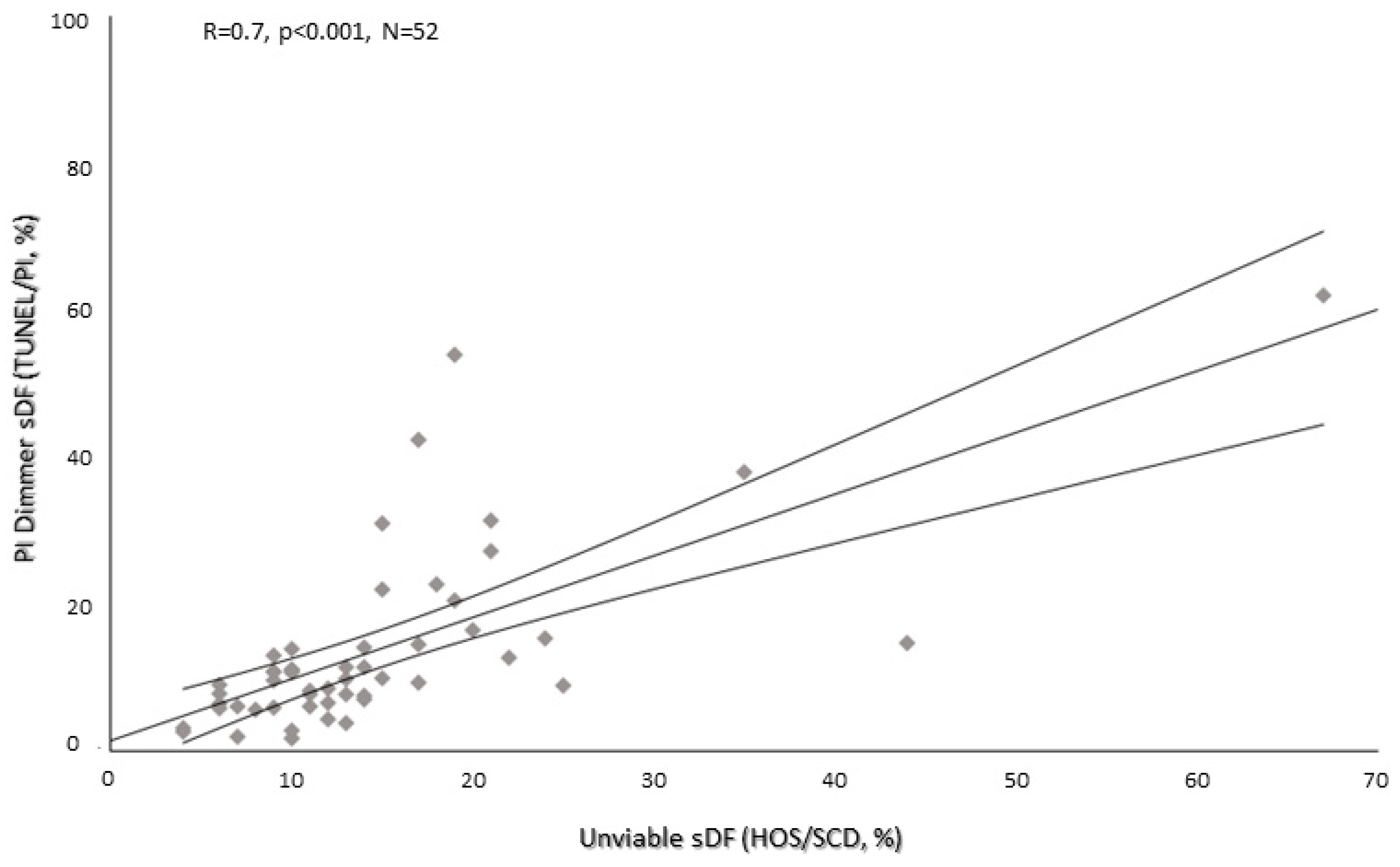
2. Results
3. Discussion
4. Materials and Methods
4.1. Semen Samples Collection and Preparation
4.2. TUNEL/PI and Flow Cytometry
4.3. SCD Test
4.4. Statistical Analysis
5. Conclusions
Author Contributions
Funding
Institutional Review Board Statement
Informed Consent Statement
Data Availability Statement
Conflicts of Interest
References
- Erenpreiss, J.; Elzanaty, S.; Giwercman, A. Sperm DNA damage in men from infertile couples. Asian J. Androl. 2008, 10, 786–790. [Google Scholar] [CrossRef]
- Sakkas, D.; Alvarez, J.G. Sperm DNA fragmentation: Mechanisms of origin, impact on reproductive outcome, and analysis. Fertil. Steril. 2010, 93, 1027–1036. [Google Scholar] [CrossRef] [PubMed]
- Andrabi, S.W.; Ara, A.; Saharan, A.; Jaffar, M.; Gugnani, N.; Esteves, S.C. Sperm DNA Fragmentation: Causes, evaluation and management in male infertility. JBRA Assist. Reprod. 2024. [Google Scholar] [CrossRef] [PubMed]
- Panner Selvam, M.K.; Ambar, R.F.; Agarwal, A.; Henkel, R. Etiologies of sperm DNA damage and its impact on male infertility. Andrologia 2021, 53, e13706. [Google Scholar] [CrossRef]
- Chatterjee, N.; Walker, G.C. Mechanisms of DNA damage, repair, and mutagenesis. Environ. Mol. Mutagen. 2017, 58, 235–263. [Google Scholar] [CrossRef]
- Carusillo, A.; Mussolino, C. DNA Damage: From Threat to Treatment. Cells 2020, 9, 1665. [Google Scholar] [CrossRef]
- Malić Vončina, S.; Golob, B.; Ihan, A.; Kopitar, A.N.; Kolbezen, M.; Zorn, B. Sperm DNA fragmentation and mitochondrial membrane potential combined are better for predicting natural conception than standard sperm parameters. Fertil. Steril. 2016, 105, 637–644. [Google Scholar] [CrossRef]
- Chen, Q.; Zhao, J.Y.; Xue, X.; Zhu, G.X. The association between sperm DNA fragmentation and reproductive outcomes following intrauterine insemination, a meta analysis. Reprod. Toxicol. 2019, 86, 50–55. [Google Scholar] [CrossRef]
- Sugihara, A.; Van Avermaete, F.; Roelant, E.; Punjabi, U.; De Neubourg, D. The role of sperm DNA fragmentation testing in predicting intra-uterine insemination outcome: A systematic review and meta-analysis. Eur. J. Obstet. Gynecol. Reprod. Biol. 2020, 244, 8–15. [Google Scholar] [CrossRef] [PubMed]
- Zhao, J.; Zhang, Q.; Wang, Y.; Li, Y. Whether sperm deoxyribonucleic acid fragmentation has an effect on pregnancy and miscarriage after in vitro fertilization/intracytoplasmic sperm injection: A systematic review and meta-analysis. Fertil. Steril. 2014, 102, 998–1005. [Google Scholar] [CrossRef]
- Osman, A.; Alsomait, H.; Seshadri, S.; El-Toukhy, T.; Khalaf, Y. The effect of sperm DNA fragmentation on live birth rate after IVF or ICSI: A systematic review and meta-analysis. Reprod. Biomed. Online 2015, 30, 120–127. [Google Scholar] [CrossRef] [PubMed]
- Deng, C.; Li, T.; Xie, Y.; Guo, Y.; Yang, Q.Y.; Liang, X.; Deng, C.H.; Liu, G.H. Sperm DNA fragmentation index influences assisted reproductive technology outcome: A systematic review and meta-analysis combined with a retrospective cohort study. Andrologia 2019, 51, e13263. [Google Scholar] [CrossRef] [PubMed]
- Robinson, L.; Gallos, I.D.; Conner, S.J.; Rajkhowa, M.; Miller, D.; Lewis, S.; Kirkman-Brown, J.; Coomarasamy, A. The effect of sperm DNA fragmentation on miscarriage rates: A systematic review and meta-analysis. Hum. Reprod. 2012, 27, 2908–2917. [Google Scholar] [CrossRef] [PubMed]
- Zidi-Jrah, I.; Hajlaoui, A.; Mougou-Zerelli, S.; Kammoun, M.; Meniaoui, I.; Sallem, A.; Brahem, S.; Fekih, M.; Bibi, M.; Saad, A.; et al. Relationship between sperm aneuploidy, sperm DNA integrity, chromatin packaging, traditional semen parameters, and recurrent pregnancy loss. Fertil. Steril. 2016, 105, 58–64. [Google Scholar] [CrossRef] [PubMed]
- Carlini, T.; Paoli, D.; Pelloni, M.; Faja, F.; Dal Lago, A.; Lombardo, F.; Lenzi, A.; Gandini, L. Sperm DNA fragmentation in Italian couples with recurrent pregnancy loss. Reprod. Biomed. Online 2017, 34, 58–65. [Google Scholar] [CrossRef] [PubMed]
- McQueen, D.B.; Zhang, J.; Robins, J.C. Sperm DNA fragmentation and recurrent pregnancy loss: A systematic review and meta-analysis. Fertil. Steril. 2019, 112, 54–60. [Google Scholar] [CrossRef] [PubMed]
- Tan, J.; Taskin, O.; Albert, A.; Bedaiwy, M.A. Association between sperm DNA fragmentation and idiopathic recurrent pregnancy loss: A systematic review and meta-analysis. Reprod. Biomed. Online 2019, 38, 951–960. [Google Scholar] [CrossRef] [PubMed]
- Douglas, C.; Parekh, N.; Kahn, L.G.; Henkel, R.; Agarwal, A. A Novel Approach to Improving the Reliability of Manual Semen Analysis: A Paradigm Shift in the Workup of Infertile Men. World J. Mens. Health 2021, 39, 172–185. [Google Scholar] [CrossRef] [PubMed]
- Guzick, D.S.; Overstreet, J.W.; Factor-Litvak, P.; Brazil, C.K.; Nakajima, S.T.; Coutifaris, C.; Carson, S.A.; Cisneros, P.; Steinkampf, M.P.; Hill, J.A.; et al. National Cooperative Reproductive Medicine Network. Sperm morphology, motility, and concentration in fertile and infertile men. N. Engl. J. Med. 2001, 345, 1388–1393. [Google Scholar] [CrossRef]
- World Health Organization. WHO Laboratory Manual for the Examination and Processing of Human Semen, 6th ed.; World Health Organization: Geneva, Switzerland, 2021. [Google Scholar]
- Ribas-Maynou, J.; García-Peiró, A.; Fernández-Encinas, A.; Abad, C.; Amengual, M.J.; Prada, E.; Navarro, J.; Benet, J. Comprehensive analysis of sperm DNA fragmentation by five different assays: TUNEL assay, SCSA, SCD test and alkaline and neutral Comet assay. Andrology 2013, 1, 715–722. [Google Scholar] [CrossRef]
- Baldi, E.; Gallagher, M.T.; Krasnyak, S.; Kirkman-Brown, J. Editorial Board Members of the WHO Laboratory Manual for the Examination and Processing of Human Semen. Extended semen examinations in the sixth edition of the WHO Laboratory Manual for the Examination and Processing of Human Semen: Contributing to the understanding of the function of the male reproductive system. Fertil. Steril. 2022, 117, 252–257. [Google Scholar] [PubMed]
- Esteves, S.C.; Zini, A.; Coward, R.M.; Evenson, D.P.; Gosálvez, J.; Lewis, S.E.M.; Sharma, R.; Humaidan, P. Sperm DNA fragmentation testing: Summary evidence and clinical practice recommendations. Andrologia 2021, 53, e13874. [Google Scholar] [CrossRef] [PubMed]
- Muratori, M.; Marchiani, S.; Tamburrino, L.; Tocci, V.; Failli, P.; Forti, G.; Baldi, E. Nuclear staining identifies two populations of human sperm with different DNA fragmentation extent and relationship with semen parameters. Hum. Reprod. 2008, 23, 1035–1043. [Google Scholar] [CrossRef] [PubMed]
- Marchiani, S.; Tamburrino, L.; Giuliano, L.; Nosi, D.; Sarli, V.; Gandini, L.; Piomboni, P.; Belmonte, G.; Forti, G.; Baldi, E.; et al. Sumo1-ylation of human spermatozoa and its relationship with semen quality. Int. J. Androl. 2011, 34 Pt 1, 581–593. [Google Scholar] [CrossRef] [PubMed]
- Muratori, M.; Marchiani, S.; Tamburrino, L.; Cambi, M.; Lotti, F.; Natali, I.; Filimberti, E.; Noci, I.; Forti, G.; Maggi, M.; et al. DNA fragmentation in brighter sperm predicts male fertility independently from age and semen parameters. Fertil. Steril. 2015, 104, 582–590. [Google Scholar] [CrossRef] [PubMed]
- Cissen, M.; Wely, M.V.; Scholten, I.; Mansell, S.; Bruin, J.P.; Mol, B.W.; Braat, D.; Repping, S.; Hamer, G. Measuring Sperm DNA Fragmentation and Clinical Outcomes of Medically Assisted Reproduction: A Systematic Review and Meta-Analysis. PLoS ONE 2016, 11, e0165125. [Google Scholar] [CrossRef] [PubMed]
- Simon, L.; Zini, A.; Dyachenko, A.; Ciampi, A.; Carrell, D.T. A systematic review and meta-analysis to determine the effect of sperm DNA damage on in vitro fertilization and intracytoplasmic sperm injection outcome. Asian J. Androl. 2017, 19, 80–90. [Google Scholar]
- Haddock, L.; Gordon, S.; Lewis, S.E.M.; Larsen, P.; Shehata, A.; Shehata, H. Sperm DNA fragmentation is a novel biomarker for early pregnancy loss. Reprod. Biomed. Online 2021, 42, 175–184. [Google Scholar] [CrossRef]
- Fernández, J.L.; Muriel, L.; Rivero, M.T.; Goyanes, V.; Vazquez, R.; Alvarez, J.G. The sperm chromatin dispersion test: A simple method for the determination of sperm DNA fragmentation. J. Androl. 2003, 24, 59–66. [Google Scholar] [CrossRef]
- Zhang, L.H.; Qiu, Y.; Wang, K.H.; Wang, Q.; Tao, G.; Wang, L.G. Measurement of sperm DNA fragmentation using bright-field microscopy: Comparison between sperm chromatin dispersion test and terminal uridine nick-end labeling assay. Fertil. Steril. 2010, 94, 1027–1032. [Google Scholar] [CrossRef]
- Javed, A.; Talkad, M.S.; Ramaiah, M.K. Corrigendum to “Evaluation of sperm DNA fragmentation using multiple methods: A comparison of their predictive power for male infertility”. Clin. Exp. Reprod. Med. 2019, 46, 211. [Google Scholar] [CrossRef] [PubMed]
- Grèze, C.; Guttmann, A.; Pons-Rejraji, H.; Vasson, M.P.; Lornage, J.; Ouchchane, L.; Brugnon, F. Can the SCD test and terminal uridine nick-end labeling by flow cytometry technique (TUNEL/FCM) be used interchangeably to measure sperm DNA damage in routine laboratory practice? Basic. Clin. Androl. 2019, 29, 17. [Google Scholar] [CrossRef]
- Chohan, K.R.; Griffin, J.T.; Lafromboise, M.; De Jonge, C.J.; Carrell, D.T. Comparison of chromatin assays for DNA fragmentation evaluation in human sperm. J. Androl. 2006, 27, 53–59. [Google Scholar] [CrossRef] [PubMed]
- García-Peiró, A.; Oliver-Bonet, M.; Navarro, J.; Abad, C.; Guitart, M.; Amengual, M.J.; Gosálvez, J.; Benet, J. Dynamics of sperm DNA fragmentation in patients carrying structurally rearranged chromosomes. Int. J. Androl. 2011, 34, e546–e553. [Google Scholar] [CrossRef]
- Feijó, C.M.; Esteves, S.C. Diagnostic accuracy of sperm chromatin dispersion test to evaluate sperm deoxyribonucleic acid damage in men with unexplained infertility. Fertil. Steril. 2014, 101, 58–63.e3. [Google Scholar] [CrossRef]
- Domínguez-Fandos, D.; Camejo, M.I.; Ballescà, J.L.; Oliva, R. Human sperm DNA fragmentation: Correlation of TUNEL results as assessed by flow cytometry and optical microscopy. Cytometry A 2007, 71, 1011–1018. [Google Scholar] [CrossRef] [PubMed]
- Cohen-Bacrie, P.; Belloc, S.; Ménézo, Y.J.; Clement, P.; Hamidi, J.; Benkhalifa, M. Correlation between DNA damage and sperm parameters: A prospective study of 1,633 patients. Fertil. Steril. 2009, 91, 1801–1805. [Google Scholar] [CrossRef] [PubMed]
- Muratori, M.; Tamburrino, L.; Tocci, V.; Costantino, A.; Marchiani, S.; Giachini, C.; Laface, I.; Krausz, C.; Meriggiola, M.C.; Forti, G.; et al. Small variations in crucial steps of TUNEL assay coupled to flow cytometry greatly affect measures of sperm DNA fragmentation. J. Androl. 2010, 31, 336–345. [Google Scholar] [CrossRef] [PubMed]
- Tamburrino, L.; Traini, G.; Marcellini, A.; Vignozzi, L.; Baldi, E.; Marchiani, S. Cryopreservation of Human Spermatozoa: Functional, Molecular and Clinical Aspects. Int. J. Mol. Sci. 2023, 24, 4656. [Google Scholar] [CrossRef]
- Aitken, R.J.; De Iuliis, G.N.; Finnie, J.M.; Hedges, A.; McLachlan, R.I. Analysis of the relationships between oxidative stress, DNA damage and sperm vitality in a patient population: Development of diagnostic criteria. Hum. Reprod. 2010, 25, 2415–2426. [Google Scholar] [CrossRef]
- Stanger, J.D.; Vo, L.; Yovich, J.L.; Almahbobi, G. Hypo-osmotic swelling test identifies individual spermatozoa with minimal DNA fragmentation. Reprod. Biomed. Online 2010, 21, 474–484. [Google Scholar] [CrossRef] [PubMed]
- Fernández, J.L.; Muriel, L.; Goyanes, V.; Segrelles, E.; Gosálvez, J.; Enciso, M.; LaFromboise, M.; De Jonge, C. Simple determination of human sperm DNA fragmentation with an improved sperm chromatin dispersion test. Fertil. Steril. 2005, 84, 833–842. [Google Scholar] [CrossRef] [PubMed]


| References | SCD/TUNEL Comparison |
|---|---|
| Ribas-Maynou et al. 2013 [21] | Similar sDF values |
| Zhang et al. 2010 [31] | SCD higher sDF values |
| Javed et al. 2019 [32] | SCD higher sDF values |
| Grèze et al. 2019 [33] | TUNEL higher sDF values |
| Chohan et al. 2006 [34] | Similar sDF values |
| Garcia-Peiro et al. 2011 [35] | Similar sDF values |
| Feijó and Esteves 2014 [36] | SCD higher sDF values |
| Present study | TUNEL higher sDF values |
| Parameter | Median [IQR] | N |
|---|---|---|
| Age (years) | 38.0 [34.0–42.0] | 71 |
| Sexual abstinence (days) | 4.0 [3.0–4.0] | 71 |
| pH | 7.8 [7.6–7.8] | 71 |
| Sperm rapid progressive motility (%) | 28.0 [16.0–36.0] | 71 |
| Sperm slow progressive motility (%) | 26.0 [20.0–33.0] | 71 |
| Sperm non-progressive motility (%) | 6.0 [5.0–7.0] | 71 |
| Sperm total motility (%) | 62.0 [52.0–69.0] | 71 |
| Sperm concentration (×106/mL) | 44.0 [20.0–74.0] | 71 |
| Sperm number (×106/ejaculate) | 145.0 [86.0–276.2] | 71 |
| Sperm viability (%) | 77.0 [65.8–85.0] | 71 |
| Sperm normal morphology (%) | 3.0 [2.0–5.0] | 71 |
Disclaimer/Publisher’s Note: The statements, opinions and data contained in all publications are solely those of the individual author(s) and contributor(s) and not of MDPI and/or the editor(s). MDPI and/or the editor(s) disclaim responsibility for any injury to people or property resulting from any ideas, methods, instructions or products referred to in the content. |
© 2024 by the authors. Licensee MDPI, Basel, Switzerland. This article is an open access article distributed under the terms and conditions of the Creative Commons Attribution (CC BY) license (https://creativecommons.org/licenses/by/4.0/).
Share and Cite
Ragosta, M.E.; Traini, G.; Tamburrino, L.; Degl’Innocenti, S.; Fino, M.G.; Dabizzi, S.; Vignozzi, L.; Baldi, E.; Marchiani, S. Sperm Chromatin Dispersion Test Detects Sperm DNA Fragmentation Mainly Associated with Unviable Spermatozoa and Underestimates the Values with Respect to TUNEL Assay. Int. J. Mol. Sci. 2024, 25, 4481. https://doi.org/10.3390/ijms25084481
Ragosta ME, Traini G, Tamburrino L, Degl’Innocenti S, Fino MG, Dabizzi S, Vignozzi L, Baldi E, Marchiani S. Sperm Chromatin Dispersion Test Detects Sperm DNA Fragmentation Mainly Associated with Unviable Spermatozoa and Underestimates the Values with Respect to TUNEL Assay. International Journal of Molecular Sciences. 2024; 25(8):4481. https://doi.org/10.3390/ijms25084481
Chicago/Turabian StyleRagosta, Maria Emanuela, Giulia Traini, Lara Tamburrino, Selene Degl’Innocenti, Maria Grazia Fino, Sara Dabizzi, Linda Vignozzi, Elisabetta Baldi, and Sara Marchiani. 2024. "Sperm Chromatin Dispersion Test Detects Sperm DNA Fragmentation Mainly Associated with Unviable Spermatozoa and Underestimates the Values with Respect to TUNEL Assay" International Journal of Molecular Sciences 25, no. 8: 4481. https://doi.org/10.3390/ijms25084481