N-(1-Deoxy-α-d-tagatopyranos-1-yl)-N-methylaniline (“d-Tagatose-N-methylaniline”)


1
Department of Biochemistry, University of Missouri, Columbia, MO 65211, USA
2
Department of Chemistry, University of Missouri, Columbia, MO 65211, USA
*
Author to whom correspondence should be addressed.
Molbank 2018, 2018(2), M994; https://doi.org/10.3390/M994
Submission received: 17 April 2018
/
Revised: 6 May 2018
/
Accepted: 7 May 2018
/
Published: 9 May 2018
(This article belongs to the Section Structure Determination)
Abstract
:Tagatosamines form in thermally-processed dairy products and contribute to the foods’ organoleptic and nutritional value. d-Tagatose-N-methylaniline (N-(1-deoxy-d-tagatos-1-yl)-N-methylaniline, 1-deoxy-1-(N-methylphenylamino)-d-tagatose) was synthesized from d-galactose via the Amadori rearrangement. In aqueous solution, it established an anomeric equilibrium consisting of 62.8% α-pyranose, 21.3% β-pyranose, 1.5% α-furanose, 8.1% β-furanose, and 6.2% acyclic keto tautomer. The crystalline α-pyranose anomer of d-tagatose-N-methylaniline adopted the 5C2 chair conformation. All hydroxyl and ring oxygen atoms and the amino nitrogen are involved in an extensive H-bonding network dominated by infinite homodromic chains. The Hirshfeld surface analysis suggests a significant contribution of non-polar intermolecular contacts to the crystal structure.
1. Introduction
Tagatosamine is a common name for N-substituted 1-amino-1-deoxy-tagatose derivatives which belong to a diverse group of early Maillard reaction intermediates that form non-enzymatically in biological systems and in foods. In heated milk, lactose undergoes partial hydrolysis with release of free d-galactose and d-glucose. Under elevated temperatures, dehydration, or prolonged storage conditions, these reducing sugars can react with free amino groups of amino acids and proteins to form glycosylamines which, in turn, undergo the Amadori rearrangement to form lactulosamines, d-tagatosamines, and d-fructosamines, along with many polymeric and volatile products of the Maillard reaction [1,2,3]. In food sciences, the Maillard reaction is recognized as one of the main chemical transformations of food carbohydrates responsible for the characteristic aromas, color and nutritional value of pasteurized, baked, or roasted foods [4], but it is also a potential health hazard [5]. Consequently, there is a significant interest in 1-amino-1-deoxy-d-tagatose structure, reactivity, and biological activity, due to its presence in processed dairy foods and formulas. In addition, the ability of synthetic d-tagatosamine derivatives to disrupt the aggregation of tumor cells in cancer metastasis models [6] suggests that a therapeutic potential for this class of compounds needs to be investigated.
Nevertheless, in comparison to an extensive fructosamine research base [7], tagatosamines have been characterized sparsely [8,9]. Only two N-substituted 1-amino-1-deoxy-d-tagatose structures [10,11] have been solved by diffraction methods, so far. As part of our program on development of antimetastatic carbohydrate-based agents [12], we prepared a tagatosamine derivative of an aromatic amine, d-tagatose-N-methylaniline (1-deoxy-1-(N-methylphenylamino)-d-tagatose, N-(1-deoxy-d-tagatos-1-yl)-N-methylaniline). We report here an analysis of its NMR and X-ray diffraction data and a Hirshfeld surface analysis of the molecules in the crystal.
2. Results and Discussion
2.1. Chemistry and Structure Description
d-Tagatose-N-methylaniline was prepared from d-galactose and N-methylaniline in one step, following a conventional Amadori rearrangement protocol [13] (Scheme 1).
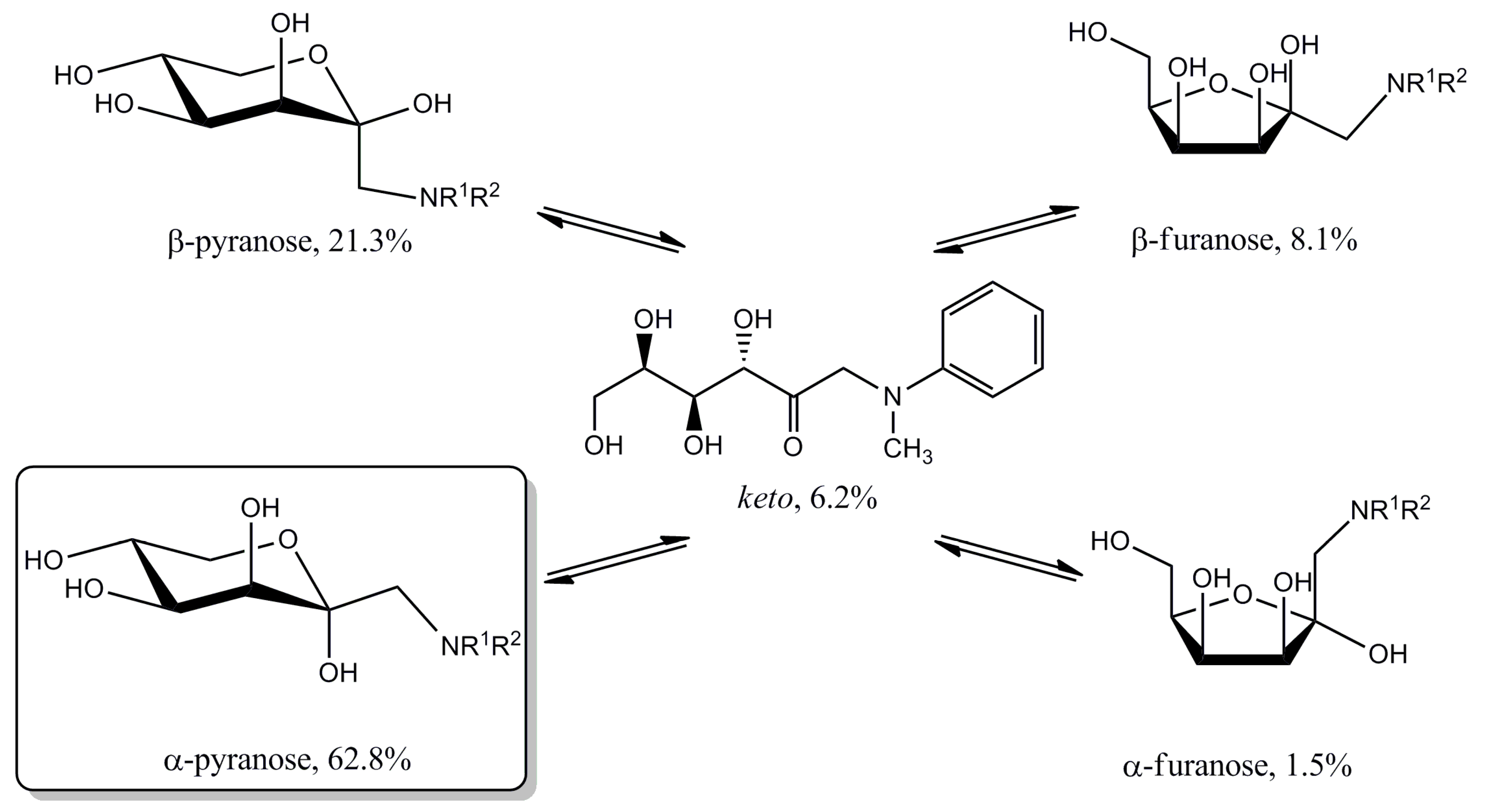
In aqueous solution, the compound establishes an equilibrium (Scheme 2), with the α-pyranose being a predominant anomeric form, followed by the β-pyranose, β-furanose, and α-furanose anomers, as would be expected for a tagatose derivative (Table 1). The predominance of α-d-tagatopyranose is expected since the pyranoid structure is preferred for reducing carbohydrates in water, while the equatorial disposition of the hydroxyl groups and the anomeric effect favor the α-anomer [14]. However, there is a notable (6.2%) proportion of the acyclic keto tautomer. Previously reported estimates for the population of the acyclic forms of tagatose or tagatosamines in aqueous solutions did not exceed 0.8% (Table 1). On the other hand, enhanced formation of the acyclic carbohydrate forms was observed in d-fructosamine derivatives with hydrophobic amino substituents that are in close proximity to the carbonyl group, and this phenomenon was attributed to the localized hydrophobic effects around the carbonyl group [13,15]. A similar, albeit smaller, effect was reported for 1-deoxy-tagatose (Table 1), as well.
2.2. Analysis of the X-ray Crystallographic Structure
The ORTEP drawing and atomic numbering are shown in Figure 2. The carbohydrate portion contains four chiral carbon atoms with the absolute configuration of the ring system (2S,3S,4S,5R) assigned on the basis of the known configuration for the starting d-galactose. The α-d-pyranose ring in crystalline d-tagatose-N-methylaniline exists in the 5C2 chair conformation, with puckering parameters [19] Q = 0.584(2) Å, θ = 1.7(2)°, and φ = 29(6)°. The related 5C2 conformations of α-d-tagatopyranose [16], N-(1-deoxy-α-d-tagatopyranos-1-yl)-amino acids [8], tagatosamines [10,11], and 1-deoxy-α-d-tagatopyranose [17,18] were found as the major or only structures in both the solution equilibria and the in crystalline state (Table 1). These conformations are, thus, very close to the sugar ring structure in N-(1-deoxy-α-d-tagatopyranos-1-yl)-N-methylaniline.
Bond distances and valence angles (Supplementary Table S4) in N-(1-deoxy-α-d-tagatopyranos-1-yl)-N-methylaniline compare well to the corresponding values found in α-d-tagatopyranose [16], 1-deoxy-α-d-tagatopyranose [17], 1-dibenzylamino-1-deoxy-α-d-tagatopyranose [10] and to average values for a number of crystalline pyranose structures [20]. The endocyclic torsions (Supplementary Table S5) do not differ significantly from the “standard” pyranoside torsions [20] with C–C–C–C(ring) at ~55°, C–C–C–O(ring)- at ~57.5°, and C–C–O–C- at ~62.5°. The exocyclic angles around the ring bonds are close to the “ideal” 60° or 180° as well.
2.3. Analysis of Molecular Packing
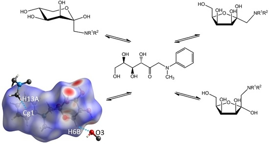
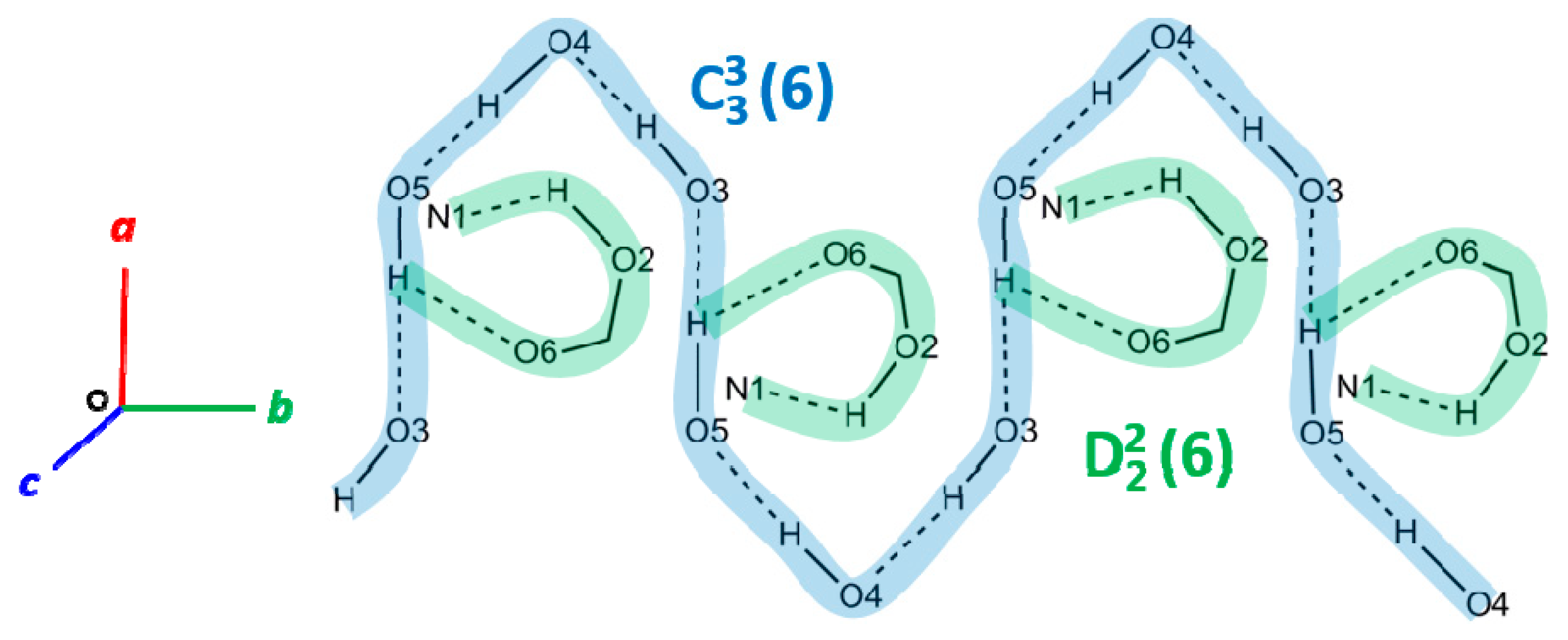
The crystal packing and intermolecular hydrogen bonds in the crystalline d-tagatose-N-methylaniline are shown in Figure 3. The intermolecular H-bonding is localized in antiparallel layers oriented along the crystallographic ac plane. Within the layers, a system of intermolecular heteroatom contacts (4 out of 5, see Table 2) is dominated by hydroxyl groups which form a system of homodromic infinite chains running along the b axis. A simplified graphical representation of the network is shown in Figure 4. There is a pattern of linked infinite C33(6) and short D22(6) chains (a topological notation according to Bernstein et al. [21]). Aromatic rings with the shortest distances between their centroids (5.410(2) Å) are located in planes with a small dihedral angle 16.7(1)°, but the rings are displaced by 3.5–4.4 Å relative to each other. Hence, π–π interactions do not appear to contribute to the crystal structure. Among candidates for the π–σ interactions, the shortest contact with the centroids is provided by the methyl group donors (Table 2).
2.4. Analysis of the Hirshfeld Surfaces
Taken together with our previous reports [13,15,22], this work completes a set of N-aryl derivatives of all four possible hexulosamines, including d-fructosamine, d-psicosamine, d-sorbosamine, and d-tagatosamine. There is one striking feature within this set that positions d-fructosamine apart from the rest of the hexulosamines. While the d-psicosamine, d-sorbosamine, and d-tagatosamine derivatives crystallize in the α-pyranose conformation, which is a dominant species in the amino sugar solutions, some N-aryl d-fructosamines tend to crystallize in a minor, acyclic conformation of the keto tautomer [13,15]. We have suggested previously that hydrophobic, non-polar interactions may play a significant role in the shaping of crystal packing forces for these molecules. One method to evaluate such interactions is to calculate and analyze the Hirshfeld surfaces, which are essentially weight functions that partition the crystal space between molecules contributing to the crystal structure [23].
A number of properties can be mapped over the Hirshfeld surface, including the electrostatic potential, topology, or interatomic contact distance functions. Figure 5 illustrates the latter, which we have calculated for the α-pyranose anomer of d-tagatose-N-methylaniline in crystal.
To determine the relative contribution of hydrophilic and hydrophobic intermolecular contacts to the packing forces in crystalline d-tagatose-N-methylaniline, the contact distance functions were plotted as 2D graphs, the so called “fingerprint plots” [24]. As shown in Figure 6, there is a large (64%) contribution of non-polar H · H contacts to the overall intermolecular interactions within the crystal. In addition, a significant proportion of the contacts (13%) are lowly polar C···H contacts. Some of these include interactions between hydrogen atoms and aromatic ring centroids, as shown in Figure 5. Only a small proportion of intermolecular contacts (21%) are due to polar O···H interactions, although not all of these qualify for conventional hydrogen bonds of the O···H–O or N · H–O types. A number of the contacts are of the O···H–C type (exemplified in Table 2 and Figure 5), which are not directional and may have a rather circumstantial significance. These numbers are in agreement with structures of crystalline N-aryl d-sorbosamine and d-psicosamine derivatives [22] (62.2% for H···H, 15% for C···H, and 22% for O···H contacts). In contrast, the acyclic conformation of crystal structures of N-aryl d-fructosamines [15] is characterized by significantly higher contributions from polar contacts (50–60% for H···H, 8–13% for C · H, and 23–32% for O···H contacts) to the packing forces.
3. Materials and Methods
3.1. Synthesis of 1-Deoxy-1-(N-methylphenyamino)-d-Tagatose
The compound was prepared following a procedure [13] previously employed for the synthesis of fructosamines. A mixture of 3.6 g (20 mmol) of d-galactose, 2.4 g (22 mmol) of N-methylaniline and 0.55 mL (6.3 mmol) of 3-mercaptopropionic acid catalyst in 8 mL of isopropanol was stirred for 16 h in screw capped glass vials at 87 °C. The reaction progress was followed by TLC. The purification steps included an ion-exchange on Amberlite IRN-77 (H+), with 0.2 M NH4OH in 50% EtOH as an eluent, and a flash filtration on a short silica column, using 5% MeOH in CH2Cl2 as an eluent. After evaporation of the eluates, crystallization of d-tagatose-N-methylaniline was aided by addition of acetone to the syrupy residue until turbidity was achieved. The crystalline material was filtered off, yielding 3.5 g (65%, based on starting d-galactose) off-white prisms, which provided monocrystals suitable for the X-ray diffraction studies. The major (α-pyranose anomer) peaks (ppm) in the 13C-NMR spectrum in 1:1 pyridine/D2O (Supplementary Figures S1 and S2) were 153.46 (C-7); 131.10 (C-9, C-11); 119.12 (C-10); 115.53 (C-8, C-12); 101.57 (C-2); 74.87 (C-4); 74.21 (C-3); 69.22 (C-5); 65.20 (C-6); 61.95 (C-1); 42.06 (C-13). See Table 1 for minor peak assignments in the spectrum and Figure 1 for the solid-state 13C-NMR spectrum. The major signals (ppm) in the 1H-NMR spectrum were 7.25 (t, 2H); 7.00 (d, 2H); 6.79 (t, 1H); 4.50 (m, 1H); 4.365 (d, 1H); 4.14 (m, 2H); 3.81 (d, 1H); 3.054 (s, 1H); 3.034 (s, 1H); 3.007 (s, 3H).
3.2. NMR
Solution 13C-NMR spectra (1:1 pyridine/D2O) were recorded at 62.9 MHz and 1H-NMR spectra (D2O) and were obtained at 250.1 MHz using a Bruker ARX250 instrument (Bruker BioSpin, Billerica, MA, USA), with TSPS as an internal standard. The solid-state 13C-NMR experiments were done at 75.5 MHz with Bruker DRX300 wide bore NMR spectrometer (Bruker BioSpin, Billerica, MA, USA) equipped with a 7 mm solid CP-MAS probe. Zirconia rotors and KEL-F caps were used. The 13C CP-MAS-TOSS spectrum of solid glycine was measured first to check the performance of the spectrometer, and the chemical shift of the carbonyl peak was set to 176.03 ppm. The 13C CP-MAS-TOSS spectrum of the subsequent sample was acquired under the same conditions and referenced to this external standard. All samples were measured at room temperature with a spin rate of 5 kHz, 1 ms of contact pulse, and a 4 s repetition delay.
3.3. X-ray Diffraction Studies
The diffraction data were collected using the Enraf-Nonius CAD4 instrument (Bruker AXS, Madison, WI, USA). Crystal data and experimental details of the crystallographic studies are given in Supplementary Table S1. Briefly, the crystal data for C13H19NO5 (M = 269.29 g/mol), which were used in all calculations, are as follows: orthorhombic, space group P212121 (no. 19), a = 6.5757(10) Å, b = 7.7698(10) Å, c = 25.0403(10) Å, V = 1279.4(3) Å3, Z = 4, T = 295 K, μ(CuKα) = 0.899 mm−1, Dcalc = 1.398 g/cm3, 1559 reflections measured (7.0° ≤ 2θ ≤ 149.1°), 1478 unique (Rint = 0.000, Rsigma = 0.029). The final R1 was 0.0290 (I > 2σ(I)) and wR2 was 0.0762 (all data). The crystal structure was solved with the direct methods program, SHELXS-97 [25], and refined by full-matrix least squares techniques with the SHELXL2017 [26] suite of programs, with the help of X-Seed [27]. Data were corrected for Lorentz and polarization effects and for absorption. Non-hydrogen atoms were refined with anisotropic thermal parameters. Hydroxyl hydrogen atoms were located in difference Fourier maps and were refined with fixed isotropic thermal parameters. The remaining H-atoms were placed at calculated positions and included in the refinement using a riding model. As a result of the unrealistic value obtained for the Flack absolute structure parameter, 0.0(2), the absolute configuration of the ring system (2S,3S,4S,5R) was assigned on the basis of the known configuration for starting d-galactose. The structure was visualized with Mercury [28].
3.4. Molecular Hirshfeld Surfaces Calculations
The Hirshfeld surfaces analyses were performed and plotted using CrystalExplorer v.17.5 software (University of Western Australia, http://hirshfeldsurface.net) [23].
4. Conclusions
In this work, we employed a classic Amadori rearrangement method for synthesis of a glycoaminoconjugate. Despite being nearly a century old, this simple protocol was useful in the preparation of a rare amino sugar structure, d-tagatosamine, from a readily available carbohydrate source, d-galactose.
d-Tagatose-N-methylaniline is conformationally unstable in solution and exists in at least five anomeric/tautomeric forms, the most abundant of which is α-pyranose. This anomer represents a single conformation of d-tagatose-N-methylaniline in the crystalline state. The crystal structure consists of interchanging “carbohydrate” and “aromatic” layers. The carbohydrate portions of the molecules participate in an extensive system of intermolecular hydrogen bonding, whereas the non-polar contacts provide a major contribution to the overall packing forces in the crystal.
Supplementary Materials
The following are available online, Figure S1: 13C-NMR spectrum of d-tagatose-N-methylaniline in 1:1 pyridine/D2O; Figure S2: An expanded carbohydrate region of 13C-NMR spectrum of d-tagatose-N-methylaniline in 1:1 pyridine/D2O showing resolution of signals for the anomeric/tautomeric forms; Table S1: Crystal data, data collection and structure refinement details; Table S2: Atomic Parameters x, y, z and B or Beq; Table S3: List of u(i,j) or U values × 100; Table S4: Bond distances (Å) and angles (°); Table S5: Torsion angles (°); checkCIF/PLATON report. The complete crystallographic data for the structural analysis have been deposited with the Cambridge Crystallographic Data Centre, CCDC # 831970. Copies of this information may be obtained free of charge from the Director, Cambridge Crystallographic Data Centre, 12 Union Road, Cambridge, CB2 1EZ, UK. (Fax: +44-1223-336033, e-mail: [email protected] or via: www.ccdc.cam.ac.uk).
Author Contributions
V.V.M. and T.P.M. conceived and designed the experiments; V.V.M. synthesized the compound; C.L.B. collected the diffraction data; V.V.M. and C.L.B. analyzed the data; V.V.M. wrote the paper. All authors read and approved the final manuscript.
Funding
This work was funded, in part, by the University of Missouri Agriculture Experiment Station Chemical Laboratories and the National Institute of Food and Agriculture, grant No. MO-HABC0002.
Acknowledgments
The authors thank Wei G. Wycoff for help in the NMR experiments.
Conflicts of Interest
The authors declare no conflict of interest.
References
- Deeth, H.C.; Lewis, M.J. High Temperature Processing of Milk and Milk Products; Wiley: Chichester, UK, 2017; 584p. [Google Scholar]
- Moeller, A.B. Chemical changes in ultra heat treated milk during storage. Prog. Food Nutr. Sci. 1981, 5, 357–368. [Google Scholar]
- Van Boekel, M.A.J.S. Effect of heating on Maillard reactions in milk. Food Chem. 1998, 62, 403–414. [Google Scholar] [CrossRef]
- Nursten, H. The Maillard Reaction: Chemistry, Biochemistry and Implications; The Royal Society of Chemistry: Cambridge, UK, 2005; 214p. [Google Scholar]
- Friedman, M. Biological effects of Maillard browning products that may affect acrylamide safety in food: Biological effects of Maillard products. Adv. Exp. Med. Biol. 2005, 561, 135–156. [Google Scholar] [PubMed]
- Denisevitch, T.V.; Semyonova-Kobzar, R.A.; Glinsky, V.V.; Mosin, V.V. The influence of synthetic aminoglycoconjugates on the aggregation ability and metastatic potential of tumor cells. Exp. Oncol. 1995, 17, 111–117. [Google Scholar]
- Mossine, V.V.; Mawhinney, T.P. 1-Amino-1-deoxy-d-fructose (“fructosamine”) and its derivatives. Adv. Carbohydr. Chem. Biochem. 2010, 64, 291–402. [Google Scholar] [PubMed]
- Kaufmann, M.; Meissner, P.M.; Pelke, D.; Mügge, C.; Kroh, L.W. Structure-reactivity relationship of Amadori rearrangement products compared to related ketoses. Carbohydr. Res. 2016, 428, 87–99. [Google Scholar] [CrossRef] [PubMed]
- Corzo-Martinez, M.; Hernandez-Hernandez, O.; Villamiel, M.; Rastall, R.A.; Moreno, F.J. In vitro bifidogenic effect of Maillard-type milk protein-galactose conjugates on the human intestinal microbiota. Int. Dairy J. 2013, 31, 127–131. [Google Scholar] [CrossRef]
- Harding, C.C.; Cowley, A.R.; Watkin, D.J.; Punzo, F.; Hotchkiss, D.; Fleet, G.W.J. 1-Amino-N,N-dibenzyl-1-deoxy-α-d-tagatopyranose methanol solvate. Acta Crystallogr. E 2005, 61, o1475–o1477. [Google Scholar] [CrossRef]
- Pérez, S.; López-Castro, A.; Márquez, R. Structure of 1-benzyl(methyl)amino-1-deoxy-α-d-lyxo-hexulopyranose. Acta Crystallogr. B 1978, 34, 2341–2344. [Google Scholar] [CrossRef]
- Mossine, V.V.; Glinsky, V.V.; Mawhinney, T.P. Antitumor effects of the early Maillard reaction products. In The Maillard Reaction: Interface between Aging, Nutrition and Metabolism; Thomas, M.C., Forbes, J., Eds.; Royal Society of Chemistry: Cambridge, UK, 2010; Volume 322, pp. 170–179. [Google Scholar]
- Mossine, V.V.; Barnes, C.L.; Chance, D.L.; Mawhinney, T.P. Stabilization of the acyclic tautomer in reducing carbohydrates. Angew. Chem. Int. Ed. 2009, 48, 5517–5520. [Google Scholar] [CrossRef] [PubMed]
- Ma, B.; Schaefer, H.F.; Allinger, N.L. Theoretical studies of the potential energy surfaces and compositions of the d-aldo- and d-ketohexoses. J. Am. Chem. Soc. 1998, 120, 3411–3422. [Google Scholar] [CrossRef]
- Mossine, V.V.; Barnes, C.L.; Mawhinney, T.P. Crystal structure of the acyclic form of 1-deoxy-1-(N-methyl-p-methoxyphenylamino)-d-fructose. Acta Crystallogr. E 2018, 74, 127–132. [Google Scholar] [CrossRef]
- Punzo, F.; Watkin, D.J.; Fleet, G.W.J. α-d-Tagatopyranose. Acta Crystallogr. E 2009, 65, o1393–o1394. [Google Scholar] [CrossRef] [PubMed]
- Jones, N.A.; Jenkinson, S.F.; Soengas, R.; Izumori, K.; Fleet, G.W.J.; Watkin, D.J. The concomitant crystallization of two polymorphs of 1-deoxy-α-d-tagatose. Acta Crystallogr. C 2007, 63, o7–o10. [Google Scholar] [CrossRef] [PubMed]
- Jones, N.A.; Jenkinson, S.F.; Soengas, R.; Fanefjord, M.; Wormald, M.R.; Dwek, R.A.; Kiran, G.P.; Devendar, R.; Takata, G.; Morimoto, K.; et al. Synthesis and NMR studies on the four diastereomeric 1-deoxy-d-keto-hexoses. Tetrahedron Asymmetry 2007, 18, 774–786. [Google Scholar] [CrossRef]
- Cremer, D.; Pople, J.A. General definition of ring puckering coordinates. J. Am. Chem. Soc. 1975, 97, 1354–1358. [Google Scholar] [CrossRef]
- Jeffrey, G.A.; Taylor, R. The application of molecular mechanics to the structures of carbohydrates. J. Comput. Chem. 1980, 1, 99–109. [Google Scholar] [CrossRef]
- Bernstein, J.; Davis, R.E.; Shimoni, L.; Chang, N.-L. Patterns in hydrogen bonding: Functionality and graph set analysis in crystals. Angew. Chem. Int. Ed. Engl. 1995, 34, 1555–1573. [Google Scholar] [CrossRef]
- Mossine, V.V.; Barnes, C.L.; Mawhinney, T.P. Molecular and crystal structure and the Hirshfeld surface analysis of 1-amino-1-deoxy-α-d-sorbopyranose and 1-amino-1-deoxy-α-d-psicopyranose (“d-sorbosamine” and “d-psicosamine”) derivatives. J. Mol. Struct. 2018, 1160, 73–79. [Google Scholar] [CrossRef]
- Spackman, M.A.; Jayatilaka, D. Hirshfeld surface analysis. CrystEngComm 2009, 11, 19–32. [Google Scholar] [CrossRef]
- Spackman, M.A.; McKinnon, J.J. Fingerprinting intermolecular interactions in molecular crystals. CrystEngComm 2002, 4, 378–392. [Google Scholar] [CrossRef]
- Sheldrick, G.M. A short history of SHELX. Acta Crystallogr. A 2008, 64, 112–122. [Google Scholar] [CrossRef] [PubMed]
- Sheldrick, G.M. Crystal structure refinement with SHELXL. Acta Crystallogr. C 2015, 71, 3–8. [Google Scholar] [CrossRef] [PubMed]
- Barbour, L.J. X-seed—A software tool for supramolecular crystallography. J. Supramol. Chem. 2003, 1, 189–191. [Google Scholar] [CrossRef]
- Macrae, C.F.; Bruno, I.J.; Chisholm, J.A.; Edgington, P.R.; McCabe, P.; Pidcock, E.; Rodriguez-Monge, L.; Taylor, R.; van de Streek, J.; Wood, P.A. Mercury CSD 2.0—New features for the visualization and investigation of crystal structures. J. Appl. Crystallogr. 2008, 41, 466–470. [Google Scholar] [CrossRef]
Scheme 1.
Synthesis of d-tagatose-N-methylaniline from d-galactose.

Scheme 2.
Tautomeric equilibrium in aqueous solution of d-tagatose-N-methylaniline.

Figure 1.
A solid-state 13C-NMR spectrum of crystalline d-tagatose-N-methylaniline.

Figure 2.
Atomic numbering and displacement ellipsoids at a 50% probability level for N-(1-deoxy-α-d-tagatopyranos-1-yl)-N-methylaniline. The intramolecular hydrogen bond is shown as a dotted line.
Figure 2.
Atomic numbering and displacement ellipsoids at a 50% probability level for N-(1-deoxy-α-d-tagatopyranos-1-yl)-N-methylaniline. The intramolecular hydrogen bond is shown as a dotted line.

Figure 3.
Crystal packing of N-(1-deoxy-α-d-tagatopyranos-1-yl)-N-methylaniline. Hydrogen bonds are shown as cyan dotted lines. Color coding for the crystallographic axes: red—a, green—b, blue—c.
Figure 3.
Crystal packing of N-(1-deoxy-α-d-tagatopyranos-1-yl)-N-methylaniline. Hydrogen bonds are shown as cyan dotted lines. Color coding for the crystallographic axes: red—a, green—b, blue—c.

Figure 4.
Hydrogen bonding pattern in crystal structure of N-(1-deoxy-α-d-tagatopyranos-1-yl)-N-methylaniline.
Figure 4.
Hydrogen bonding pattern in crystal structure of N-(1-deoxy-α-d-tagatopyranos-1-yl)-N-methylaniline.

Figure 5.
Views of the Hirshfeld surface for N-(1-deoxy-α-d-tagatopyranos-1-yl)-N-methylaniline, with the vdW-normalized contact distance function (dnorm) mapped in colors encoding intermolecular contacts, from closest (red) to the most distant (blue), in the range of −0.67 to 1.28. Shown are molecular fragments involved in the C–H···π interaction and the shortest C–H···O contact.

Figure 6.
(a) The full two-dimensional plot for the de and di contact distance functions and those delineated for the specific contacts: (b) O···H; (c) C···H; (d) H···H. The contributions of specific contacts to the intermolecular interactions are shown as percentages. Other contact type contributions not shown here: C···O (2.0%) and C···C (0.1%).


Table 1.
Tentative assignments of chemical shifts (ppm) in 13C-NMR spectra and percentage of d-tagatose-N-methylaniline (d-TagNMa) tautomers in 1:1 pyridine/D2O and in solid state at 25 °C. For comparison, the tautomeric compositions of d-tagatose [8,16], 1-deoxy-d-tagatose [17,18], d-tagatose-l-proline [8], and d-fructose-N-methylaniline (d-FruNMa) [13] are given.
| Carbon | α-Pyranose | β-Pyranose | α-Furanose | β-Furanose | Acyclic Keto | Crystalline State |
|---|---|---|---|---|---|---|
| C-1 | 61.95 | 61.00 | 59.84 | 58.80 | 62.68 | 62.7 |
| C-2 | 101.57 | 102.63 | 108.10 | 106.63 | 213.84 | 97.61 |
| C-3 | 74.21 | 72.24 | 79.16 | 74.42 | 76.62 | 71.50 |
| C-4 | 74.87 | 74.14 | 73.90 | 73.64 | 74.1 | 72.78 |
| C-5 | 69.22 | 67.39 | 81.64 | 82.30 | 73.06 | 69.12 |
| C-6 | 65.20 | 62.40 | 62.6 | 62.40 | 65.30 | 62.7 |
| % for d-TagNMa | 62.8 | 21.3 | 1.5 | 8.1 | 6.2 | 100 α-pyr |
| % for d-Tag | 78.4 | 14.6 | 1.8 | 4.7 | 0.5 | 100 α-pyr |
| % for 1-deoxy-d-Tag | 75 | 19 | 2 | 2 | 2 | 100 α-pyr |
| % for d-Tag-l-Pro | 70.2 | 19.4 | 5.6 | 4.1 | 0.8 | - |
| % for d-FruNMa | 2.3 | 51.2 | 4.6 | 31.9 | 10.1 | 100 acyclic |
Table 2.
Hydrogen bonding geometry (Å, °) in the crystal structure of N-(1-deoxy-α-d-tagatopyranos-1-yl)-N-methylaniline.
Table 2.
Hydrogen bonding geometry (Å, °) in the crystal structure of N-(1-deoxy-α-d-tagatopyranos-1-yl)-N-methylaniline.
| Contact | D–H | H···A | D···A | D–H···A | Symmetry Code 2 |
|---|---|---|---|---|---|
| O2–H···N1 | 0.79(2) | 2.05(2) | 2.816(1) | 164(2) | intra |
| O3–H···O4 | 0.88(3) | 2.03(3) | 2.878(1) | 162(2) | −1−x,−1/2+y,−1/2−z |
| O4–H···O5 | 0.75(3) | 2.43(3) | 3.114(1) | 153(3) | −1−x,−1/2+y,−1/2−z |
| O5–H···O3 | 0.75(3) | 2.53(3) | 2.885(1) | 111(2) | −2−x,1/2+y,−1/2−z |
| O5–H···O6 | 0.83(3) | 2.23(3) | 3.058(1) | 176(3) | −2−x,1/2+y,−1/2−z |
| Suspected contacts | |||||
| C6–HA···O2 | 0.97 | 2.44 | 2.771(2) | 100 | intra |
| C6–HB···O3 | 0.97 | 2.50 | 3.289(2) | 138 | −2-x,1/2+y,−1/2−z |
| C13–HB···O6 | 0.96 | 2.39 | 3.060(3) | 127 | intra |
| C13–HA···Cg1 1 | 0.96 | 2.93 | 3.859(3) | 162 | −1/2+x,−1/2−y,−z |
1 Cg1 refers to the benzene ring center of gravity. 2 Symmetry code refers to the acceptor atoms.
© 2018 by the authors. Licensee MDPI, Basel, Switzerland. This article is an open access article distributed under the terms and conditions of the Creative Commons Attribution (CC BY) license (http://creativecommons.org/licenses/by/4.0/).
Share and Cite
MDPI and ACS Style
Mossine, V.V.; Barnes, C.L.; Mawhinney, T.P. N-(1-Deoxy-α-d-tagatopyranos-1-yl)-N-methylaniline (“d-Tagatose-N-methylaniline”). Molbank 2018, 2018, M994. https://doi.org/10.3390/M994
AMA Style
Mossine VV, Barnes CL, Mawhinney TP. N-(1-Deoxy-α-d-tagatopyranos-1-yl)-N-methylaniline (“d-Tagatose-N-methylaniline”). Molbank. 2018; 2018(2):M994. https://doi.org/10.3390/M994
Chicago/Turabian StyleMossine, Valeri V., Charles L. Barnes, and Thomas P. Mawhinney. 2018. "N-(1-Deoxy-α-d-tagatopyranos-1-yl)-N-methylaniline (“d-Tagatose-N-methylaniline”)" Molbank 2018, no. 2: M994. https://doi.org/10.3390/M994
Note that from the first issue of 2016, this journal uses article numbers instead of page numbers. See further details here.