Review

Selected Case Studies on Fastidious Eukaryotic Microorganisms: Issues and Investigation Strategies

by
Benedetta Turchetti
1,*,
Annamaria Bevivino
2,
Patrizia Casella
3,
Claudia Coleine
4,
Giovanna E. Felis
5,
Carolina Elena Girometta
6,
Antonio Molino
3,
Iolanda Perugini
7,
Antonino Pollio
8,
Valeria Prigione
7,
Laura Selbmann
4,9,
Giovanna Cristina Varese
7 and
Pietro Buzzini
1
1
Department of Agricultural, Food and Environmental Sciences & Industrial Yeasts Collection DBVPG, University of Perugia, 06121 Perugia, PG, Italy
2
Department for Sustainability, ENEA—Italian National Agency for New Technologies, Energy and Sustainable Economic Development—ENEA Casaccia, 00123 Santa Maria di Galeria, RM, Italy
3
Department for Sustainability, ENEA—Italian National Agency for New Technologies, Energy and Sustainable Economic Development—ENEA Portici, 80055 Portici, NA, Italy
4
Department of Ecological and Biological Sciences, University of Tuscia, 01100 Viterbo, VT, Italy
5
Department of Biotechnology & VUCC-DBT Verona University Culture Collection, University of Verona, 37129 Verona, VR, Italy
6
Department of Earth and Environmental Sciences, University of Pavia, 27100 Pavia, PV, Italy
7
Department of Life Sciences and Systems Biology, Mycotheca Universitatis Taurinensis, University of Torino, 10124 Torino, TO, Italy
8
Department of Biology, University of Naples “Federico II”, 80138 Naples, NA, Italy
9
Italian National Antarctic Museum (MNA), Mycological Section, 16121 Genoa, GE, Italy
*
Author to whom correspondence should be addressed.
Diversity 2023, 15(7), 862; https://doi.org/10.3390/d15070862
Submission received: 12 May 2023 / Revised: 8 July 2023 / Accepted: 12 July 2023 / Published: 16 July 2023
(This article belongs to the Special Issue Feature Papers in Microbial Diversity and Culture Collections)

Abstract

:
The concept of fastidious microorganisms currently found in scientific literature is mainly related to the difficulty of isolating/culturing/preserving bacteria. Eukaryotes are investigated much less in this respect, although they represent a fundamental part of the microbial world. Furthermore, not only isolation, but also identification and culturing (in the perspective of long-term preservation) should be considered key aspects often impacting on the study of fastidious microorganisms, especially in terms of preservation in culture collections and biotechnological exploitation. The present review aimed to investigate the current state of the art on fastidious eukaryotes, with special emphasis on the efforts to improve their isolation, identification, culturing and long-term preservation in culture collections practices. A few case studies focused on some fastidious eukaryotic microorganisms (including possible customized solutions to overcome specific issues) are also presented: isolation and preservation of slow-growing fungi, culturing of Haematococcus lacustris, isolation of unialgal strains of Cyanidiophytina (Rhodophyta), identification of Metschnikowia pulcherrima clade yeasts, isolation and preservation of Pyricularia species, preservation of Halophytophtora spp.

1. Introduction

1.1. General Concept of Fastidious Microorganisms

Although an unequivocal definition of fastidious microorganisms is challenging to find in the literature, the conventional concept is often associated with the general difficulty of some microorganisms to be cultured under standard laboratory conditions [1]. Studies published to date reported, with great accuracy, the different meanings of the word “fastidious” in the microbial world. The number of articles (papers, book chapters and conference proceedings) indexed in Google Scholar (June 2023, using “with the exact phrase” in Google Scholar advanced search mask excluding “including citations”) and that exhibit specific contiguous couple of words is reported in Table 1. The word “fastidious” is largely associated with the word “bacteria” rather than the more generic one “microorganism” either in the title, keywords or text of the papers. Only 139 papers showed the presence of the couple of words “fastidious fungi” and only 12 showed “fastidious algae”. Unexpectedly, the combination “fastidious eukaryotes” was not present in publications indexed in Google Scholar. These results showed how the association of the word “fastidious” with “eukaryotes” and, in particular, “fungi” and “algae” is neglected in comparison to the association with “bacteria”. Using the combinations of “fastidious bacteria” + “pathogen” (present in the text but not necessarily next to “fastidious bacteria”) or “fastidious bacteria” + “medium” (present in the text but not necessarily next to “fastidious bacteria”), a slightly lower number of publications were found. The combination “fastidious bacteria” + “environment” is less occurring, which demonstrates how the clinical and culture issues related to fastidious bacteria have been much more studied to date than environmental ones. Moreover, the very low number of publications showing the simultaneous presence of the words “fastidious bacteria” + “culture collections” demonstrates the need to implement the studies on the difficulties encountered by collections to successfully approach the preservation of fastidious microorganisms in their ordinary practices.
Isolation and culturing are basic techniques of conventional microbiology. They are essential steps for the preservation of microbial diversity under viable form. Additionally, the correct identification of microbial strains at the species level is an essential condition for all microbiological studies. According to Kock’s postulates, both steps are crucial prerequisites in clinical applications (where successful treatment of infectious diseases needs the correct species assignment of each pathogenic organism) and in testing the efficacy of different drugs for defining the correct antimicrobial therapy [2,3,4]. Due to the fundamental importance of the diagnosis of infections through culturing of microorganisms, new (or optimized) culture media have recently been developed to increase the chances of success in the culturability of fastidious pathogens [1].
From a biotechnological point of view, the correct identification of microbial strains is required for their patent coverage or for defining their biosafety level, which is a prerequisite for their use in food and feed chains (e.g., see guidelines of the European Food and Safety Authority [5].
Finally, the correct identification of strains preserved inside culture collection should be the prerequisite for offering certified biological material to third parties.
Long-term preservation of microbial diversity maintaining high vitality is also a critical point. As the current techniques for preserving microbial biodiversity do not ensure the perpetuation of all sorts of microorganisms (including microbiomes), the risk of losing a more or less consistent part of ecologically and industrially relevant components of microbial community appears to be very high [6].
The erroneous idea that the issue related to fastidious microorganisms is a problem almost exclusively restricted to the difficulty of culturing bacteria (especially pathogenic species) under standard laboratory conditions may underestimate the true extent of the phenomenon.
Within this framework, the purpose of this review was to report on the state-of-the-art fastidious eukaryotes, with special emphasis on the efforts to improve their isolation, identification, culturing and long-term preservation in culture collections practices. Several case studies focused on different fastidious eukaryotic microorganisms describing possible customized solutions to overcome specific issues will also be presented.

1.2. General Concepts on Fastidious Bacteria

According to the general description reported above, the word “fastidious” is typically related to some bacteria, whose complex species-dependent nutritional and physiological requirements (e.g., the presence of definite nutrients and/or buffering agents, as well as the need to grow them under specific ranges of temperature, pH, O2/CO2 concentration, osmotic conditions, pressure, etc.) make their growth difficult using standard laboratory conditions [7,8]. Some ecological limitations, e.g., the involvement in metabolic networks, as well as the failure to interact with other (more versatile) microbial species, apparently indicate that some fastidious bacteria may have adapted their life to extreme habitats characterized by a low degree of biological diversity [7,8].
From an ecological point of view, fastidious bacteria should not be considered “weak” organisms, because they sometimes exhibit an efficient growth within their specific niches, characterized by peculiar conditions of temperature, acidity/alkalinity, oligotrophy/eutrophy and, sometimes, deficit of biological competitors [5]. The pathogenic bacterial species Treponema pallidum, which causes syphilis in humans, is challenging to be cultured under standard laboratory conditions, essentially because it probably needs to find specific conditions simulating its own natural habitat [7,8].
Most of the bacteria defined as “fastidious” are pathogens responsible for serious infections. Therefore, early detection and monitoring are important steps for increasing the chances of their easy eradication through the definition of correct pharmacological therapies [2,3,4,5].

1.3. General Concepts on Fastidious Eukaryotic Microorganisms

A number of fastidious organisms can also be found in the eukaryotic domain. Although a few protozoa are considered very fastidious in their growth requirements [9], most studies on fastidious eukaryotes have been focused on pathogenic fungi of clinical or phytopathological relevance, especially in relation to the efforts made by scientists worldwide to improve their isolation, culturing (e.g., the development of new culture media for both research and diagnosis), long-term cryopreservation and post-cryopreservation recovery [10,11,12,13,14,15,16,17].
Culturing of some fastidious fungi requires the supplementation of culture media with definite ingredients stimulating cell growth [18,19], e.g., the addition of oleic acid to conventional culture media significantly improves their efficiency in isolating the lipid-assimilating yeast Malassezia spp. [10]. As reported above, the difficult isolation of some fungal species from clinical and plant specimens even represents an obstacle capable of confuting Kock’s postulates. Ali et al. [20] bypassed this problem by suggesting the concept of sequence-based identification of fastidious fungal pathogens through the determination of the transcriptome sequence of both symptomatic and asymptomatic hosts [21]. Furthermore, while the development of culture-independent molecular techniques (i.e., next generation sequencing) have simplified since early 2000s, the detection of fastidious (or even not-yet-cultured) fungal species [22,23,24], their cultivation and preservation still remains in most cases an unsolved problem.

2. Case Studies

As reported above, overcoming the problems given by fastidious eukaryotic microorganisms is a matter that cannot be addressed in a generalized way, because each taxon often needs customized solutions. This is, of course, determined by the general variability existing in the microbial world, not only at the domain/kingdom level but also among different genera/species, or even at the intraspecific level due to the high degree of phenotypic variability frequently found within taxa characterized by a high level of cosmopolitanism.
Long-term cryopreservation of fastidious eukaryotic microorganisms frequently failed [25,26]. Although a few studies [11,17] suggested that some modifications of standard protocols can improve the efficiency of post-cryopreservation recovery of some fungal species, a few authors [12,13] underlined the extreme difficulty in establishing a unique protocol allowing the optimal recovery for all fastidious fungi. The same authors also suggested that culture collections should establish custom-made suitable protocols to enhance the efficiency of long-term cryopreservation of their own fastidious strains. Gleason et al. [13] reported that many fastidious eukaryotic microorganisms failing to survive (or poorly surviving) to long-term cryopreservation also exhibit a common limited tolerance to deviations from their preferred growth conditions. Consequently, the knowledge of such tolerance mechanisms could increase their chances of post-cryopreservation recovery [13].
In the following paragraphs, some case studies focusing on the difficulties encountered in the isolation, culture and long-term preservation (including recovery) of some fastidious eukaryotic microorganisms, as well as the description of taxon-specific solutions to mitigate (or even overcome) some specific problems, are reported.

2.1. Case-Study 1: Isolation and Long-Term Preservation of Slow-Growing Fungi from Extreme Environments

Slow fungal growth is often associated with the ability to survive under extreme conditions (high energy is required for adaptations). This ability is not always a mere consequence of the limitations imposed by extreme environments (e.g., both generalist or specialist fungi can exhibit the ability to slow their metabolic rates under unfavorable conditions), but may frequently be a true adaptive response (e.g., highly specialized guilds are often unable to reverse their growth rate upon restoration of the former physical–chemical conditions) [27,28]. Typical fastidious fungi exhibiting slow growth as a permanent feature (e.g., some obligate halophilic species, namely Wallemia ichthyophaga (Wallemiomycetes, Wallemiales,) or Hortaea werneckii (Dothideomycetes, Capnodiales), psychrophilic yeast-like species as Aureobasidium subglaciale (Dothideomycetes, Dothideales) or psychro-xerophilic rock-inhabiting Antarctic species as Cryomyces antarcticus (Dothideomycetes, incertae sedis) and Friedmanniomyces endolithicus (Dothideomycetes, Capnodiales) are specialist species which acquired the ability to express stress responses [28,29,30,31].
Inconveniences encountered in studying slow-growing fungi involve isolation, culturing and long-term preservation. Isolation may be difficult if specific procedures are not meticulously followed because slow-growing hyphae are easily outcompeted and overgrown by fast-growing (and more competitive) fungi, which are very difficult to eradicate, albeit still possible. A successful isolation of slow-growing fungi from environmental samples requires reproduction at the laboratory scale of a lot of physical-chemical stresses stimulating the development of a specific guild, as well as to prevent the growth of fast-growing fungi. The most recommended procedures basically imply:
(i)
Repeated washings (when possible) of the environmental samples with sterile water added with 0.1 Tween 20 to remove potential contaminants [32];
(ii)
Inoculation of a small portion (or dilution) of environmental samples on properly “poisoned” media (e.g., Rose–Bengal agar medium) to significantly deplete the growth of fast-growing fungi [33];
(iii)
Incubation for a long time (even for months) through reproduction of the stress to which slow-growing fungi are adapted (e.g., low temperatures, i.e., 4–15 °C, for the isolation of psychrophilic fungi);
(iv)
Continuous inspection of cultures to remove possible contaminants and even to transfer slow-growing colonies onto new media.
Some species (i.e., Antarctolichenia onofrii (Arthoniomycetes, Lichenostigmatales), Leucosporidium creatinivorum (Microbotryomycetes, Leucosporidiales)) characterized by peculiar nutritional requirements or by a close dependence from the metabolism of other microorganisms [34] may rapidly lose their viability if maintained in metabolically active forms. Therefore, customized long-term preservation protocols should be applied. A. onofrii can be successfully cryopreserved in 20% glycerol at −150 °C. Since highly costly processes are avoided under demanding conditions, these fungi basically reproduce asexually with minimal development of micronematous conidiophores in Ascomycota, which are often not observed at all. Sexual reproduction is instead often lost in some extremophilic fungi [35,36]. Therefore, the best option for long-term maintenance of slow-growing fungi is their cryopreservation at −80 °C or −150 °C.

2.2. Case-Study 2: Culturing of Haematococcus lacustris (Girod-Chantrans) Rostafinski

Haematococcus lacustris (Girod-Chantrans) Rostafinski (Chlorophycae, Volvocales), is a unicellular freshwater microalga belonging to the class Chlorophyceae. In some publications, H. lacustris was called also H. pluvialis [37]. Its life cycle includes two phases that depends on the physiology of the microalga:
(i)
Motile phase;
(ii)
Non motile phase.
Some unfavorable environmental conditions can lead to oxidative stress and to intensive light or nutrient deficiency and the formation of spores called haematocysts. This can determine an increased production of astaxanthin, which is accumulated during metabolic decline and transition towards haematocysts [38]. H. lacustris could be defined as a fastidious microalgal species due to its peculiar physiological and nutritional requirement, which make its culturing challenging under standard laboratory conditions. The fastidious growth of H. lacustris makes this species a good source of astaxanthin, but the difficulty lies in controlling the motile and non-motile growth phases and experimenting with the best unfavorable growth conditions for astaxanthin accumulation. As is well known, two-stage growth strategies are required to allow the growth of H. lacustris cells, which, under unfavorable conditions in the form of zoospores/aplanospores, increase astaxanthin content. However, one of the unresolved issues is high mortality of cells once grown under unfavorable conditions and astaxanthin accumulation [39]. H. lacustris is cultured on BG-11 (Blue-Green) medium containing a complex mixture of macro- and micronutrients [40,41]. The original composition of BG-11 medium included NaNO3 (1.5 g/L), K2HPO4 (0.04 g/L), MgSO4·7H2O (0.075 g/L), CaCl2 ·2H2O (0.036 g/L), citric acid (0.006 g/L), ferric ammonium citrate (0.006 g/L), EDTA disodium magnesium salt (0.001 g/L), Na2CO3 (0.02 g/L) and micronutrient solution.
Under normal growth conditions, cells are in motile phase. They are characterized by a high motility due to isocont flagella. The motile cell can divide by mitosis during asexual reproduction to generate sporangium that can contain a maximum of eight zoospores/aplonospore within the cell wall [42].
When grown under adverse conditions, motile cells lose the two flagella and they transform into a non-motile cell whose size can be variable and which is initially green in color with a low secondary carotenoid content. The non-motile cells are able to reproduce by asexual division producing up to 20–32 aplanospores within each sporangium. When the condition is favorable, the aplonospore can become motile cells. On the other hand, under stressing conditions (e.g., an excessive light intensity and/or salinity, oligotrophy, excessive oxidative stress), the zoospores begin to thicken the cell wall until the formation of an ultra-resistant coating (composed by a trilaminar sheath and by a secondary wall, resistant to acetolysis), which allows them to survive under extreme chemical–physical conditions. The haematocysts (i.e., aplanospores) begins to accumulate large amounts of secondary carotenoids (under the form of esters of fatty acids) instead of the photosynthetic apparatus shrinking, allowing the cell protection against damages caused by irradiation and oxidative stress [40,43,44].
Recent studies reported the culturing of H. lacustris in the BG-11 medium at 28 °C, pH 7.5–8.5 C, using the white light LED (4000 lux intensity, 100 mol/photons/m2/s) with an air flow rate of 300 mL/min by using Packed Bed Reactors (PBRs) characterized by a volume/surface ratio of 56.5 L/m2. Under such operative conditions, the motile phase was well-defined and the growth appeared consistent with several research studies [45]. Two different blue LED lights intensities (low and high: 55 µmol/s/m2 = 500 lux and 280 µmol/s/m2 = 2500 lux, respectively) were used to evaluate the effect of oxidative stress on H. lacustris cells. The effect of both high and low intensity of blue LED light in the terms of carotenoids and fatty acids content, including the switch to haematocysts form, is reported in Figure 1 [46,47].

2.3. Case-Study 3: Isolation of Unialgal Strains from Mixed Populations of Cyanidiophytina (Rhodophyta)

“If one has spent much time culturing and isolating blue-green algae, turning to Cyanidium is a refreshing experience. Here is an alga that lives in nature as a unialgal culture and that develops rapidly in enrichment cultures [48]”. This idyllic statement seems to be far from the definition of “fastidious” that characterizes all the eukaryotic microorganisms included in this review. The landmarking studies of Brock and coworkers opened an avenue of research on extremophilic algae belonging to the genus Cyanidium, but after several years, it became clear that, under the appearance of a unialgal culture, the majority of field “Cyanidium” populations hide an unexpected diversity. At present, four different genera of unicellular Cyanidiophytina (Rhodophyta) have been described: Cyanidium, Cyanidioschyzon, Cyanidiococcum and Galdieria [49]. The species Cyanidium caldarium, Galdieria sulphuraria (both round-shaped, with a thick cell wall) and Cyanidioschyzon merolae (club-shaped, without cell wall) often live together, forming mats all along hydrothermal springs worldwide characterized by temperatures and pH values ranging from 30 to 50 °C and from 0.8 to 2.0, respectively [50].
Cyanidiophytina samples collected in the field have been cultured under laboratory conditions using a restricted number of culture media, all based on the inorganic liquid medium acidified with H2S04 to pH 1.5–2.0 [51]. Other media elaborated afterward differed from the above media in several aspects, namely the concentration and the formulation of some macroelements, especially N and Fe, and the presence of several microelements, such as V, Co and Cu [51,52,53,54,55].
The simultaneous presence of two species in the “Cyanidium” field population was described for the first time by De Luca and Taddei [56]. They provisionally identified them as C. caldarium form A and C. caldarium form B, which could apparently be isolated thanks to their relevant ecophysiological differences: C. caldarium form A exhibited an obligate autotrophic aptitude, whereas C. caldarium form B can also switch its metabolism to a heterotrophic ability and showed a pronounced bleaching when grown in the dark. Accordingly, the isolation of form A (the “true” C. caldarium) from B (subsequently named G. sulphuraria) [57] appeared simple and effective. Unfortunately, despite its obligate autotrophic aptitude, form A can survive for prolonged periods in the dark.
To make this frame even more complicated, De Luca et al. [58] reported the ability of the new species Cyanidioschyzon merolae to grow intermingled with both forms (A and B) in samples collected in European, American and Asian hot springs. The possibility to isolate pure cultures of each species from the above mixed populations has been struggled with using the traditional method of serial dilutions [59]. Anyway, although most pure cultures have been obtained in this way, recent developments of metagenomic techniques revealed that many of these isolates still contain traces of the other Cyanidiales species. Del Mondo [60] proposed a cytofluorimetric and sorting analysis to discriminate among these species and designed novel species-specific primers to confirm the identity of the sorted strains by polymerase chain reaction. Compared to the traditional time-consuming isolating techniques, this approach resulted relatively fast, but required facilities (both instruments and skilled personnel) that could not be available in any laboratory. Therefore, strains deposited in different culture collections should be checked using this approach to obtain pure Cyanidiales isolates. The case of the genus Cyanidium (and related genera) is probably not unpreceded. Algal collections, therefore, represent an untapped source of hidden diversity that deserve more attention and efforts to be fully explored.

2.4. Case-Study 4: Identification of Yeasts of the Metschnikowia pulcherrima Clade

Metschnikowia is an ascomycetous yeast genus including either pathogenic or biotech-related species [61,62,63]. Among them, Metschnikowia pulcherrima is certainly the most exploited species due to its eclectic biotechnological potential [64,65]. Taxonomically speaking, the family Metschnikowiaceae belongs to order Serinales, while class is either uncertain in the subphylum Saccharomycotina (e.g., in Mycobank, taxon # 3147) or, more recently, it has been included into class Pichiomycetes on the basis of phylogenomic analysis [66] following the phylogenomic approach proposed for fungi [67]. In addition to the diverse class-level placements over time, the assignment at the species level is also challenging, and directly impacts the reliability of biological material provided by culture collection. Indeed, the scarcity of typical morphological and physiological features and the limited applicability of the standard molecular markers for species belonging to the M. pulcherrima clade (i.e., M. andauensis, M. citriensis, M. fructicola, M. leonuri, M. persimmonesis, M. pulcherrima, M. rubicola, M. shanxiensis, M. sinensis, M. zizyphicola and M. chrysoperlae) due to the presence of multiple copies of the primary (e.g., ITS segments and D1/D2 domain of 26S rRNA) and secondary (e.g., RPB2, TEF1α, ACT1 and EF2) barcodes, as well as their intragenomic sequence heterogeneity (comparable with or higher than the inter-species diversity), make clearly identifying Metschnikowia species extremely difficult (sometimes impossible) [61,68,69,70]. A recent study [71] revealed that all possible phylogenetic markers were present in multiple copies in the genome of M. pulcherrima strains, and the different copies exhibited a relatively high intragenomic variability. Therefore, these markers appear to fail when used for species identification within the M. pulcherrima clade. Due to the above-described limitations, Sipiczki [70] recently concluded that species of the M. pulcherrima clade cannot be, at present, distinguished from each other by the criteria of any of the phenotypic, phylogenetic and biological species concepts.
The whole-genome sequencing (including the free access to genome data) recently assumed increasing relevance as a current taxonomy tool for fungal identification [72,73,74,75]. Phylogenomic analysis may resolve problems related to the species identification, but this approach is still scarcely used in yeast taxonomy (especially if compared with bacterial one) and the limited availability of yeast genome sequences so far deposited in public databases appear to be the main bottleneck. Actually, only seven genomes of strains of the M. pulcherrima clade are publicly available [69,76,77,78,79]. Further data and the wider dissemination of genomic analysis could allow new insights into this complex species identification in the future.
As for cryopreservation, Metschnikowia pulcherrima-related strains are not particularly difficult to maintain in standard conditions of glycerol 25% (v/v) at −80 °C, while a study on a single strain showed that culture age and density, as well as type and concentration of protective agents, are relevant elements for a successful freeze-drying, a useful technique also for possible subsequent applications [80].

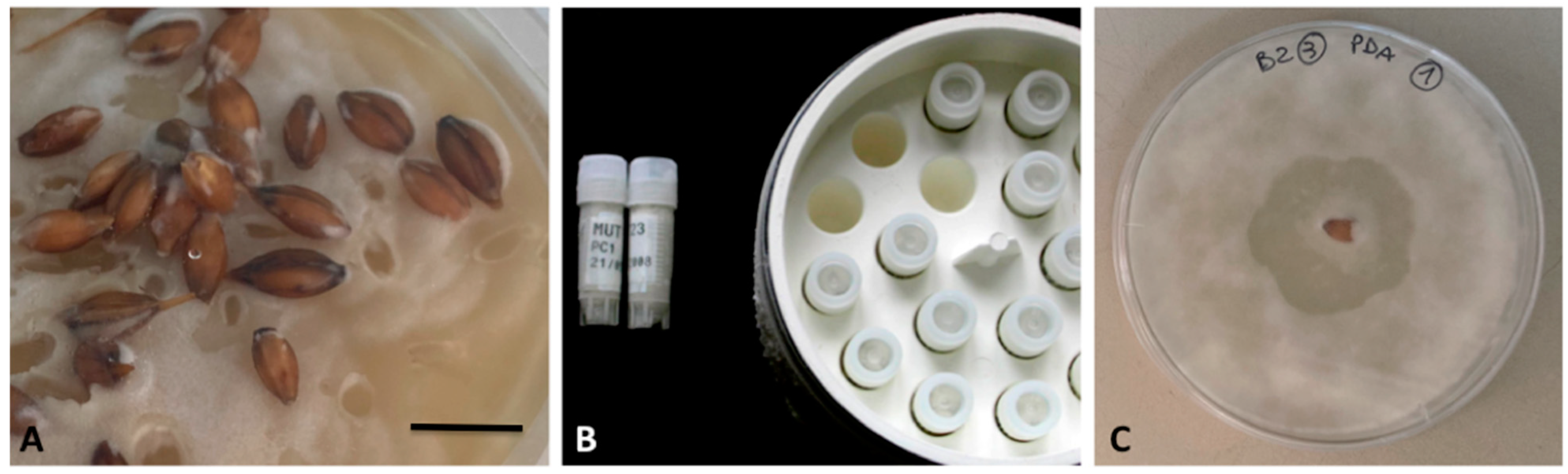
2.5. Case-Study 5: Isolation and Preservation of Pyricularia Species

Rice blast is the major rice disease [81] caused by fungi of the genus Pyricularia (Sordariomycetes, Magnaporthales) belonging to the Pyricularia grisea complex [82,83]. This species complex provisionally accommodates the problematic status of P. grisea sensu stricto and Pyricularia oryzae.
Culture collections of Pyricularia species are fundamental for studying their inter- and intraspecific diversity, taxonomy and phylogeny [84]. Host infection caused by P. oryzae is neither easily inducible and reproducible in the greenhouse, nor in mesocosm, outside of standard conditions of temperature and humidity [85]. Accordingly, conservation in pure culture should be preferred. Anyhow, Pyricularia strains are difficult to isolate and to preserve as pure cultures, due to their low and short-termed survival rate under laboratory standard media, at least under the form of monosporic cultures.
Pyricularia species exhibit a hemibiotrophic aptitude, which is strongly shifted towards biotrophism [86,87]. Consequently, the isolation of strains is very difficult. A protocol reported by Valent et al. [88] still constitutes the “gold standard” of successful methods for long-term preservation, despite it only relying on sterile mycelium instead of conidia. Conidia are hardly produced in culture, although this phenomenon is probably strain-dependent [89,90].
In addition to the possible presence of microbial contaminants, the need of having monosporic cultures (resulting from the germination of a single macroconidium) is one more issue to be taken into consideration. Even in pure and (expectedly) haploid cultures, mycelia from different individuals may be casually mixed in the same sample. Therefore, the simultaneous presence of different individuals may finally induce parasexual phenomena and increase the difficulty in the characterization of the single strain. Monosporic culture should be consequently preferred as a way to certify strain quality. The protocol for obtaining monosporic cultures can be summarized as follows:
(i)
Storage of portions of infected plants at −20 °C;
(ii)
Washing of samples in water [91], disinfection by 3% NaClO, re-washing in sterile water and plate spreading in tap water agar (TWA) solid medium as a humid chamber [92,93];
(iii)
Incubation of unsealed plates at room temperature and under light;
(iv)
Periodical checking of plates to detect the presence of conidiophores bearing macroconidia [94];
(v)
Isolation of mycelia in potato dextrose agar (PDA) solid medium for the production of macroconidia;
(vi)
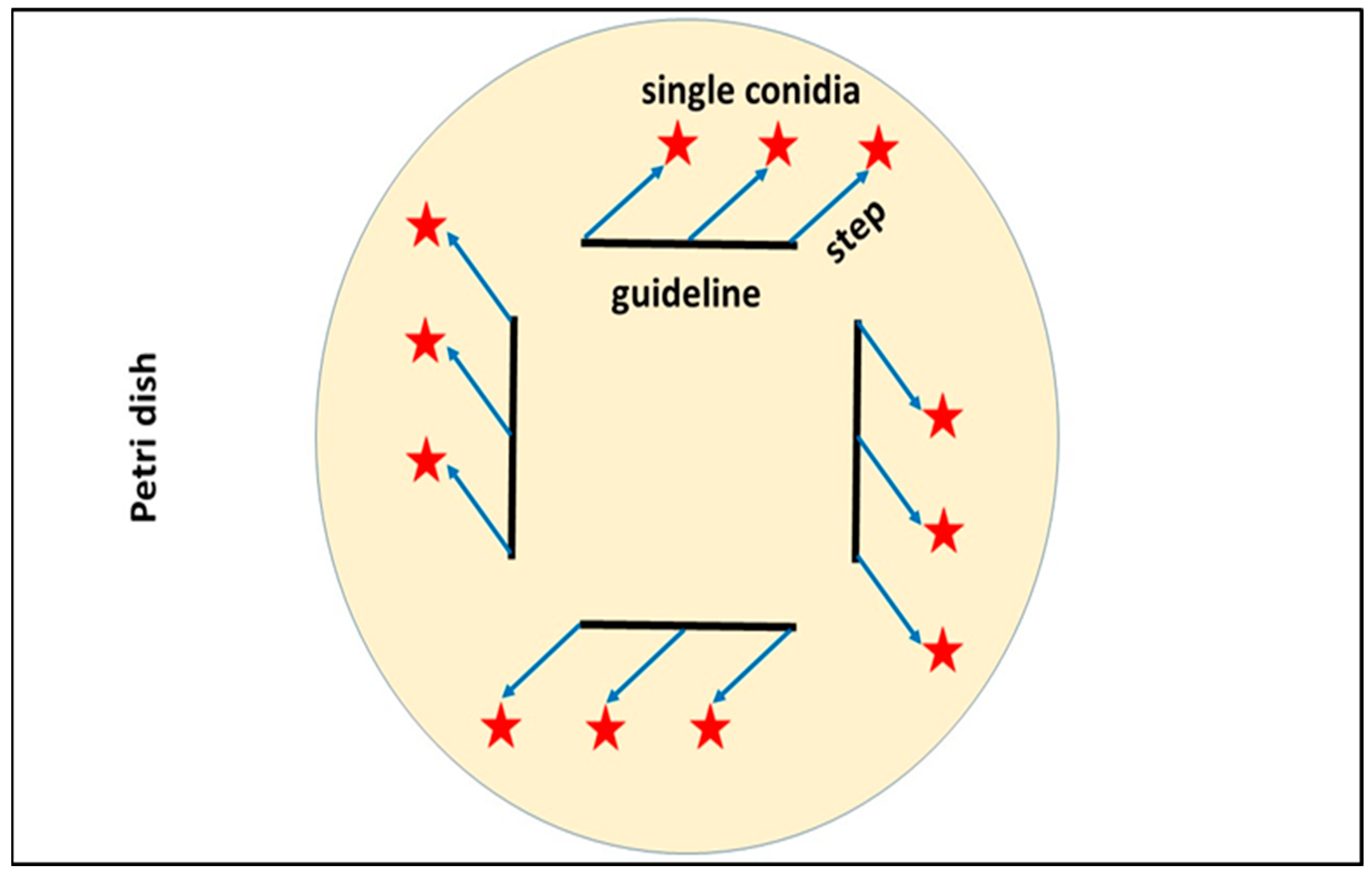
Transfer of single macroconidia in TWA solid medium; spatial set to obtain monosporic cultures is shown in Figure 2;
(vii)
Checking of conidia germination after 24 h;
(viii)
Transfer of new colonies in modified rice–meal–yeast extract agar (RYA) solid medium [84], where colonization typically takes 15–25 days to give ripe conidiophores.
Figure 2. Basic scheme for monosporic cultures of Pyricularia species. Guidelines are cut into the medium to facilitate the equidistant inoculation of conidia as well as to easily track them when checking the early stages of germination; stars represent single macroconidia inoculated.
Diversity 15 00862 g002
RYA supports short-term conservation only. Paper discs were aseptically placed onto the colony mat grown for 14 days on RYA at room temperature. After full mycelial colonization, discs were aseptically drawn from the plate, dried at 37 °C for 48 h, and stored at −20 °C [88]. Periodical inocula in fresh media and viability check on PDA or RYA are suggested.

2.6. Case-Study 6: Preservation of Halophytophtora spp. (Peronosporomycetes) from Marine Environment

Halophytophthora is a sister genus of Phytophthora (Peronosporomycetes, Peronosporales) mainly found in marine, mangrove and estuarine habitats. Although its saprotrophic aptitude was considered an established evidence for decades [95,96], some studies suggested that this genus might be involved in the widespread decline of the seagrass Zostera marina [97,98]. The long-term preservation protocols for pathogenic Peronosporomycetes mainly include cryopreservation, storage under mineral or paraffin oil, or storage in sterile water at room temperature [16]. However, the viability and/or genetic stability of these organisms are generally difficult to maintain for long periods of time [99]. Indeed, a number of marine Halophytophthora strains cannot be maintained in conditions of active growth for a long time, nor can they be preserved using standard long-term cryopreservation protocols.
A recent study on the isolation of Halophytophthora strains revealed that, after the first sub-culturing on both corn meal agar (CMA) supplemented with pimaricin, ampicillin and rifampicin, and PDA, about 40% of isolates were found to be no longer viable [90]. The protocol for improving long-term cryopreservation of Halophytophthora strains can be summarized as follows:
(i)
Collection of 2 mm mycelium plugs from colonies pre-grown on PDA in a 10% glycerol solution;
(ii)
Fast controlled with the glycerol solution;
(iii)
As an alternative, freezing (−1 °C/minute up to −80 °C and then transfer at −152 °C) of the material collected in the cryovials direct submersion of colonies in 10% glycerol solution followed by an overnight incubation at 5 °C before freezing (to overcome the difficulties encountered in collecting mycelium plugs).
Post cryopreservation recovery proved to fail in some of these Peronosporomycetes, with a survival rate of less than 10%. To overcome this problem, the following protocol could be suggested (Figure 3):
(i)
Inoculum of H. lusitanica strains on PDA plates and incubation at their optimal temperature for growth (15 °C) for two weeks;
(ii)
Addition of sterile wheat grains (about 30 for each strain) to the surface of actively growing colonies and incubation at 15 °C for about 3 weeks;
(iii)
Transfer of wheat grains colonized by H. lusitanica strains into cryotubes containing 10% glycerol aqueous solution;
(iv)
Six months cryopreservation at both −80 °C and −152 °C.
An excellent post cryopreservation recovery (survival rate = 100%) was observed.
Figure 3. (A) wheat grains colonized by Halophytophthora lusitanica (bar, 10 mm); (B) cryovials containing grains; (C) actively growing colony after revitalization (plate diameter, 90 mm).
Diversity 15 00862 g003

3. Conclusions

Although little considered in the current literature, fastidious eukaryotes may represent a problem in the routine work inside worldwide microbiological laboratories. This aspect is much more remarkable in culture collections where a large diversity of species (needing sometimes customized techniques of cultivation, identification, long-term preservation and revitalization) is preserved [100].
The present review underlined that using standard models and protocols to overcome the above issues is challenging because the single genus/species exhibits peculiarities limiting any generalization. On the other hand, the continuous research in this field and the publication of new successful techniques (or the upgrade of the existing ones) are crucial to share the knowledge suggesting similar solutions to the global scientific body.
In recent years, with the development of cheaper and simpler genome sequencing, metagenomic and proteomic studies, the understanding of microbial community structure, microbial guilts, and also peculiar needs of single species, became possible also without any isolation (culture independent molecular approach). Recent studies revealed that over >90% of eukaryotic diversity is not yet cultured [101,102], although it is recognized to be responsible of ecological, clinical and industrial implications. Along with the development of “OMICS” techniques, the importance of isolation, identification, cultivation and long-term preservation in culture collections of eukaryotes (including fastidious ones) should always be a clear landmark for all microbial studies through the development of new and customized tools.

Author Contributions

Conceptualization, B.T. and P.B.; investigation, all authors; writing—original draft preparation, all authors; writing—review and editing, B.T. and P.B. All authors have read and agreed to the published version of the manuscript.

Funding

This research received no external funding.

Institutional Review Board Statement

Not applicable.

Data Availability Statement

No new data were created.

Acknowledgments

The authors thank the European Commission—NextGenerationEU, Project “Strengthening the MIRRI Italian Research Infrastructure for Sustainable Bioscience and Bioeconomy”, code n. IR0000005 for the support. The authors are also grateful to Anna Maria Picco, University of Pavia, and to Marinella Rodolfi, University of Pavia, for technical support. The authors thank Teresa Emma Cutrona for English revision.

Conflicts of Interest

The authors declare no conflict of interest. The funders had no role in the design of the study; in the collection, analyses, or interpretation of data; in the writing of the manuscript; or in the decision to publish the results.

References

  1. Mobed, A.; Baradaran, B.; de la Guardia, M.; Agazadeh, M.; Hasanzadeh, M.; Rezaee, M.A.; Mosafer, J.; Mokhtarzadeh, A.; Hamblin, M.R. Advances in detection of fastidious bacteria: From microscopic observation to molecular biosensors. Trends Anal. Chem. 2019, 113, 157–171. [Google Scholar] [CrossRef]
  2. Kamagata, Y.; Tamaki, H. Culturing of uncultured fastidious microbes. Microbes Environ. 2005, 20, 85–91. [Google Scholar] [CrossRef] [Green Version]
  3. Fournier, P.E.; Dubourg, G.; Raoult, D. Clinical detection and characterization of bacterial pathogens in the genomics era. Genome Med. 2014, 6, 114. [Google Scholar] [CrossRef] [Green Version]
  4. Zarecki, R.; Oberhardt, M.A.; Reshef, L.; Gophna, U.; Ruppin, E. A novel nutritional predictor links microbial fastidiousness with lowered ubiquity, growth rate, and cooperativeness. PLoS Comput. Biol. 2014, 10, e1003726. [Google Scholar] [CrossRef] [Green Version]
  5. Available online: https://www.efsa.europa.eu/en (accessed on 3 March 2023).
  6. Prakash, O.; Nimonkar, Y.; Desai, D. A recent overview of microbes and microbiome preservation. Indian J. Microbiol. 2020, 60, 297–309. [Google Scholar] [CrossRef] [PubMed]
  7. Doern, G.V. Detection of selected fastidious bacteria. Clin. Infect. Dis. 2000, 30, 166–173. [Google Scholar] [CrossRef] [PubMed] [Green Version]
  8. Rishmawi, N.; Ghneim, R.; Kattan, R.; Ghneim, R.; Zoughbi, M.; Abu-Diab, A.; Turkuman, S.; Dauodi, R.; Shomali, I.; Issa, A.R.; et al. Survival of fastidious and nonfastidious aerobic bacteria in three bacterial transport swab systems. J. Clin. Microbiol. 2007, 45, 1278–1283. [Google Scholar] [CrossRef] [Green Version]
  9. Visvesvara, G.S.; Garcia, L.S. Culture of protozoan parasites. Clin. Microbiol. Rev. 2002, 15, 327–328. [Google Scholar] [CrossRef] [Green Version]
  10. Mayser, P.; Haze, P.; Papavassilis, C.; Pickel, M.; Gfuender, K.; Guého, E. Differentiation of Malassezia species: Selectivity of Cremophor EL, castor oil and ricinoleic acid for M. furfur. British J. Dermatol. 1997, 137, 208–213. [Google Scholar] [CrossRef]
  11. Kitamoto, Y.; Suzuki, A.; Shimada, S.; Yamanaka, K. A new method for the preservation of fungus stock cultures by deep freezing. Mycoscience 2002, 43, e143–e149. [Google Scholar] [CrossRef]
  12. Bardin, M.; Suliman, M.E.; Sage-Palloix, A.M.; Mohamed, Y.F.; Nicot, P.C. Inoculum production and long-term conservation methods for cucurbits and tomato powdery mildews. Mycol. Res. 2007, 111, 740–747. [Google Scholar] [CrossRef]
  13. Gleason, F.H.; Mozley-Standridge, S.E.; Porter, D.; Boyle, D.G.; Hyatt, A.D. Preservation of Chytridiomycota in culture collections. Mycol. Res. 2007, 111, 129–136. [Google Scholar] [CrossRef]
  14. Kocharin, K.; Wongsa, P. Semi-defined medium for in vitro culturing of the fastidious insect pathogenic fungus Cordyceps unilateralis. Mycopathologia 2006, 161, 255–260. [Google Scholar] [CrossRef]
  15. Huffnagle, G.B.; Noverr, M.C. The emerging world of the fungal microbiome. Trends Microbiol. 2013, 21, 334–341. [Google Scholar] [CrossRef] [Green Version]
  16. Bittar, F.; Gouriet, F.; Khelaifia, S.; Raoult, D.; Ranque, S. FastFung: A novel medium for the culture and isolation of fastidious fungal species from clinical samples. J. Microbiol. Methods 2021, 180, 106108. [Google Scholar] [CrossRef] [PubMed]
  17. Houseknecht, J.L.; Suh, S.O.; Zhou, J.J. Viability of fastidious Phytophthora following different cryopreservation treatments. Fungal Biol. 2012, 116, 1081–1089. [Google Scholar] [CrossRef]
  18. Ben Salah, I.; Makni, F.; Cheikhrouhou, F.; Neji, S.; Sellami, H.; Ayadi, A. Malassezia species: Pathology, isolation and identification media. J. Mycol. Med. 2010, 20, 53–60. [Google Scholar] [CrossRef]
  19. Shah, A.; Koticha, A.; Ubale, M.; Wanjare, S.; Mehta, P.; Khopkar, U. Identification and speciation of Malassezia in patients clinically suspected of having Pityriasis versicolor. Indian J. Dermatol. 2013, 58, 239. [Google Scholar] [CrossRef] [PubMed]
  20. Ali, S.S.; Asman, A.; Shao, J.; Firmansyah, A.P.; Susilo, A.W.; Rosmana, A.; McMahon, P.; Junaid, M.; Guest, D.; Kheng, T.Y.; et al. Draft genome sequence of fastidious pathogen Ceratobasidium theobromae, which causes vascular-streak dieback in Theobroma cacao. Fungal Biol. Biotechnol. 2019, 30, 14. [Google Scholar] [CrossRef]
  21. Fredericks, D.; Relman, D.A. Sequence-based identification of microbial pathogens: A reconsideration of Koch’s postulates. Clin. Microbiol. Rev. 1996, 9, 18–33. [Google Scholar] [CrossRef]
  22. Ott, S.J.; Kühbacher, T.; Musfeldt, M.; Rosenstiel, P.; Hellmig, S.; Rehman, A.; Drews, O.; Weichert, W.; Timmis, K.N.; Schreiber, S. Fungi and inflammatory bowel diseases: Alterations of composition and diversity. Scand. J. Gastroenterol. 2008, 43, 831–841. [Google Scholar] [CrossRef] [PubMed]
  23. Scanlan, P.D.; Marchesi, J.R. Micro-eukaryotic diversity of the human distal gut microbiota: Qualitative assessment using culture-dependent and -independent analysis of faeces. ISME J. 2008, 2, 1183–1193. [Google Scholar] [CrossRef] [PubMed]
  24. Hamad, I.; Sokhna, C.; Raoult, D.; Bittar, F. Molecular detection of eukaryotes in a single human stool sample from Senegal. PLoS ONE 2012, 7, e40888. [Google Scholar] [CrossRef] [PubMed] [Green Version]
  25. Humber, R.A. Special considerations for operating a culture collection of fastidious fungal pathogens. J. Ind. Microbiol. 1994, 13, 195–196. [Google Scholar] [CrossRef]
  26. Smith, D.; Onions, A.H.S. The Preservation and Maintenance of Living Fungi, 2nd ed.; CAB International: Wallingford, UK, 1994. [Google Scholar]
  27. Gostinčar, C.; Grube, M.; De Hoog, S.; Zalar, P.; Gunde-Cimerman, N. Extremotolerance in fungi: Evolution on the edge. FEMS Microbiol. Ecol. 2009, 71, 2–11. [Google Scholar] [CrossRef] [Green Version]
  28. Gostinčar, C.; Zalar, P.; Gunde-Cimerman, N. No need for speed: Slow development of fungi in extreme environments. Fungal Biol. Rev. 2022, 39, 1–14. [Google Scholar] [CrossRef]
  29. Turk, M.; Gostinčar, C. Glycerol metabolism genes in Aureobasidium pullulans and Aureobasidium subglaciale. Fungal Biol. 2018, 122, 63–73. [Google Scholar] [CrossRef]
  30. Zalar, P.; Zupančič, J.; Gostinčar, C.; Zajc, J.; de Hoog, G.S.; De Leo, F.; Azua-Bustos, A.; Gunde-Cimerman, N. The extremely halotolerant black yeast Hortaea werneckii—A model for intraspecific hybridization in clonal fungi. IMA Fungus 2019, 10, 10. [Google Scholar] [CrossRef] [Green Version]
  31. Coleine, C.; Stajich, J.E.; Selbmann, L. Fungi are key players in extreme ecosystems. Trends Ecol. Evol. 2022, 37, 517–528. [Google Scholar] [CrossRef]
  32. Selbmann, L.; Isola, D.; Egidi, E.; Zucconi, L.; Gueidan, C.; De Hoog, G.S.; Onofri, S. Mountain tips as reservoirs for new rock-fungal entities: Saxomyces gen. nov. and four new species from the Alps. Fungal Divers. 2014, 65, 167–182. [Google Scholar] [CrossRef] [Green Version]
  33. Chander, A.M.; Singh, N.K.; Simpson, A.C.; Seuylemezian, A.; Mason, C.E.; Venkateswaran, K. Draft genome sequences of fungi isolated from Mars 2020 spacecraft assembly facilities. Microbiol. Resour. Announc. 2022, 1, e00464-22. [Google Scholar] [CrossRef] [PubMed]
  34. Muggia, L.; Coleine, C.; De Carolis, R.; Cometto, A.; Selbmann, L. Antarctolichenia onofrii gen. nov. sp. nov. from Antarctic endolithic communities untangles the evolution of rock-inhabiting and lichenized fungi in Arthoniomycetes. J. Fungi 2021, 7, 935. [Google Scholar] [CrossRef] [PubMed]
  35. De Hoog, G.S.; Beguin, H.; Batenburg-van de Vegte, W.H. Phaeotheca triangularis, a new meristematic black yeast from a humidifier. Antonie Leeuwenhoek 1997, 71, 289–295. [Google Scholar] [CrossRef] [PubMed]
  36. Sun, S.; Heitman, J. Is sex necessary? BMC Biol. 2011, 9, 56. [Google Scholar] [CrossRef] [Green Version]
  37. Nakada, T.; Ota, S. What is the correct name for the type of Haematococcus Flot. (Volvocales, Chlorophyceae)? Taxon 2016, 65, 343–348. [Google Scholar] [CrossRef]
  38. Chekanov, K. Diversity and Distribution of Carotenogenic Algae in Europe: A Review. Mar. Drugs 2023, 21, 108. [Google Scholar] [CrossRef]
  39. Li, F.; Cai, M.; Lin, M.; Huang, X.; Wang, J.; Ke, H.; Wang, C.; Zheng, X.; Cheng, D.; Yang, S. Enhanced biomass and astaxanthin production of Haematococcus pluvialis by a cell transformation strategy with optimized initial biomass density. Mar. Drugs 2020, 18, 341. [Google Scholar] [CrossRef]
  40. Cheng, J.; Li, K.; Zhu, Y.; Yang, W.; Zhou, J.; Cen, K. Transcriptome sequencing and metabolic pathways of astaxanthin accumulated in Haematococcus pluvialis mutant under 15% CO2. Bioresour. Technol. 2017, 228, 99–105. [Google Scholar] [CrossRef]
  41. Stanier, R.Y.; Kunisawa, R.; Mandel, M.C.B.G.; Cohen-Bazire, G. Purification and properties of unicellular blue-green algae (order Chroococcales). Bacteriol. Rev. 1971, 35, 171–205. [Google Scholar]
  42. Zhang, C.; Liu, J.; Zhang, L. Cell cycles and proliferation patterns in Haematococcus pluvialis. Chin. J. Oceanol. Limnol. 2017, 35, 1205–1211. [Google Scholar] [CrossRef]
  43. Molino, A.; Mehariya, S.; Iovine, A.; Larocca, V.; Di Sanzo, G.; Martino, M.; Casella, P.; Chianese, S.; Musmarra, D. Extraction of astaxanthin and lutein from microalga Haematococcus pluvialis in the red phase using CO₂ supercritical fluid extraction technology with ethanol as co-solvent. Mar. Drugs 2018, 16, 432. [Google Scholar] [CrossRef] [Green Version]
  44. Molino, A.; Rimauro, J.; Casella, P.; Cerbone, A.; Larocca, V.; Chianese, S.; Karatza, D.; Mehariya, S.; Ferraro, A.; Hristoforou, E.; et al. Extraction of astaxanthin from microalga Haematococcus pluvialis in red phase by using generally recognized as safe solvents and accelerated extraction. J. Biotechnol. 2018, 283, 51–61. [Google Scholar] [CrossRef] [PubMed]
  45. Oslan, S.N.H.; Shoparwe, N.F.; Yusoff, A.H.; Rahim, A.A.; Chang, C.S.; Tan, J.S.; Oslan, S.N.; Arumugam, K.; Ariff, A.B.; Sulaiman, A.Z.; et al. A review on Haematococcus pluvialis bioprocess optimization of green and red stage culture conditions for the production of natural astaxanthin. Biomolecules 2021, 11, 256. [Google Scholar] [CrossRef] [PubMed]
  46. Casella, P.; Rimauro, J.; Iovine, A.; Mehariya, S.; Musmarra, D.; Molino, A. Characterization of extracts from Haematococcus pluvialis red phase by using accelerated solvent extraction. Chem. Eng. Trans. 2019, 74, 1417–1422. [Google Scholar]
  47. Mehariya, S.; Sharma, N.; Iovine, A.; Casella, P.; Marino, T.; Larocca, V.; Molino, A.; Musmarra, D. An integrated strategy for nutraceuticals from Haematoccus pluvialis: From culturing to extraction. Antioxidants 2020, 9, 825. [Google Scholar] [CrossRef] [PubMed]
  48. Brock, T.D. Thermophilic Microorganisms and Life at High Temperatures; Springer: New York, NY, USA, 1978. [Google Scholar]
  49. Liu, S.L.; Chiang, Y.R.; Yoon, H.S.; Fu, H.Y. Comparative genome analysis reveals Cyanidiococcus gen. nov., a new extremophilic red algal genus sister to Cyanidioschyzon (Cyanidioschyzonaceae, Rhodophyta). J. Phycol. 2020, 56, 1428–1442. [Google Scholar] [CrossRef] [PubMed]
  50. Albertano, P.; Ciniglia, C.; Pinto, G.; Pollio, A. The taxonomic position of Cyanidium, Cyanidioschyzon and Galdieria: An update. Hydrobiologia 2000, 433, 137–143. [Google Scholar] [CrossRef]
  51. Allen, M.B. Studies with Cyanidium caldarium, an anomalously pigmented chlorophyte. Arch. Mikrobiol. 1959, 32, 270–277. [Google Scholar] [CrossRef]
  52. Ascione, R.; Southwick, W.; Fresco, J.R. Laboratory culturing of a thermophilic alga at high temperature. Science 1966, 153, 752–755. [Google Scholar] [CrossRef]
  53. Vona, V.; Di Martino Rigano, V.; Esposito, S.; Di Martino, C.; Rigano, C. Growth, photosynthesis, respiration, and intracellular free amino acid profiles in the unicellular alga Cyanidium caldarium. Effect of nutrient limitation and resupply. Physiol. Plant. 1992, 85, 652–658. [Google Scholar] [CrossRef]
  54. Minoda, A.; Sakagami, R.; Yagisawa, F.; Kuroiwa, T.; Tanaka, K. Improvement of culture conditions and evidence for nuclear transformation by homologous recombination in a red alga, Cyanidioschyzon merolae 10D. Plant Cell Physiol. 2004, 45, 667–671. [Google Scholar] [CrossRef] [PubMed] [Green Version]
  55. Ohnuma, M.; Yokoyama, T.; Inouye, T.; Sekine, Y.; Tanaka, K. Polyethylene glycol (PEG)-mediated transient gene expression in a red alga, Cyanidioschyzon merolae 10D. Plant Cell Physiol. 2008, 49, 117–120. [Google Scholar] [CrossRef]
  56. De Luca, P.; Taddei, R. Due alghe delle fumarole acide dei Campi Flegrei (Napoli): Cyanidium caldarium? Delpinoa 1970, 10, 79–89. [Google Scholar]
  57. Merola, A.; Castaldo, R.; De Luca, P.; Gambardella, R.; Musacchio, A.; Taddei, R. Revision of Cyanidium caldarium. Three species of acidophilic algae. Plant Biosyst. 1981, 115, 189–195. [Google Scholar] [CrossRef]
  58. De Luca, P.; Taddei, R.; Varano, L. Cyanidioschyzon merolae: A new alga of thermal acidic environments. Webbia 1978, 33, 37–44. [Google Scholar] [CrossRef]
  59. Blaize, J.F.; Suter, E.; Corbo, C.P. Serial Dilutions and Plating: Microbial Enumeration. JoVE-Protocol-1050. 2019. Available online: https://www.jove.com/v/10507/serial-dilutions-and-plating-microbial-enumeration (accessed on 3 March 2023).
  60. Del Mondo, A. Development of Non-Invasive Diagnostic Methods for Monitoring Biodeterioration of Monuments. Ph.D. Thesis, Doctorate School in Biology, University of Naples, Federico II, Naples, Italy, 2017; pp. 30–39. [Google Scholar]
  61. Lachance, M.A. Metschnikowia: Half tetrads, a regicide and the fountain of youth. Yeast 2016, 33, 563–574. [Google Scholar] [CrossRef]
  62. Turland, N.J.; Wiersema, J.H.; Barrie, F.R.; Greuter, W.; Hawksworth, D.L.; Herendeen, P.S.; Knapp, S.; Kusber, W.H.; Li, D.Z.; Marhold, K.; et al. International Code of Nomenclature for Algae, Fungi, and Plants (Shenzhen Code). In Proceedings of the Nineteenth International Botanical Congress, Shenzhen, China, 23–29 July 2017; [Regnum Vegetabile no. 159]. Koeltz Botanical Books: Glashütten, Germany, 2018. [Google Scholar]
  63. Jiang, H.; Bao, J.; Xing, Y.; Li, X.; Chen, Q. Comparative genomic analyses provide insight into the pathogenicity of Metschnikowia bicuspidata LNES0119. Front. Microbiol. 2022, 13, 939141. [Google Scholar] [CrossRef]
  64. Morata, A.; Loira, I.; Escott, C.; Del Fresno, J.M.; Bañuelos, M.A.; Suárez-Lepe, J.A. Applications of Metschnikowia pulcherrima in wine biotechnology. Fermentation 2019, 5, 63. [Google Scholar] [CrossRef] [Green Version]
  65. Vicente, J.; Ruiz, J.; Belda, I.; Benito-Vázquez, I.; Marquina, D.; Calderón, F.; Santos, A.; Benito, S. The genus Metschnikowia in enology. Microorganisms 2020, 8, 1038. [Google Scholar] [CrossRef]
  66. Groenewald, M.; Hittinger, C.; Bensch, K.; Opulente, D.; Shen, X.-X.; Li, Y.; Liu, C.; LaBella, A.; Zhou, X.; Limtong, S.; et al. A genome-informed higher rank classification of the biotechnologically important fungal subphylum Saccharomycotina. Stud. Mycol. 2023, 105, 1–22. [Google Scholar] [CrossRef]
  67. Li, Y.; Steenwyk, J.L.; Chang, Y.; Wang, Y.; James, T.Y.; Stajich, J.E.; Spatafora, J.W.; Groenewald, M.; Dunn, C.W.; Hittinger, C.T.; et al. A Genome-Scale Phylogeny of the Kingdom Fungi. Curr. Biol. 2021, 31, 1653–1665. [Google Scholar] [CrossRef] [PubMed]
  68. Sipiczki, M. Metschnikowia pulcherrima and related pulcherrimin-producing yeasts: Fuzzy species boundaries and complex antimicrobial antagonism. Microorganisms 2020, 8, 1029. [Google Scholar] [CrossRef] [PubMed]
  69. Sipiczki, M. When barcoding fails: Genome chimerisation (admixing) and reticulation obscure phylogenetic and taxonomic relationships. Mol. Ecol. Resour. 2022, 22, 1762–1785. [Google Scholar] [CrossRef] [PubMed]
  70. Sipiczki, M. Taxonomic revision of the pulcherrima clade of Metschnikowia (Fungi): Merger of species. Taxonomy 2022, 2, 107–123. [Google Scholar] [CrossRef]
  71. Troiano, E.; Larini, I.; Binati, R.L.; Gatto, V.; Torriani, S.; Buzzini, P.; Turchetti, B.; Salvetti, E.; Felis, G.E. Finding a correct species assignment for a Metschnikowia strain: Insights from the genome sequencing of strain DBT012. FEMS Yeast Res. 2023, 23, foad024. [Google Scholar] [CrossRef]
  72. Capella-Gutierrez, S.; Kauff, F.; Gabaldon, T. A phylogenomics approach for selecting robust sets of phylogenetic markers. Nucleic Acids Res. 2014, 42, 54. [Google Scholar] [CrossRef]
  73. Libkind, D.; Čadež, N.; Opulente, D.A.; Langdon, Q.K.; Rosa, C.A.; Sampaio, J.P.; Gonçalves, P.; Hittinger, C.T.; Lachance, M.A. Towards yeast taxogenomics: Lessons from novel species descriptions based on complete genome sequences. FEMS Yeast Res. 2020, 20, foaa042. [Google Scholar] [CrossRef]
  74. James, T.Y.; Kauff, F.; Schoch, C.L.; Matheny, P.B.; Hofstetter, V.; Cox, C.J.; Celio, G.; Gueidan, C.; Fraker, E.; Miadlikowska, J.; et al. Reconstructing the early evolution of Fungi using a six-gene phylogeny. Nature 2006, 443, 818–822. [Google Scholar] [CrossRef]
  75. Boekhout, T.; Aime, M.C.; Begerow, D.; Gabaldón, T.; Heitman, J.; Kemler, M.; Khayhan, K.; Lachance, M.A.; Louis, E.J.; Sun, S.; et al. The evolving species concepts used for yeasts: From phenotypes and genomes to speciation networks. Fungal Div. 2021, 109, 27–55. [Google Scholar] [CrossRef]
  76. Piombo, E.; Sela, N.; Wisniewski, M.; Hoffmann, M.; Gullino, M.L.; Allard, M.W.; Levin, E.; Spadaro, D.; Droby, S. Genome sequence, assembly and characterization of two Metschnikowia fructicola strains used as biocontrol agents of postharvest diseases. Front. Microbiol. 2019, 9, 593. [Google Scholar] [CrossRef] [Green Version]
  77. Venkatesh, A.; Murray, A.L.; Boyle, A.B.; Quinn Farrington, L.; Maher, T.J.; Ó’Gaora, P.; Wolfe, K.H.; O’Brien, C.E.; Butler, G. Draft genome sequence of a highly heterozygous yeast strain from the Metschnikowia pulcherrima subclade, UCD127. Genome Announc. 2018, 6, e00550-18. [Google Scholar] [CrossRef] [PubMed] [Green Version]
  78. Rahmat, E.; Park, I.; Kang, Y. The whole-genome sequence of the novel yeast species Metschnikowia persimmonesis isolated from medicinal plant Diospyros kaki Thunb. G3 Genes Genomes Gen. 2021, 11, jkab246. [Google Scholar] [CrossRef] [PubMed]
  79. Wang, S.; Zhang, H.; Ruan, C.; Yi, L.; Deng, L.; Zeng, K. Metschnikowia citriensis FL01 antagonize Geotrichum citriaurantii in citrus fruit through key action of iron depletion. Int. J. Food Microbiol. 2021, 357, 109384. [Google Scholar] [CrossRef] [PubMed]
  80. Spadaro, D.; Ciavorella, A.A.; Lopez-Reyes, J.G.; Garibaldi, A.; Gullino, M.L. Effect of culture age, protectants, and initial cell concentration on viability of freeze-dried cells of Metschnikowia pulcherrima. Can. J. Microbiol. 2010, 56, 809–815. [Google Scholar] [CrossRef] [PubMed]
  81. Nalley, L.; Tsiboe, F.; Durand-Morat, A.; Shew, A.; Thoma, G. Economic and environmental impact of rice blast pathogen (Magnaporthe oryzae) alleviation in the United States. PLoS ONE 2016, 11, e0167295. [Google Scholar] [CrossRef] [Green Version]
  82. Zhang, H.; Zheng, X.; Zhang, Z. The Magnaporthe grisea species complex and plant pathogenesis. Mol. Plant Pathol. 2016, 17, 796. [Google Scholar] [CrossRef] [Green Version]
  83. Zhang, N.; Luo, J.; Rossman, A.Y.; Aoki, T.; Chuma, I.; Crous, P.W.; Dean, R.; de Vries, R.P.; Donofrio, N.; Hyde, K.D.; et al. Generic names in Magnaporthales. IMA Fungus 2016, 7, 155–159. [Google Scholar] [CrossRef]
  84. Piotti, E.; Rigano, M.M.; Rodino, D.; Rodolfi, M.; Castiglione, S.; Picco, A.M.; Sala, F. Genetic structure of Pyricularia grisea (Cooke) Sacc. isolates from Italian paddy fields. J. Phytopathol. 2005, 153, 80–86. [Google Scholar] [CrossRef]
  85. Debona, D.; Rodrigues, F.Á.; Rios, J.A.; Nascimento, K.J.T. Biochemical changes in the leaves of wheat plants infected by Pyricularia oryzae. Phytopathology 2012, 102, 1121–1129. [Google Scholar] [CrossRef] [Green Version]
  86. Valent, B. Understanding effector biology and biotrophic strategies used by the ascomycete fungus Magnaporthe (Pyricularia) oryzae to cause the devastating rice blast disease. In Proceedings of the American Phytopathological Society (APS) Annual Meeting, S. Antonio, TX, USA, 5–9 August 2017. [Google Scholar]
  87. Tian, D.; Yang, F.; Niu, Y.; Lin, Y.; Chen, Z.; Li, G.; Luo, Q.; Wang, F.; Wang, M. Loss function of SL (sekiguchi lesion) in the rice cultivar Minghui 86 leads to enhanced resistance to (hemi)biotrophic pathogens. BMC Plant. Biol. 2020, 20, 507. [Google Scholar] [CrossRef]
  88. Valent, B.; Crawford, M.S.; Weaver, C.G.; Chumley, F.G. Genetic studies of fertility and phatogenicity in Magnaphorte grisea (Pyricularia oryzae). Iowa State J. Res. 1986, 60, 569–594. [Google Scholar]
  89. Thierry, M.; Charriat, F.; Milazzo, J.; Adreit, H.; Ravel, S.; Cros-Arteil, S.; Borron, S.; Sella, V.; Kroj, T.; Ioos, R. Maintenance of divergent lineages of the Rice Blast fungus Pyricularia oryzae through niche separation, loss of sex and post-mating genetic incompatibilities. PLoS Pathogens. 2022, 18, e1010687. [Google Scholar]
  90. Hosseyni-Moghaddam, M.; Soltani, J. An investigation on the effects of photoperiod, aging and culture media onvegetative growth and sporulation of rice blast pathogen Pyricularia oryzae. Prog. Biol. Sci. 2013, 3, 135–143. [Google Scholar]
  91. Dickinson, C.H. Cultural studies of leaf saprophytes. In Ecology of Leaf Surface Microorganisms; Preece, T.F., Dickinson, C.H., Eds.; Academic Press: London, UK, 1971; pp. 129–137. [Google Scholar]
  92. Harris, J.L. Modified method for fungal slide culture. J. Clin. Microbiol. 1986, 24, 460–461. [Google Scholar] [CrossRef]
  93. Waller, J.M. Detection and isolation of fungal and bacterial pathogens. In Plant Pathologist’s Pocketbook; Waller, J.M., Lenne, J.M., Waller, S.J., Eds.; CABI Publishing: Oxon, UK, 2002; pp. 208–215. [Google Scholar]
  94. Zhang, H.; Wu, Z.; Wang, C.; Li, Y.; Xu, J.R. Germination and infectivity of microconidia in the rice blast fungus Magnaporthe oryzae. Nat. Commun. 2014, 5, 4518. [Google Scholar] [CrossRef] [PubMed] [Green Version]
  95. Maia, C.; Horta Jung, M.; Carella, G.; Milenković, I.; Janoušek, J.; Tomšovský, M.; Mosca, S.; Schena, L.; Cravador, A.; Moricca, S.; et al. Eight new Halophytophthora species from marine and brackish-water ecosystems in Portugal and an updated phylogeny for the genus. Persoonia 2022, 48, 54–90. [Google Scholar] [CrossRef]
  96. Adl, S.M.; Bass, D.; Laned, C.E.; Luke, J.; Schochg, C.L.; Smirnovh, A.; Agathai, S.; Berneyj, C.; Brownk, M.W.; Burkim, F.; et al. Revisions to the Classification, Nomenclature, and Diversity of Eukaryotes. J. Eukaryot. Microbiol. 2019, 66, 4–119. [Google Scholar] [CrossRef] [Green Version]
  97. Govers, L.L.; Man in ‘t Veld, W.A.; Meffert, J.P.; Bouma, T.J.; van Rijswick, P.C.J.; Heusinkveld, J.H.T.; Orth, R.J.; van Katwijk, M.M.; van der Heide, T. Marine Phytophthora species can hamper conservation and restoration of vegetated coastal ecosystems. Proc. R. Soc. B Biol. Sci. 2016, 283, 20160812. [Google Scholar] [CrossRef]
  98. Man in ‘t Veld, W.A.; Rosendahl, K.C.H.M.; Van Rijswick, P.C.J.; Meffert, J.P.; Boer, E.; Westenberg, M.; van der Heide, T.; Govers, L.L. Multiple Halophytophthora spp. and Phytophthora spp. including P. gemini, P. inundata and P. chesapeakensis sp. nov. isolated from the seagrass Zostera marina in the Northern hemisphere. Eur. J. Plant Pathol. 2019, 153, 341–357. [Google Scholar] [CrossRef]
  99. Cui, H.; Ren, X.; Yun, L.; Hou, Q.; Feng, F.; Liu, H. Simple and inexpensive long-term preservation methods for Phytophthora infestans. J. Microbiol. Methods 2018, 152, 80–85. [Google Scholar] [CrossRef]
  100. De Vero, L.; Boniotti, M.B.; Budroni, M.; Buzzini, P.; Cassanelli, S.; Comunian, R.; Gullo, M.; Logrieco, A.F.; Mannazzu, I.; Musumeci, R.; et al. Preservation, characterization and exploitation of microbial biodiversity: The perspective of the Italian network of Culture Collections. Microorganisms 2019, 7, 685. [Google Scholar] [CrossRef] [PubMed] [Green Version]
  101. Blackwell, M. The fungi: 1, 2, 3… 5.1 million species? Am. J. Bot. 2011, 98, 426–438. [Google Scholar] [CrossRef] [PubMed]
  102. Hawksworth, D.L.; Crous, P.W.; Redhead, S.A.; Reynolds, D.R.; Samson, R.A.; Seifert, K.A.; Taylor, J.W.; Wingfield, M.J.; Abaci, O.; Aime, C.; et al. The Amsterdam declaration on fungal nomenclature. IMA Fungus 2011, 2, 105–111. [Google Scholar] [CrossRef] [PubMed] [Green Version]
Figure 1. Microscope observation (400×) of morphological changes during the growth of the H. lacustris red phase: (a) a first switch from motile to non-motile phase at 2500 lux (280 mol/photons/s/m2) of blue light; (b) a second switch from motile to non-motile phase at 500 lux (55 mol/photons/s/m2) of blue light [40].
Diversity 15 00862 g001
Table 1. Number of scientific papers indexed in Google Scholar that exhibit specific words either in the title, keywords or text.
Words Present Anywhere in the Articles
+Pathogen+Medium+Environment+Culture Collection
Fastidious microorganisms3500160029002120390
Fastidious bacteria10,100755076106280857
Fastidious fungi13911313212831
Fastidious algae12/11/
Fastidious eukaryotes/////
Disclaimer/Publisher’s Note: The statements, opinions and data contained in all publications are solely those of the individual author(s) and contributor(s) and not of MDPI and/or the editor(s). MDPI and/or the editor(s) disclaim responsibility for any injury to people or property resulting from any ideas, methods, instructions or products referred to in the content.

