Hysteretic Photochromic Switching (HPS) in Doubly Doped GaN(Mg):Eu—A Summary of Recent Results

 ,
, 
Abstract
1. Introduction
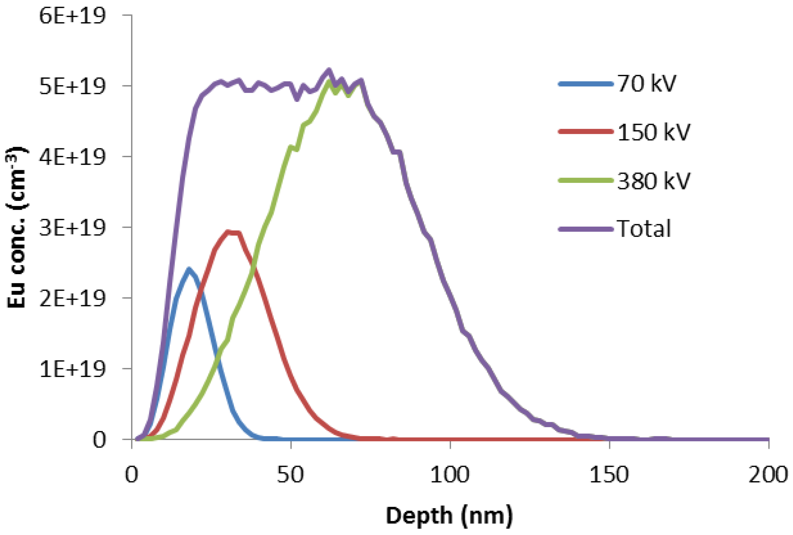
2. Materials and Methods
3. Results
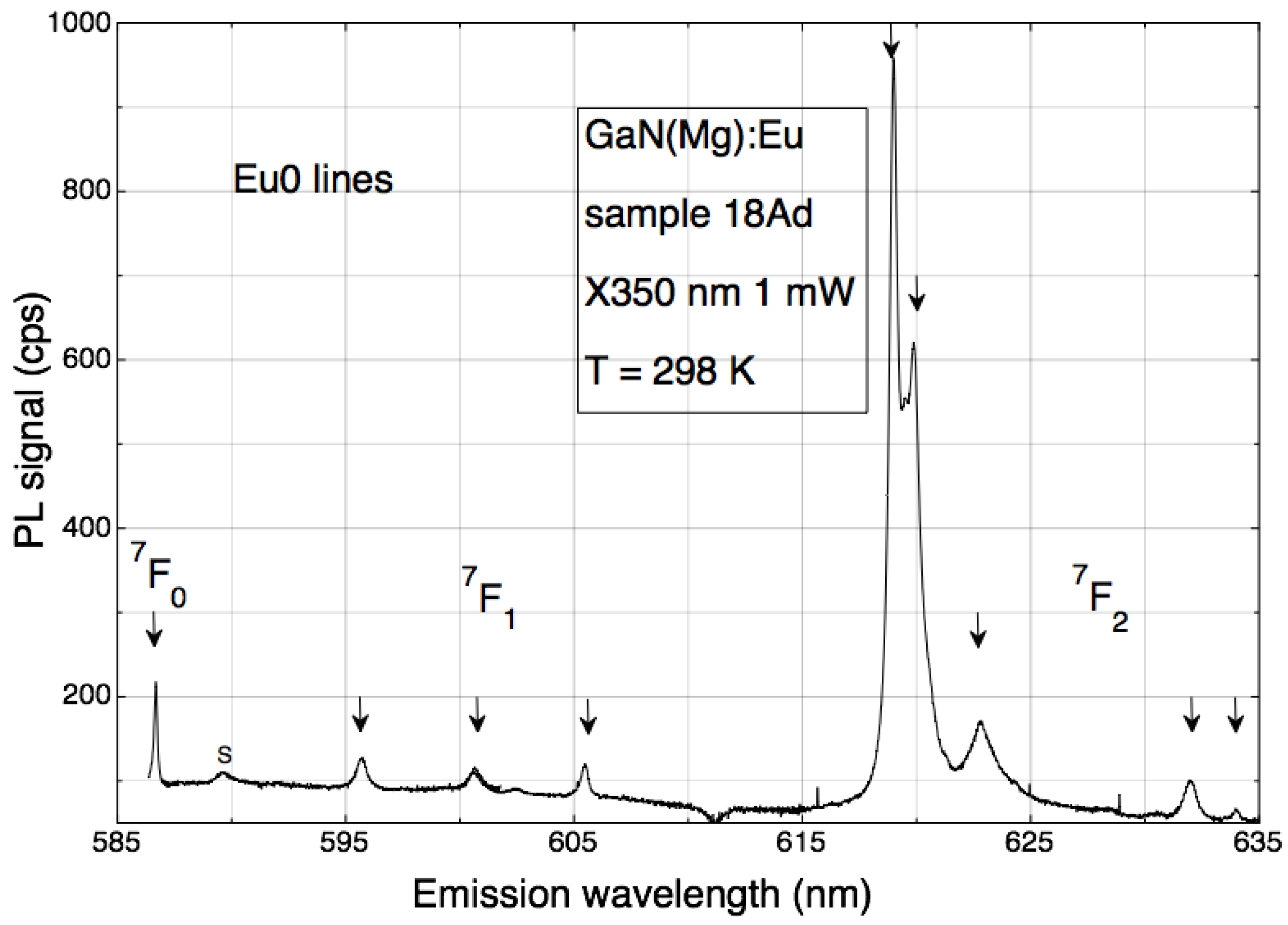
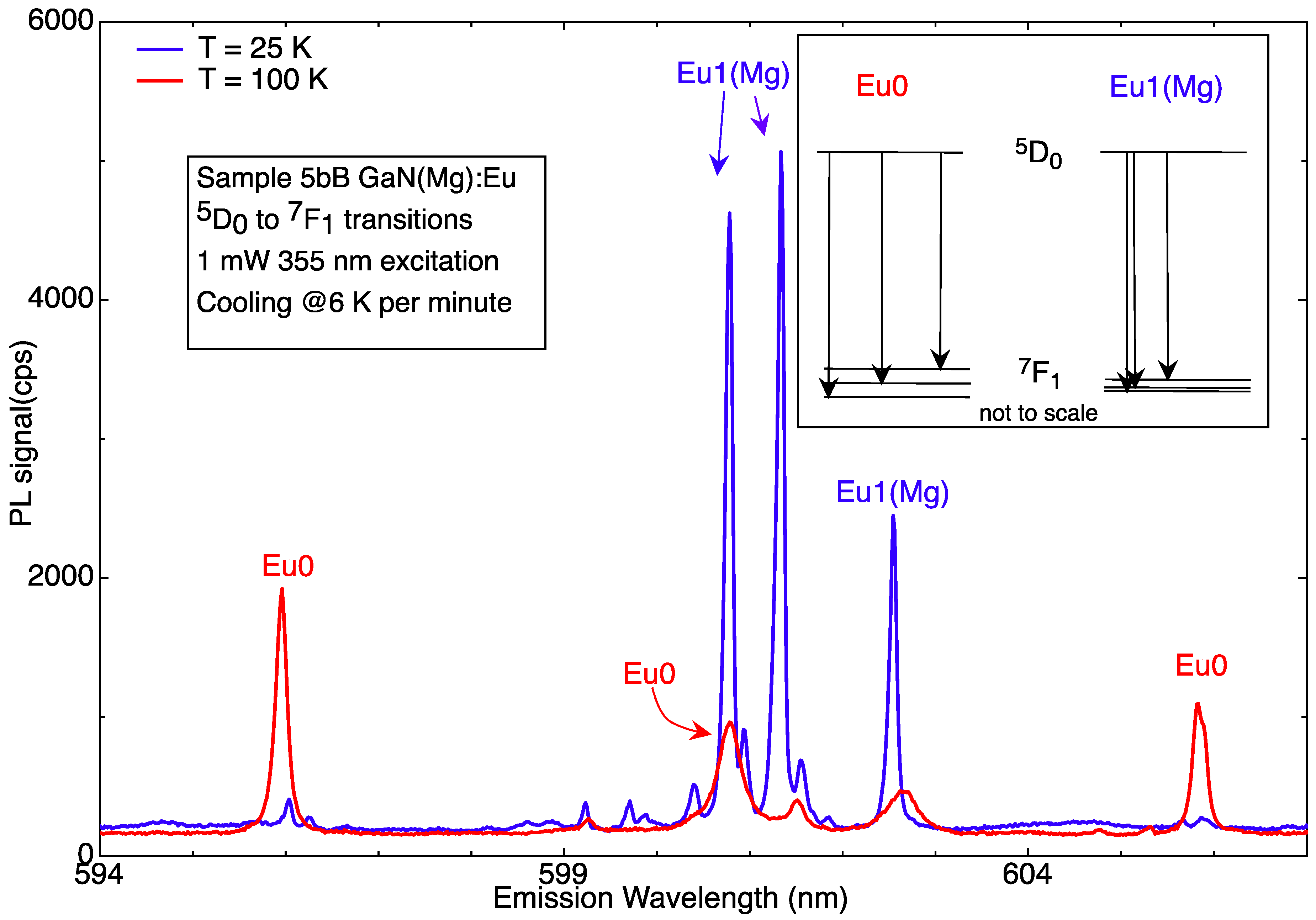
3.1. Eu0 Spectrum
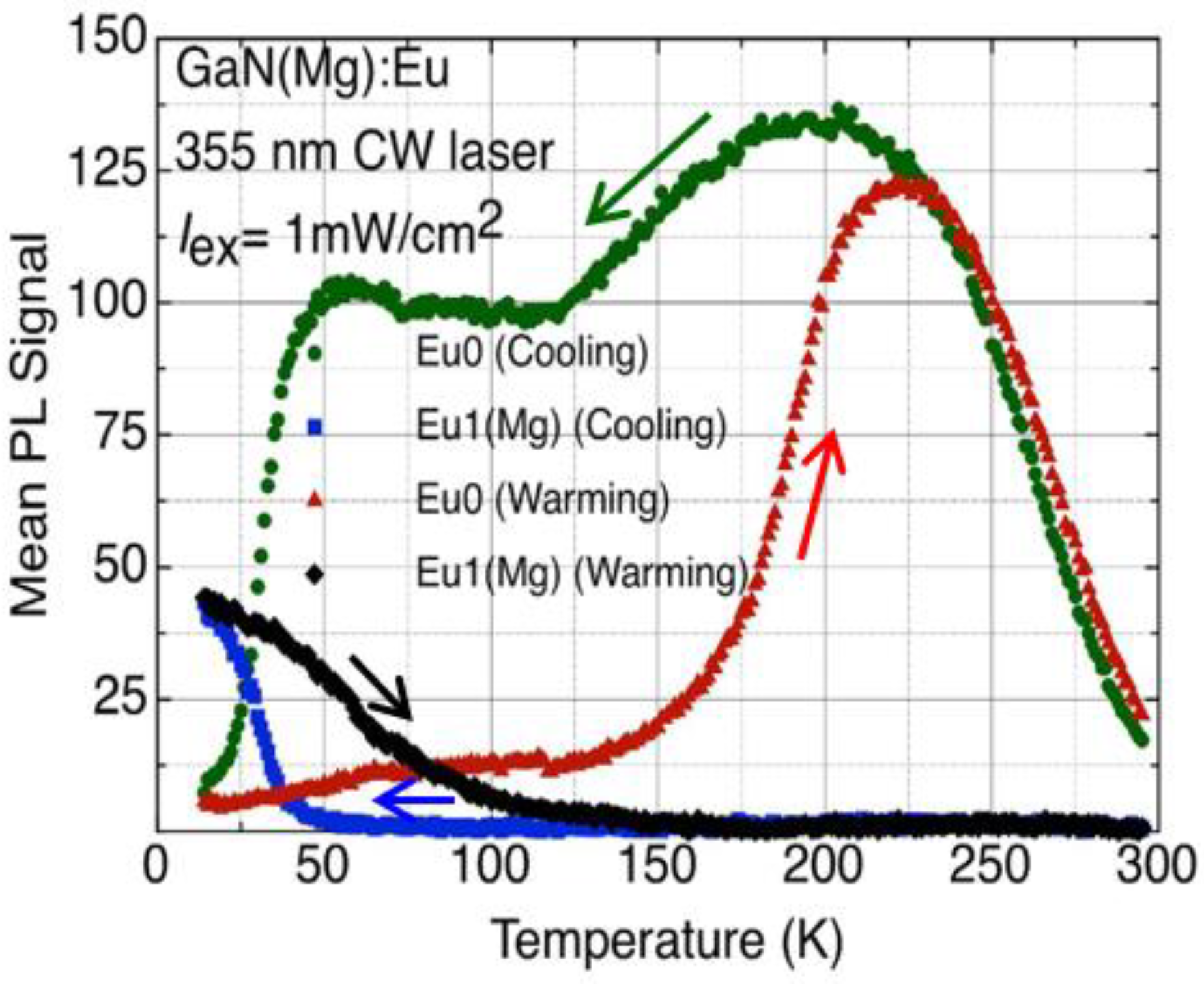
3.2. HPS
3.3. Quantifying Photochromism
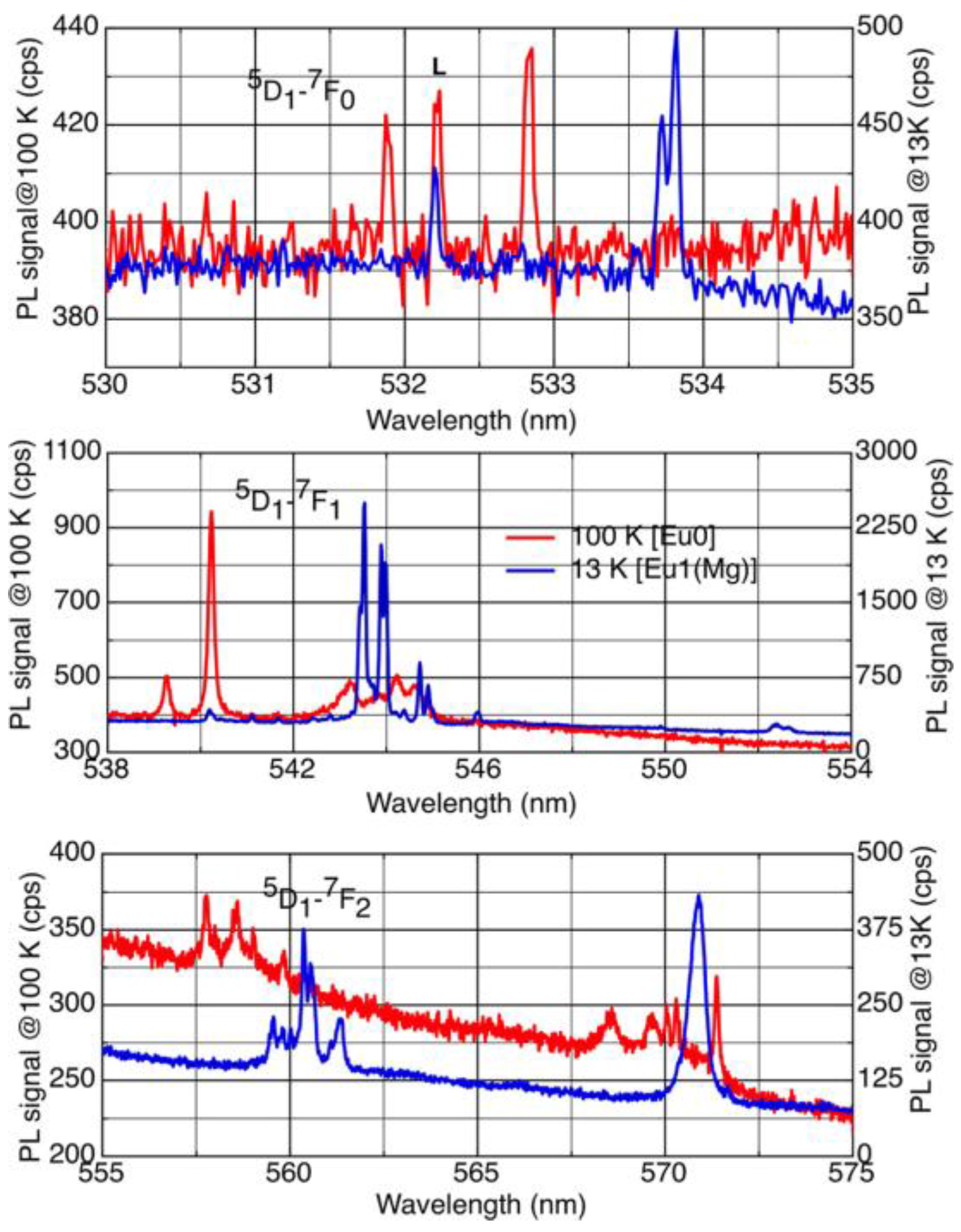
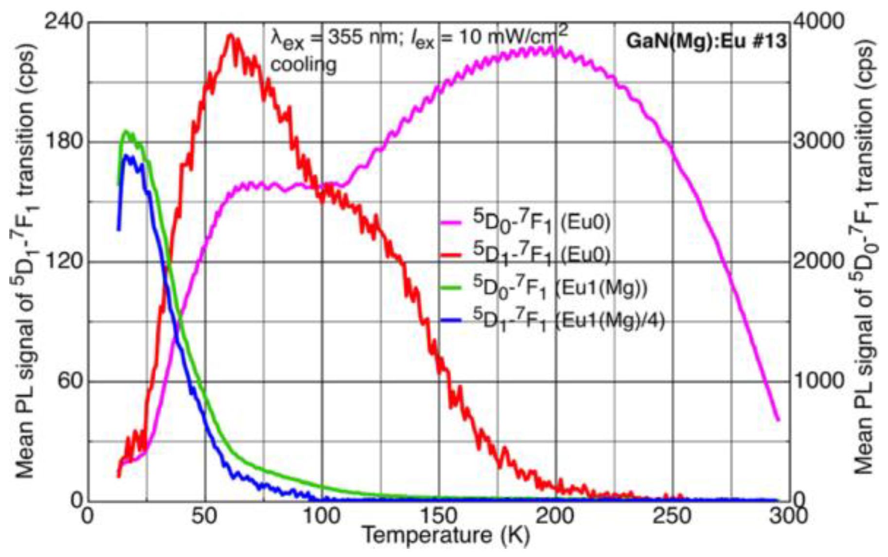
3.4. Transitions from 5D1 Levels
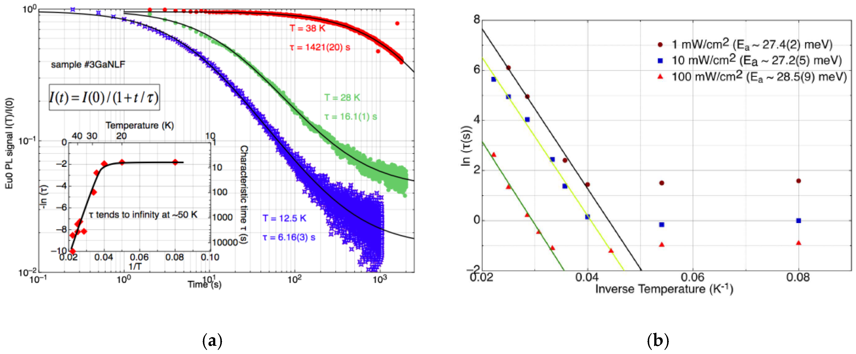
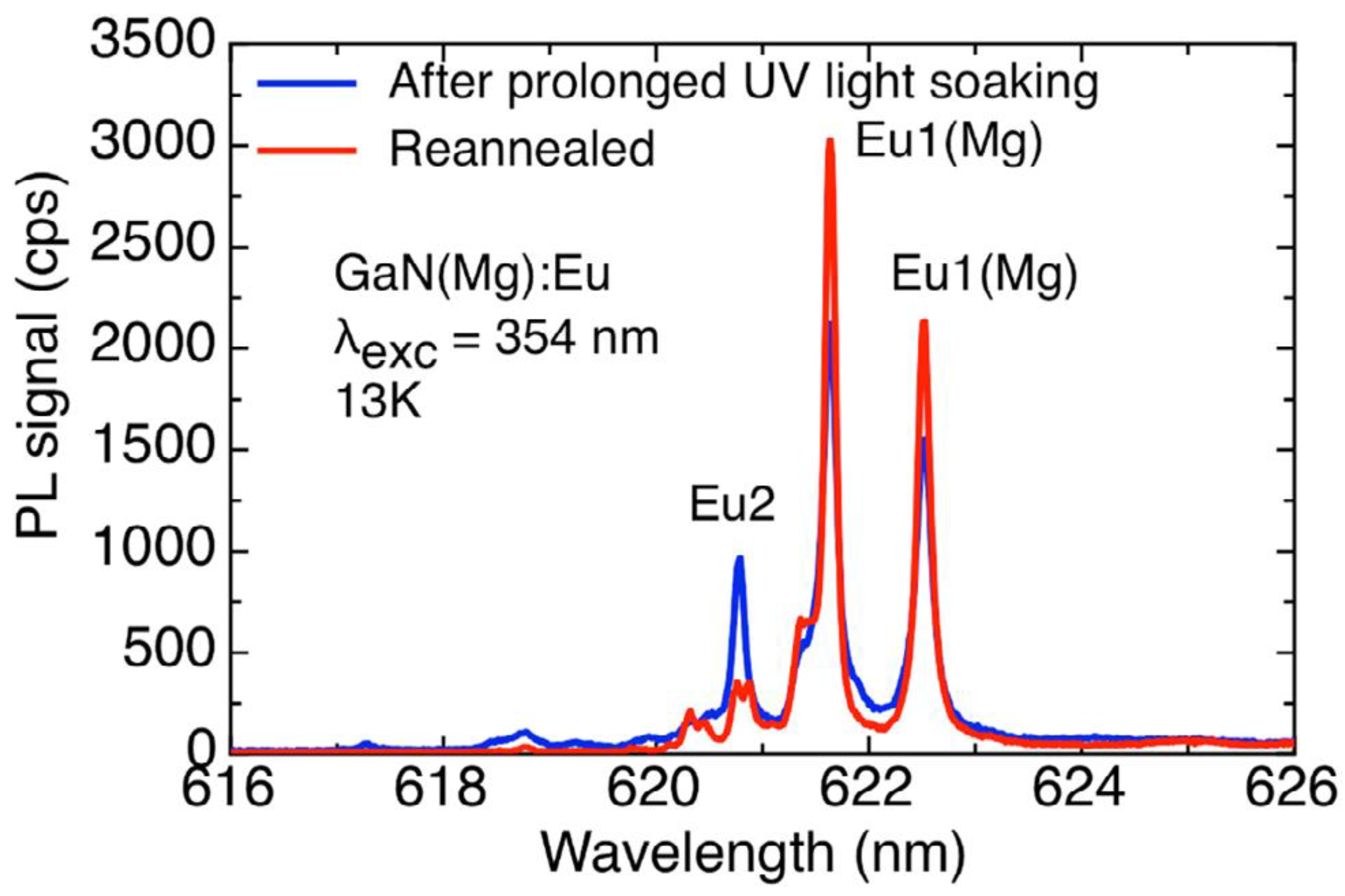
3.5. Photodissociation
4. Discussion
5. Conclusions
Author Contributions
Funding
Conflicts of Interest
References
- O’Donnell, K.P.; Dierolf, V. (Eds.) Rare-Earth Doped III-Nitrides for Optoelectronic and Spintronic Applications. In Topics in Applied Physics 124; Springer: Dordrecht, The Netherlands, 2010; ISBN 978-90-481-2876-1. [Google Scholar]
- O’Donnell, K.P. 2012 UKNC Winter Meeting; University of Bath: Bath, UK, Unpublished talk; 2012. [Google Scholar]
- Singh, A.K.; O’Donnell, K.P.; Edwards, P.R.; Lorenz, K.; Kappers, M.J.; Boćkowski, M. Hysteretic photochromic switching of Eu-Mg defects in GaN links the shallow transient and deep ground states of the Mg acceptor. Sci. Rep. 2017, 7, 41982. [Google Scholar] [CrossRef] [PubMed]
- Lany, S.; Zunger, A. Dual nature of acceptors in GaN and ZnO: The curious case of the shallow MgGa deep state. Appl. Phys. Lett. 2010, 96, 142114. [Google Scholar] [CrossRef]
- Lyons, J.L.; Janotti, A.; Van de Walle, C.G. Shallow versus deep nature of Mg acceptors in nitride semiconductors. Phys. Rev. Lett. 2012, 108, 156403. [Google Scholar] [CrossRef] [PubMed]
- Singh, A.K.; O’Donnell, K.P.; Edwards, P.R.; Cameron, D.; Lorenz, K.; Kappers, M.J.; Boćkowski, M.; Yamaga, M.; Prakash, R. Luminescence of Eu3+ in GaN(Mg, Eu): Transitions from the 5D1 level. Appl. Phys. Lett. 2017, 111, 241105. [Google Scholar] [CrossRef]
- Singh, A.K.; O’Donnell, K.P.; Edwards, P.R.; Lorenz, K.; Leach, J.H.; Boćkowski, M. Eu-Mg defects and donor-acceptor pairs in GaN: Photodissociation and the excitation transfer problem. J. Phys. D. 2018, 51, 65106. [Google Scholar] [CrossRef]
- O’Donnell, K.P.; Hourahine, B. Rare earth doped III-nitrides for optoelectronics. Eur. Phys. J. Appl. Phys. 2006, 36, 91–103. [Google Scholar] [CrossRef]
- O’Donnell, K.P.; Edwards, P.R.; Yamaga, M.; Lorenz, K.; Kappers, M.J.; Boćkowski, M. Crystalfield symmetries of luminescent Eu3+ centers in GaN: The importance of the 5D0 to 7F1 transition. Appl. Phys. Lett. 2016, 108, 022102. [Google Scholar] [CrossRef]
- Wang, K.; Martin, R.W.; O’Donnell, K.P.; Katchkanov, V.; Nogales, E. Selectively excited photoluminescence from Eu-implanted GaN. Appl. Phys. Lett. 2005, 87, 112107. [Google Scholar] [CrossRef]
- Roqan, I.S.; O’Donnell, K.P.; Martin, R.W.; Edwards, P.R.; Song, S.F.; Vantomme, A.; Lorenz, K.; Alves, E.; Boćkowski, M. Identification of the prime optical center in GaN:Eu3+. Phys. Rev. B 2010, 81, 085209. [Google Scholar] [CrossRef]
- Mitchell, B.; Poplawsky, J.; Lee, D.; Koizumi, A.; Fujiwara, Y.; Dierolf, V. The role of donor-acceptor pairs in the excitation of Eu-ions in GaN:Eu epitaxial layers. J. Appl. Phys. 2014, 115, 204501. [Google Scholar] [CrossRef]
- Lorenz, K.; Miranda, S.M.C.; Alvesa, E.; Roqan, I.S.; O’Donnell, K.P.; Boćkowski, M. High pressure annealing of europium implanted GaN. Proc. SPIE 2012, 2012, 82620C-1. [Google Scholar] [CrossRef]
- O’Donnell, K.P.; Edwards, P.R.; Kappers, M.J.; Lorenz, K.; Alves, E.; Boćkowski, M. Europium-doped GaN(Mg): Beyond the limits of the light-emitting diode. Phys. Status Solidi C 2014, 11, 662–665. [Google Scholar] [CrossRef]
- O’Donnell, K.P. 2015 UKNC Winter Meeting; University of Nottingham: Nottingham, UK, Unpublished talk; 2015. [Google Scholar]
- O’Donnell, K.P. 2016 UKNC Winter Meeting; University of Cambridge: Cambridge, UK, Unpublished talk; 2016. [Google Scholar]


© 2018 by the authors. Licensee MDPI, Basel, Switzerland. This article is an open access article distributed under the terms and conditions of the Creative Commons Attribution (CC BY) license (http://creativecommons.org/licenses/by/4.0/).
Share and Cite
Edwards, P.R.; O’Donnell, K.P.; Singh, A.K.; Cameron, D.; Lorenz, K.; Yamaga, M.; Leach, J.H.; Kappers, M.J.; Boćkowski, M. Hysteretic Photochromic Switching (HPS) in Doubly Doped GaN(Mg):Eu—A Summary of Recent Results. Materials 2018, 11, 1800. https://doi.org/10.3390/ma11101800
Edwards PR, O’Donnell KP, Singh AK, Cameron D, Lorenz K, Yamaga M, Leach JH, Kappers MJ, Boćkowski M. Hysteretic Photochromic Switching (HPS) in Doubly Doped GaN(Mg):Eu—A Summary of Recent Results. Materials. 2018; 11(10):1800. https://doi.org/10.3390/ma11101800
Chicago/Turabian StyleEdwards, Paul R., Kevin P. O’Donnell, Akhilesh K. Singh, Douglas Cameron, Katharina Lorenz, Mitsuo Yamaga, Jacob H. Leach, Menno J. Kappers, and Michal Boćkowski. 2018. "Hysteretic Photochromic Switching (HPS) in Doubly Doped GaN(Mg):Eu—A Summary of Recent Results" Materials 11, no. 10: 1800. https://doi.org/10.3390/ma11101800
APA StyleEdwards, P. R., O’Donnell, K. P., Singh, A. K., Cameron, D., Lorenz, K., Yamaga, M., Leach, J. H., Kappers, M. J., & Boćkowski, M. (2018). Hysteretic Photochromic Switching (HPS) in Doubly Doped GaN(Mg):Eu—A Summary of Recent Results. Materials, 11(10), 1800. https://doi.org/10.3390/ma11101800