Article

Ferromagnetic Properties of N-Doped and Undoped TiO2 Rutile Single-Crystal Wafers with Addition of Tungsten Trioxide

1
College of Physics and Materials Science, Henan Normal University, Xinxiang 453007, China
2
School of Physics and Technology, Wuhan University, Wuhan 430072, China
*
Author to whom correspondence should be addressed.
Materials 2018, 11(10), 1934; https://doi.org/10.3390/ma11101934
Submission received: 23 August 2018 / Revised: 25 September 2018 / Accepted: 30 September 2018 / Published: 11 October 2018

Abstract

:
In this work, undoped, N-doped, WO3-loaded undoped, and WO3-loaded with N-doped TiO2 rutile single-crystal wafers were fabricated by direct current (DC) magnetron sputtering. N-doping into TiO2 and WO3 loading onto TiO2 surface were used to increase and decrease oxygen vacancies. Various measurements were conducted to analyze the structural and magnetic properties of the samples. X-ray diffraction results showed that the N-doping and WO3 loading did not change the phase of all samples. X-ray photoelectron spectroscopy results revealed that W element loaded onto rutile single-crystal wafers existed in the form of WO3. UV-Vis spectrometer results showed that the absorption edge of WO3-loaded undoped and WO3-loaded with N-doped TiO2 rutile single-crystal wafers had red shift, resulting in a slight decrease in the corresponding band gap. Photoluminescence spectra indicated that oxygen vacancies existed in all samples due to the postannealing atmosphere, and oxygen vacancies density increased with N-doping, while decreasing with WO3 loading onto TiO2 surface. The magnetic properties of the samples were investigated, and the saturation magnetization values were in the order N-doped > WO3-loaded with N-doped > undoped > WO3-loaded undoped rutile single-crystal wafers, which was the same order as the oxygen vacancy densities of these samples. N-doping improved the saturation magnetization values, while WO3-loaded decreased the saturation magnetization values. This paper reveals that the magnetic properties of WO3-loaded with N-doped rutile single-crystal wafers originate from oxygen vacancies.

1. Introduction

Since 1998, when Prinz [1] first found novel applications of dilute magnetic semiconductors (DMSs) in spintronic devices, such as spin light-emitting diode, magnetic random access memory, and spin field emission transistor [2,3], a growing number of researchers have studied the ferromagnetic behavior of DMSs and the origin of this ferromagnetism. Many relevant experiments have been successfully conducted to study the room temperature ferromagnetic behavior of DMSs, especially TiO2-based DMSs [4,5,6,7]. Researchers studying TiO2-based DMSs ferromagnetism have mainly concentrated on element doping, such as Co, Mn, Ru, Ni, Fe, Cu, Sm, S, N [8,9,10,11,12,13,14,15,16,17], and so on. Based on these studies, several models have emerged to illustrate DMSs’ room temperature ferromagnetic origin, such as the Stoner-type model [18], the band-coupling model [19], and the Ruderman–Kittel–Kasuya–Yosida model. Furthermore, researchers have also recently proposed oxygen vacancies as the origin of room temperature ferromagnetism (RTFM) [6,20,21]. The true origin of RTFM is therefore difficult to find. Wang et al. [22] suggested that RTFM in nitrogen-doped rutile TiO2 powders are rooted on oxygen vacancies (Vos) induced by N-doping. As N-doping is an effective method to produce Vos, it is known that WO3 is a very stable metal oxide [23] and can protect TiO2 wafer surface after loading onto it. In this work, we selected WO3 loading and N-doping method to adjust and control Vos densities in TiO2 wafers to study the origin of TiO2’s room temperature ferromagnetic behavior.
Undoped, N-doped, WO3-loaded undoped, and WO3-loaded with N-doped TiO2 rutile single-crystal wafers were fabricated by magnetron sputtering. The magnetic properties and optical properties of the prepared samples were studied. The largest saturation magnetization (Ms) values were found in N-doped rutile single-crystal wafers. The Ms values were adjusted by N-doping and WO3 loading. The N-doping increased the Ms values by increasing Vos density, while WO3-loading decreased the Ms values by decreasing Vos density. The room temperature ferromagnetic property was adjusted by controlling the Vos.

2. Materials and Methods

TiO2 rutile single-crystal wafers oriented (100) (named as sample 1) in the size of 5 mm × 5 mm × 0.5 mm were used as substrates, which were bought from Beijing Scistar Technology Co., Ltd. (Beijing, China). The N-doped TiO2 single-crystal wafers (named as sample 2) were obtained by annealing sample 1 in N2 and NH3 mixed atmosphere at 500 °C for 2 h. WO3-loaded TiO2 single-crystal wafers (named as sample 3) were obtained by wafers grown on TiO2 substrates by direct current (DC) magnetron sputtering with a W target in a small ion sputter apparatus (Cressington sputter coater 108) at room temperature. After loading the W element, samples were postannealed in air at 450 °C for 2 h to get sample 3. Following the N-doping process for sample 3, WO3-loaded with N-doped TiO2 single-crystal wafers (named as sample 4) were obtained. The structural properties of the samples were characterized by X-ray diffraction (XRD, Bruker D8 Discover, Karlsruhe, Germany) with 2θ scanning range from 20° to 80°, and the morphology of the samples was observed by scanning electron microscopy (SEM, S-4800, Hicathi, Tokyo, Japan). The surface element composition and chemical valence states were measured with X-ray photoelectron spectroscopy (XPS, ESCALAB 250Xi, Thermo Fisher Scientific, Waltham, MA, USA). The optical properties and band gap were studied using the absorption spectra from a UV-Vis spectrometer (UV 3600, Shimadzu, Kyoto, Japan). The photoluminescence (PL) spectra were obtained by He–Cd laser (Persee, Beijing, China) with 280 nm wavelength of the excitation light source. The magnetic properties of samples were characterized at room temperature by vibrating sample magnetometer (VSM, Quantum Design PPMS-9, San Diego, CA, USA) with a magnetic field from −8 K Oe to 8 K Oe.

3. Results and Discussion

The crystal structures of the samples were characterized by XRD, and the XRD patterns of all samples are shown in Figure 1a (the Y-axis is a logarithmic scale). As can be seen in the figure, a primary peak appeared at 39.16°, which corresponded to the TiO2 rutile (200) plane, while a weak peak appeared at 35.18° corresponding to the TiO2 rutile (200) Kβ [24]. Only (200) diffraction peaks were detected in all samples. Therefore, all samples were mainly oriented in the (100) direction. The peak intensities of sample 3 were weaker than those of other samples, but the relative intensities were the same as other samples. The XRD patterns had no peaks of products for tungsten and tungsten oxide due to the low sputtering concentration of WO3. These results were similar to Sajjad et al.’s. report, where no peaks related to W element were observed even with a W sputtering concentration up to 4% [25]. The content of W element was 2.17% in our experiment, which was determined by XPS measurements.
The surface morphology of samples 1 and 3 are shown in Figure 1b. From sample 3, it was obvious that many ash-colored particles were loaded on the sample surface compared to sample 1, where the surface was smooth and clean. The diameters of the deposited particles varied from 10 to 80 nm. The deposited particles might be related to W oxide in the special process of forming sample 3.
To verify the surface elemental composition and elemental chemical valence state, XPS measurements were conducted for sample 4. The binding energy (BE) of all peaks in spectral line was calibrated by C 1s at 284.8 eV. Figure 2a–d displays Ti 2p, O 1s, N 1s, and W 4f core-level spectra for sample 4, respectively. It is known that there are some rules in peak splitting of XPS results: (i) The intensity ratio between the doublets of p, d, and f atomic orbital should be 2:1, 3:2, and 4:3, respectively. (ii) The full width at half maximum (FWHM) values of doublets in the same valence state of the same element should be equal. (iii) The FWHM of the high valence state of the same element should be greater than or equal to the FWHM of low valence state. All spectral lines in our experiment were fitted with Gaussian–Lorentzian function. The red lines were the background, and the lines with symbols were the original experiment data. As can be seen in Figure 2a, peak A and peak B appeared at BE of 458.2 eV and 464.2 eV, respectively, which corresponded to Ti4+ standard peak according to the handbook of XPS [26], and two weak peaks appeared at BE of 457.7 eV and 463.8 eV. The BE of Ti2+ in TiO appears at around 455 eV and Ti4+ in TiO2 appears at around 459 eV, according to the XPS handbook, which means that Peak C and peak D corresponded to the Ti3+ standard peak. The fitted doublets of Ti4+ and Ti3+ met the aforementioned peak splitting rules. The XPS result of Ti meant that little of Ti3+ coexisted with Ti4+ states in sample 4. As can be seen in Figure 2b, there were four O 1s peaks, meaning there were four different chemical environments in sample 4. A relatively strong peak appeared at BE of 529.20 eV, which was attributed to lattice oxygen in TiO2 [27,28]. A small peak that appeared at 531.9 eV corresponded to Ti-N-O band in sample 4 due to the doping of N element in the sample; this finding is in agreement with Chan and Lu’s report, which showed that the BE of Ti-N-O band is 531.5 eV [29]. One weak peak appeared at BE of 532.3 eV, which could be ascribed to the C=O band and the hydroxyl groups due to the contamination of the sample surface [30,31]. Another peak appeared at BE of 531.1 eV, which could be attributed to the W-O band in WO3 [32,33]. As can be seen in Figure 2c, a weak peak was observed at 399.02 eV, which could be attributed to N in the Ti-N-O group [34]. The N 1s peak intensity was apparently not high because the NH3 content was only 5% in the N2/NH3 atmosphere in the experimental procedure. In Figure 2d, it can be seen that the peak of purple spectral line appeared at BE of 35.1 eV and 37.7 eV, which could be attributed to W 4f 7/2 and 5/2 doublets in WO3, respectively. The distance between the doublets was 2.5 eV, which was close to the standard distance of 2.15 eV [26]. The fitted doublets of W 4f met the aforementioned peak splitting rules. Besides, a peak appeared at BE of 36.3 eV. In Yamashita and Hayes’ report, the BE of Ti 3p appeared at 36.0 eV inTi2O3 and 37.5 eV in TiO2 [35]. This meant that the peak that appeared at BE of 36.3 eV could be assigned as Ti 3p. The result verified the existence of Ti4+ and Ti3+ in the sample, consistent with previous analysis. The XPS spectra result of W 4f core level indicated that W element existed on the surface of WO3-loaded with N-doped samples in the form of +6 chemical valence states. This means that WO3 was loaded on the TiO2 sample surface rather than impregnated into the TiO2 lattice. The loading of WO3 on the wafer surface did not alter the chemical state of the Ti or O element of the wafer.
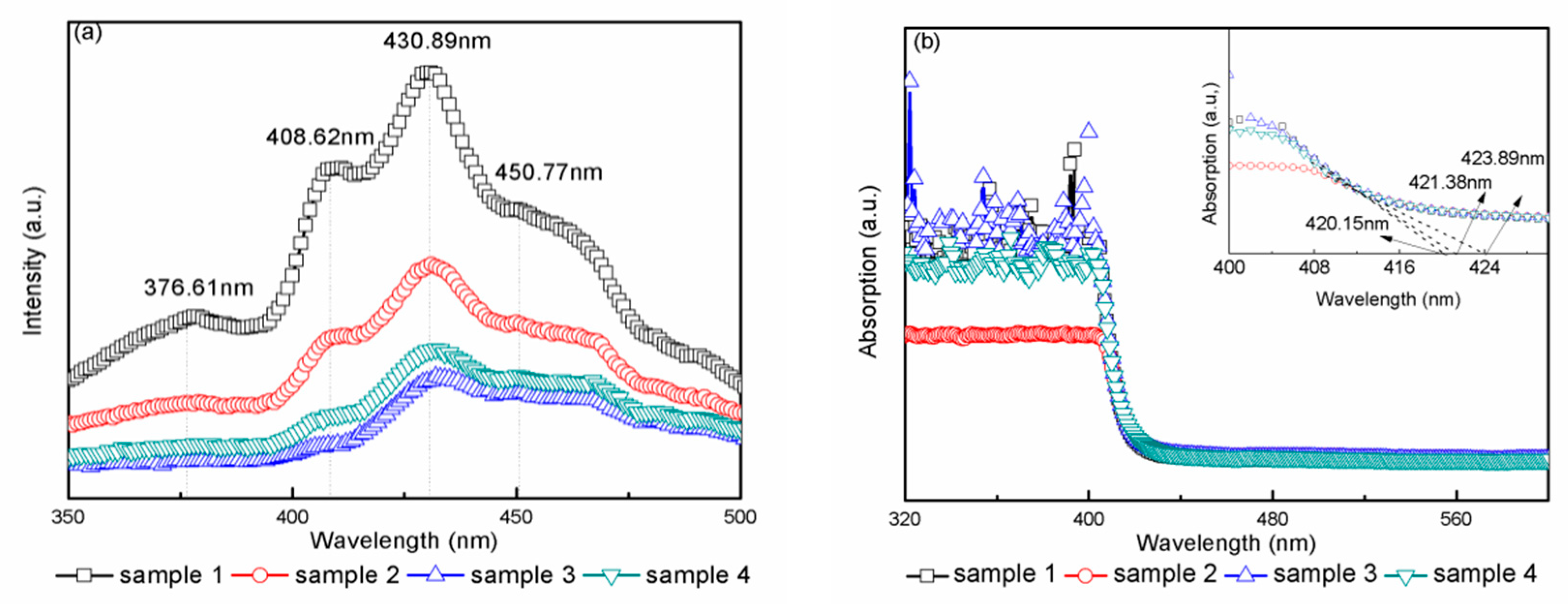
To investigate the electron–hole recombination rate and the existence of Vos, PL spectra of all samples were collected at the wavelength range of 350–500 nm and are shown in Figure 3a. In the wavelengths from 350 to 410 nm, all samples had two emission peaks, which were attributed to band edge emission. The emission peak position at 376.61 nm originated from charge recombination at the shallow-trap surface states [36]. The emission peak appearing at 408.62 nm corresponded to a band gap transition from conduction band to valence band, which was equal to the band gap of rutile TiO2 (3.02 eV). The emission peak at 430.89 nm might have been due to the self-trapped excitons in the TiO6 octahedra [37]. The emission peak at 450.77 nm and 476 nm were related to Vos in the samples due to the recombination of photogenerated holes and electrons [38,39].
As for intensity, sample 1 had the strongest peak intensity compared to the other three samples. The weak PL intensity in sample 3 indicated the low radiative recombination rate of the electrons and holes. Photogenerated electrons were transfer from TiO2 conduction band to tungsten oxide conduction band, and the holes accumulated in the TiO2 valence band. As a result, photogenerated electrons and holes were effectively separated. In addition, when the tungsten oxide concentration was lower than its optimal ratio, the tungsten energy level would be a separation center [25]. All these reasons contributed to a low PL intensity in samples 3 and 4. The concentration of Vo in sample 2 increased due to the annealed atmosphere, the excited electrons were easily trapped by Vos, and the holes were trapped by the doped nitrogen atoms [40], leading to the separation of electrons and holes.
The optical properties and band gap of the prepared samples were investigated by UV-Vis spectrophotometer. Figure 3b displays the absorption spectra of samples 1–4. All samples had absorption edges ranging from 400 to 425 nm due to the band-to-band transition from the Ti 2p valence band to the conduction band in the TiO2 single crystals. The absorption edge of samples 1–4 was 420.15 nm, 423.89 nm, 420.15 nm, and 421.38 nm, respectively. It was obvious that samples 2 and 4 had red shift, which was similar to other reports [41,42]. The band gap was calculated by the equation: Eg = hc/λ, where c is the light velocity equal to 3 × 10⁸ m/s, h is Planck’s constant equal to 6.626 × 10⁻³⁴ J s, λ is the corresponding wavelength of the samples, Eg is the calculated result (band gap) of the samples. The calculated band gap of samples 1–4 was 2.96 eV, 2.93 eV, 2.96 eV, and 2.95 eV, respectively. Samples 1 and 3 had the same band gap values, which was due to WO3 being simply deposited on the single-crystal wafers. Compared to sample 1, the band gap of samples 2 and 4 was slightly decreased, as in the report by Chen et al. [32].
To further investigate the magnetic property of the samples, the magnetic moment vs. the magnetic field curves were collected at room temperature, as shown in Figure 4. The enlarged figure in the inset near zero magnetic field has been provided to distinctly illustrate the coercive force (Hc). The figure was plotted after deducting diamagnetic substrate. All the samples had a typical magnetic hysteresis loop, indicating that all samples were ferromagnetic materials. As can be seen in Figure 4, sample 2 yielded the highest Ms value (15.15 × 10⁻⁴ emu/cm2), while sample 3 had the lowest Ms value (4.16 × 10⁻⁴ emu/cm2). Sample 4 had a larger Ms value of 9.42 × 10⁻⁴ emu/cm2 than sample 1, which had a value of 6.05 × 10⁻⁴ emu/cm2. Furthermore, the Ms values were in the order of sample 2 > sample 4 > sample 1 > sample 3. The coercive force values (Hc) of samples 1–4 were 54.72 Oe, 98.50 Oe, 54.72 Oe, and 111.37 Oe, respectively.
As in Kim et al.’s report [43], the undoped rutile single-crystal wafer had a small Ms in our experiment. In addition, Kim et al. reported that Vos induces a lattice distortion in rutile TiO2 and introduces ferromagnetism in undoped TiO2 wafers due to the charge distribution. Sample 2 had the largest Ms values compared to others due to the high oxygen vacancy concentration, which indicated that the Vos was the main source of ferromagnetic behavior. For samples 3 and 4, the WO3 loading on the wafer surface caused lower Ms compared to samples 1 and 2. The demagnetizing field could be formed due to polarization by the addition of WO3, resulting in an increase in magnetic loss [23]. Therefore, WO3 loading decreased the Ms values.
In this study, WO3 did not show any ferromagnetism, which would rule out any ferromagnetic contribution related to the W second phase. The ferromagnetism of sample 3 must have been intrinsic and originated from the oxygen vacancy. For sample 4, the WO3 deposited on the sample’s surface formed the demagnetizing field, resulting in an increase in magnetic loss. Therefore, the WO3-loaded undoped samples had the lowest Ms values compared to the others. For N-doped samples, due to an increase in Vos, N-doped samples had the highest Ms values compared to others. Considering the existence of many Vos, the influence of Vos on ferromagnetism can be explained by the carrier-mediated bound magnetic polaron model (BMP). The ferromagnetic coupling between two Ti3+ ions was formed through Vos. When the Vos concentration exceeded a certain value, the bound magnetic polaron caused the ferromagnetic coupling [44,45,46]. The weak ferromagnetic property of sample 4 was due to the low concentration of oxygen as well as the defects of the nanoparticles loaded on the sample surface, which might have decreased ferromagnetic coupling. Briefly, the more Vos present in the samples, the more apparent was the ferromagnetic behavior [47].

4. Conclusions

In conclusion, undoped, N-doped, WO3-loaded undoped, and WO3-loaded with N-doped TiO2 rutile single-crystal wafers were prepared by magnetron sputtering. Oxygen vacancy was increased by N-doping into TiO2 single-crystal wafers. XPS results showed that W element existed in the form of WO3 loaded onto wafer surface, and PL spectra indicated that WO3 loaded onto wafers decreased oxygen vacancy concentration. The N-doped TiO2 rutile single-crystal wafers had the maximum magnetic saturation value. The magnetic properties of the samples were in accordance with the oxygen vacancy concentration, which meant that the magnetic properties of the samples were sensitive to oxygen vacancy. Oxygen vacancies play a dominant role in the origin of WO3 loaded, N-doped TiO2 rutile single-crystal wafers. This work provides a fundamental understanding of the origin of the ferromagnetic properties of DMSs, which is critical for the incorporation of DMSs in future device applications.

Author Contributions

Methodology, J.X. and H.W.; Software, Z.Zh. (Zhongpo Zhou); Formal Analysis, J.X., H.W., and Z.Zo. (Zhaorui Zou); Investigation, J.X.; Writing—Original Draft Preparation, J.X.; Writing—Review & Editing, J.X. and H.W.; Funding Acquisition, H.W.

Funding

This research was funded by [NSFC] grant numbers [11404100, 11474086, 11175135, 10904116 and 11304083], [the Young Scholar Foundation of Henan Normal University] grant number [5101029470616], and [the Postdoctoral Research Foundation of Henan Province] grant number [5201026500204]. And The APC was funded by [Henan Normal Uviversity].

Conflicts of Interest

The authors declare no conflict of interest.

References

  1. Prinz, G.A. Magnetoelectronics. Science 1998, 282, 1660–1663. [Google Scholar] [CrossRef] [PubMed]
  2. Fiederling, R.; Keim, M.; Reuscher, G.; Ossau, W.; Schmidt, G.; Waag, A.; Molenkamp, L.W. Injection and detection of a spin-polarized current in a light-emitting diode. Nature 1999, 402, 787–790. [Google Scholar] [CrossRef]
  3. Han, H.F.; Wen, Z.C.; Wei, H.X. Nanoring magnetic tunnel junction and its application in magnetic random access memory demo devices with spin-polarized current switching (invited). J. Appl. Phys. 2008, 103, 07E933. [Google Scholar] [CrossRef]
  4. Manivannan, A.; Seehra, M.S.; Majumder, S.B.; Katiyar, R.S. Magnetism of Co-doped titania thin films prepared by spray pyrolysis. Appl. Phys. Lett. 2003, 83, 111–113. [Google Scholar] [CrossRef]
  5. Kim, J.Y.; Park, J.H.; Park, B.G.; Noh, H.J.; Oh, S.J.; Yang, J.; Kim, D.H.; Bu, S.; Noh, T.W.; Lin, H.; et al. Ferromagnetism induced by clustered Co in Co-doped anatase TiO2 thin films. Phys. Rev. Lett. 2003, 90, 017401. [Google Scholar] [CrossRef] [PubMed]
  6. Wang, X.; Song, Y.; Tao, L.L.; Feng, J.F.; Sui, Y.; Tang, J.; Song, B.; Wang, Y.; Wang, Y.; Zhang, Y.; et al. Origin of ferromagnetism in aluminum-doped TiO2 thin films: Theory and experiments. Appl. Phys. Lett. 2014, 105, 262402. [Google Scholar] [CrossRef]
  7. Xiao, L.; Deng, M.; Zeng, W.; Zhang, B.; Xu, Z.; Yi, C.; Liao, G. Novel robust superhydrophobic coating with self-cleaning properties in air and oil based on rare earth metal oxide. Ind. Eng. Chem. Res. 2017, 56, 12354–12361. [Google Scholar] [CrossRef]
  8. Cuaila, J.L.S.; Alayo, W.; Avellaneda, C.O. Ferromagnetism in spin-coated cobalt-doped TiO2 thin films and the role of crystalline phases. J. Magn. Magn. Mater. 2017, 442, 212–217. [Google Scholar] [CrossRef]
  9. Kumar, A.; Kashyap, M.K.; Sabharwal, N.; Kumar, S.; Kumar, P.; Asokan, K. Structural, optical and weak magnetic properties of Co and Mn codoped TiO2 nanoparticles. Solid State Sci. 2017, 73, 19–26. [Google Scholar] [CrossRef]
  10. Xu, J.; Zhou, Z.P.; Wang, H.Y. Origin of ferromagnetism in Ru and N codoped TiO2 nanotubes: Experiments and theory investigations. J. Nanomater. 2017, 2, 1–7. [Google Scholar] [CrossRef]
  11. Elilarassi, R.; Chandrasekaran, G. Influence of nickel doping on the structural, optical and magnetic properties of TiO2 diluted magnetic semiconductor nanoparticles prepared by high energy ball-milling technique. J. Mater. Sci. Mater. Electron. 2017, 28, 14536–14542. [Google Scholar] [CrossRef]
  12. Ahmed, S.A. Structural, optical, and magnetic properties of Cu-doped TiO2 samples. Cryst. Res. Technol. 2017, 52, 1600335. [Google Scholar] [CrossRef]
  13. Tseng, L.T.; Luo, X.; Li, S.; Yi, J.B. Magnetic properties of Sm-doped rutile TiO2 nanorods. J. Alloys Compd. 2016, 687, 294–299. [Google Scholar] [CrossRef]
  14. Shi, B.; Liu, Y.; Song, C.; Han, G. First-principles investigation of the band structure of S-doped TiO2. Rare Met. Mater. Eng. 2008, 37, 638–640. [Google Scholar]
  15. Vasu, K.; Sreedhara, M.B.; Ghatak, J.; Rao, C.N.R. Atomic Layer Deposition of p-Type Epitaxial Thin Films of Undoped and N-Doped Anatase TiO2. ACS Appl. Mater. Interfaces 2016, 8, 7897–7901. [Google Scholar] [CrossRef] [PubMed]
  16. Ahmed, S.A. Ferromagnetism in Cr-, Fe-, and Ni-doped TiO2 samples. J. Magn. Magn. Mater. 2017, 442, 152–157. [Google Scholar] [CrossRef]
  17. Kumar, S.; Park, J.S.; Kim, D.J.; Lee, M.H.; Song, T.K.; Gautam, S.; Chae, K.H.; Kim, S.S.; Kim, M.H. Electronic structure and magnetic properties of Co doped TiO2 thin films using X-ray absorption spectroscopy. Ceram. Int. 2015, 41, S370–S375. [Google Scholar] [CrossRef]
  18. Coey, J.M.D.; Stamenov, P.; Gunning, R.D.; Venkatesan, M.; Paul, K. Ferromagnetism in defect-ridden oxides and related materials. New. J. Phys. 2010, 12, 053025. [Google Scholar] [CrossRef] [Green Version]
  19. Peng, H.; Xiang, H.J.; Wei, S.H.; Li, S.S.; Xia, J.B.; Li, J. Origin and enhancement of hole-induced ferromagnetism in first-row d0 semiconductors. Phys. Rev. Lett. 2009, 102, 017201. [Google Scholar] [CrossRef] [PubMed]
  20. Bapna, K.; Choudhary, R.J.; Pandey, S.K.; Phase, D.M.; Sharma, S.K.; Knobel, M. Electronic depictions of magnetic origin in undoped and Fe doped TiO2-d epitaxial thin films. Appl. Phys. Lett. 2011, 99, 112502. [Google Scholar] [CrossRef]
  21. Song, Y.L.; Wang, X.J.; Tao, L.L.; Song, B.Q.; Zhang, L.L.; Zhang, Y.; Sui, Y.; Liu, Z.G.; Tang, J.K.; Han, X.F. Effect of Ga-doping and oxygen vacancies on the ferromagnetism of TiO2 thin films. J. Alloys Compd. 2017, 694, 929–934. [Google Scholar] [CrossRef]
  22. Wang, J.B.; Wu, K.C.; Mi, J.W.; Luo, C.W.; Wu, K.H.; Uen, T.M.; Lin, J.Y.; Juang, J.Y.; Liu, S.J. Room-temperature ferromagnetism in carbon- and nitrogen-doped rutile TiO2. Appl. Phys. A 2015, 118, 725–731. [Google Scholar] [CrossRef]
  23. Park, K.S.; Nam, J.H.; Oh, J.H. Magnetic properties of with addition of tungsten trioxide. J. Magn. Magn. Mater. 2001, 226–230, 1415–1417. [Google Scholar] [CrossRef]
  24. Ding, Y.; Yuan, C.; Wang, Z.; Liu, S.; Shi, J.; Xiong, R.; Yin, D.; Lu, Z. Improving thermostability of Cr2O3 thin films by doping with Sn. Appl. Phys. Lett. 2014, 105, 092401. [Google Scholar] [CrossRef]
  25. Sajjad, A.K.L.; Shamaila, S.; Tian, B.; Chen, F.; Zhang, J. One step activation of WOx/TiO2 nanocomposites with enhanced photocatalytic activity. Appl. Catal. B Environ. 2009, 91, 397–405. [Google Scholar] [CrossRef]
  26. Wagner, C.D.; Riggs, W.M.; Davis, L.E.; Moulder, J.F.; Muilenberg, G.E. Handbook of X-ray Photoelectron Spectroscopy; Muilenberg, G.E., Ed.; Perkin-Eimer Corporation: Eden Prairie, MN, USA, 1979. [Google Scholar]
  27. Jabbari, V.; Hamadanian, M.; Reisi-Vanani, A.; Razi, P.; Hoseinifardc, S.; Villagr´an, D. In, V-codoped TiO2 nanocomposite prepared via a photochemical reduction technique as a novel highefficiency visible-light-driven nanophotocatalyst. RSC Adv. 2015, 5, 78128–78135. [Google Scholar] [CrossRef]
  28. Zhou, Z.; Wang, H.; Zou, Z.; Du, M.; Guo, J.; Yang, Z. Investigations on the origin of ferromagnetism of Cu doped anatase TiO2 nanotubes. Mater. Res. Bull. 2017, 86, 287–294. [Google Scholar] [CrossRef]
  29. Chan, M.; Lu, F. Preparation of titanium oxynitride thin films by reactive sputtering using air/Ar mixtures. Surf. Coat. Technol. 2008, 203, 614–618. [Google Scholar] [CrossRef]
  30. Mohanty, P.; Mishra, N.C.; Choudhary, R.J.; Banerjee, A.; Shripathi, T.; Lalla, N.P.; Annapoorni, S.; Rath, C. Oxygen vacancy induced phase formation and room temperature ferromagnetism in un-doped and Co-doped TiO2 thin films. J. Phys. D Appl. Phys. 2012, 45, 1418–1420. [Google Scholar] [CrossRef]
  31. Sharma, S.; Chaudhary, S.; Kashyap, S.C.; Sharma, S.K. Room temperature ferromagnetism in Mn doped TiO2 thin films: Electronic structure and Raman investigations. J. Appl. Phys. 2011, 109, 083905. [Google Scholar] [CrossRef]
  32. Chen, L.; Qu, Y.; Yang, X.; Liao, B.; Xue, W.; Cheng, W. Characterization and first-principles calculations of WO3/TiO2 composite films on titanium prepared by microarc oxidation. Mater. Chem. Phys. 2017, 201, 311–322. [Google Scholar] [CrossRef]
  33. Rajagopal, S.; Nataraj, D.; Mangalaraj, D.; Djaoued, Y.; Robichaud, J.; Khyzhun, O.Y. Controlled growth of WO(3) nanostructures with three different morphologies and their structural, optical, and photodecomposition studies. Nanoscale Res. Lett. 2009, 4, 1335–1342. [Google Scholar] [CrossRef] [PubMed]
  34. Burda, C.; Lou, Y.; Chen, X.; Samia, A.C.S.; Stout, J.; Gole, J.L. Enhanced nitrogen doping in TiO2 nanoparticles. Nano Lett. 2003, 3, 1049–1051. [Google Scholar] [CrossRef]
  35. Yamashita, T.; Hayes, P. Analysis of XPS spectra of Fe2+ and Fe3+ ions inoxide materials. Appl. Surf. Sci. 2008, 254, 2441–2449. [Google Scholar] [CrossRef]
  36. Yoon, M.; Seo, M.; Jeong, C.; Jang, J.H.; Jeon, K.S. Synthesis of Liposome-templated titania nanodisks:  optical properties and photocatalytic activities. Chem. Mater. 2005, 17, 6069–6079. [Google Scholar] [CrossRef]
  37. Ahmed, S.A. Annealing effects on structure and magnetic properties of Mn-doped TiO2. J. Magn. Magn. Mater. 2016, 402, 178–183. [Google Scholar] [CrossRef]
  38. Patel, S.K.S.; Gajbhiye, N.S. Room temperature magnetic properties of Cu-doped titanate, TiO2 (B) and anatasenanorods synthesized by hydrothermal method. Mater. Chem. Phys. 2012, 132, 175–179. [Google Scholar] [CrossRef]
  39. Huang, C.; Guo, Y.; Liu, X.; Wang, Y. Structural and optical properties of Ti1−xCoxO2 films prepared by sol–gel spin coating. Thin Solid Films 2006, 505, 141–144. [Google Scholar] [CrossRef]
  40. Irie, H.; Watanabe, Y.; Hashimoto, K. Nitrogen-concentration dependence on photocatalytic activity of TiO2−xNx Powders. J. Phys. Chem. B 2003, 107, 5483–5486. [Google Scholar] [CrossRef]
  41. Gomez-Polo, C.; Larumbe, S.; Monge, M. Room temperature ferromagnetism and absorption red-shift in nitrogen-doped TiO2 nanoparticles. J. Alloys Compd. 2014, 612, 450–455. [Google Scholar] [CrossRef]
  42. Zhou, S.; Lv, J.; Guo, L.K.; Xu, G.Q.; Wang, D.M.; Zheng, Z.X.; Wu, Y.C. Preparation and photocatalytic properties and N-doped nano-TiO2/muscovite composites. Appl. Surf. Sci. 2012, 258, 6136–6141. [Google Scholar] [CrossRef]
  43. Kim, D.; Hong, J.; Park, Y.R.; Kim, K.J. The origin of oxygen vacancy induced ferromagnetism in undoped TiO2. J. Phys. Condens. Matter 2009, 21, 195405. [Google Scholar] [CrossRef] [PubMed]
  44. Patel, S.K.S.; Gajbhiye, N.S. Oxygen deficiency induced ferromagnetism in Cr-doped TiO2 nanorods. J. Magn. Magn. Mater. 2013, 330, 21–24. [Google Scholar] [CrossRef]
  45. Patel, S.K.S.; Gajbhiye, N.S. Intrinsic room-temperature ferromagnetism of V-doped TiO2 (B) nanotubes synthesized by the hydrothermal method. Solid State Commun. 2011, 151, 1500–1503. [Google Scholar] [CrossRef]
  46. Tolea, F.; Grecu, M.N.; Kuncser, V.; Constantinescu, S.G.; Ghica, D. On the role of Fe ions on magnetic properties of doped TiO2 nanoparticles. Appl. Phys. Lett. 2015, 106, 142404. [Google Scholar] [CrossRef]
  47. Santara, B.; Giri, P.K.; Imakitab, K.; Fujii, M. Evidence of oxygen vacancy induced room temperature ferromagnetism in solvothermally synthesized undoped TiO2 nanoribbons. Nanoscale 2013, 5, 5476–5488. [Google Scholar] [CrossRef] [PubMed]
Figure 1. (a) XRD spectra of samples 1–4 prepared by magnetron sputtering. (b) The top view of SEM microphotograph of sample 1 (left) and sample 3 (right).
Materials 11 01934 g001
Figure 2. XPS spectra for (a) Ti 2p, (b) O 1s, (c) N 1s, and (d) W 4f core-level for sample 4, respectively.
Materials 11 01934 g002
Figure 3. (a) Photoluminescence (PL) spectra and (b) absorption spectra for samples 1–4. The insert in (b) shows a zoomed-in view of the surrounding of the absorption edge.
Materials 11 01934 g003
Figure 4. Magnetic field vs. moment curve of samples 1–4 measured at room temperature. The insert shows a zoomed-in view of the small field.
Materials 11 01934 g004