Electrochemically Enhanced Drug Delivery Using Polypyrrole Films

 ,
,  , ,
, ,  and
and 
Abstract
:1. Introduction
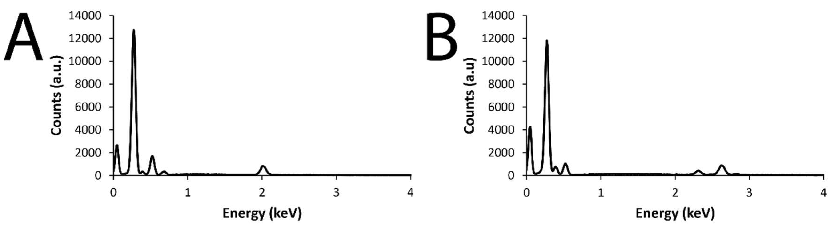
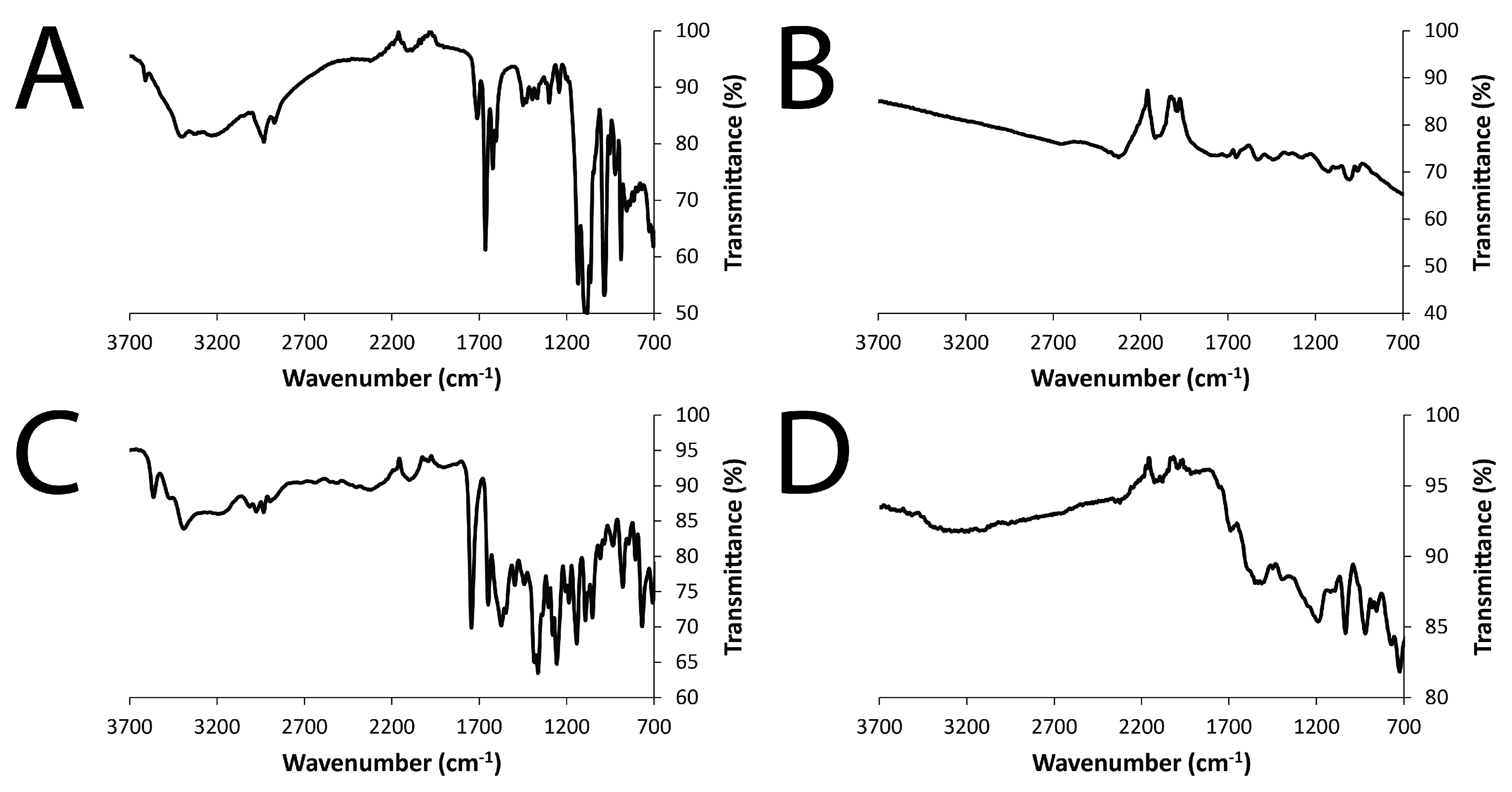
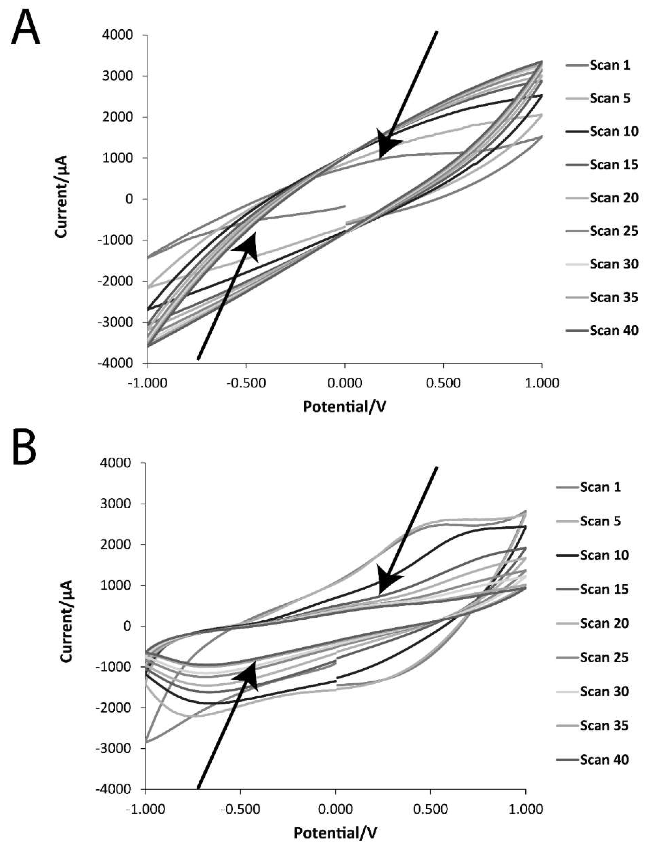
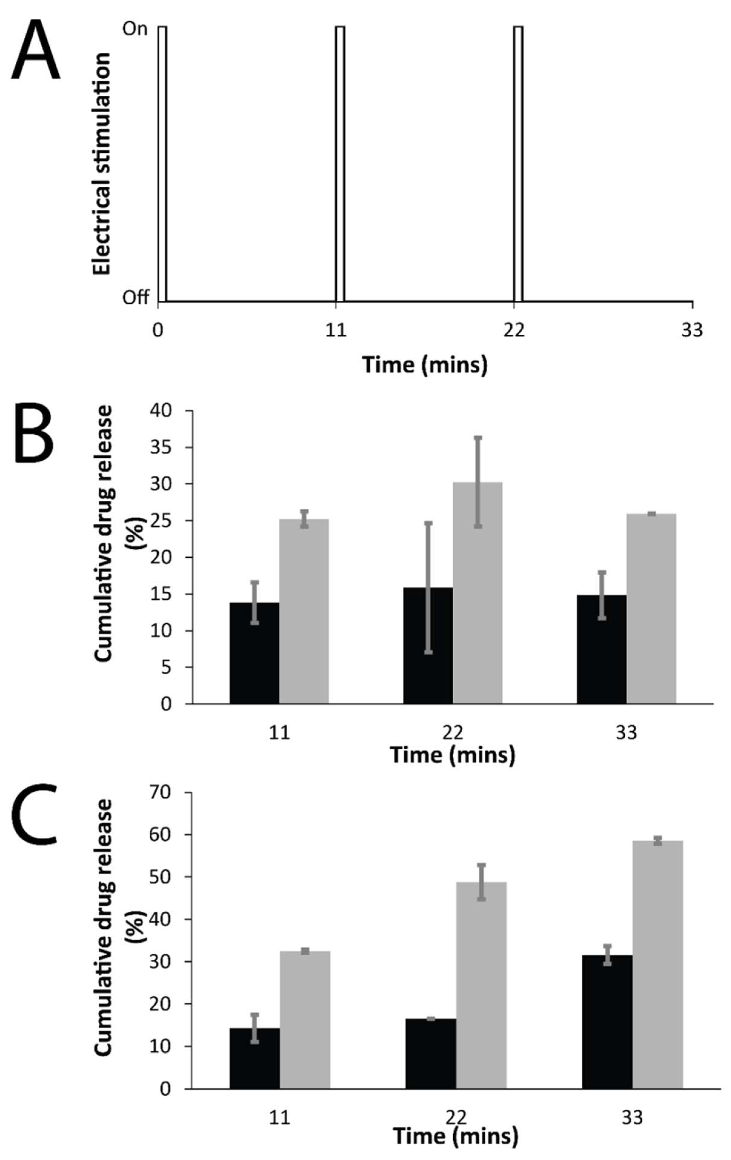
2. Results
3. Discussion
4. Materials and Methods
4.1. Materials
4.2. Preparation of Films via Electropolmerization
4.3. Scanning Electron Microscopy (SEM) Studies
4.4. Fourier-Transform Infrared (FTIR) Spectroscopy Studies
4.5. X-ray Diffraction (XRD) Studies
4.6. Electrochemical Characterization of Films
4.7. Drug-Delivery Studies
4.8. Calculating the Main Physical Descriptors of the Investigated Drugs
5. Conclusions
Author Contributions
Funding
Acknowledgments
Conflicts of Interest
References
- Barbe, C.; Bartlett, J.; Kong, L.; Finnie, K.; Calleja, G.; Lin, H.Q.; Larkin, M.; Calleja, S.; Bush, A. Silica Particles: A novel drug-delivery System. Adv. Mater. 2004, 16, 1959–1966. [Google Scholar] [CrossRef]
- Sahoo, S.K.; Labhsetwar, V. Nanotech approaches to drug delivery and imaging. Drug Discov. Today 2003, 8, 1112–1120. [Google Scholar] [CrossRef]
- Morgan, N.B. Medical shape memory alloy applications—The market and its products. Mater. Sci. Eng. A 2004, 378, 16–23. [Google Scholar] [CrossRef]
- Wei, X.; Liu, J. Power sources and electrical recharging strategies for implantable medical devices. Front. Energy Power Eng. 2008, 2, 1–13. [Google Scholar] [CrossRef]
- Khan, W.; Muntimadugu, E.; Jaffe, M.; Domb, A.J. Implantable Medical Devices. In Focal Controlled Drug Delivery; Domb, A., Khan, W., Eds.; Springer: Boston, MA, USA, 2014. [Google Scholar]
- Kurtz, S.M.; Devine, J.N. PEEK biomaterials in trauma, orthopedic, and spinal implants. Biomaterials 2007, 28, 4845–4869. [Google Scholar] [CrossRef] [PubMed] [Green Version]
- McAllister, B.S.; Haghighat, K. Bone augmentation techniques. J. Periodontol. 2007, 78, 377–396. [Google Scholar] [CrossRef] [PubMed]
- Qiu, Y.; Park, K. Environment-sensitive hydrogels for drug delivery. Adv. Drug Deliv. Rev. 2001, 53, 321–339. [Google Scholar] [CrossRef]
- Hoffman, A.S. Intelligent Polymers. In Controlled Drug Delivery: Challenge and Strategies; Park, K., Ed.; American Chemical Society: Washington, DC, USA, 1997; pp. 485–497. [Google Scholar]
- Bae, Y.H. Stimuli-Sensitive Drug Delivery. In Controlled Drug Delivery: Challenge and Strategies; Park, K., Ed.; American Chemical Society: Washington, DC, USA, 1997; pp. 147–160. [Google Scholar]
- Suzuki, M. Amphoteric polyvinyl alcohol hydrogel and electrohydrodynamic control method for artificial muscles. In Polymer Gels; DeRossi, D., Ed.; Plenum Press: New York, NY, USA, 1991; pp. 221–236. [Google Scholar]
- Kishi, R.; Ichijo, O.; Hirasa, O. Thermo-responsive devices using poly(vinylmethyl ether) hydrogels. J. Intell. Mater. Syst. Struct. 1993, 4, 533–537. [Google Scholar] [CrossRef]
- Kajiwara, K.; Ross-Murphy, S.B. Synthetic gels on the move. Nature 1992, 355, 208–209. [Google Scholar] [CrossRef]
- Osada, Y.; Okuzaki, H.; Hori, H. A polymer gel with electrically driven motility. Nature 1992, 355, 242–244. [Google Scholar] [CrossRef]
- Ueoka, Y.; Gong, J.; Osada, Y. Chemomechanical polymer gel with fish-like motion. J. Intell. Mater. Syst. Struct. 1997, 8, 465–471. [Google Scholar] [CrossRef]
- Zhao, Y.; Tavares, A.C.; Gauthier, M.A. Nano-engineered electro-responsive drug delivery systems. J. Mater. Chem. B 2016, 4, 3019–3030. [Google Scholar] [CrossRef]
- Clancy, K.F.A.; Hardy, J.G. Gene Delivery with Organic Electronic Biomaterials. Curr. Pharm. Des. 2017, 23, 3614–3625. [Google Scholar] [CrossRef] [PubMed]
- Qian, C.G.; Chen, Y.L.; Feng, P.J.; Xiao, X.Z.; Dong, M.; Yu, J.C.; Hu, Q.Y.; Shen, Q.D.; Gu, Z. Conjugated polymer nanomaterials for theranostics. Acta Pharmacol. Sin. 2017, 38, 764–781. [Google Scholar] [CrossRef] [PubMed]
- Repenko, T.; Rix, A.; Ludwanowski, S.; Go, D.; Kiessling, F.; Lederle, W.; Kuehne, A.J.C. Bio-degradable highly fluorescent conjugated polymer nanoparticles for bio-medical imaging applications. Nat. Commun. 2017, 8, 470. [Google Scholar] [CrossRef] [PubMed] [Green Version]
- Bendrea, A.D.; Cianga, L.; Cianga, I. Review paper: progress in the field of conducting polymers for tissue engineering applications. J. Biomater. Appl. 2011, 26, 3–84. [Google Scholar] [CrossRef] [PubMed]
- Guimard, N.K.; Gomez, N.; Schmidt, C.E. Conducting polymers in biomedical engineering. Prog. Polym. Sci. 2007, 32, 876–921. [Google Scholar] [CrossRef]
- Guiseppi-Elie, A. Electroconductive hydrogels: Synthesis, characterization and biomedical applications. Biomaterials 2010, 31, 2701–2716. [Google Scholar] [CrossRef] [PubMed]
- Hardy, J.G.; Lee, J.Y.; Schmidt, C.E. Biomimetic conducting polymer-based tissue scaffolds. Curr. Opin. Biotechnol. 2013, 24, 847–854. [Google Scholar] [CrossRef] [PubMed]
- Staples, N.A.; Goding, J.A.; Gilmour, A.D.; Aristovich, K.Y.; Byrnes-Preston, P.; Holder, D.S.; Morley, J.W.; Lovell, N.H.; Chew, D.J.; Green, R.A. Conductive Hydrogel Electrodes for Delivery of Long-Term High Frequency Pulses. Front. Neurosci. 2018, 11, 748. [Google Scholar] [CrossRef] [PubMed]
- Svirskis, D.; Travas-Sejdic, J.; Rodgers, A.; Garg, S. Electrochemically controlled drug delivery based on intrinsically conducting polymers. J. Control. Release 2010, 146, 6–15. [Google Scholar] [CrossRef] [PubMed]
- Pillay, V.; Tsai, T.-S.; Choonara, Y.E.; du Toit, L.C.; Kumar, P.; Modi, G.; Naidoo, D.; Tomar, L.K.; Tyagi, C.; Ndesendo, V.M.K. A review of integrating electroactive polymers as responsive systems for specialized drug delivery applications. J. Biomed. Mater. Res. Part A 2013, 102, 2039–2054. [Google Scholar] [CrossRef] [PubMed]
- Ma, H.; Liu, M.S.; Jen, A.K.-Y. Interface-tailored and nanoengineered polymeric materials for (opto)electronic devices. Polym. Int. 2009, 58, 594–619. [Google Scholar] [CrossRef]
- Stenger-Smith, J.D. Intrinsically electrically conducting polymers, synthesis, characterization, and their applications. Prog. Polym. Sci. 1998, 23, 57–79. [Google Scholar] [CrossRef]
- Gurunathan, K.; Murugan, A.V.; Marimuthu, R.; Mulik, U.P.; Amalnerkar, D.P. Electrochemically synthesised conducting polymeric materials for applications towards technology in electronics, optoelectronics and energy storage devices. Mater. Chem. Phys. 1999, 61, 173–191. [Google Scholar] [CrossRef]
- Long, Y.Z.; Li, M.M.; Gu, C.Z.; Wan, M.X.; Duvail, J.L.; Liu, Z.W.; Fan, Z.Y. Recent advances in synthesis, physical properties and applications of conducting polymer nanotubes and nanofibers. Prog. Polym. Sci. 2011, 36, 1415–1442. [Google Scholar] [CrossRef]
- Wallace, G.G.; Higgins, M.J.; Moulton, S.E.; Wang, C. Nanobionics: The impact of nanotechnology on implantable medical bionic devices. Nanoscale 2012, 4, 4327–4347. [Google Scholar] [CrossRef] [PubMed]
- Wadhwa, R.; Lagenaur, C.F.; Cui, X.T. Electrochemically controlled release of dexamethasone from conducting polymer polypyrrole coated electrode. J. Control. Release 2006, 110, 531–541. [Google Scholar] [CrossRef] [PubMed] [Green Version]
- Szunerits, S.; Teodorescu, F.; Boukherroub, R. Electrochemically triggered release of drugs. Eur. Polym. J. 2016, 83, 467–477. [Google Scholar] [CrossRef]
- Uppalapati, D.; Sharma, M.; Aqrawe, Z.; Coutinho, F.; Rupenthal, I.D.; Boyd, B.J.; Travas-Sejdic, J.; Svirskis, D. Micelle directed chemical polymerization of polypyrrole particles for the electrically triggered release of dexamethasone base and dexamethasone phosphate. Int. J. Pharm. 2018, 543, 38–45. [Google Scholar] [CrossRef] [PubMed]
- Ramtin, A.; Seyfoddin, A.; Coutinho, F.P.; Waterhouse, G.I.; Rupenthal, I.D.; Svirskis, D. Cytotoxicity considerations and electrically tunable release of dexamethasone from polypyrrole for the treatment of back-of-the-eye conditions. Drug. Deliv. Transl. Res. 2016, 6, 793–799. [Google Scholar] [CrossRef] [PubMed]
- Seyfoddin, A.; Chan, A.; Chen, W.T.; Rupenthal, I.D.; Waterhouse, G.I.; Svirskis, D. Electro-responsive macroporous polypyrrole scaffolds for triggered dexamethasone delivery. Eur. J. Pharm. Biopharm. 2015, 94, 419–426. [Google Scholar] [CrossRef] [PubMed]
- Schmidt, C.E.; Shastri, V.R.; Vacanti, J.P.; Langer, R. Stimulation of neurite outgrowth using an electrically conducting polymer. Proc. Natl. Acad. Sci. USA 1997, 94, 8948–8953. [Google Scholar] [CrossRef] [PubMed] [Green Version]
- Wang, Z.; Roberge, C.; Dao, L.H.; Wan, Y.; Shi, G.; Rouabhia, M.; Guidoin, R.; Zhang, Z. In vivo evaluation of a novel electrically conductive polypyrrole/poly(d,l-lactide) composite and polypyrrole-coated poly(d,l-lactide-co-glycolide) membranes. J. Biomed. Mater. Res. A 2004, 70, 28–38. [Google Scholar] [CrossRef] [PubMed]
- Mihardja, S.S.; Sievers, R.E.; Lee, R.L. The effect of polypyrrole on arteriogenesis in an acute rat infarct model. Biomaterials 2008, 29, 4205–4210. [Google Scholar] [CrossRef] [PubMed] [Green Version]
- Durgam, H.; Sapp, S.; Deister, C.; Khaing, Z.; Chang, E.; Luebben, S.; Schmidt, C.E. Novel degradable co-polymers of polypyrrole support cell proliferation and enhance neurite out-growth with electrical stimulation. J. Biomater. Sci. Polym. Ed. 2010, 21, 1265–1282. [Google Scholar] [CrossRef] [PubMed]
- George, P.M.; Lyckman, A.W.; LaVan, D.A.; Hegde, A.; Leung, Y.; Avasare, R.; Testa, C.; Alexander, P.M.; Langer, R.; Sur, M. Fabrication and biocompatibility of polypyrrole implants suitable for neural prosthetics. Biomaterials 2005, 26, 3511–3519. [Google Scholar] [CrossRef] [PubMed]
- Ludwig, K.A.; Uram, J.D.; Yang, J.; Martin, D.C.; Kipke, D.R. Chronic neural recordings using silicon microelectrode arrays electrochemically deposited with a poly(3,4-ethylenedioxythiophene) (PEDOT) film. J. Neural Eng. 2006, 3, 59–70. [Google Scholar] [CrossRef] [PubMed]
- Luo, S.C.; Mohamed Ali, E.; Tansil, N.C.; Yu, H.H.; Gao, S.; Kantchev, E.A.; Ying, J.Y. Poly(3,4-ethylenedioxythiophene) (PEDOT) nanobiointerfaces: Thin, ultrasmooth, and functionalized PEDOT films with in vitro and in vivo biocompatibility. Langmuir 2008, 24, 8071–8077. [Google Scholar] [CrossRef] [PubMed]
- Mattioli-Belmonte, M.; Giavaresi, G.; Biagini, G.; Virgili, L.; Giacomini, M.; Fini, M.; Giantomassi, F.; Natali, D.; Torricelli, P.; Giardino, R. Tailoring biomaterial compatibility: In vivo tissue response versus in vitro cell behavior. Int. J. Artif. Organs. 2003, 26, 1077–1085. [Google Scholar] [CrossRef] [PubMed]
- Wang, C.H.; Dong, Y.Q.; Sengothi, K.; Tan, K.L.; Kang, E.T. In-vivo response to polyaniline. Synth. Met. 1999, 102, 1313–1314. [Google Scholar] [CrossRef]
- Ru, X.; Shi, W.; Huang, X.; Cui, X.; Ren, B.; Ge, D. Synthesis of polypyrrole nanowire network with high adenosine triphosphate release efficiency. Electrochim. Acta 2011, 56, 9887–9892. [Google Scholar] [CrossRef]
- Available online: https://www.edax.com/resources/interactive-periodic-table (accessed on 16 June 2018).
- Sirivisoot, S.; Pareta, R.; Webster, T.J. Electrically controlled drug release from nanostructured polypyrrole coated on titanium. Nanotechnology 2011, 22, 085101. [Google Scholar] [CrossRef] [PubMed]
- Chougule, M.A.; Pawar, S.G.; Godse, P.R.; Mulik, R.N.; Sen, S.; Patil, V.B. Synthesis and Characterization of Polypyrrole (PPy) Thin Films. Soft Nanosci. Lett. 2011, 1, 3660. [Google Scholar] [CrossRef]
- Hardy, J.G.; Cornelison, R.C.; Sukhavasi, R.C.; Saballos, R.J.; Vu, P.; Kaplan, D.L.; Schmidt, C.E. Electroactive Tissue Scaffolds with Aligned Pores as Instructive Platforms for Biomimetic Tissue Engineering. Bioengineering 2015, 2, 15–34. [Google Scholar] [CrossRef] [PubMed]
- Hardy, J.G.; Hernandez, D.S.; Cummings, D.M.; Edwards, F.A.; Shear, J.B.; Schmidt, C.E. Multiphoton microfabrication of conducting polymer-based biomaterials. J. Mater. Chem. B 2015, 3, 5001–5004. [Google Scholar] [CrossRef] [Green Version]
- Hardy, J.G.; Villancio-Wolter, M.K.; Sukhavasi, R.C.; Mouser, D.J.; Aguilar, D., Jr.; Geissler, S.A.; Kaplan, D.L.; Schmidt, C.E. Electrical stimulation of human mesenchymal stem cells on conductive nanofibers enhances their differentiation towards osteogenic outcomes. Macromol. Rapid Commun. 2015, 36, 1884–1890. [Google Scholar] [CrossRef] [PubMed]
- Hardy, J.G.; Khaing, Z.Z.; Xin, S.; Tien, L.W.; Ghezzi, C.E.; Mouser, D.J.; Sukhavasi, R.C.; Preda, R.C.; Gil, E.S.; Kaplan, D.L.; et al. Into The Groove: Instructive Silk-Polypyrrole Films With Topological Guidance Cues Direct DRG Neurite Outgrowth. J. Biomater. Sci. Polym. Ed. 2015, 26, 1327–1342. [Google Scholar] [CrossRef] [PubMed] [Green Version]
- Cai, L.; Jiang, H.; Wang, L. Enhanced photo-stability and photocatalytic activity of Ag3PO4 via modification with BiPO4 and polypyrrole. Appl. Surf. Sci. 2017, 420, 43–52. [Google Scholar] [CrossRef]
- Allen, N.S.; Murray, K.S.; Fleming, R.J.; Saunders, B.R. Physical properties of polypyrrole films containing trisoxalatometallate anions and prepared from aqueous solution. Synth. Met. 1997, 87, 237–247. [Google Scholar] [CrossRef]
- Roozbahani, M.; Kharaziha, M.; Emadi, R. pH sensitive dexamethasone encapsulated laponite nanoplatelets: Release mechanism and cytotoxicity. Int. J. Pharm. 2017, 518, 312–319. [Google Scholar] [CrossRef] [PubMed]
- Shaker, M.A.; Shaaban, M.I. Formulation of carbapenems loaded gold nanoparticles to combat multi-antibiotic bacterial resistance: In vitro antibacterial study. Int. J. Pharm. 2017, 525, 71–84. [Google Scholar] [CrossRef] [PubMed]
- Zhao, D.M.; Zhang, X.H.; Feng, L.J.; Jia, L.; Wang, S.F. Simultaneous determination of hydroquinone and catechol at PASA/MWNTs composite film modified glassy carbon electrode. Colloids Surf. B Biointerfaces 2009, 74, 317–321. [Google Scholar] [CrossRef] [PubMed]
- Jiang, S.; Sun, Y.; Cui, X.; Huang, X.; He, Y.; Ji, S.; Shi, W.; Ge, D. Enhanced drug loading capacity of polypyrrole nanowire network for controlled drug release. Synth. Met. 2013, 163, 19–23. [Google Scholar] [CrossRef] [Green Version]
- Moulton, S.E.; Higgins, M.J.; Kapsa, R.M.I.; Wallace, G.G. Organic Bionics: A New Dimension in Neural Communications. Adv. Funct. Mater. 2012, 22, 2003–2014. [Google Scholar] [CrossRef]
- Nguyen, D.N.; Yoon, H. Recent Advances in Nanostructured Conducting Polymers: From Synthesis to Practical Applications. Polymers 2016, 8, 118. [Google Scholar] [CrossRef]
- Guarino, V.; Zuppolini, S.; Borriello, A.; Ambrosio, L. Electro-Active Polymers (EAPs): A Promising Route to Design Bio-Organic/Bioinspired Platforms with on Demand Functionalities. Polymers 2016, 8, 185. [Google Scholar] [CrossRef]
- Kim, S.; Jeong, J.-O.; Lee, S.; Park, J.-S.; Gwon, H.-J.; Jeong, S.I.; Hardy, J.G.; Lim, Y.-M.; Lee, J.Y. Effective gamma-ray sterilization and characterization of conductive polypyrrole biomaterials. Sci. Rep. 2018, 8, 3721. [Google Scholar] [CrossRef] [PubMed]
- Hardy, J.G.; Mouser, D.J.; Arroyo-Currás, N.; Geissler, S.; Chow, J.K.; Nguy, L.; Kim, J.M.; Schmidt, C.E. Biodegradable electroactive polymers for electrochemically-triggered drug delivery. J. Mater. Chem. B. 2014, 2, 6809–6822. [Google Scholar] [CrossRef]
- Hardy, J.G.; Amend, M.N.; Geissler, S.; Lynch, V.M.; Schmidt, C.E. Peptide-directed assembly of functional supramolecular polymers for biomedical applications: electroactive molecular tongue-twisters (oligoalanine-oligoaniline-oligoalanine) for electrochemically enhanced drug delivery. J. Mater. Chem. B. 2015, 3, 5005–5009. [Google Scholar] [CrossRef] [Green Version]
- Hathout, R.M.; El-Ahmady, S.H.; Metwally, A.A. Curcumin or bisdemethoxycurcumin for nose-to-brain treatment of Alzheimer disease? A bio/chemo-informatics case study. Nat. Prod. Res. 2017, 12, 1–10. [Google Scholar] [CrossRef] [PubMed]


| Drug | Dipole | Number of Hydrogen Bond Acceptors | Number of Hydrogen Bond Donors | Globularity | Flexibility | LogP (octanol/water) | Molecular Weight (Da.) |
|---|---|---|---|---|---|---|---|
| DMP | 1.7033 | 8 | 5 | 0.1110 | 5.3661 | 1.2640 | 472.4460 |
| MER | 9.2305 | 9 | 7 | 0.0265 | 8.8623 | −0.5960 | 437.5140 |
© 2018 by the authors. Licensee MDPI, Basel, Switzerland. This article is an open access article distributed under the terms and conditions of the Creative Commons Attribution (CC BY) license (http://creativecommons.org/licenses/by/4.0/).
Share and Cite
Shah, S.A.A.; Firlak, M.; Berrow, S.R.; Halcovitch, N.R.; Baldock, S.J.; Yousafzai, B.M.; Hathout, R.M.; Hardy, J.G. Electrochemically Enhanced Drug Delivery Using Polypyrrole Films. Materials 2018, 11, 1123. https://doi.org/10.3390/ma11071123
Shah SAA, Firlak M, Berrow SR, Halcovitch NR, Baldock SJ, Yousafzai BM, Hathout RM, Hardy JG. Electrochemically Enhanced Drug Delivery Using Polypyrrole Films. Materials. 2018; 11(7):1123. https://doi.org/10.3390/ma11071123
Chicago/Turabian StyleShah, Sayed Ashfaq Ali, Melike Firlak, Stuart Ryan Berrow, Nathan Ross Halcovitch, Sara Jane Baldock, Bakhtiar Muhammad Yousafzai, Rania M. Hathout, and John George Hardy. 2018. "Electrochemically Enhanced Drug Delivery Using Polypyrrole Films" Materials 11, no. 7: 1123. https://doi.org/10.3390/ma11071123