Article

Lanthanum Ferrite Ceramic Powders: Synthesis, Characterization and Electrochemical Detection Application

1
Faculty of Industrial Chemistry and Environmental Engineering, Politehnica University Timisoara, Piata Victoriei No. 2, RO-300006 Timisoara, Romania
2
Department of Oxide Materials Science and Engineering, Faculty of Applied Chemistry and Materials Science, Polytehnic University of Bucharest, Gh. Polizu Street no.1-7, 011061 Bucharest, Romania
*
Authors to whom correspondence should be addressed.
Materials 2020, 13(9), 2061; https://doi.org/10.3390/ma13092061
Submission received: 29 March 2020 / Revised: 22 April 2020 / Accepted: 27 April 2020 / Published: 29 April 2020

Abstract

:
The perovskite-type lanthanum ferrite, LaFeO3, has been prepared by thermal decomposition of in situ obtained lanthanum ferrioxalate compound precursor, LaFe(C2O4)3·3H2O. The oxalate precursor was synthesized through the redox reaction between 1,2-ethanediol and nitrate ion and characterized by chemical analysis, infrared spectroscopy, and thermal analysis. LaFeO3 obtained after the calcination of the precursor for at least 550–800 °C/1 h have been investigated by X-ray diffraction (XRD), field emission scanning electron microscopy (FE-SEM), transmission electron microscopy (TEM), and high-resolution transmission electron microscopy (HRTEM). A boron-doped diamond electrode (BDD) modified with LaFeO3 ceramic powders at 550 °C (LaFeO3/BDD) by simple immersion was characterized by cyclic voltammetry and tested for the voltammetric and amperometric detection of capecitabine (CCB), which is a cytostatic drug considered as an emerging pollutant in water. The modified electrode exhibited a complex electrochemical behaviour by several redox systems in direct relation to the electrode potential range. The results obtained by cyclic voltammetry (CV), differential-pulsed voltammetry (DPV), and multiple-pulsed amperometry proved the electrocatalytic effect to capecitabine oxidation and reduction and allowed its electrochemical detection in alkaline aqueous solution.

Graphical Abstract

1. Introduction

Perovskite-type oxides have received growing attention due to its diversity and performance [1,2,3,4,5]. Among the perovskite materials, lanthanum ferrite has been investigated intensively due to its potential applications in biosensors [6], (photo)catalysis [7], chemical sensors [8,9], electrochemistry field [10,11,12,13,14,15], magnetic, optical, and ferroelectric properties [16,17], environmentally-friendly pigments [18], spin electronic devices [19,20], and more.
In general, electrochemical behaviors of perovskite-type oxides LaFeO3 have been investigated for various applications due to its high oxidation–reduction characteristics and electrical conductivity, e.g., electrocatalysts for oxygen evolution [11], photoelectrochemical water oxidation [12], hydrogen storage [13], gas sensor [14,15,21], voltammetric/amperometric detection of biomolecules [22], solid oxide fuel cells, and electrode materials [23,24].
Many methods have been studied and developed for obtaining LaFeO3: sol-gel method [25,26], mechanochemical [27], hydrothermal processes [28], molten salt synthesis [29], combustion method [30,31], microwave-assisted synthesis [32], co-precipitation of hydroxides [33], and polymerizable complex method [34]. Up to date, the precise control of perovskite-type oxides size, shape, and surface is still a challenge. The thermal decomposition of complex compounds precursors represent a simple, efficient, and reliable method for the synthesis of mixed oxides characterized by small particles at a relatively low temperature that depend on the system composition and properties [35,36]. However, it is required to find well-defined redox reaction conditions for precursor generating, which represents the key element to get an effective synthesis process of the mixed oxides characterized through desired advanced properties [37,38]. This is the reason for studying the optimum synthesis conditions (e.g., pH, reaction temperature and time, reactant molar ratio) for each system and corresponding mixed oxides. For example, it is possible to conduct the oxidation of 1,2-ethanediol either glyoxylate anion [39,40] or oxalate anion, in the absence or presence of nitric acid, depending on the working conditions.
Herein, we reported an original method of in situ synthesis LaFe(C2O4)3·3H2O oxalate compound, as a precursor of LaFeO3. This consists of a redox reaction of 1,2-ethanediol with metallic nitrates, in the presence of nitric acid, which was suitable for synthesis of other metallic oxides precursors [37,41,42,43,44,45,46]. The polyheterometallic oxalates, coordinative compounds in which the ligands are tetradentate, are more important because their thermal decomposition generate a large amounts of gases, which leads to nanomaterials with porous structures, high surface area, and homogeneous distribution, which are essential properties for practical sensing applications. The electrocatalytic characteristics in direct relation to the intrinsic morphostructural properties of LaFeO3 toward the electrooxidation or electroreduction process of the target analytes are responsible for the performance of the electrochemical detection. Its utility has been reported for gas sensing, e.g., SO2, formaldehyde, ethanol [21], and only a few for amperometric/voltammetric of biomolecules, e.g., guanine and uric acid [22], which allows the development of high sensitivity-electrochemical detection.
Capecitabine (CCB), chemically N4-pentoxycarbonyl-5’-deoxy-5-fluorocytidine, is a pyrimidine analogue, which acts inside the body as 5-fluorouracil, and commonly used cytostatic drugs in chemotherapy for treating colorectal and breast cancer [47,48]. Since the number of cancer patients has increased considerably, the CCB consumption increased continuously. For example, one-fold from the year 2004 to 2008 in France, and the environmental concentration of 3 ng L−1 was reported [49], which led to its presence into the water environment at a higher concentration. Few analytical methods including HPLC (high-performance liquid chromatography), LC-MS (liquid chromatography and mass spectrometry), and LC-MS/MS have been reported for CCB determination in biological samples [50,51,52]. Recently, electrochemical methods have been developed for CCB determination in biological samples using unmodified/modified carbon-based electrode materials, which considered the reduction process of CCB [47,52,53]. It is important to explore new electrode materials and techniques to develop sensitive, fast, simple, and cheap method for CCB determination. In this context, LaFeO3 was tested as an electrocatalyst in relation to its electrochemical behavior including the presence of redox systems to allow the CCB detection at LaFeO3 modified commercial BDD electrode material. To the best of our knowledge, no study related to CCB detection using LaFeO3-based electrode material has been reported. LaFeO3-modified boron doped diamond electrode (LaFeO3/BDD) was successfully used for the voltammetric and amperometric detection of CCB including both processes of CCB oxidation and reduction.

2. Materials and Methods

2.1. Materials

Pure LaFeO3 was obtained through the calcination in the temperature range of 550 to 800 °C of the LaFe(C2O4)3·3H2O precursor, which was obtained previously. It was used as reagents: La(NO3)3·6H2O, (Fe(NO3)3·9H2O, 1,2-ethanediol, 2 M HNO3 solution from Merck (Darmstadt, Germany). NaOH and capecitabine (CCB) used was an analytical-grade reagent from Merck. The doubly distilled and deionised water were used for preparing all solutions.

2.2. Synthesis of LaFe(C2O4)3·3H2O Oxalate Precursor and LaFeO3

An aqueous solution containing lanthanum nitrate, iron nitrate, 1,2-ethanediol, and 2 M nitric acid in a molar ratio of 1:1:3:2 that assured the solution pH of 3 was heated for 20 min at about 100 °C in a water bath. The synthesis conditions related to acidic pH of 3 were set-up based on the stoichiometry of the redox reaction related to the molar ratio. The temperature of about 100 °C is aqueous solution boiling temperature and the reaction time of 20 min was considered as the time for consuming reactants. The obtained precursor was purified by washing with acetone and dried in air. VARIAN SpectrAA 110 atomic absorption spectrophotometer (Varian, Palo Alto, CA, USA) was used to determine the metal content and a Carlo Erba 1108 elemental analyzer (Carlo Erba, Milan, Italy) was used for carbon and hydrogen analyzing. LaFeC6O15H6, calc./found: La%: 27.09/27.21, Fe%: 10.89/10.62, C%: 14.04/13.95, and H%: 1.17/1.22. The precursor was calcinated in air under the temperature range from 550 to 800 °C with a heating rate of 10 °C min−1 for time duration of one hour to get the LaFeO3 ceramic powders.

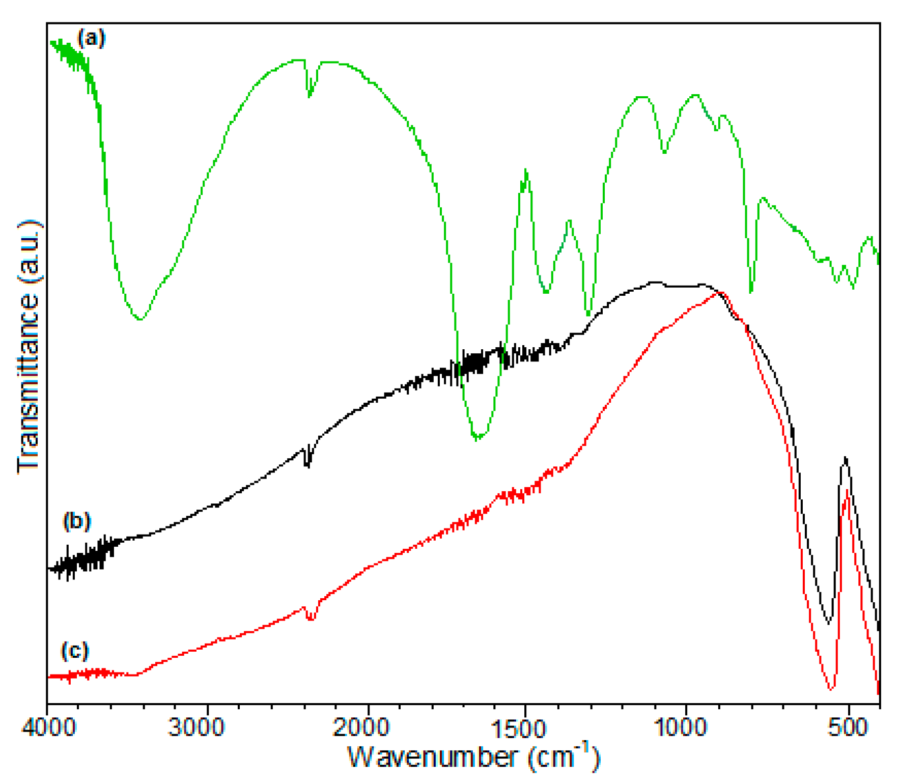
2.3. Characterization of LaFe(C2O4)3·3H2O Oxalate Precursor and LaFeO3

The comparative Fourier transform infrared spectroscopy (FTIR) spectra of the LaFeO3 and oxalate precursor were recorded based on KBr pellets using a Jasco FT-IR spectrophotometer (Jasco, Tokyo, Japan) under the range of 4000–400 cm−1. Thermal measurements (TG, DTG, DSC) were carried out in artificial air flow of 20 mL min−1 and heating rate of 10 K min−1 using a NETZSCH-STA 449C instrument (Netzsch, Selb, Germany) under the temperature range of 25–1000 °C, using alumina crucibles that were performed on the precursor.
X-ray diffraction (XRD) analyses performed at room temperature by a Rigaku Ultima IV diffractometer (Rigaku Co., Tokyo, Japan), using Ni-filtered CuKα radiation (λ = 1.5418 Ǻ) to characterize the phase purity and crystal structure of calcined powders. To refine the lattice parameters, the Rietveld method using the HighScore Plus 3.0e software (Rigaku Co., Tokyo, Japan) was applied.
Scanning electron microscopy (FE-SEM), using a high-resolution FEI QUANTA INSPECT F microscope (FEI Co., Eindhoven, The Netherlands) with a field emission gun, was used to assess the size and the agglomeration tendency of the LaFeO3 particles. In addition, transmission electron microscopy (TEM/HR-TEM) and selected area electron diffraction (SAED) investigations were performed for a high-accuracy assessment of the morphology and crystallinity degree of the constitutive LaFeO3 particles. Moreover, a TecnaiTM G2 F30 S-TWIN transmission electron microscope (FEI Co., Eindhoven, The Netherlands) was used to collect the bright-field and high-resolution images.

2.4. Electrochemical Detection Application

In order to easily test the electrochemical behaviour of LaFeO3 in the presence of capecitabine, the commercial BDD electrode produced by Windsor Scientific Ltd. (Slough, UK). For electroanalytic use, with a boron content of about 0.1, it was modified by simple immersion in 5 mg mL−1 LaFeO3 suspension. Unmodified and LaFeO3-modified boron-doped diamond electrode (LaFeO3/BDD) were electrochemically characterized and tested for detection using Autolab Pontentiostat/Galvanostat PGStat 302 (EcoChemie, Utrecht, The Netherlands) controlled with GPES 4.9 software (EcoChemie, Utrecht, The Netherlands) and a classical three-electrode cell. The saturated calomel electrode as a reference (SCE), the platinum electrode as a counter-electrode and the LaFeO3 modified/unmodified boron-doped diamond electrode modified (LaFeO3/BDD and BDD) as working electrodes were used for the electrochemical applications. The LaFeO3/BDD electrode was prepared by simple immersion prior to each electrochemical experiment running. The reproducibility of the modified surface was checked after stabilization by cyclic voltammetry and confirmed by a complete overlay of the cyclic voltammograms.
After electrode immersion, 10 continuous repetitive cyclic voltammograms within the various potential ranges related to each application conditions were applied for the electrochemical stabilization of the electrode. The cyclic voltammetry, differential-pulsed voltammetry, and multiple-pulsed amperometry were applied for the electrochemical characterization and the detection applications.
All measurements were carried out in 0.1 M sodium hydroxide supporting electrolyte at room temperature without a temperature control.

3. Results and Discussion

3.1. Characterization of LaFe(C2O4)3·3H2O Oxalate Precursor

The method of the synthesis of the oxalate complex compound is based on the oxidation reaction of 1,2-ethanediol by the nitrate ion.
3C2H4(OH)2 + (La3+ + 3NO3) + (Fe3+ + 3NO3) + 2(H++NO3) → LaFe(C2O4)3·3H2O(s) + 8NO(g) + 7H2O(g)
NO(g) + ½ O2(g) → NO2(g)
The IR spectrum of oxalate precursor (Figure 1a) reveals the presence of water [3410 cm−1OH, νH2O), 794 cm−1 (lattice water)] [41,54], oxalate anions [as bidentate ligand: 1642 cm−1asym (O=C–O)) + δsym (HOH), 1308 cm−1sym (O=C–O), 1051 cm−1(C–O))] [42]; as tetradentate ligand: 1445 cm−1sym (OCO)), 915 cm−1(OCO))] [55], and metal-oxygen linkages [600–400 cm−1: ν(La–O) and ν(Fe–O) vibrations]. The IR spectra of LaFeO3 obtained after calcination at 550 °C (Figure 1b) and 700 °C (Figure 1c) exhibit only the bands characteristic for vibrations ν(La–O) and ν(Fe–O) in the range of 600–400 cm−1 [43,44].
No other bands characteristic to the presence of the water and the oxalate anion can be noticed due to thermal decomposition of the precursor, known as the LaFe(C2O4)3·3H2O oxalate compound, which are further detailed and presented.
La(III)-Fe(III) oxalate trihydrate decomposes (Figure 2) via three steps with the formation of a carbonate intermediate.
The first step of thermal decomposition of LaFe(C2O4)3·3H2O (25–160 °C, mass loss: found 11.10%, calcd. 10.53%) is associated with an endothermic effect, and the mass loss is attributed to the evolving of the lattice water [43].
The next decomposition step (160–280 °C) characterized by an endothermic effect is due to the degradation of the terminal oxalate anions (mass loss, found 10.40%, calcd. 10.92%) with the formation of a carbonate intermediate, [CO3La(C2O4)FeCO3] [44,45].
The strong exothermic effect that characterizes the third step (280–550 °C) is attributed to the oxidative degradation of CO 3 2 and the last C 2 O 4 2 with the formation of the lanthanum ferrite, which retains small amounts of carbon dioxide (mass loss: found 34.03%, calcd. 34.32%). In the temperature range of 550–700 °C, the mass loss corresponds to the removal of carbon dioxide.
Simultaneously, with this endothermic decomposition step, an exothermic process of crystallization, involving the formation of the perovskite skeleton from an amorphous phase also takes place, so that, only a small and flattened endothermic peak, as a resultant of the two opposite processes, can be observed on the DSC curve [45,46].
The thermal analysis suggests the following sequence for decomposition of LaFe(C2O4)3·3H2O in static air atmosphere.
LaFe ( C 2 O 4 ) 3 · 3 H 2 O ( s )   ( I ) 3   H 2 O ( g )   +   LaFe ( C 2 O 4 ) 3 ( s )
LaFe ( C 2 O 4 ) 3 ( s )   ( II )   2   C O ( g ) + CO 3 LaC 2 O 4 FeC O 3 ( s )
CO 3 LaC 2 O 4 FeC O 3 ( s )   + 1 / 2 O 2   ( III )   4   CO 2 ( g ) + LaFeO 3 ( s )

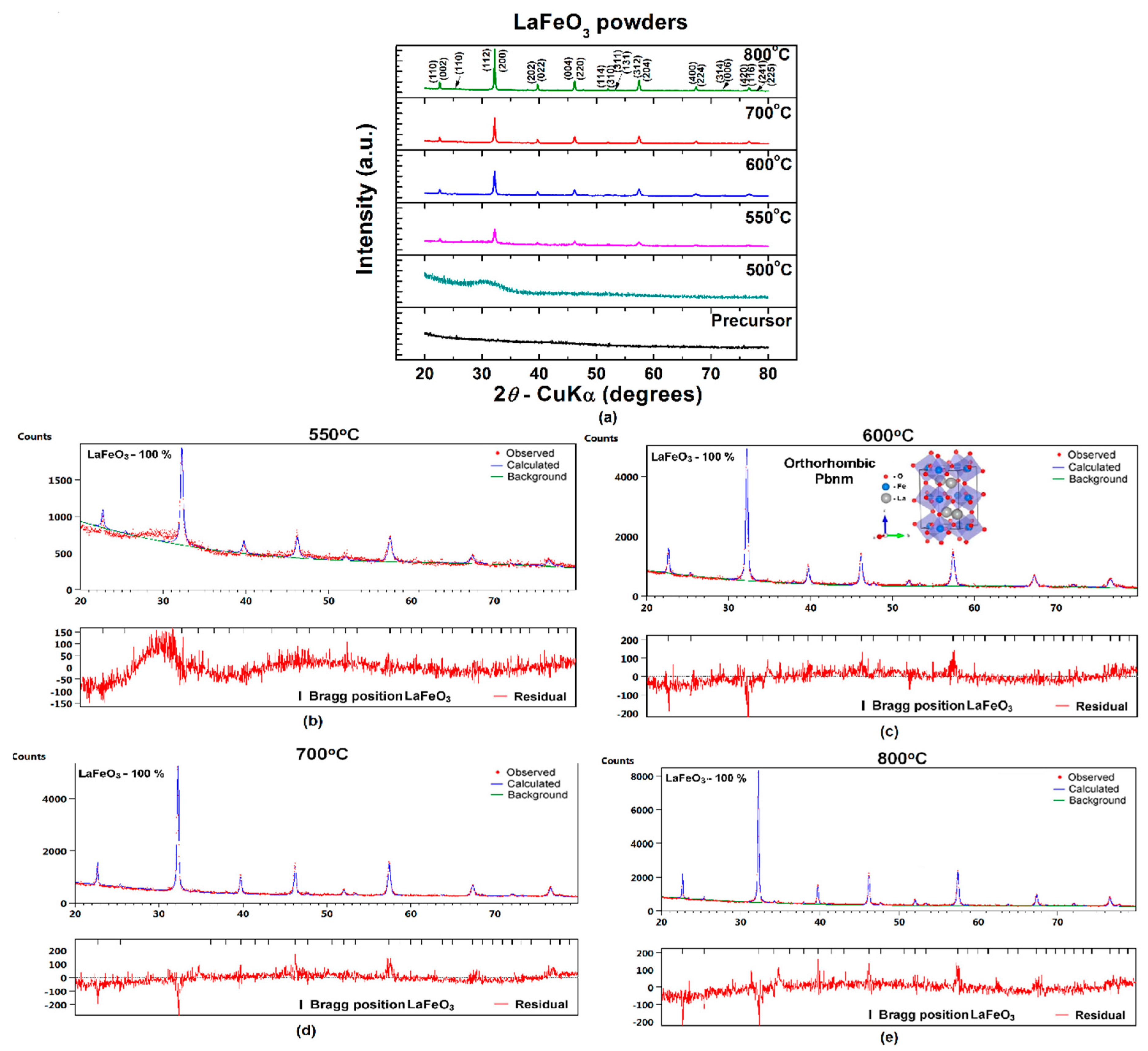
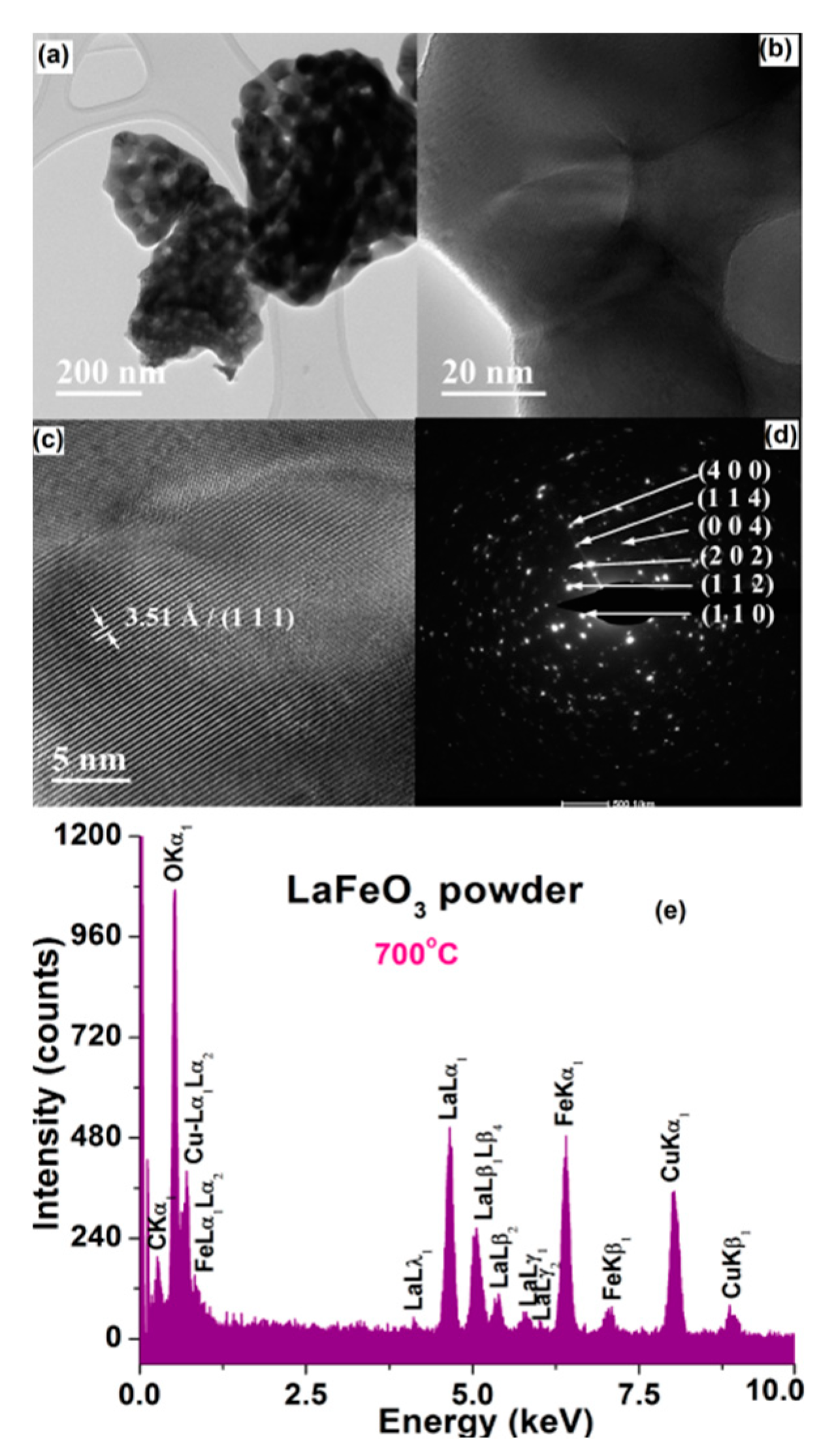
3.2. Characterization of LaFeO3 Powders

The room-temperature X-ray diffraction patterns presented in Figure 3a show that the as-prepared precursor, as well as the powder that resulted after calcination at 500 °C, are amorphous, while, in the powders obtained at higher temperatures (T ≥ 550 °C), the main reflections specific to the LaFeO3 perovskite phase with orthorhombic Pbnm(62) structure were detected. For the powder calcined at 550 °C, the broader profile and the lower intensity of the diffraction peaks as well as the higher background of the corresponding diffraction pattern suggested that a significant amount of an amorphous phase still persists in the sample. The increase of the annealing temperature from 550 to 800 °C induces the reduction of the amount of amorphous phase, concurrently with the gradual increase of the crystallintiy degree and purity of the perovskite phase reflected in the enhancement of intensity of the main diffraction lines of LaFeO3.
The results of the Rietveld analysis of the room temperature diffraction data of LaFeO3 powders thermally treated at various temperatures in the temperature range of 550–800 °C are presented in Figure 3b–e. The quality of the samples is indicated by the parameters provided by the Rietveld refinement, R expected (Rexp), R profile (Rp), weighted R profile (Rwp), and goodness of fit (χ2), which show values in good agreement with other literature data [56] (Table 1).
The dependence of the lattice parameters, a, b, and c and, consequently, of the unit cell volume V, on the calcination temperature, exhibits a decreasing trend (Table 1). The progress of the crystallization process involves a clear contraction of the unit cell, as shown in Figure 4a.
Even if the coarsening process evolves with the increase of annealing temperature, the values of the average crystallite size, also determined from the XRD data, are kept in the nanometric range, varying from 17.5 nm for the LaFeO3 powder calcined at 550 °C to 36.7 nm in the case of the powder calcined at 800 °C. The significantly lower rate of increasing both the proportion of crystalline phase with respect to the amount of amorphous phase and the crystallite size with the temperature increase for LaFeO3 particles investigated in this study, in comparison to the steeper increase of these characteristics in the case of BiFeO3 powders calcined in the same temperature range reported earlier, which can be related to the higher refractoriness of lanthanum relative to that of Bi in the ferrite-type compounds [44]. As expected, the increase of the crystallite size determines a lattice relaxation proved by the clear decrease in the lattice micro-strains when increasing the calcination temperature (Figure 4b). Therefore, the increase of the crystallite size involves the reduction in the inter-atomic spacing, which results in the contraction of the unit cell.
For all the powders under investigation, the FE-SEM analyses showed the clear tendency of the LaFeO3 particles to form large (of few tens of microns), non-uniform (as shape and size) aggregates, with a spongeous aspect, as indicated by the FE-SEM overall images of Figure 5a,c,e,g.
The higher magnification FE-SEM images have taken in order to notice the structuring inside the aggregates showed that the powder calcined at 550 °C consists of very small-sized particles (most of them below 15 nm). The value of the average particle size is difficult to be estimated mainly because of the presence of a significant amount of amorphous phase (as XRD data indicated), which impeded the clear distinction of the particle’s boundaries (Figure 5b). The structuring inside the aggregates becomes better highlighted in the case of the powders that resulted after annealing at 600 and especially at 700 °C (Figure 5d,f). The presence of the amorphous phase is still noticed along with the crystalline aggregates in the LaFeO3 powder calcined at 600 °C (Figure 5d), while the aggregates of the powder calcined at 600 °C seems to consist of porous agglomerates of 300–500 nm, inside which nanometric particles (of 25–35 nm), linked together by necks and form 3D networks are observed (Figure 5f). Taking into account the values of the average crystallite size determined from the XRD data, one can assume that the nanoparticles of the LaFeO3 powders resulted after annealing in the temperature range of 550–700 °C are single crystals. The FE-SEM detail image of Figure 5h indicates that the LaFeO3 powder calcined at 800 °C consists of partially-sintered blocks, formed of nano-sized crystalline grains (with an average grain size <DG> = 69.4 nm) with well-defined boundaries and triple junctions, between which a small amount of intergranular porosity is detected. In some regions, the grain growth process, favored by the higher annealing temperature, induced a coalescence of some smaller grains, with the formation of the larger ones (of 200–300 nm) as well as the concurrent modification of some intergranular pores into intragranular pores (Figure 5h). Considering the value of the average crystallite size obtained from the XRD data, the values of the grain/particle size inside the agglomerates estimated from the FE-SEM observations suggest that the grains of the LaFeO3 powder calcined at 800 °C exhibit a polycrystalline nature. Thus, we were able to estimate that the smaller grains consist of 2–3 crystallites, while the larger ones are formed from 6–10 crystallites.
The FE-SEM observations are sustained by the results of TEM investigations. Thereby, the lower magnification TEM images of Figure 6a and Figure 7a clearly indicate the presence of the submicronic aggregates of various sizes and with irregular shapes. The higher magnification TEM images emphasized the structuring of the aggregates, which are formed by particles of 10–20 nm in the case of the powder calcined at 550 °C (Figure 6b) and of ~40–50 nm for powder calcined at 700 °C (Figure 7b).
The HRTEM image of the LaFeO3 sample calcined at 550 °C indicate the presence of some small particles with sizes of 6.5–10 nm. Inside these particles, oriented fringes spaced at 1.60 Å corresponding to the crystalline plane (2 0 4) were noticed (Figure 6c). It is difficult to notice these small particles because they are usually embedded into an amorphous matrix. In the case of the powder calcined at 700 °C, the lower amount of amorphous phase allowed to better visualize the long-range ordered fringes, so that the HRTEM image clearly emphasizes long-range ordered fringes spaced at 3.51 Å, corresponding to the crystalline plane (1 1 1) (Figure 7c).
For both powders, the high crystallinity degree of the randomly oriented particles is also pointed out by the bright spots, forming concentric diffraction rings, assigned to several crystalline planes of the perovskite LaFeO3 phase in the specific SAED (selected area electron diffraction) patterns of Figure 6d and Figure 7d.
EDX (energy-dispersive X-ray analysis) analyses were also performed in order to determine the elemental composition of the LaFeO3 powders under investigation. The EDX spectra show only the presence of La, Fe, and O species, which indicates that no contamination took place during the powders processing (Figure 6e and Figure 7e).
The morphostructural properties of the LaFeO3 are different functions of the calcination temperature, and the lowest particles were obatined at a relatively low temperature of 550 °C in comparison with other materials synthesized by the same method [43,44,45,46].

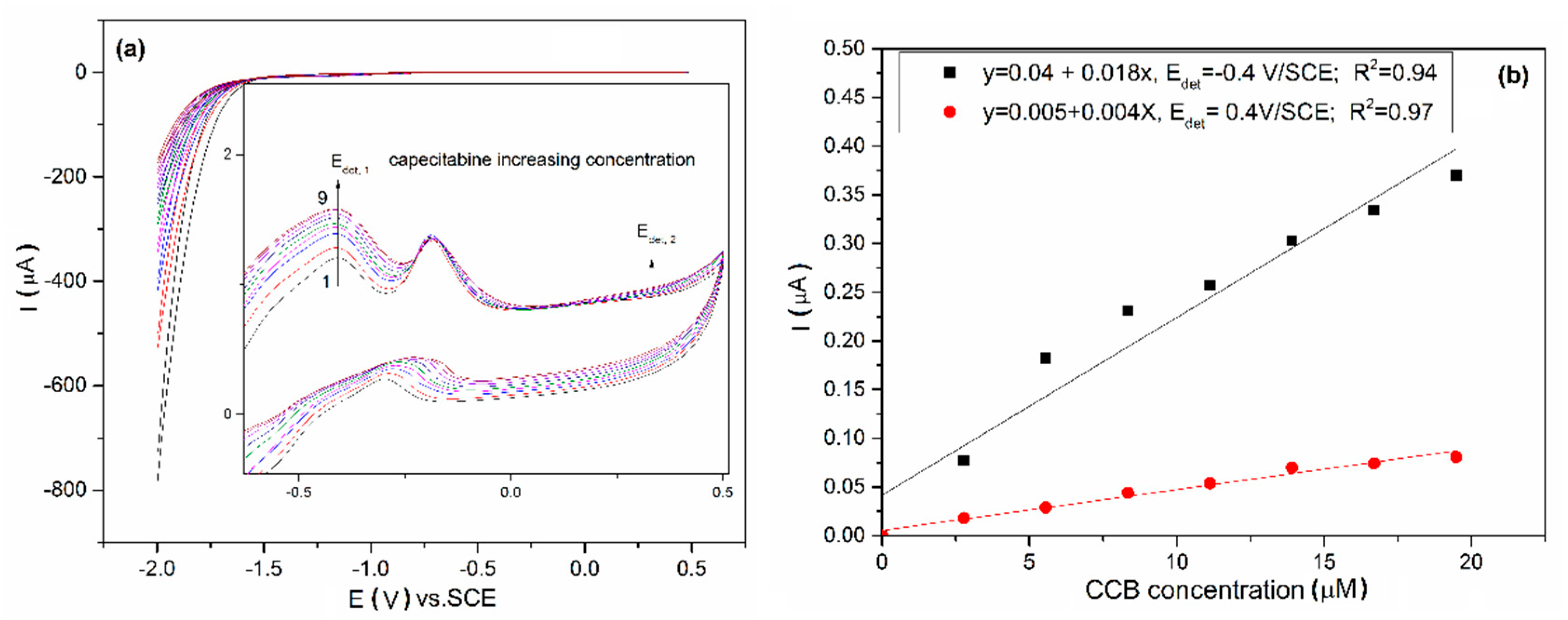
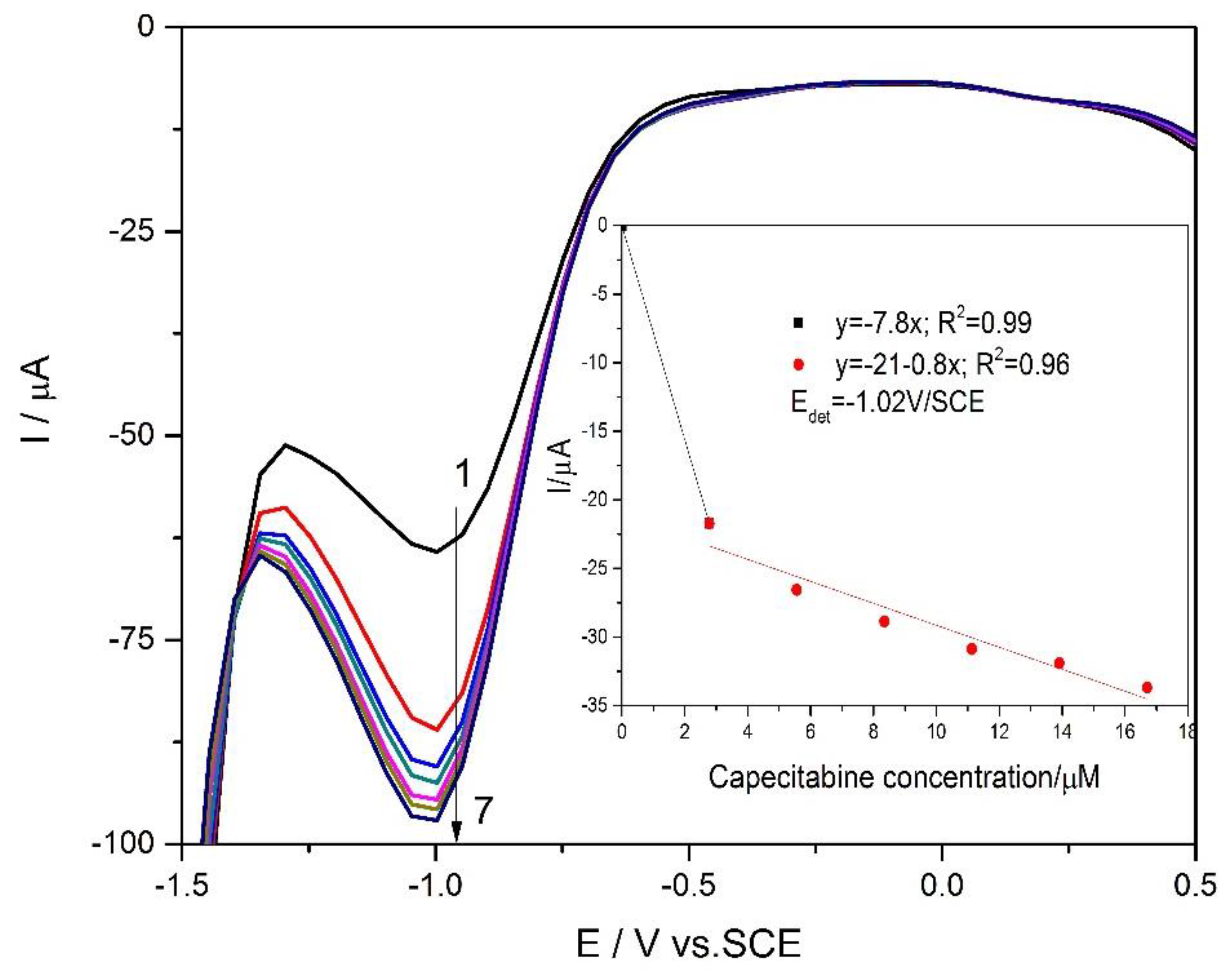
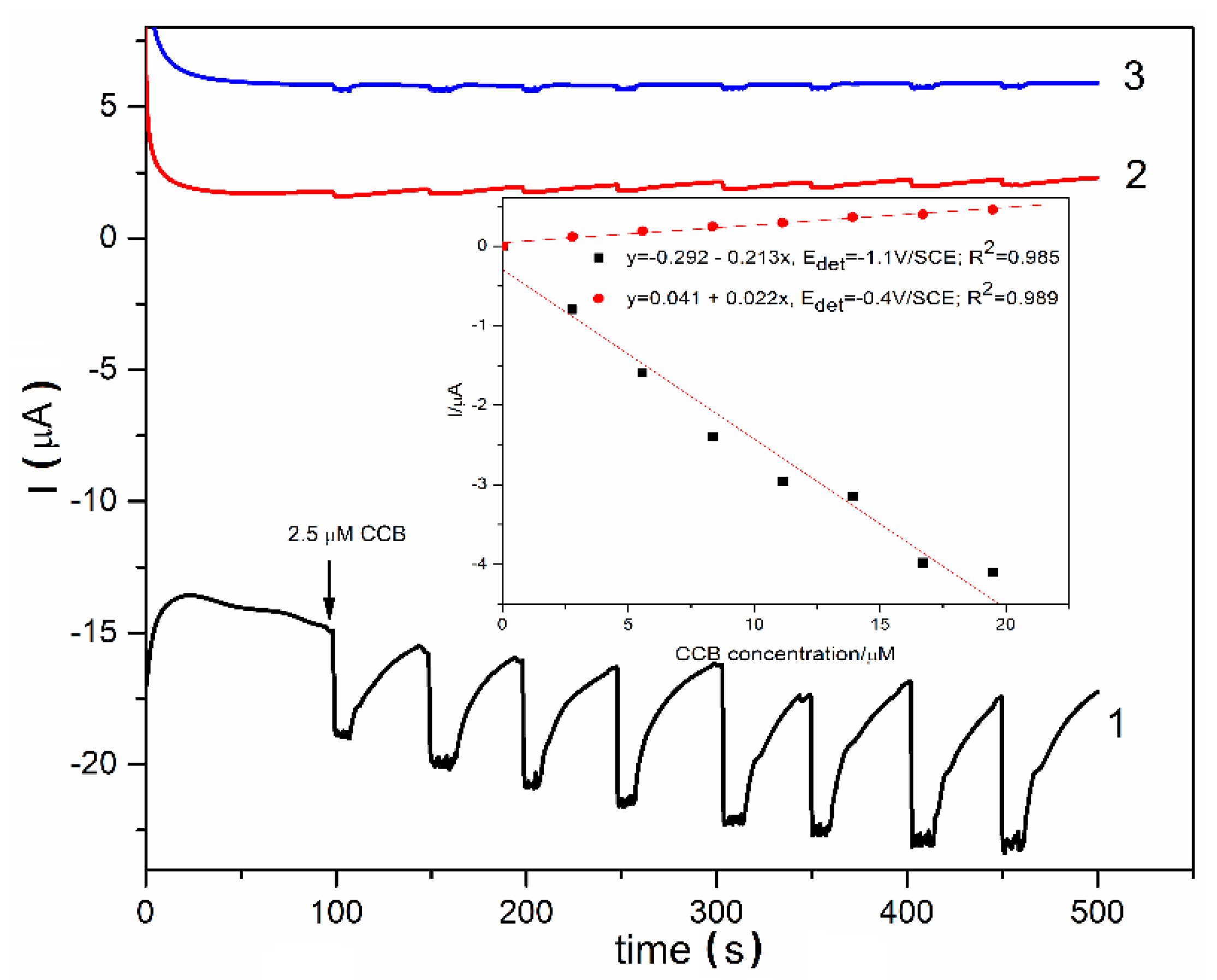
3.3. Electrochemical Characterization and Detection Application

Based on the morphostructural properties of LaFeO3 ceramic powders, which were presented above and discussed in relation with the electroactivity capacity and electro-catalytical effect, for testing in electrochemical detection application LaFeO3 synthesized at 550 °C. The comparative electrochemical behaviours of unmodified and LaFeO3-modified BDD electrodes in alkaline medium was studied by cyclic voltammetry (CV) and the results are presented in Figure 8a,b. It can be noticed that the oxidation and coupled reduction peak for LaFeO3/BDD, which can be attributed to Fe2+/Fe3+ and Fe3+/Fe4+ redox systems according to the literature [12]. In addition, capacitive component of the current is much larger in comparison with the unmodified BDD electrode, which is expected for the electrocatalyst electrode material. Thus, a depolarization effect toward the oxygen evolution reaction and a polarization effect on the hydrogen evolution reaction were manifested through LaFeO3 on the BDD electrode surface. The electrocatalytic activity of the LaFeO3 sample is relevant with the crystalline size and the rate of oxygen migration from bulk towards the surface [57,58], which relates to the applied potential range.
Both unmodified and LaFeO3/BDD electrodes were tested in the presence of 5 µM CCB and no signal was found at an unmodified BDD electrode (the results are not shown here). The behavior of the LaFeO3/BDD electrode in the presence of various CCB concentrations ranged from 2.5 to 22.5 µM is shown in Figure 9a and it can be noticed that the current grows with CCB concentration increasing for two potential values, which can be considered as detection potentials. The redox systems manifested to −0.4 V/SCE and +0.4 V/SCE showed a strong promoting effect and high stability towards the electrochemical oxidation of CCB. The linear dependence between the current and CCB concentration was found at the potential value of −0.4 V/SCE and 0.4 V/SCE (Figure 9b). Under this potential range, no increase of any cathodic reduction peak was noticed for the LaFeO3/BDD electrode, which informed that the CCB oxidation is not reversible.
Taking into account that differential-pulsed voltammetry technique (DPV) exhibit the advantages of the higher sensitivity in comparison with CV, this technique was tested in various working conditions related to modulation amplitude and the step potential. The detection results depended by the direction of the applied potential scanning. No reproducible results were achieved for anodic scanning, which suggest that this technique is not appropriate for the CCB detection based on its oxidation process. However, very good results were reached by potential scanning in a cathodic sense under a modulation amplitude of 0.2 V and step potential of 0.05 V (Figure 10). Cathodic peak characteristics to CCB reduction was found at the potential value of −1.02 V/SCE, which should be due to the oxidation-based activation of the electrode surface. The cathodic peak current increased linearly with CCB concentration for two concentration ranges below 2.5 µM CCB and ranged from 2.5 µM to 17 µM CCB (see inset of Figure 10).
One of the most desired technique for electro-detection is chronoamperometry (CA) due to its simplicity and easy application, which was tested for the LaFeO3/BDD electrode in CCB detection. A very low signal was achieved (the results are not shown here), which led to testing three-levels potential based multiple-pulsed amperometry (MPA) as a variant of CA. The potential levels were selected based on the results of voltammetry techniques of both CV and DPV, considering the oxidation and reduction processes related to CCB. According to the literature [59], the oxidation steps of the CCB to 5″-deoxy-5-fluorocytudine and further to five fluorouracil and its reduction to dihydrofluorouracil should be considered based on the previous results of CV and DPV related to the potential range and scanning sense. Amperograms recorded by MPA are presented in Figure 11. The pulses were applied continuously using the following scheme:
(a) −1.1 V/SCE for a duration of 50 ms, where CCB reduction occurred,
(b) −0.4 V/SCE for a duration of 50 ms, where CCB oxidation occurred involving Fe2+/Fe3+ redox system,
(c) 0.4 V/SCE for a duration of 50 ms, where further CCB oxidation occurred involving Fe3+/Fe4+ redox system besides an oxygen evolution reaction starting.
The electroanalytical parameters for CCB detection related to sensitivity, the lowest limit of detection, and the limit of quantification determined for each above-mentioned technique are gathered in Table 2.
In comparison with reported results related to CCB electrochemical detection presented in Table 3, it can be noticed that the superiority of this electrode in relation with a very good limit of detection (LOD) and considering its availability for either CCB oxidation or CCB reduction with the mention that the literature reported only the detection procedure based on CCB reduction.

4. Conclusions

In the present work, LaFeO3 powders were synthesized using a new method, based on the thermal decomposition of in situ obtained LaFe(C2O4)3·3H2O compound.
Single phase LaFeO3 powders with orthorombic Pbnm perovskite structure were obtained after annealing at temperatures ranging between 550 and 800 °C. The crystallinity degree reflected in the values of the average crystallite size increases as the annealing temperature increased. Concurrently, a contraction of the unit cell volume is induced by the increase of the annealing temperature. Small particles, with sizes below 20 nm and with a high aggregation tendency, were obtained after annealing at 550 °C. Even if the increase of the annealing temperature at 800 °C induces a coarsening process, the size of the LaFeO3 particles was kept in the nanometric range (~70 nm). However, a clear tendency to form partially-sintered blocks due to the coalescence of the small particles into larger, polyhedral grains with well-defined boundaries was noticed for the LaFeO3 sample calcined at 800 °C.
The LaFeO3/BBD electrode exhibited the electrocatalytic activity toward the capecitabine (CCB) oxidation and reduction depending on the applied potential range and scanning direction that influenced the iron redox systems, considered as a basis for developing detection methods. Cyclic voltammetry (CV) and differential-pulsed voltammetry (DPV) techniques operated under specific working conditions allowed us to develop the CCB voltammetric detection method. The best analytical performance was reached using DPV based on the CCB reduction mechanism. Multiple-pulsed amperometry operated under three potential levels implied CCB oxidation and a reduction mechanism, which allows CCB detection in the same time at the two potential values of −1.1 V/SCE and −0.4 V/SCE corresponding to CCB reduction and respective oxidation. The results of electroanalytical performance for CCB detection and its flexibility for anodic and/or cathodic detection made the LaFeO3/BDD electrode have great potential for selective or simultaneous detection of CCB in aqueous matrix.

Author Contributions

Methodology, R.D. and F.M. Investigation, S.N., C.P., B.V., and A.S.; Data curation, R.D., S.N., A.I., C.P., B.V., A.S., and F.M. Writing—original draft preparation, R.D., A.I., and F.M. Writing—review and editing, R.D. and F.M. All authors have read and agreed to the published version of the manuscript.

Acknowledgments

This work was supported partially by research grant GNaC2018-ARUT, no. 1350/01.02.2019, financed by Politehnica University of Timisoara and partially by a grant of the Romanian Ministery of Research and Innovation, project number PN-III-P1-1.2-PCCDI-2017-0245/26 PCCDI/2018 (SUSTENVPRO), within PNCDI III.

Conflicts of Interest

The authors declare no conflict of interest.

References

  1. Addabbo, T.; Bertocci, F.; Fort, A.; Gregorkiewitz, M.; Mugnaini, M.; Spinicci, R.; Vignoli, V. Gas sensing properties and modeling of YCoO3 based perovskite materials. Sens. Actuators B Chem. 2015, 221, 1137–1155. [Google Scholar] [CrossRef]
  2. Natile, M.M.; Ponzoni, A.; Concina, I.; Glisenti, A. Chemical tuning versus microstructure features in solid-state gas sensors: LaFe1−xGaxO3, a case study. Chem. Mater. 2014, 26, 1505–1513. [Google Scholar] [CrossRef]
  3. Kersen, U. Microstructural and surface characterization of solid state sensor based on LaFeO3−σ oxide for detection of NO. Analyst 2001, 126, 1377–1381. [Google Scholar] [CrossRef] [PubMed]
  4. Phan, T.T.N.; Nikoloski, A.N.; Bahri, P.A.; Li, D. Facile fabrication of perovskite-incorporated hierarchically mesoporous/macroporous silica for efficient photoassisted-Fenton degradation of dye. Appl. Surf. Sci. 2019, 491, 488–496. [Google Scholar] [CrossRef]
  5. Takalkar, G.; Bhosale, R.; AlMomani, F. Combustion synthesized A0.5Sr0.5MnO3−δ perovskites (where, A = La, Nd, Sm, Gd, Tb, Pr, Dy and Y) as redox materials for thermochemical splitting of CO2. Appl. Surf. Sci. 2019, 489, 80–91. [Google Scholar] [CrossRef]
  6. Wang, G.; Sun, J.; Zhang, W.; Jiao, S.; Fang, B. Simultaneous determination of dopamine, uric acid and ascorbic acid with LaFeO3 nanoparticles modified electrode. Microchim. Acta 2009, 164, 357–362. [Google Scholar] [CrossRef]
  7. Kumar, D.; Jayavel, R. Facile hydrothermal synthesis and characterization of LaFeO3 nanospheres for visible light photocatalytic applications. J. Mater. Sci. Mater. Electron. 2014, 25, 3953–3961. [Google Scholar] [CrossRef]
  8. Bai, S.L.; Shi, B.J.; Ma, L.J.; Yang, P.C.; Liu, Z.Y.; Li, D.Q.; Chen, A.F. Synthesis of LaFeO3 catalytic materials and their sensing properties. Sci. China Ser. B Chem. 2009, 52, 2106–2113. [Google Scholar] [CrossRef]
  9. Ciambelli, P.; Cimino, S.; Rossi, S.D. AFeO3 (A = La, Nd, Sm) and LaFe1xMgxO3 perovskites as methane combustion and CO oxidation catalysts: Structural, redox and catalytic properties. Appl. Catal. B-Environ. 2001, 29, 239–250. [Google Scholar] [CrossRef]
  10. Kuščer, D.; Hrovat, M.; Holc, J.; Bernik, S.; Kolar, D. Some characteristics of Al2O3− and CaO-modified LaFeO3-based cathode materials for solid oxide fuel cells. J. Power Sources 1996, 61, 161–165. [Google Scholar] [CrossRef]
  11. Grygar, T. Electrochemical reactions of La(Ni,Cr)O3 in acidic aqueous solutions. J. Solid State Electrochem. 1999, 3, 412–416. [Google Scholar] [CrossRef]
  12. Peng, Q.; Shan, B.; Wen, Y.; Chen, R. Enhanced charge transport of LaFeO3 via transition metal (Mn, Co, Cu) doping for visible light photoelectrochemical water oxidation. Int. J. Hydrog. Energy 2015, 40, 15423–15431. [Google Scholar] [CrossRef]
  13. Yuan, Y.; Dong, Z.; Li, Y.; Zhang, L.; Zhao, Y.; Wang, B.; Han, S. Electrochemical properties of LaFeO3-rGO composite. Proc. Nat. Sci. Mater. 2017, 27, 88–92. [Google Scholar] [CrossRef]
  14. Zhang, Q.; Shan, X.; Fu, Y.; Liu, P.; Li, X.; Liu, B.; Zhang, L.; Li, D. Electrochemical determination of the anticancer drug capecitabine based on a graphene-gold nanocomposite-modified glassy carbon electrode. Int. J. Electrochem. Sci. 2017, 12, 10773–10782. [Google Scholar] [CrossRef]
  15. Haron, W.; Wisitsoraatb, A.; Wongnawa, S. Nanostructured perovskite oxides—LaMO3 (M=Al, Co, Fe) prepared by co-precipitation method and their ethanol-sensing characteristics. Ceram. Int. 2017, 43, 5032–5040. [Google Scholar] [CrossRef]
  16. Phokha, S.; Pinitsoontorn, S.; Maensiri, S.; Rujirawat, S. Structure, optical and magnetic properties of LaFeO3 nanoparticles prepared by polymerized complex method. J. Sol-Gel Sci. Technol. 2014, 71, 333–341. [Google Scholar] [CrossRef]
  17. Mitra, A.; Mahapatra, A.S.; Mallick, A.; Shaw, A.; Ghosh, M.; Chakrabarti, P.K. Simultaneous enhancement of magnetic and ferroelectric properties of LaFeO3 by co-doping with Dy3+ and Ti4+. J. Alloys Compd. 2017, 726, 1195–1204. [Google Scholar] [CrossRef]
  18. Dohnalová, Ž.; Šulcová, P.; Trojan, M. Synthesis and characterization of LnFeO3 pigments. J Therm. Anal. Calorim. 2008, 91, 559–563. [Google Scholar] [CrossRef]
  19. Anajafi, Z.; Naseri, M.; Neri, G. Optical, magnetic and gas sensing properties of LaFeO3 nanoparticles synthesized by different chemical methods. J. Electron. Mater. 2019, 48, 6503–6511. [Google Scholar] [CrossRef]
  20. Dai, Z.; Lee, C.S.; Kim, B.Y.; Kwak, C.H.; Yoon, J.W.; Jeong, H.M.; Lee, J.H. Honeycomb-like periodic porous LaFeO₃ thin film chemiresistors with enhanced gas-sensing performances. ACS Appl. Mater. Interfaces 2014, 6, 16217–16226. [Google Scholar] [CrossRef]
  21. Malik, R.; Tomer, V.K.; Mishra, Y.K.; Lin, L. Functional gas sensing nanomaterials: A panoramic view. Appl. Phys. Rev. 2020, 7, 021301. [Google Scholar] [CrossRef] [Green Version]
  22. Kumar, Y.; Pradhan, S.; Pramanik, S.; Bandyopadhyay, R.; Das, D.K.; Pramanik, P. Efficient electrochemical detection of guanine, uric acid and their mixture by composite of nano-particles of lanthanides ortho-ferrite XFeO3 (X = La, Gd, Pr, Dy, Sm, Ce and Tb). J. Electroanal. Chem. 2018, 830–831, 95–105. [Google Scholar] [CrossRef]
  23. Bidrawn, F.; Kim, G.; Aramrueang, N.; Vohs, J.M.; Gorte, R.J. Dopants to enhance SOFC cathodes based on Sr-doped LaFeO3 and LaMnO3. J. Power Sources 2010, 195, 720–728. [Google Scholar] [CrossRef]
  24. Murata, K.; Fukui, T.; Abe, H.; Naito, M.; Nogi, K. Morphology control of La(Sr)Fe(Co)O3−a cathodes for IT-SOFCs. J. Power Sources 2005, 145, 257–261. [Google Scholar] [CrossRef]
  25. Yang, Z.; Huang, Y.; Dong, B.; Li, H.L. Controlled synthesis of highly ordered LaFeO3 nanowires using a citrate-based sol-gel route. Mater. Res. Bull. 2006, 41, 274–281. [Google Scholar] [CrossRef]
  26. Lebid, M.; Omari, M. Synthesis and electrochemical properties of LaFeO3 oxides prepared via sol–gel method. Arab. J. Sci. Eng. 2014, 39, 147–152. [Google Scholar] [CrossRef]
  27. Zhang, Q.; Saito, F. Effect of Fe2O3 crystallite size on its mechanochemical reaction with La2O3 to form LaFeO3. J. Mater. Sci. 2001, 36, 2287–2290. [Google Scholar] [CrossRef]
  28. Kemeng, J.; Hongxing, D.; Jiguang, D.; Song, L.; Shaohua, X.; Han, W. Glucose assisted hydrothermal preparation of porous LaFeO3 for toluene combustion. J. Solid State Chem. 2013, 199, 164–170. [Google Scholar] [CrossRef]
  29. Yang, J.; Li, R.; Zhou, J.; Li, X.; Zhang, Y.; Long, Y.; Li, Y. Synthesis of LaMO3 (M = Fe, Co, Ni) using nitrate or nitrite molten salts. J. Alloys Compd. 2010, 508, 301–308. [Google Scholar] [CrossRef]
  30. Wang, Y.; Zhu, J.; Zhang, L.; Jang, X.; Lu, L.; Wang, X. Preparation and characterization of perovskite LaFeO3 nanocrystals. Mater. Lett. 2006, 60, 1767–1770. [Google Scholar] [CrossRef]
  31. Zhu, C.; Nobuta, A.; Nakatsugawa, I.; Akiyama, T. Solution combustion synthesis of LaMO3 (M = Fe, Co, Mn) perovskite nanoparticles and the measurement of their electrocatalytic properties for air cathode. Int. J. Hydrog. Energy 2013, 38, 13238–13248. [Google Scholar] [CrossRef]
  32. Prado-Gonjal, J.; Arévalo-López, Á.M.; Morán, E. Microwave-assisted synthesis: A fast and efficient route to produce LaMO3 (M = Al, Cr, Mn, Fe, Co) perovskite materials. Mater. Res. Bull. 2011, 46, 222–230. [Google Scholar] [CrossRef]
  33. Fabian, F.A.; Pedra, P.P.; Filho, J.L.S.; Duque, J.G.S.; Meneses, C.T. Synthesis and characterization of La(Cr, Fe, Mn)O3 nanoparticles obtained by co-precipitation method. J. Magn. Magn. Mater. 2015, 379, 80–83. [Google Scholar] [CrossRef]
  34. Popa, M.; Franti, J.; Kakihama, M. Lanthanum ferrite LaFeO3+d nanopowders obtained by the polymerizable complex method. Solid State Ionics 2002, 154–155, 437–445. [Google Scholar] [CrossRef]
  35. Patron, L.; Budrugeac, P.; Balu, A.; Carp, O.; Diamandescu, L.; Feder, M. Thermal analysis of some polynuclear coordination compounds. Ligand tartarate, precursors of LnFeO3 perovskites. J. Therm. Anal. Calorim 2007, 88, 273–277. [Google Scholar] [CrossRef]
  36. Carp, O. Materials obtained by solid-state thermal decomposition of coordination compounds and metal-organic coordination polymers. In Reactions and Mechanisms in Thermal Analysis of Advanced Materials; Tiwari, A., Raj, B., Eds.; Scrivener Publishing LLC: Beverly, MA, USA, 2015; pp. 63–84. [Google Scholar] [CrossRef]
  37. Dumitru, R.; Manea, F.; Pacurariu, C.; Lupa, L.; Pop, A.; Cioabla, A.; Surdu, A.; Ianculescu, A. Synthesis, characterization of nanosized ZnCr2O4 and its photocatalytic performance in the degradation of humic acid from drinking water. Catalysts 2018, 8, 210. [Google Scholar] [CrossRef] [Green Version]
  38. Niculescu, M.; Birzescu, M.; Dumitru, R.; Sisu, E.; Budrugeac, P. Co(II)-Ni(II) heteropolynuclear coordination compound obtained through the reaction of 1,2-propanediol with metallic nitrates as precursor for mixed oxide of spinel type NiCo2O4. Thermochim. Acta 2009, 493, 1–5. [Google Scholar] [CrossRef]
  39. Stefanescu, M.; Sasca, V.; Birzescu, M. Thermal behaviour of the homopolynuclear glyoxylate complex combinations with Cu(II) and Cr(III). J. Therm. Anal. Calorim. 2003, 72, 515–524. [Google Scholar] [CrossRef]
  40. Caizer, C.; Stefanescu, M.; Muntean, C.; Hrinca, I. Studies and magnetic properties of Ni-Zn ferrite synthesized from the glyoxilates complex combination. J. Optoelectron. Adv. Mater. 2001, 3, 919–924. [Google Scholar]
  41. Bîrzescu, M.; Niculescu, M.; Dumitru, R.; Budrugeac, P.; Segal, E. Copper(II) oxalate obtained through the reaction of 1,2-ethanediol with Cu(NO3)2·3H2O. J. Therm. Anal. Calorim. 2008, 94, 297–303. [Google Scholar] [CrossRef]
  42. Bîrzescu, M.; Niculescu, M.; Dumitru, R.; Carp, O.; Segal, E. Synthesis, structural characterization and thermal analysis of the cobalt(II) oxalate obtained through the reaction of 1,2-ethanediol with Co(NO3)2·6H2O. J. Therm. Anal. Calorim. 2009, 96, 979–986. [Google Scholar] [CrossRef]
  43. Dumitru, R.; Manea, F.; Lupa, L.; Păcurariu, C.; Ianculescu, A.; Baciu, A.; Negrea, S. Synthesis, characterization of nanosized CoAl2O4 and its electrocatalytic activity for enhanced sensing application. J. Therm. Anal. Calorim. 2017, 128, 1305–1312. [Google Scholar] [CrossRef]
  44. Dumitru, R.; Ianculescu, A.; Păcurariu, C.; Lupa, L.; Pop, A.; Vasile, B.; Surdu, A.; Manea, F. BiFeO3-synthesis, characterization and its photocatalytic activity towards doxorubicin degradation from water. Ceram. Int. 2019, 45, 2789–2802. [Google Scholar] [CrossRef]
  45. Dumitru, R.; Papa, F.; Balint, I.; Culita, D.; Munteanu, C.; Stanica, N.; Ianculescu, A.; Diamandescu, L.; Carp, O. Mesoporous cobalt ferrite: A rival of platinum catalyst in methane combustion reaction. Appl. Catal. A Gen. 2013, 467, 178–186. [Google Scholar] [CrossRef]
  46. Patrinoiu, G.; Dumitru, R.; Culita, D.C.; Munteanu, C.; Birjega, R.; Calderon-Moreno, J.M.; Cucos, A.; Pelinescu, D.; Chifiriuc, M.C.; Bleotu, C.; et al. Self-assembled zinc oxide hierarchical structures with enhanced antibacterial properties from stacked chain-like zinc oxalate compounds. J. Colloid Interface Sci. 2019, 552, 258–270. [Google Scholar] [CrossRef]
  47. Kalanur, S.S.; Seetharamappa, J.; Mamatha, G.P.; Hadagali, M.D.; Kandagal, P.B. Electrochemical behavior of an anti-cancer drug at glassy carbon electrode and its determination in pharmaceutical formulations. Int. J. Electrochem. Sci. 2008, 3, 756–767. [Google Scholar]
  48. Barişçi, S.; Turkay, O.; Ulusoy, E.; Şeker, M.G.; Yüksel, E. Electro-oxidation of cytostatic drugs: Experimental and theoretical identification of by-products and evaluation of ecotoxicological effects. Chem. Eng. J. 2018, 334, 1820–1827. [Google Scholar] [CrossRef]
  49. Xie, H. Occurrence, ecotoxicology, and treatment of anticancer agents as water contaminants, environmental&analytical toxicology. J. Environ. Anal. Toxicol. 2012, S2, 002. [Google Scholar] [CrossRef] [Green Version]
  50. Madrakian, T.; Ghasemi, H.; Haghshenas, E.; Afkhami, A. Preparation of a ZnO nanoparticles/multiwalled carbon nantubes/carbon paste electrode a sensitive tool for capecitabine determination in real samples. RSC Adv. 2016, 6, 33851–33856. [Google Scholar] [CrossRef]
  51. Deng, P.; Ji, C.; Dai, X.; Zhong, D.; Ding, L.; Chen, X. Simultaneous determination of capecitabine and its three nucleoside metabolites in human plasma by high performance liquid chromatography–tandem mass spectrometry. J. Chromatogr. 2015, 989, 71–79. [Google Scholar] [CrossRef]
  52. Zhang, G.H.; Chen, Q.; Deng, X.Y.; Jiao, H.Y.; Wang, P.Y.; Gengzang, D.J. Synthesis and characterization of In-doped LaFeO3 hollow nanofibers with enhanced formaldehyde sensing properties. Mater. Lett. 2017, 236, 229–232. [Google Scholar] [CrossRef]
  53. Teixeira, P.R.S.; Teixeira, A.S.D.M.; Farias, E.A.D.; Filho, E.C.S.; Cunha, H.N.; Santos Junior, J.R.; Nunes, L.C.C.; Lima, H.R.S.; Eiras, C. Development of a low-cost electrochemical sensor based on babassu mesocarp (Orbignya phalerata) immobilized on a flexible gold electrode for applications in sensors for 5-fluorouracil chemotherapeutics. Anal. Bioanal. Chem. 2019, 411, 659–667. [Google Scholar] [CrossRef] [PubMed]
  54. Fujita, J.; Nakamoto, K.; Kobayshi, M. Infrared spectra of metallic complexes. II. The absorption bands of coördinated water in aquo complexes. J. Am. Chem. Soc. 1956, 78, 3963–3965. [Google Scholar] [CrossRef]
  55. Nakamoto, K. Infrared and Raman Spectra of Inorganic and Coordination Compounds; Wiley: New York, NY, USA, 1986. [Google Scholar]
  56. Bellakki, M.B.; Manivannan, V.; McCurdy, P.; Kohli, S. Synthesis, and measurement of structural and magnetic properties, of La1−xNaxFeO3 (0.0≤x≤0.3) perovskite oxides. J. Rare Earth 2009, 27, 691–697. [Google Scholar] [CrossRef]
  57. Dai, X.; Yu, C.; Li, R.; Wu, Q.; Shi, K.; Hao, Z. Effect of calcination temperature and reaction conditions on methane partial oxidation using lanthanum-based perovskite as oxygen donor. J. Rare Earths 2008, 26, 341–345. [Google Scholar] [CrossRef]
  58. Shivakumara, C. Low temperature synthesis and characterization of rare earth orthoferrites LnFeO3 (Ln = La, Pr and Nd) from molten NaOH flux. Solid State Commun. 2006, 139, 165–169. [Google Scholar] [CrossRef]
  59. Ljoncheva, M.; Kosjek, T.; Isidori, M.; Heath, E. 5-Fluorouracil and its prodrug capecitabine: Occurrence, fate and effect in environment. In Fate and Effect of Anticancer Drugs in the Environment; Isidori, M., Kosjek, T., Filipic, M., Eds.; Springer: Cham, Switzerland, 2020; pp. 331–375. [Google Scholar] [CrossRef]
Figure 1. IR vibrational spectra of LaFe(C2O4)3·3H2O compound (a), LaFeO3 550 °C (b), and LaFeO3 700 °C (c).
Materials 13 02061 g001
Figure 2. Thermal curves TG (thermogravimetric), DTG (derivative thermogravimetric), and DSC (differential scanning calorimetry) of LaFe(C2O4)3·3H2O compound.
Materials 13 02061 g002
Figure 3. (a) Room-temperature XRD patterns and (be) results of Rietveld analysis of XRD data for LaFeO3 powders calcined at various temperatures: (b) 550, (c) 600, (d) 700, and (e) 800 °C.
Materials 13 02061 g003
Figure 4. Annealing temperature dependence of: (a) unit cell volume and crystallinity and (b) average crystallite size and lattice microstrains.
Materials 13 02061 g004
Figure 5. FE-SEM overall (a,c,e,g) and detail (b,d,f,h) images of LaFeO3 powders calcined at various temperatures: (a,b)—550 °C, (c,d)—600 °C, (e,f)—700 °C, and (g,h)—800 °C (green circles A—groups of nano-sized grains, yellow B—larger grains formed by the coalescence of the smaller ones).
Materials 13 02061 g005
Figure 6. (a,b) TEM images of various magnifications: (c) HRTEM image, (d) SAED pattern, and (e) EDX spectrum for LaFeO3 powder calcined at 550 °C.
Materials 13 02061 g006
Figure 7. (a,b) TEM images of various magnification, (c) HRTEM image, (d) SAED pattern, and (e) EDX spectrum for LaFeO3 powder calcined at 700 °C.
Materials 13 02061 g007
Figure 8. (a) Cyclic voltammetry recorded at an unmodified BDD electrode (curve 1) and LaFeO3/BDD electrode (curve 2) in 0.1 M NaOH supporting the electrolyte, scan rate of 0.05 Vs−1. (b) Detail of cyclic voltammetry recorded at an unmodified BDD electrode (curve 1) and LaFeO3/BDD electrode (curve 2) in 0.1 M NaOH supporting electrolyte.
Materials 13 02061 g008
Figure 9. (a) Cyclic voltammetry recorded at LaFeO3/BDD electrode in 0.1 M NaOH (curve 1) and in the various CCB concentrations ranged from 2.5 µM to 22.5 µM (curves 2–9), scan rate of 0.05 Vs−1. (b) Calibration plots of anodic peak with various CCB concentrations ranged from 2.5 µM to 22.5 µM (curves 2–9).
Materials 13 02061 g009
Figure 10. Differential-pulsed voltammograms recorded at LaFeO3/BDD electrode in 0.1 M NaOH (curve 1) and in the various CCB concentrations ranged from 2.5 µM to 17 µM (curves 2–7), scan rate of 0.05 Vs−1, modulation amplitude of 0.2 V, and step potential of 0.05 V.
Materials 13 02061 g010
Figure 11. Multiple-pulsed amperograms (MPAs) recorded at the LaFeO3/BDD electrode in a 0.1 M NaOH supporting electrolyte and adding 2.5 µM CCB concentrations at three potential levels applied for time duration of 50 ms: −1.1 V/SCE (curve 1), −0.4 V/SCE (curve 2), and 0.4 V/SCE (curve 3). Inset: Calibration plots of current vs CCB concentrations for −1.1 V/SCE and −0.4 V/SCE.
Materials 13 02061 g011
Table 1. Rietveld refined structural parameters for LaFeO3 powders annealed at various temperatures.
CompoundLaFeO3
Annealing Temperature (°C)550600700800
Crystal SystemOrthorhombic (ICCD no. 04-013-6775)
Space GroupPbnm (62)
Lattice parameters (Å)A5.562 ± 0.0115.562 ± 0.0035.561 ± 0.0025.561 ± 0.002
B7.856 ± 0.0167.856± 0.0047.856 ± 0.0037.856 ± 0.002
C5.558 ± 0.0125.558 ± 0.0035.558 ± 0.0025.558 ± 0.001
Unit cell volume, V3)242.9242.8242.8242.8
R factors (%)Rexp5.5205.6125.8925.664
Rp5.2444.8145.4885.041
Rwp6.4165.8456.5786.057
χ21.3511.0851.2461.143
Table 2. The electroanalytical parameters for CCB determination using LaFeO3/BDD electrode.
TechniqueMechanism DetectionDetection Potential/
V vs. SCE
Sensitivity/
µA µM−1 cm−2
Correlation Coefficient/R2LOD */µMLQ/µMRSD **
%
CVCCB oxidation−0.400.2570.9710.0210.0380.10
0.400.0500.9660.5901.9701.02
DPVCCB reduction−1.02111.0 (CCB concentration lower than 2.5 µM);0.9900.0100.2502.50
11.43 (CCB concentration higher than 2.5 µM)0.9600.7342.4462.50
MPACCB reduction and oxidation−1.13.0400.9850.1580.5270.84
−0.40.3140.9890.1030.3430.68
* The limit of detection was measured under 3 Sb/m. ** relative standard deviation was determined for three replicates.
Table 3. Comparison of the limit of detection for CCB electrochemical detection using a different electrode material.
ElectrodeLimit of Detection/µMReference
Glassy carbon0.113[47]
ZnO/MWCNT/CPE0.030[50]
AuNPs/SGNF/GCE0.017[52]
LaFeO3/BDD0.010This work

