
Impact of ZrO2 Content on the Formation of Sr-Enriched Phosphates in Al2O3/ZrO2 Nanocomposites for Bone Tissue Engineering
 , , , , and
Abstract
:1. Introduction
2. Materials and Methods
2.1. Chemical
2.2. Obtention of Nanocomposites and Surface Treatment
2.3. Characterizations before Biomimetic Coating
2.4. Characterizations after Biomimetic Coating
2.5. Cytotoxicity Assays
2.6. Adhesion and Proliferation of rMSCs
2.7. Statistical Analyses
3. Results and Discussion
3.1. Characterization of Nanocomposites
3.2. Characterization of Nanocomposites after Biomimetic Coating
3.3. Cell Viability, Adhesion, and Proliferation of rMSCs
4. Conclusions
Author Contributions
Funding
Institutional Review Board Statement
Informed Consent Statement
Data Availability Statement
Conflicts of Interest
References
- Gaddam, A.; Brazete, D.S.; Neto, A.S.; Nan, B.; Fernandes, H.R.; Ferreira, J.M.F. Robocasting and Surface Functionalization with Highly Bioactive Glass of ZrO2 Scaffolds for Load Bearing Applications. J. Am. Ceram. Soc. 2021, 105, 1753–1764. [Google Scholar] [CrossRef]
- Wang, M.; Xu, P.; Lei, B. Engineering Multifunctional Bioactive Citrate-Based Biomaterials for Tissue Engineering. Bioact. Mater. 2023, 19, 511–537. [Google Scholar] [CrossRef] [PubMed]
- Palmero, P. Structural Ceramic Nanocomposites: A Review of Properties and Powders’ Synthesis Methods. Nanomaterials 2015, 5, 656–696. [Google Scholar] [CrossRef] [PubMed]
- Poznyak, A.; Knörnschild, G.; Karoza, A.; Norek, M.; Pligovka, A. Peculiar Porous Aluminum Oxide Films Produced via Electrochemical Anodizing in Malonic Acid Solution with Arsenazo-I Additive. Materials 2021, 14, 5118. [Google Scholar] [CrossRef]
- Poznyak, A.A.; Knörnschild, G.H.; Pligovka, A.N.; Larin, T.D. Anodic alumina prepared in aqueous solutions of chelating complex zinc and cobalt compounds. Tech. Phys. 2022, 67, 411–422. [Google Scholar] [CrossRef]
- Cuevas, A.L.; Dominguez, A.; Zamudio-García, J.; Vega, V.; González, A.S.; Marrero-López, D.; Prida, V.M.; Benavente, J. Optical and Electrochemical Properties of a Nanostructured ZnO Thin Layer Deposited on a Nanoporous Alumina Structure via Atomic Layer Deposition. Materials 2024, 17, 1412. [Google Scholar] [CrossRef]
- Phan, N.T.T.; Sato, M.; Kobayashi, T. Silica Scaling Inhibition in Water Treatment Process Using Fibrous Al2O3-Nylon 6 Adsorbents. Fibers 2024, 12, 11. [Google Scholar] [CrossRef]
- Abbas, M.K.G.; Ramesh, S.; Tasfy, S.F.H.; Lee, K.Y.S. A State-of-the-Art Review on Alumina Toughened Zirconia Ceramic Composites. Mater. Today Commun. 2023, 37, 106964. [Google Scholar] [CrossRef]
- Ravikumar, K.; Sarkar, D.; Basu, B. ZrO2-Toughened Al2O3 Composites with Better Fracture and Wear Resistance Properties. J. Biomater. Appl. 2017, 32, 1174–1186. [Google Scholar] [CrossRef]
- Benalcázar-Jalkh, E.B.; Bergamo, E.T.P.; Campos, T.M.B.; Coelho, P.G.; Sailer, I.; Yamaguchi, S.; Alves, L.M.M.; Witek, L.; Tebcherani, S.M.; Bonfante, E.A. A Narrative Review on Polycrystalline Ceramics for Dental Applications and Proposed Update of a Classification System. Materials 2023, 16, 7541. [Google Scholar] [CrossRef]
- Cavalu, S.; Ratiu, C.; Ponta, O.; Simon, V.; Rugina, D.; Miclaus, V.; Akin, I.; Goller, G. Improving osseointegration of alumina/zirconia ceramic implants by fluoride surface treatment. Dig. J. Nanomater. Biostruct. 2014, 9, 797–808. [Google Scholar]
- Ojaimi, C.L.; Ferreira, J.A.; Santos, F.A.; Chinelatto, A.L.; Pallone, E.M.J.A.; Chinelatto, A.S.A. Mechanical Characterisation and Hydrothermal Degradation of Al2O3-15 vol% ZrO2 Nanocomposites Consolidated by Two-Step Sintering. Ceram. Int. 2018, 44, 16128–16136. [Google Scholar] [CrossRef]
- Ho, K.-N.; Chen, L.-W.; Kuo, T.-F.; Chen, K.-S.; Lee, S.-Y.; Wang, S.-F. Surface Modification of Zirconia Ceramics through Cold Plasma Treatment and the Graft Polymerization of Biomolecules. J. Dent. Sci. 2023, 18, 73–80. [Google Scholar] [CrossRef] [PubMed]
- Barrere, F.; van Blitterswijk, C.A.; de Groot, K.; Layrolle, P. Influence of Ionic Strength and Carbonate on the Ca-P Coating Formation from SBF×5 Solution. Biomaterials 2002, 23, 1921–1930. [Google Scholar] [CrossRef] [PubMed]
- Kokubo, T.; Matsushita, T.; Takadama, H.; Kizuki, T. Development of Bioactive Materials Based on Surface Chemistry. J. Eur. Ceram. Soc. 2009, 29, 1267–1274. [Google Scholar] [CrossRef]
- Yilmaz, B.; Pazarceviren, A.E.; Tezcaner, A.; Evis, Z. Historical Development of Simulated Body Fluids Used in Biomedical Applications: A Review. Microchem. J. 2020, 155, 104713. [Google Scholar] [CrossRef]
- Sousa, L.M.R.; Virgens Santana, M.; Silva, B.P.; Marques, T.O.; Peña-Garcia, R.R.; Hidalgo, A.A.; Vega, M.L.; Viana, B.C.; Stocco, T.D.; Vasconcellos, L.M.R.; et al. Surface Modification of Ti6Al7Nb Alloy by Al2O3 Nanofilms and Calcium Phosphate Coatings. Surf. Coat. Technol. 2023, 456, 129249. [Google Scholar] [CrossRef]
- Santos, K.H.; Ferreira, J.A.; Osiro, D.; Conceição, G.J.A.; Filho, R.B.; Colnago, L.A.; Pallone, E.M.J.A. Influence of Different Chemical Treatments on the Surface of Al2O3/ZrO2 Nanocomposites during Biomimetic Coating. Ceram. Int. 2017, 43, 4272–4279. [Google Scholar] [CrossRef]
- Sartori, T.A.I.C.; Ferreira, J.A.; Osiro, D.; Colnago, L.A.; Pallone, E.M.J.A. Formation of Different Calcium Phosphate Phases on the Surface of Porous Al2O3-ZrO2 Nanocomposites. J. Eur. Ceram. Soc. 2018, 38, 743–751. [Google Scholar] [CrossRef]
- Arjunan, A.; Baroutaji, A.; Robinson, J.; Praveen, A.S.; Pollard, A.; Wang, C. Future Directions and Requirements for Tissue Engineering Biomaterials. Encycl. Smart Mater. 2022, 1, 195–218. [Google Scholar] [CrossRef]
- Hou, X.; Zhang, L.; Zhou, Z.; Luo, X.; Wang, T.; Zhao, X.; Lu, B.; Chen, F.; Zheng, L. Calcium Phosphate-Based Biomaterials for Bone Repair. J. Funct. Biomater. 2022, 13, 187. [Google Scholar] [CrossRef] [PubMed]
- Dorozhkin, S.V. Calcium Orthophosphate (CaPO4)-Based Bioceramics: Preparation, Properties, and Applications. Coatings 2022, 12, 1380. [Google Scholar] [CrossRef]
- Bohner, M.; Santoni, B.L.G.; Döbelin, N. β-Tricalcium Phosphate for Bone Substitution: Synthesis and Properties. Acta Biomater. 2020, 113, 23–41. [Google Scholar] [CrossRef] [PubMed]
- Lavagnini, I.R.; Campos, J.V.; Osiro, D.; Ferreira, J.A.; Colnago, L.A.; Pallone, E.M.J.A. Influence of Alumina Substrates Open Porosity on Calcium Phosphates Formation Produced by the Biomimetic Method. Prog. Biomater. 2022, 11, 263–271. [Google Scholar] [CrossRef] [PubMed]
- Eliaz, N.; Metoki, N. Calcium Phosphate Bioceramics: A Review of Their History, Structure, Properties, Coating Technologies and Biomedical Applications. Materials 2017, 10, 334. [Google Scholar] [CrossRef] [PubMed]
- Mosas, K.K.A.; Chandrasekar, A.R.; Dasan, A.; Pakseresht, A.; Galusek, D. Recent Advancements in Materials and Coatings for Biomedical Implants. Gels 2022, 8, 323. [Google Scholar] [CrossRef] [PubMed]
- Furukawa, A. The Formation of Strontium Apatites through Alkaline Hydrolysis of Strontium Hydrogen Phosphate and Their Crystallographic Characterization. Ceram. Int. 2021, 47, 21848–21861. [Google Scholar] [CrossRef]
- Olivier, F.; Rochet, N.; Delpeux-Ouldriane, S.; Chancolon, J.; Sarou-Kanian, V.; Fayon, F.; Bonnamy, S. Strontium Incorporation into Biomimetic Carbonated Calcium-Deficient Hydroxyapatite Coated Carbon Cloth: Biocompatibility with Human Primary Osteoblasts. Mater. Sci. Eng. C 2020, 116, 111192. [Google Scholar] [CrossRef] [PubMed]
- Yan, M.-D.; Ou, Y.-J.; Lin, Y.-J.; Liu, R.-M.; Fang, Y.; Wu, W.-L.; Zhou, L.; Yao, X.; Chen, J. Does the Incorporation of Strontium into Calcium Phosphate Improve Bone Repair? A Meta-Analysis. BMC Oral Health 2022, 22, 62. [Google Scholar] [CrossRef]
- Pelepenko, L.E.; Marciano, M.A.; Francati, T.M.; Bombarda, G.; Bessa Marconato Antunes, T.; Sorrentino, F.; Martin, R.A.; Boanini, E.; Cooper, P.R.; Shelton, R.M.; et al. Can Strontium Replace Calcium in Bioactive Materials for Dental Applications? J. Biomed. Mater. Res. Part A 2022, 110, 1892–1911. [Google Scholar] [CrossRef]
- Nguyen, T.-D.T.; Jang, Y.-S.; Lee, M.-H.; Bae, T.-S. Effect of Strontium Doping on the Biocompatibility of Calcium Phosphate-Coated Titanium Substrates. J. Appl. Biomater. Funct. Mater. 2019, 17, 228080001982651. [Google Scholar] [CrossRef]
- Ressler, A.; Cvetnić, M.; Antunović, M.; Marijanović, I.; Ivanković, M.; Ivanković, H. Strontium Substituted Biomimetic Calcium Phosphate System Derived from Cuttlefish Bone. J. Biomed. Mater. Res. Part B Appl. Biomater. 2019, 108, 1697–1709. [Google Scholar] [CrossRef]
- Faga, M.G.; Vallée, A.; Bellosi, A.; Mazzocchi, M.; Thinh, N.N.; Martra, G.; Coluccia, S. Chemical Treatment on Alumina–Zirconia Composites Inducing Apatite Formation with Maintained Mechanical Properties. J. Eur. Ceram. Soc. 2012, 32, 2113–2120. [Google Scholar] [CrossRef]
- Macan, J.; Sikirić, M.D.; Deluca, M.; Bermejo, R.; Baudin, C.; Plodinec, M.; Salamon, K.; Čeh, M.; Gajović, A. Mechanical Properties of Zirconia Ceramics Biomimetically Coated with Calcium Deficient Hydroxyapatite. J. Mech. Behav. Biomed. Mater. 2020, 111, 104006. [Google Scholar] [CrossRef]
- Santos, K.H.; Ferreira, J.A.; Osiro, D.; Carvalho, R.A.; Alberto Colnago, L.; Alves Júnior, C.; Pallone, E.M.J.A. Influence of the Cold Plasma Treatment on the Al2O3/ZrO2 Nanocomposites Surfaces. Appl. Surf. Sci. 2020, 531, 147206. [Google Scholar] [CrossRef]
- Belwanshi, M.; Jayaswal, P.; Aherwar, A. A Study on Tribological Effect and Surface Treatment Methods of Bio-Ceramics Composites. Mater. Today Proc. 2021, 44, 4131–4137. [Google Scholar] [CrossRef]
- Cunha, W.; Carvalho, O.; Henriques, B.; Silva, F.S.; Özcan, M.; Souza, J.C.M. Surface Modification of Zirconia Dental Implants by Laser Texturing. Lasers Med. Sci. 2022, 37, 77–93. [Google Scholar] [CrossRef]
- Cavalu, S.; Banica, F.; Simon, V.; Akin, I.; Goller, G. Surface Modification of Alumina/ Zirconia Ceramics Upon Different Fluoride-Based Treatments. Int. J. Appl. Ceram. Technol. 2013, 11, 402–411. [Google Scholar] [CrossRef]
- Chinelatto, A.S.A.; Chinelatto, A.L.; Ojaimi, C.L.; Ferreira, J.A.; Pallone, E.M.J.A. Effect of Sintering Curves on the Microstructure of Alumina–Zirconia Nanocomposites. Ceram. Int. 2014, 40, 14669–14676. [Google Scholar] [CrossRef]
- ASTM C373-88; Standard Test Method for Water Absorption, Bulk Density, Apparent Porosity, and Apparent Specific Gravity of Fired Whiteware Products. American Society for Testing and Materials: West Conshohocken, PA, USA, 2006; pp. 1–2.
- NIST: Atomic Spectra Database Lines Form; National Institute of Standards and Technology (NIST): Gaithersburg, MD, USA, 2018.
- Abdel-Fattah, W.I.; El-Sayed, E.-S.M.; Talaat, M.S.; Adawy, A. Comparative Study of Sr+2 and Zn+2 Incorporation in the Biomimetic Coating of a Prosthetic Alloy. Open Biomater. 2011, 3, 4–13. [Google Scholar] [CrossRef]
- Nunes, F.C.; Maia, M.A.; Santos, K.H.; Conceição, G.J.A.; Ferreira, J.A.; Pallone, E.M.J.A. Influence of Sr2+ in Calcium Phosphates Formation on the Surface of Al2O3/ZrO2 Nanocomposites. Ceram. Int. 2021, 47, 30685–30690. [Google Scholar] [CrossRef]
- Silva, A.D.R.; Rigoli, W.R.; Mello, D.C.R.; Vasconcellos, L.M.R.; Pallone, E.M.J.A.; Lobo, A.O. Porous Alumina Scaffolds Chemically Modified by Calcium Phosphate Minerals and Their Application in Bone Grafts. Int. J. Appl. Ceram. Technol. 2018, 16, 562–573. [Google Scholar] [CrossRef]
- Joint Committee For Powder Diffraction Studies (JCPDS); International Center for Diffraction Data; American Society for Testing and Materials. Powder Diffraction File; Card Numbers: 09-432, 09-348, 09-169, 25-1137, 85-502, 89-5631, and 70-5511; ICDD: Square, PA, USA, 2017. [Google Scholar]
- Koutsopoulos, S. Synthesis and Characterization of Hydroxyapatite Crystals: A Review Study on the Analytical Methods. J. Biomed. Mater. Res. 2002, 62, 600–612. [Google Scholar] [CrossRef]
- Santos, K.H.; Ferreira, J.A.; Osiro, D.; Conceição, G.J.A.; Colnago, L.A.; Alves Júnior, C.; Pallone, E.M.J.A. Plasma Surface Treatments of Al2O3/ZrO2 Nanocomposites and Their Influence on the Formation and Adhesion of Calcium Phosphates. Appl. Surf. Sci. 2018, 456, 552–560. [Google Scholar] [CrossRef]
- Taylor, E.A.; Mileti, C.J.; Ganesan, S.; Kim, J.H.; Donnelly, E. Measures of Bone Mineral Carbonate Content and Mineral Maturity/Crystallinity for FT-IR and Raman Spectroscopic Imaging Differentially Relate to Physical–Chemical Properties of Carbonate-Substituted Hydroxyapatite. Calcif. Tissue Int. 2021, 109, 77–91. [Google Scholar] [CrossRef] [PubMed]
- ISO 10993-5; Biological Evaluation of Medical Devices—Part 5: Tests for In Vitro Cytotoxicity. International Organization for Standardization: Geneva, Switzerland, 2009. [CrossRef]
- Aina, V.; Bergandi, L.; Lusvardi, G.; Malavasi, G.; Imrie, F.E.; Gibson, I.R.; Cerrato, G.; Ghigo, D. Sr-Containing Hydroxyapatite: Morphologies of HA Crystals and Bioactivity on Osteoblast Cells. Mater. Sci. Eng. C 2013, 33, 1132–1142. [Google Scholar] [CrossRef] [PubMed]
- Yuan, Z.; Bi, J.; Wang, W.; Sun, X.; Wang, L.; Mao, J.; Yang, F. A Novel Synthesis Method and Properties of Calcium-Deficient Hydroxyapatite/α-TCP Biphasic Calcium Phosphate. J. Biomater. Appl. 2022, 36, 1712–1719. [Google Scholar] [CrossRef] [PubMed]
- Somers, N.; Jean, F.; Lasgorceix, M.; Preux, N.; Delmotte, C.; Boilet, L.; Petit, F.; Leriche, A. Fabrication of Doped β-Tricalcium Phosphate Bioceramics by Direct Ink Writing for Bone Repair Applications. J. Eur. Ceram. Soc. 2023, 43, 629–638. [Google Scholar] [CrossRef]
- Arıcı, Ş.; Kaçmaz, E.G.; Kamali, A.R.; Ege, D. Influence of Graphene Oxide and Carbon Nanotubes on Physicochemical Properties of Bone Cements. Mater. Chem. Phys. 2023, 293, 126961. [Google Scholar] [CrossRef]
- Collin, M.S.; Sharma, A.; Bhattacharya, A.; Sasikumar, S. Synthesis of Strontium Substituted Hydroxyapatite by Solution Combustion Route. J. Indian Chem. Soc. 2021, 98, 100191. [Google Scholar] [CrossRef]
- Rey, C.; Marsan, O.; Combes, C.; Drouet, C.; Grossin, D.; Sarda, S. Characterization of Calcium Phosphates Using Vibrational Spectroscopies. In Advances in Calcium Phosphate Biomaterials; Springer Series in Biomaterials Science and Engineering; Springer: Berlin/Heidelberg, Germany, 2014; pp. 229–266. [Google Scholar] [CrossRef]
- Boanini, E.; Gazzano, M.; Nervi, C.; Chierotti, M.R.; Rubini, K.; Gobetto, R.; Bigi, A. Strontium and Zinc Substitution in β-Tricalcium Phosphate: An X-ray Diffraction, Solid State NMR and ATR-FTIR Study. J. Funct. Biomater. 2019, 10, 20. [Google Scholar] [CrossRef] [PubMed]
- Ravi, N.D.; Balu, R.; Sampath Kumar, T.S. Strontium-Substituted Calcium Deficient Hydroxyapatite Nanoparticles: Synthesis, Characterization, and Antibacterial Properties. J. Am. Ceram. Soc. 2012, 95, 2700–2708. [Google Scholar] [CrossRef]
- Spirandeli, B.R.; Ribas, R.G.; Amaral, S.S.; Martins, E.F.; Esposito, E.; Vasconcellos, L.M.R.; Campos, T.M.B.; Thim, G.P.; Trichês, E.S. Incorporation of 45S5 Bioglass via Sol-Gel in β-TCP Scaffolds: Bioactivity and Antimicrobial Activity Evaluation. Mater. Sci. Eng. C 2021, 131, 112453. [Google Scholar] [CrossRef] [PubMed]
- Horváthová, R.; Müller, L.; Helebrant, A.; Greil, P.; Müller, F.A. In Vitro Transformation of OCP into Carbonated HA under Physiological Conditions. Mater. Sci. Eng. C 2008, 28, 1414–1419. [Google Scholar] [CrossRef]
- Geng, Z.; Cui, Z.; Li, Z.; Zhu, S.; Liang, Y.; Liu, Y.; Li, X.; He, X.; Yu, X.; Wang, R.; et al. Strontium Incorporation to Optimize the Antibacterial and Biological Characteristics of Silver-Substituted Hydroxyapatite Coating. Mater. Sci. Eng. C 2016, 58, 467–477. [Google Scholar] [CrossRef] [PubMed]
- Veiga, A.; Castro, F.; L Oliveira, A.; Rocha, F. High Efficient Strategy for the Production of Hydroxyapatite/Silk Sericin Nanocomposites. J. Chem. Technol. Biotechnol. 2020, 96, 241–248. [Google Scholar] [CrossRef]
- AL-Wafi, R.; Jafer, R.; Yahia, I.S.; Al-Ghamdi, A.A.; Al-ghamdi, M.A.; El-Naggar, A.M. Fast and Easy Synthesis of Novel Strontium Apatite Nanostructured Phase: Structure, Spectroscopy, and Dielectric Analysis. Ceram. Int. 2017, 43, 17153–17159. [Google Scholar] [CrossRef]
- Karampour, H.; Parsa, M.A.; Moghadam, A.H.; Pourhasan, B.; Ashiri, R. Facile Solution-Based Synthesis of Impurity-Free Hydroxyapatite Nanocrystals at Ambient Conditions. J. Mater. Res. Technol. 2022, 16, 656–674. [Google Scholar] [CrossRef]
- Vidane, A.S.; Nunes, F.C.; Ferreira, J.A.; Fukumasu, H.; Freitas, S.H.; Pallone, E.M.J.A.; Ambrósio, C.E. Biocompatibility and interaction of porous alumina-zirconia scaffolds with adipose-derived mesenchymal stem cells for bone tissue regeneration. Heliyon 2023, 9, e20128. [Google Scholar] [CrossRef]
- Vidane, A.S.; Zomer, H.D.; Oliveira, B.M.M.; Guimarães, C.F.; Fernandes, C.B.; Perecin, F.; Silva, L.A.; Miglino, M.A.; Meirelles, F.V.; Ambrósio, C.E. Reproductive Stem Cell Differentiation: Extracellular Matrix, Tissue Microenvironment, and Growth Factors Direct the Mesenchymal Stem Cell Lineage Commitment. Reprod. Sci. 2013, 20, 1137–1143. [Google Scholar] [CrossRef]
- Carvalho, A.; Cangueiro, L.; Oliveira, V.; Vilar, R.; Fernandes, M.H.; Monteiro, F.J. Femtosecond laser microstructured Alumina toughened Zirconia: A new strategy to improve osteogenic differentiation of hMSCs. Appl. Surf. Sci. 2018, 435, 1237–1245. [Google Scholar] [CrossRef]
- Inui, T.; Haneda, S.; Sasaki, M.; Furuoka, H.; Ito, M.; Yanagawa, M.; Hiyama, M.; Tabata, Y.; Sasaki, N. Enhanced chondrogenic differentiation of equine bone marrow-derived mesenchymal stem cells in zirconia microwell substrata. Res. Vet. Sci. 2019, 125, 345–350. [Google Scholar] [CrossRef] [PubMed]


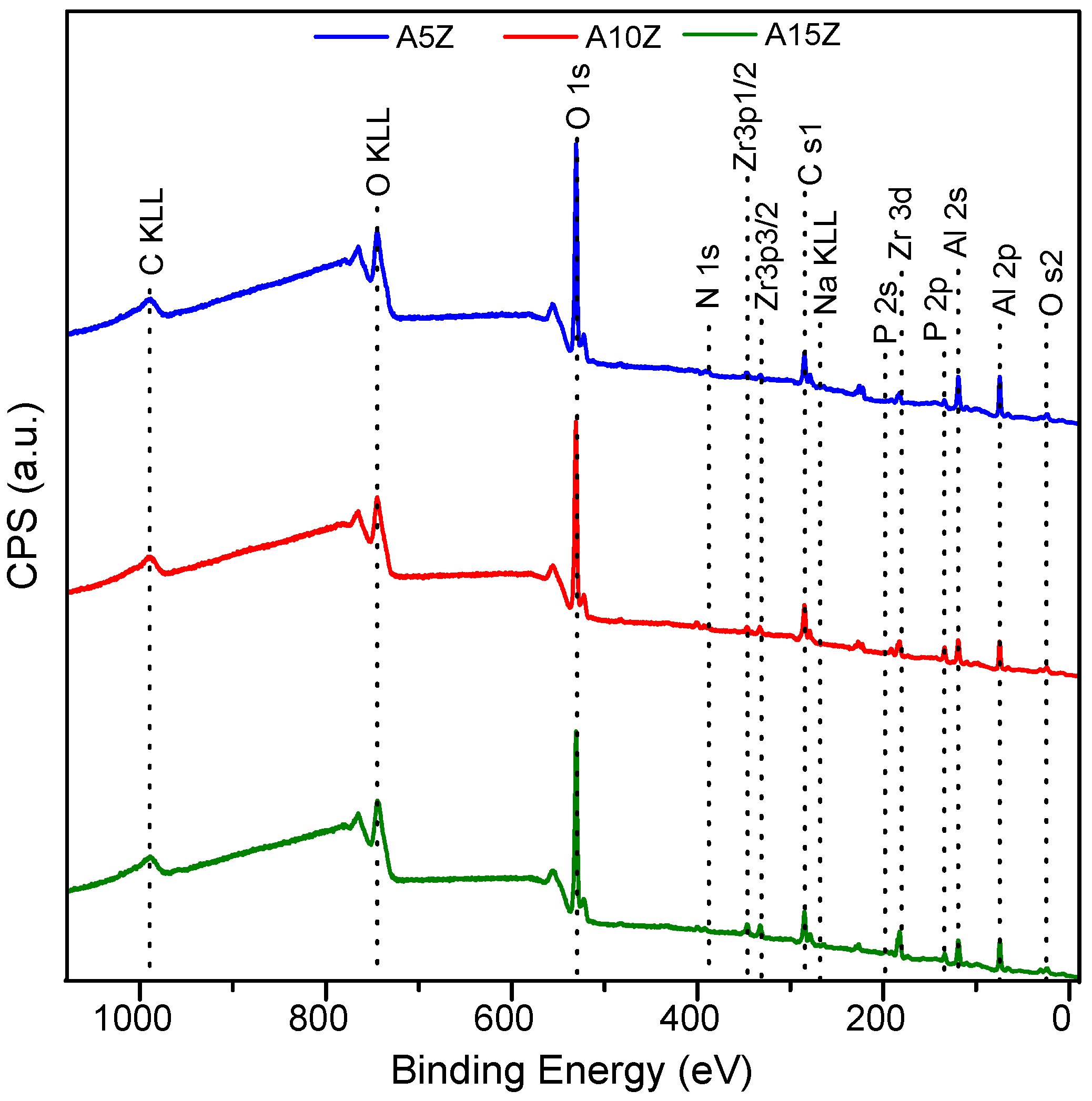
| Element | % Atomic Composition (±5%) | ||
|---|---|---|---|
| A5Z | A10Z | A15Z | |
| Oxygen (O 1s) | 44.6 | 43.8 | 44.3 |
| Carbon (C 1s) | 17.7 | 22.7 | 19.8 |
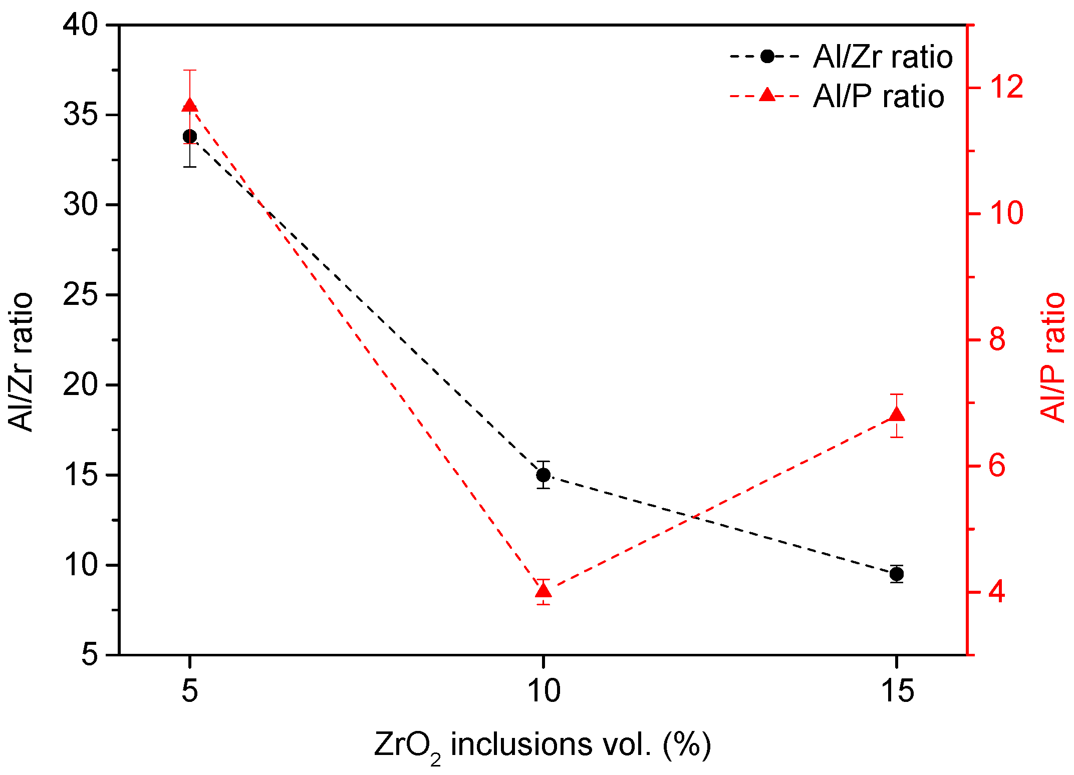
| Zirconium (Zr 3d) | 1.0 | 1.7 | 3.0 |
| Aluminum (Al 2p) | 33.8 | 25.5 | 28.7 |
| Phosphorus (P 2p) | 2.9 | 6.4 | 4.2 |
| A5Z | A10Z | A15Z | |||||
|---|---|---|---|---|---|---|---|
| Assignments | Pos. | Abs. Area | Pos. | Abs. Area | Pos. | Abs. Area | |
| O 1s | ZrO2 | 529.14 | 5448.7 | 529.56 | 14,284.5 | 529.47 | 22,754.4 |
| Al2O3 | 530.43 | 72,827.2 | 530.52 | 58,935.4 | 530.39 | 48,952.7 | |
| –PO4, –OH | 531.03 | 40,612.1 | 531.29 | 39,953.0 | 531.12 | 41,779.6 | |
| O–C | 532.27 | 21,968.1 | 532.35 | 18,900.7 | 532.32 | 15,607.3 | |
| O–C=O | 533.46 | 3671.0 | 533.41 | 4098.8 | 533.50 | 4086.2 | |
| Zr 3d | Zr 3d5/2 | 182.26 | 4639.1 | 182.25 | 7236.8 | 182.15 | 12,317.5 |
| Zr 3d3/2 | 184.23 | 3063.2 | 184.60 | 4778.5 | 184.52 | 8133.2 | |
| Al 2p | Al 2p | 74.59 | 20,052.7 | 74.58 | 13,766.2 | 74.57 | 15,049.3 |
| P 2p | H3PO4 | 134.01 | 3226.4 | 133.98 | 7429.3 | 134.08 | 4452.1 |
| C 1s | C–H | 284.93 | 16,721.9 | 284.90 | 18,490.9 | 284.85 | 16,619.6 |
| C–O | 286.70 | 2401.2 | 286.73 | 4151.9 | 286.66 | 3114.0 | |
| O–C=O | 288.43 | 1452.6 | 288.75 | 1412.0 | 288.77 | 805.2 | |
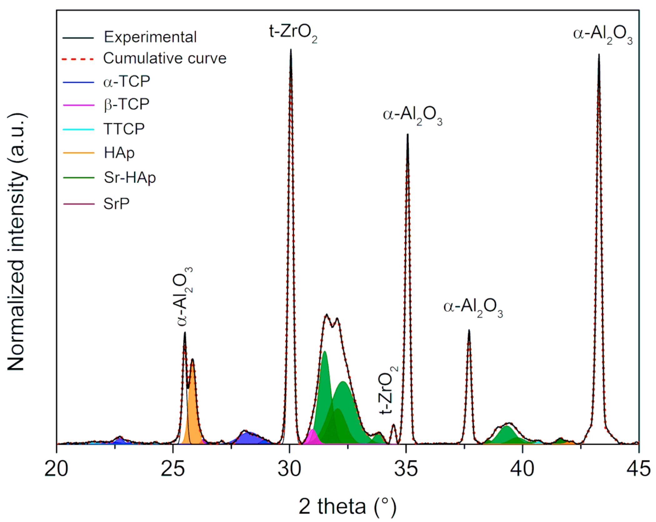
| Phosphates Phases | A5Z | A10Z | A15Z |
|---|---|---|---|
| α-TCP | 17.26 | 25.36 | 20.64 |
| β-TCP | 4.82 | 7.82 | 4.64 |
| TTCP | 15.81 | 4.57 | 5.81 |
| HAp | 16.41 | 21.47 | 27.46 |
| Sr-HAp | 44.21 | 38.82 | 39.28 |
| Sr-Phosphate | 1.49 | 1.96 | 2.17 |
Disclaimer/Publisher’s Note: The statements, opinions and data contained in all publications are solely those of the individual author(s) and contributor(s) and not of MDPI and/or the editor(s). MDPI and/or the editor(s) disclaim responsibility for any injury to people or property resulting from any ideas, methods, instructions or products referred to in the content. |
© 2024 by the authors. Licensee MDPI, Basel, Switzerland. This article is an open access article distributed under the terms and conditions of the Creative Commons Attribution (CC BY) license (https://creativecommons.org/licenses/by/4.0/).
Share and Cite
Nunes, F.C.; Santos, S.I.P.; Colnago, L.A.; Hammer, P.; Ferreira, J.A.; Ambrósio, C.E.; Pallone, E.M.J.A. Impact of ZrO2 Content on the Formation of Sr-Enriched Phosphates in Al2O3/ZrO2 Nanocomposites for Bone Tissue Engineering. Materials 2024, 17, 1893. https://doi.org/10.3390/ma17081893
Nunes FC, Santos SIP, Colnago LA, Hammer P, Ferreira JA, Ambrósio CE, Pallone EMJA. Impact of ZrO2 Content on the Formation of Sr-Enriched Phosphates in Al2O3/ZrO2 Nanocomposites for Bone Tissue Engineering. Materials. 2024; 17(8):1893. https://doi.org/10.3390/ma17081893
Chicago/Turabian StyleNunes, Fabio Caixeta, Sarah Ingrid Pinto Santos, Luiz Alberto Colnago, Peter Hammer, Julieta Adriana Ferreira, Carlos Eduardo Ambrósio, and Eliria Maria Jesus Agnolon Pallone. 2024. "Impact of ZrO2 Content on the Formation of Sr-Enriched Phosphates in Al2O3/ZrO2 Nanocomposites for Bone Tissue Engineering" Materials 17, no. 8: 1893. https://doi.org/10.3390/ma17081893