Article

Matrix-Assisted Laser Desorption/Ionization Time-of-Flight Mass Spectrometry: An Innovative Tool for Rapid Identification of Hylurgus ligniperda, an Invasive Pest

1
State Key Laboratory to Efficient Production of Forest Resources, Beijing Forestry University, Beijing 100083, China
2
Beijing Xinhui Purui Technology Development Co., Ltd., Beijing 102200, China
*
Author to whom correspondence should be addressed.
Forests 2024, 15(4), 729; https://doi.org/10.3390/f15040729
Submission received: 7 March 2024 / Revised: 6 April 2024 / Accepted: 18 April 2024 / Published: 22 April 2024
(This article belongs to the Section Forest Health)

Abstract

:
Hylurgus ligniperda is an imported quarantine plant pest in China. Its identification is usually based on morphological characteristics; therefore, species identification needs high professional requirements of staff and professionals with high experience accumulated through long-term training. Matrix-assisted laser desorption/ionization time-of-flight mass spectrometry (MALDI-TOF MS) is a rapid identification technology, which is based on protein profiles of species. It has been widely used for the identification of pathogenic microorganisms. Many studies have reported the identification of mosquitoes, ticks, and other arthropods. The application of MALDI-TOF MS in the identification of H. ligniperda can improve the identification efficiency of H. ligniperda, preventing and control its harm and further spread. To construct a spectra database for H. ligniperda, we analyzed the effect of different factors, such as different body parts, developmental stages, populations, and preservation conditions, on its protein spectrum. We collected protein spectrum profiles from 19 specimens of H. ligniperda and its related species, obtaining 211 protein spectra to construct a reference database and validate identification. The protein spectrum from the chest specimens of H. ligniperda showed many peaks, high intensity, and a stable signal, indicating a successful data establishment. The difference in protein spectra between different regions of the same species was less, but did not affect the identification results. Clear differences were observed in the protein spectrum across many developmental stages. The database established by the adult specimens protein spectrum can accurately identify Dendroctonus valens, Tomicus piniperda, and H. ligniperda. MALDI-TOF MS technology can be used for the rapid identification of H. ligniperda. This method is rapid and direct, and the identification results are robust. It does not require specialized entomological expertise and can be used for customs interception and field investigations.

1. Introduction

Hylurgus ligniperda is an important forest pest, which belongs to the order Coleoptera, family Curculionidae, subfamily Scolytinae. It is also known as red-haired pine bark beetle. It is native to southern Europe and the Mediterranean coast of northern Africa. However, the rapid pace of commercial and social globalization creates unprecedented opportunities for species to migrate to new regions of the world. Due to the development of global international trade [1], this pest has now spread to all continents, including all of Europe; northern Africa; the southern regions of Asia, including Japan, Turkey, and Sri Lanka; Australia and New Zealand in Oceania; Brazil, Chile, and Uruguay in South America; and the United States in North America. H. ligniperda is a major pest to Pinus conifers and is a quarantine pest of imported plants in China [2]. It has a strong natural transmission ability and can be transmitted over long distances via the transportation and freight of wood, wood packaging materials, and bedding materials [3]. The parasitic range of H. ligniperda is broad, encompassing pine and spruce. It mainly endangers newly cut trees and causes damage to healthy trees [4]. H. ligniperda has invaded China, specifically colonized Yantai, Shandong Province, China, and seriously endangered Pinus thunbergii. Since then, H. ligniperda has been discovered in Qingdao, Tai’an, Rizhao, and Weifang. H. ligniperda is highly pathogenic, and adult beetles can directly enter the soil and invade the roots of trees. They can even invade fine roots that are 0.5 cm away from the foundation of trees of approximately 7 m or more. Furthermore, they can pre-infringe sub-healthy trees and kill them [5]. China has a vast territory, rich in pine resources, and many foreign pines are widely distributed in the form of artificial pure forests. Because the geographical location and climatic conditions of China are similar to the distribution of the insect, the adaptability of H. ligniperda is extremely strong [6]. Therefore, the diffusion and spread of H. ligniperda can considerably affect the forestry ecology of China and cause major economic losses. Accurate identification of species is the foundation for the study of H. ligniperda. H. ligniperda are appearing in many regions of China. Therefore, effective control and forest protection is important. This can also help in achieving the rapid and precise identification of species and understanding the diffusion dynamics of H. ligniperda in depth.
H. ligniperda is frequently intercepted at ports in China. In 2006, Wang Fang et al. provided a detailed introduction to the morphological characteristics of H. ligniperda to enhance accurate identification and improve detection rates, aiding port inspection and quarantine personnel in identifying it [3]. Presently, the identification of quarantine-intercepted insects is largely based on morphological methods. Traditional insect morphological identification methods have some limitations. For instance, most of the morphological identification of insects is based on complete adults, whereas the early morphological structure of insects (eggs, larvae, and pupae) and the incomplete body of insects are difficult to identify. Therefore, identification is very challenging [7]. Additionally, species classification and identification depend on the professional identification of biological and morphological characteristics and the cognition of species concepts. This requires professional training, long-term scientific research, and experience [8]. In the concept of entry–exit inspection and quarantine, the identification of unknown pests intercepted in the quarantine process lacks accuracy and timeliness. This is due to the absence of professional technical personnel for classification and identification [7].
Molecular markers have become an ideal new form of genetic marker for domestic invasive organisms with the development of molecular biotechnology. In 2021, Ren Lili et al. conducted morphological and molecular identification, as well as DNA barcode analysis based on CO I and 28S gene sequences and found that the sample of H. ligniperda collected in Yantai, Shandong was the H. ligniperda. This is the first time that the beetle has been colonized in China. The main morphological identification characteristics of H. ligniperda have been clarified, including those of the Tribe Tomicini, the differences between H. ligniperda and common similar species, and the morphological characteristics of different insect states of H. ligniperda. This provides an important theoretical basis and practical guidance for the identification of H. ligniperda samples collected in other regions of China [5]. Compared with classical taxonomy, gene barcoding has the advantages of higher accuracy, efficiency, and convenience in species classification and identification. However, these steps require training of the operator. To obtain the sequence information of the selected gene before PCR, the whole process is time-consuming and costly, which cannot be easily applied to the rapid identification and classification of specimens [9,10]. Indeed, cox1 DNA barcoding was not able to differentiate members of the Culex pipiens group, Cx. quinquefasciatus and Cx. pipiens molestus, but these specimens were correctly identified using MALDI-TOF MS [11]. Therefore, developing a novel method that requires minimal time and is cost-effective is required. This can enable the identification of arthropod species without relying on genetic sequence information [10].
Matrix-assisted laser desorption/ionization time-of-flight mass spectrometry (MALDI-TOF MS) is another technique for species identification [12]. It is a new type of biological soft ionization mass spectrometry technology developed in recent years, suitable for the detection and analysis of mixtures and biological macromolecules. Species identification based on MALDI-TOF MS involves three steps: first, locate the specimen on a specially designed metal plate called a target plate, then measure the MALDI-TOF MS, and finally infer the species by matching the spectrum with a known or well-defined spectral database [10]. Compared with traditional morphological identification and molecular methods, this method requires low reagent cost and relatively simple specimen preparation. It allows rapid and direct data collection and analysis (usually less than 30 min) [10,13,14]. No reference genome or protein sequence is required. It can yield reliable identification results and also does not require professional knowledge. The technology has been applied to the detection and identification of microbial diagnosis, virology, mycology, and other fields [10,13,15,16]. Once a mass spectrometry reference database is established, any laboratory equipped with a MALDI-TOF MS system can transfer and use it directly [17].
Over the past decade, MALDI-TOF MS technology has significantly advanced in the identification of arthropods, including flies [18,19,20], mosquitoes [11,21,22,23], ticks [24,25], and fleas [26,27]. When assessing the feasibility and accuracy of MALDI-TOF MS technology, researchers typically integrate morphological and genetic identification with MALDI-TOF MS technology. The outcomes consistently demonstrate that the samples yield informative, reproducible, and species-specific protein mass spectra. MALDI-TOF MS technology can successfully allow the identification of arthropods.
Using some tissues of insects, such as those of feet, we can accurately identify biting mosquitoes collected in the field, including hidden species, mosquito species, and mosquito larvae. The laboratory-reared specimens and field mosquito larvae have been successfully identified. Furthermore, the tool is applied to field mosquito larvae monitoring [12,17,28]. Due to the structural characteristics of mosquito legs, maintaining their integrity as samples becomes challenging, thereby posing significant obstacles to the accuracy and reliability of the database. Vega-Rúa et al. improved mosquito identification through MALDI-TOF MS bio-typing by utilizing protein signatures from two body parts. The chest and legs of mosquitoes were selected as experimental samples, and the experimental results showed that the MS spectra thus obtained had high specificity, species specificity, and species repeatability. Therefore, even if the legs of mosquitoes are prone to damage, the protein characteristics of their chest and legs can still accurately identify the species of mosquitoes [29]. Additionally, bed bug specimens preserved in ethanol were identified up to the species level using head extracts from adult samples and head and chest specimens from immature samples [27]. This aforementioned research suggests that MALDI-TOF MS can be applied to the species identification of arthropods and allows rapid identification. Up to now, the application of MALDI-TOF MS technology in species identification of Coleoptera insects has not been reported. Accurately and quickly identifying pest species is a key link in epidemic prevention and pest control, which is crucial for timely response measures, containment of pest spread, and mitigation of damage caused. Determining the feasibility of this technology in the species identification of Coleoptera insects is of immense significance for the quarantine and control of related Coleoptera forestry pests [1].
In this study, we aimed to identify the protein extracts of H. ligniperda and its related species by MALDI-TOF MS to identify different species. We used a two-part classification method to compare performance between morphological identification and MALDI-TOF MS. We aimed to establish a reference database of H. ligniperda and perform blind detection to evaluate the feasibility and accuracy of the MALDI-TOF MS method in the identification of H. ligniperda. In the future, the protein spectrum can be used to improve the detection rate of forest pests and diseases in the customs rapid inspection system.

2. Materials and Methods

2.1. Specimens Collection

The information regarding specimen collection utilized in this experiment is delineated in Table 1 and Table 2. The adult specimens of H. ligniperda were primarily obtained from six funnel-shaped traps. Initially, we proceeded to the designated site and installed the traps; each trap was suspended at intervals of 100 m, with the bottom positioned 0.3 m below ground level. The central component of the trap is a plant-derived attractant, comprising α-pinene and ethanol in a mass ratio of (2–3):1. Alternatively, it may include α-pinene and ethanol supplemented with one or more of 3-carene and β-pinene [30]. These traps are meticulously crafted to effectively attract and capture adult H. ligniperda specimens. Then the specimens in the trap were inspected and collected once a week.
The trap for H. ligniperda is mainly designed for adults. Therefore, when collecting the eggs, larvae and pupae of H. ligniperda, the focus was mainly on the weak trees and stumps that had been infected. The larvae mainly live under the bark for feeding activities and, as they gradually grow and develop to the mature stage, they pupate and fly out in spring. Invasive holes are usually found at the base and root of the trunk, and reddish-brown worm casts are excreted after feeding in the tree. The habitats of eggs, larvae and pupae of H. ligniperda usually choose places where the phloem is seriously eaten. These places have formed an ideal living environment due to long-term feeding, which provides convenient conditions for their reproduction and growth. Under the skin of the root, we can observe the reddish-brown debris-like feces. These feces are not only eye-catching in color, but also loose in texture and easy to identify, so they become an important basis for selecting collection targets. The eggs, larvae and pupae of H. ligniperda are all white in appearance, making them easy to distinguish in the reddish-brown dung, which is conducive to finding hidden eggs, larvae and pupae.
The eggs, larvae, and pupae of Hylotrupes ligniperda are in a delicate stage, particularly during the pupal phase. Their shells are thin, and mishandling can lead to pupal mortality. Hence, meticulous care and caution are imperative during the collection process. It is advisable to refrain from employing sharp or coarse tools during harvesting to prevent mechanical harm to the insects.
Furthermore, adult specimens of Dendroctonus valens were primarily acquired through root excavation and bark removal from trees, while Tomicus piniperda specimens were captured using local traps. Following the collection of the aforementioned samples, meticulous examination was conducted to identify detailed morphological characteristics and determine species attribution [3,5,31,32]. These collected insect specimens were preserved in a medical-grade freezer (model: MDF-86V588E) at −80 °C.

2.2. Instruments and Reagents

The main instruments used in this experiment are shown in Table 3.
The main reagents used in this experiment included lysis solution and matrix, and the preparation method is as follows [10,17,33].
Preparation of lysis solution: The same amount of 50% acetonitrile and 70% formic acid was added to a 1.5 mL centrifuge tube and mixed evenly for later use.
Preparation of CHCA matrix: 100% acetonitrile, 100% trifluoroacetic acid, and sterile water were added to a 1.5 mL centrifuge tube, then excess α-cyano-4-hydroxycinnamic acid was added. The resulting solution was sonicated at room temperature (25 °C) for 30 min. The final concentration of CHCA matrix composed of 50% acetonitrile and 2.5% trifluoroacetic acid was obtained for later use.

2.3. Preparation of MALDI-TOF MS Specimens

The 
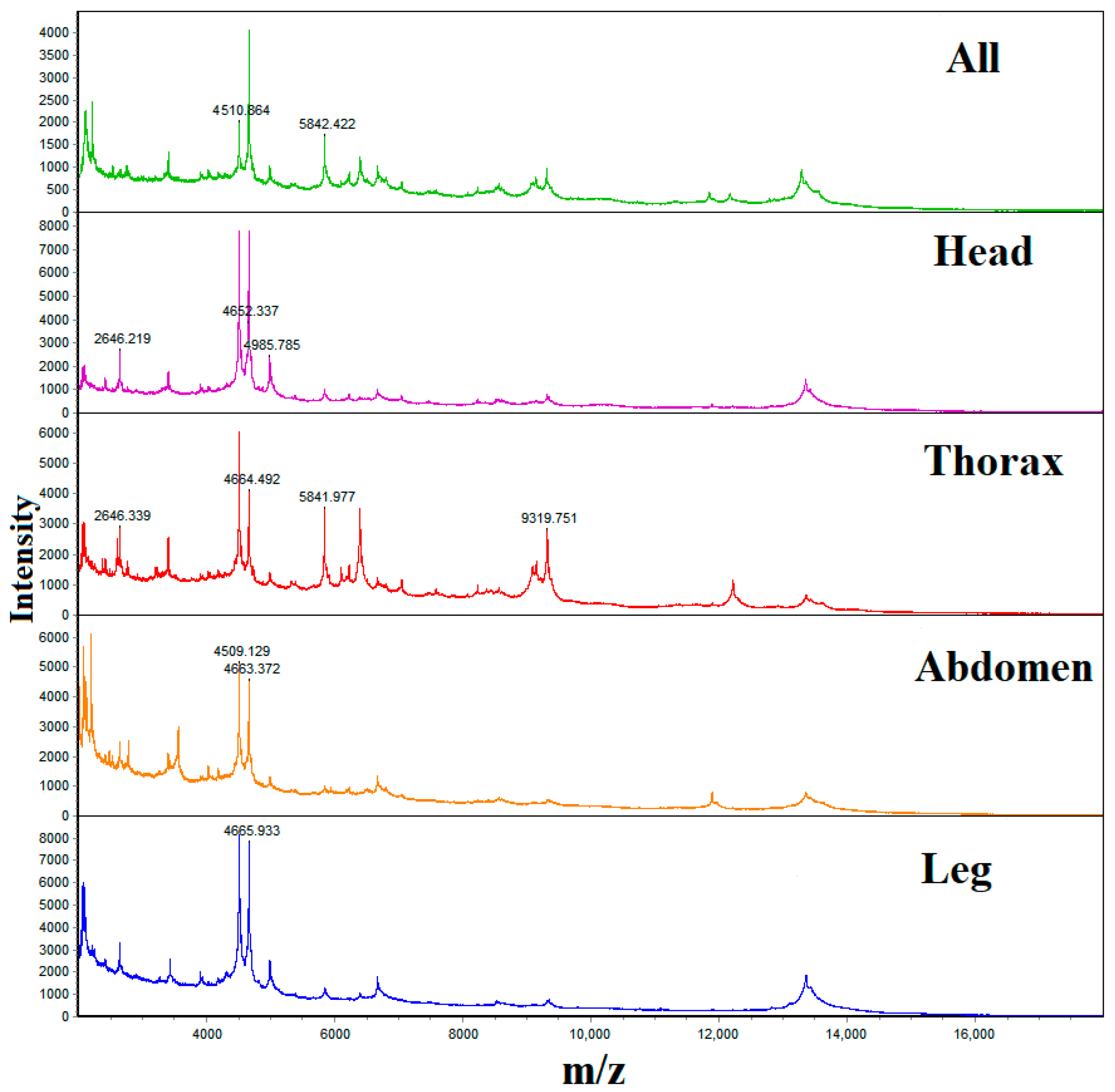
H. ligniperda contains three pairs of legs, while they are often missing in samples obtained from the wild and those intercepted at customs. Consequently, we dissected the H. ligniperda beetle into several parts, including the head, chest, abdomen, and legs, and compared them with intact specimens to identify those yielding the most abundant proteins for mass spectrometry experiments, which are not readily depleted. Initially, a series of preliminary experiments was conducted to identify the optimal lysis solution, as outlined in Table 4. Using adults of H. ligniperda from Yantai as specimens, each adult was decomposed into various parts, including the head, chest, abdomen, and legs, with each part taken as a separate sample. Additionally, a complete beetle was taken as a sample, resulting in five samples in total, totaling three groups.
The specimens were added to a 1.5 mL centrifuge tube, and an appropriate amount of prepared lysis solution was added. Then it was manually homogenized using a grinding gun. The homogenate was centrifuged at 12,000 rpm for 5 min, and 1 μL of the supernatant of each specimen was transferred onto the steel target plate. Each specimen was divided into three points. Subsequently, each specimen was covered on the target plate, dried at room temperature (25 °C) for a few min. Then add 1 μL of lysis solution dropwise at each point and dry at room temperature (25 °C) for a few minutes, and introduced into the MALDI-TOF MS instrument for analysis. The experiment included three replicate groups. The amount of lysis solution added in other groups of experiments should refer to this group, and the amount of lysis solution should be increased or decreased according to the volume of the specimen.
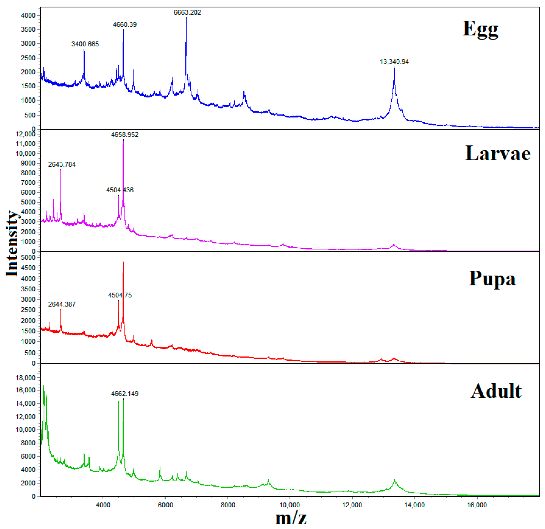
For the experiment for different insect states of H. ligniperda, we used complete samples for research. In this experiment, MALDI-TOF MS was used to analyze specimens at various developmental stages of H. ligniperda, including eggs, larvae, pupae, and adults. A total of five groups of repeated experiments were performed, and the experimental design is detailed in Table 5.
In the study of different populations and their related species of H. ligniperda, we specifically selected the part with the most abundant protein mass spectrometry for the experiment. The chests of various H. ligniperda populations were used as experimental specimens for the study. The main specimens were adult H. ligniperda from Yantai, Weihai, and Tai’an. The collection information is detailed in Table 1, and the experimental design is presented in Table 6. Three groups of repeated experiments were performed.
The specimens from various species were collected and subjected to MALDI-TOF MS analysis. Different species include the T. piniperda, H. ligniperda, and D. valens, important forest pests, which belong to the order Coleoptera. The collection details for specimens are outlined in Table 1, and the experimental design is presented in Table 7, comprising a total of five groups of repeated experiments.

2.4. MALDI-TOF MS Parameters

The protein profile was obtained using the CPRO-180 real-time workstation system. Linear positive ion mode was used to collect specimens in the mass range of 2–20 kDa. Each target plate was externally calibrated using the reference strain Escherichia coli CICC 10389, one of the key steps to ensure the accuracy and reliability of the experimental results. Each mass spectrum corresponds to the ions obtained from 300 laser emissions from six regions at the same point. The Spectrum Viewer software was then used to analyze and compare the obtained spectra.

2.5. Spectrogram Analysis and Reference Database Creation

The repeatability of different spectra of each specimen was analyzed using Xinhui technology cluster analysis software. Four specimens within the species were selected, and the common characteristic peaks were extracted by the cluster analysis software. These characteristic peaks were then imported into the CPRO-180 real-time workstation system to construct the species database.

3. Results and Analysis

3.1. Protein Spectrum Analysis

3.1.1. Spectra Analysis of Different Parts of H. ligniperda

Figure 1 shows that the main peak positions of the protein spectra of all parts of H. ligniperda were consistent, but the MALDI-TOF MS mass spectrum of the chest of the H. ligniperda is more abundant than the peak information from other body parts, suggesting that using the chest provides more accurate identification results. The chest is also the only remaining body part that is not easily degraded or lost during collection, transportation, or storage, and does not affect the MALDI-TOF MS atlas results, which can be used for species identification [29]. Therefore, subsequent experiments were performed on the chest of the specimens.
Considering that the chest of H. ligniperda contains three pairs of legs, the legs are breakable, and the loss of one or several legs occurs frequently during H. ligniperda sampling, transportation or storage. If only three or fewer legs are available for a single specimen, the identification by MALDI-TOF MS could be compromised. An additional control experiment was introduced. It is verified that MALDI-TOF MS spectra of chests with and without legs differed, emphasizing the need to ensure that specimens used in subsequent experiments do not contain legs. This result successfully overcame the problem of missing legs in the sample of H. ligniperda, providing more accurate and reliable data support for subsequent experimental research, not only applicable to the study of H. ligniperda, but also providing reference for the study of other insect groups.

3.1.2. Spectra Analysis of Different Developmental Stages of H. ligniperda

The developmental process of H. ligniperda follows complete metamorphosis, encompassing the following four stages: egg, larva, pupa, and adult. Different developmental stages exhibit distinct protein profiles, which directly affect the use of the database. As shown in Figure 2, the MALDI-TOF MS mass spectrometry results from different developmental stages of H. ligniperda show that the protein peaks of eggs are the most abundant and have the greatest number, with peak positions significantly differing from those of other developmental stages. Pupae, larvae, and adults share the same main peak, but differences were found in the secondary peaks. Consequently, a protein database established using adult protein spectra can only be applied to the identification of adults. To identify eggs, pupae, and larvae, it is necessary to establish a distinct protein database for each developmental period.

3.1.3. Spectra Analysis of Different Populations of H. ligniperda

As shown in Figure 3, the main peak positions of each population were at 4.5 kDa, and the secondary peak was also basically consistent. It can be concluded that the protein spectra from different populations of H. ligniperda exhibit high consistency, repeatability, and reproducibility. This indicates that the protein spectra of the same species possess significant similarity, enabling their application in the rapid identification of H. ligniperda.

3.1.4. H. ligniperda and Other Species

As shown in Figure 4, the main peaks of the protein mass spectra of T. piniperda, H. ligniperda, and D. valens are located at 9.5 kDa, 4.5 kDa, and 2.5 kDa, and noticeable differences were observed in MALDI-TOF MS spectra among different species, exhibiting rich and species-specific information. The three MALDI-TOF MS mass spectra obtained from each specimen within a species exhibit high consistency, repeatability, and reproducibility, supporting the construction of a reference database.

3.2. Reference Database Creation

MALDI-TOF MS maps were collected from a total of 19 specimens of three species involved in the construction of the database, with at least 9 protein maps collected for each specimen to ensure the adequacy and reliability of the data. At the same time, it was verified that the spectra of different species have high species specificity, while the mass spectra of the same species have high consistency, repeatability, and reproducibility, resulting in a total of 211 effective spectra. Then, the MALDI-TOF MS spectra of H. ligniperda, D. valens, and T. piniperda were exported to construct a reference database. This database was used to preliminarily verify whether different species, such as H. ligniperda, can be successfully identified (Table 8).

3.3. Research Verification Test

The test is divided into the following two parts: one part involves testing different species, whereas the other part involves testing specimens of H. ligniperda preserved under different conditions. The aim is to determine the accuracy of identification results for different species and the effect of different storage times on the identification results of H. ligniperda at different temperatures. The specimens were stored under different conditions for 1 day, 3 days, 6 days, and 10 days at room temperature (25 °C), 4 °C, and −20 °C, with each group undergoing repetitive experiments thrice (Table 9).
The MALDI-TOF MS mass spectrometry results (Figure 5) showed that the mass spectra of the specimens under various storage conditions were satisfactory, with minimal differences observed. The signal intensity matching the values of the mass spectrum corresponding to the reference mass spectrum was high, enabling the successful identification of the specimens as H. ligniperda.
From the entire experiment, we found that the specimens were relatively dry and shriveled, and the protein mass spectra obtained showed a significant decrease in richness, even though the main peaks were consistent. The specimens of H. ligniperda used for testing in Figure 5 were all stored in self-sealing bags to avoid light. Therefore, we speculate that it may be because H. ligniperda lured by the traps were not collected and preserved in a timely manner, resulting in prolonged exposure to the outdoor environment and partial protein breakdown. Therefore, the specimens cannot be exposed to outdoor for a long time, because the forest environment is relatively harsh, especially in summer, with high temperature, humidity, and sufficient sunlight.

4. Discussion

We have proposed for the first time the use of MALDI-TOF MS spectroscopy to achieve rapid detection and identification of an invasive species, H. ligniperda, which forms the basis for developing effective management strategies and quarantine measures [1].
Using the MALDI-TOF MS method to achieve rapid identification of species, it is important to establish a standardized experimental plan before creating and sharing a species protein fingerprint database [16], including specimen site selection, developmental period selection, lysis solution selection, and formulation of spectral quality control parameters. Therefore, we first optimized the experimental system.
Our study revealed that using chest protein specimens results in a large number of spectral peaks, high peak values, and high reproducibility. This is consistent with the findings in the mosquito protein profile establishment, suggesting that the protein content in the chest is high and more stable [16,29,34]. Therefore, the MALDI-TOF-MS mass spectrometry of the protein extract from the chest of the H. ligniperda is a suitable part for establishing a reference database for identification. In addition, by analyzing the MALDI-TOF MS spectra of sample proteins from different developmental stages of H. ligniperda, it was found that pupae, larvae, and adults all contain the same main peak, but there are still differences in the secondary peak. Among them, it is worth mentioning that the protein mass spectrometry peaks of a single egg of H. ligniperda also show extremely high richness. If protein fingerprinting can be established using eggs as specimens, rapid identification of eggs by MALDI-TOF MS can be achieved, which is impossible for morphological identification and gene barcode technology [11]. In the MALDI-TOF MS atlas of chest protein extraction from samples of different populations of H. ligniperda in China, we found that the protein atlas differences between different regions of the same species were small, which did not affect the identification results. We speculate that, with sufficiently fresh samples and standardized experimental procedures, based on species identification, the specific geographic location of the population to which the sample belongs can even be identified.
In order to construct a reference database, mass spectra of chest proteins were collected from the H. ligniperda and its related species, the D. valens and T. piniperda. They all belong to the order Coleoptera. The results showed that significant differences were observed in the MALDI-TOF MS spectra between different species, displaying rich species-specific information. The mass spectra of the same species showed high consistency, repeatability, and reproducibility. The experimental results confirmed the construction of a reference database using MALDI-TOF MS mass spectrometry of the long forest beetle and its related species, and the sample chest protein extract is sufficient to identify species categories using the MALDI-TOF MS method. The results obtained using this method confirm the identification results obtained using morphological methods. The validity of the database was established by a blind test; in this test, when there is a corresponding reference spectrum in the database, the specimen can be correctly identified by MALDI-TOF MS [10].
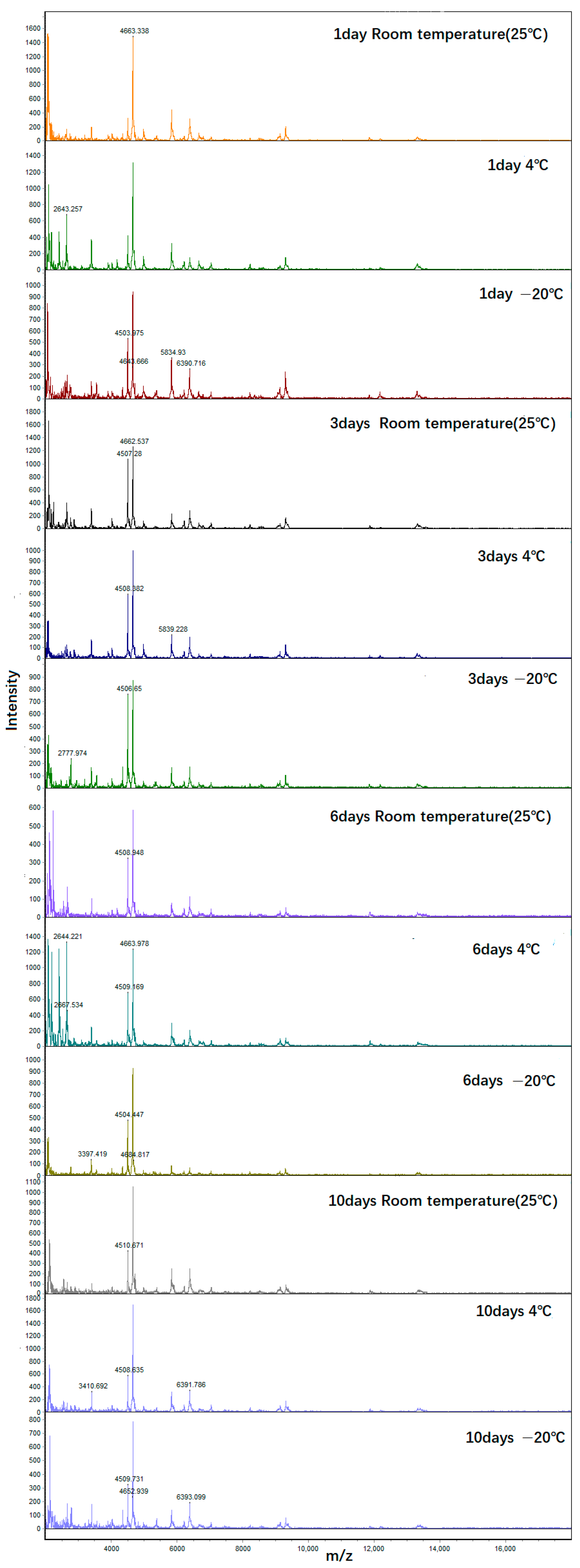
Species identification using protein mass spectrometry is challenged by the poor stability of proteins compared to nucleic acids. However, MALDI-TOF MS identification uses ribosomal proteins, providing relatively high stability. In our experiment, H. ligniperda specimens preserved at room temperature (25 °C) for up to 10 days still produced abundant peaks, eliminating limitations related to specimen integrity and freshness. This is particularly beneficial for quarantine work, no longer limited to the completeness and freshness of the sample.
While our study established the MALDI-TOF MS identification system for Scolytinae, it is currently limited by the availability of specimens from different species, in this study we focused on establishing the protein spectrum database for H. ligniperda and its related species, D. valens, and T. piniperda adults.
Compared to morphological identification and molecular biology methods, MALDI-TOF MS represents an interesting method for identifying arthropods. This method has a simple sample preparation method, short processing time, and fast data analysis. The identification results can be obtained in a short period of time, and the identification results are reliable. The test cost is low, and professional entomological knowledge is not required [10,35]. Moreover, once a mass spectrometry reference database is constructed, any laboratory equipped with a MALDI-TOF instrument can use it for related species identification [17]. Future applications of this method should extend to the use of mass spectrometers for on-the-spot measurements and species identification through the constructed database, including customs interception and field investigations. This identification method is particularly important for pests such as H. ligniperda intercepted by customs and field investigations, as it can ensure rapid and accurate identification of pest species, providing strong support for subsequent prevention and control measures [1]. In the future, it can also be applied to species identification of other forestry pests of the order Coleoptera, and will play a more important role in future scientific research and practical application.

5. Conclusions

The application of MALDI-TOF MS identification technology and the creation of mass spectrometry reference databases have brought great convenience to quarantine work. This database not only enables laboratories to quickly and accurately conduct various measurements. The mass spectrometry reference database can also be applied to species identification of other Coleoptera forestry pests. By comparing the information in the database, species identification can be efficiently carried out. This means that, as long as the mass spectrometry information of known or unknown pest species is included in the database, they can be quickly identified using the MALDI-TOF instrument.
Looking forward to the future, the application prospects of mass spectrometry reference databases are very broad. Especially in the prevention and control of forestry pests, this identification method will become a powerful assistant for researchers and practitioners, improving the accuracy and efficiency of pest identification, helping them to better understand the types and distribution of pests, to formulate more scientific and effective prevention and control strategies, to provide more comprehensive and systematic support for the comprehensive management of forestry pests, and to make greater contributions to the protection of China’s forest resources and ecological environment.

Author Contributions

Conceptualization, J.W. and J.T.; methodology, J.W. and Z.D.; software, J.W. and Z.D.; validation, J.Z.; formal analysis, J.W.; investigation, J.W.; resources, Z.D. and J.Z.; data curation, J.W. and Z.D.; writing—original draft preparation, J.W.; writing—review and editing, J.T. and Z.D.; visualization, J.Z.; project administration, J.T.; funding acquisition, J.T. All authors have read and agreed to the published version of the manuscript.

Funding

This research was supported by National Key Research and Development Program of China (2021YFD1400300).

Data Availability Statement

The datasets generated and analyzed during the current study are not publicly available, but they are available from the corresponding author upon reasonable request.

Acknowledgments

We are very grateful to Lihong Bi and the staff of Yantai Forest Resources Monitoring and Protection Service Center for their kind assistance during the fieldwork.

Conflicts of Interest

Authors Zhijun Dong and Jiaqiang Zhu were employed by the company Beijing Xinhui Purui Technology Development Co., Ltd. The remaining authors declare that the research was conducted in the absence of any commercial or financial relationships that could be construed as a potential conflict of interest.

References

  1. Venette, R.C.; Hutchison, W.D. Invasive insect species: Global challenges, strategies & opportunities. Front. Insect Sci. 2021, 1, 650520. [Google Scholar]
  2. Dong, W.; Lin, G.; Chen, Y. An important forest pest H. ligniperda raptured at Fujian Port. J. Fujian For. Sci. Technol. 2010, 37, 112–114. [Google Scholar] [CrossRef]
  3. Wang, F.; Chen, S.; Zhou, W. Identification of Hylurgus ligniperda Fabricius. Plant Quar. 2006, 1, 30–31. (In Chinese) [Google Scholar]
  4. Liang, Z.; Zhang, J.; Yang, D.; Chen, N. Overview of alien insects intercepted at China ports from 2005 to 2015. Plant Quar. 2017, 31, 64–68. [Google Scholar]
  5. Ren, L.; Tao, J.; Wu, H.; Zong, S.; Wang, C.; Hua, D.; Shi, J.; Liu, Y.; Luo, Y. The first discovery and infective characteristics of a major invasive pest Hylurgus ligniperda (Coleoptera: Scolytidae) in China. Sci. Silvae Sin. 2021, 57, 140–150. [Google Scholar] [CrossRef]
  6. Teng, K.; Zhang, W.; Du, X.; Ou, W. Highly alert to the invasion of Hylurgus ligniperda Fabriciu with New Zealand coniferous logs. Plant Quar. 2012, 26, 90. [Google Scholar]
  7. Liu, Y.; Song, Y.; Li, X. DNA barcode technology based on mitochondrial CO I gene in Application in insect molecular identification. Plant Quar. 2010, 24, 46–50. [Google Scholar] [CrossRef]
  8. Ding, S.M. The application of DNA barcode technology in the identification of harmful insects in archives. China Arch. 2022, 8, 66–68. (In Chinese) [Google Scholar]
  9. Sauer, S.; Freiwald, A.; Maier, T.; Kube, M.; Reinhardt, R.; Kostrzewa, M.; Geider, K. Classification and identification of bacteria by mass spectrometry and computational analysis. PLoS ONE 2008, 3, e2843. [Google Scholar] [CrossRef]
  10. Yssouf, A.; Almeras, L.; Raoult, D.; Parola, P. Emerging tools for identification of arthropod vectors. Future Microbiol. 2016, 11, 549–566. [Google Scholar] [CrossRef]
  11. Lawrence, A.L.; Batovska, J.; Webb, C.E.; Lynch, S.E.; Blacket, M.J.; Slapeta, J.; Parola, P.; Laroche, M. Accurate identification of Australian mosquitoes using protein profiling. Parasitology 2019, 146, 462–471. [Google Scholar] [CrossRef] [PubMed]
  12. Kaufmann, C.; Schaffner, F.; Ziegler, D.; Pflüger, V.; Mathis, A. Identification of field-caught Culicoides biting midges using matrix-assisted laser desorption/ionization time of flight mass spectrometry. Parasitology 2012, 139, 248–258. [Google Scholar] [CrossRef]
  13. Singhal, N.; Kumar, M.; Kanaujia, P.K.; Virdi, J.S. MALDI-TOF mass spectrometry: An emerging technology for microbial identification and diagnosis. Front. Microbiol. 2015, 6, 791. [Google Scholar] [CrossRef]
  14. Schaffner, F.; Kaufmann, C.; Pfluger, V.; Mathis, A. Rapid protein profiling facilitates surveillance of invasive mosquito species. Parasit Vectors 2014, 7, 142. [Google Scholar] [CrossRef]
  15. Freiwald, A.; Sauer, S. Phylogenetic classification and identification of bacteria by mass spectrometry. Nat. Protoc. 2009, 4, 732–742. [Google Scholar] [CrossRef]
  16. Bamou, R.; Costa, M.M.; Diarra, A.Z.; Martins, A.J.; Parola, P.; Almeras, L. Enhanced procedures for mosquito identification by MALDI-TOF MS. Parasites Vectors 2022, 15, 240. [Google Scholar] [CrossRef]
  17. Yssouf, A.; Socolovschi, C.; Flaudrops, C.; Ndiath, M.O.; Sougoufara, S.; Dehecq, J.S.; Lacour, G.; Berenger, J.M.; Sokhna, C.S.; Raoult, D.; et al. Matrix-assisted laser desorption ionization—Time of flight mass spectrometry: An emerging tool for the rapid identification of mosquito vectors. PLoS ONE 2013, 8, e72380. [Google Scholar] [CrossRef] [PubMed]
  18. Lafri, I.; Almeras, L.; Bitam, I.; Caputo, A.; Yssouf, A.; Forestier, C.L.; Izri, A.; Raoult, D.; Parola, P. Identification of Algerian Field-Caught Phlebotomine Sand Fly Vectors by MALDI-TOF MS. PLoS Neglected Trop. Dis. 2016, 10, e0004351. [Google Scholar] [CrossRef]
  19. Hoppenheit, A.; Murugaiyan, J.; Bauer, B.; Steuber, S.; Clausen, P.H.; Roesler, U. Identification of Tsetse (Glossina spp.) Using Matrix-Assisted Laser Desorption/Ionisation Time of Flight Mass Spectrometry. PLoS Neglected Trop. Dis. 2013, 7, e2305. [Google Scholar] [CrossRef] [PubMed]
  20. Hoppenheit, A.; Murugaiyan, J.; Bauer, B.; Clausen, P.H.; Roesler, U. Analysis of and from two distant locations in Burkina Faso using MALDI TOF MS. Parasitol. Res. 2014, 113, 723–726. [Google Scholar] [CrossRef]
  21. Tahir, D.; Almeras, L.; Varloud, M.; Raoult, D.; Davoust, B.; Parola, P. Assessment of MALDI-TOF mass spectrometry for filariae detection in mosquitoes. PLoS Neglected Trop. Dis. 2017, 11, e0006093. [Google Scholar] [CrossRef] [PubMed]
  22. Yssouf, A.; Parola, P.; Lindström, A.; Lilja, T.; L’Ambert, G.; Bondesson, U.; Berenger, J.M.; Raoult, D.; Almeras, L. Identification of European mosquito species by MALDI-TOF MS. Parasitol. Res. 2014, 113, 2375–2378. [Google Scholar] [CrossRef]
  23. Fall, F.K.; Diarra, A.Z.; Bouganali, C.; Sokhna, C.; Parola, P. Using MALDI-TOF MS to Identify Mosquitoes from Senegal and the Origin of Their Blood Meals. Insects 2023, 14, 785. [Google Scholar] [CrossRef]
  24. Boyer, P.H.; Almeras, L.; Plantard, O.; Grillon, A.; Talagrand-Reboul, E.; McCoy, K.; Jaulhac, B.; Boulanger, N. Identification of closely related Ixodes species by protein profiling with MALDI-TOF mass spectrometry. PLoS ONE 2019, 14, e0223735. [Google Scholar] [CrossRef]
  25. Karger, A.; Kampen, H.; Bettin, B.; Dautel, H.; Ziller, M.; Hoffmann, B.; Süss, J.; Klaus, C. Species determination and characterization of developmental stages of ticks by whole-animal matrix-assisted laser desorption/ionization mass spectrometry. Ticks Tick-Borne Dis. 2012, 3, 78–89. [Google Scholar] [CrossRef]
  26. Yssouf, A.; Socolovschi, C.; Leulmi, H.; Kernif, T.; Bitam, I.; Audoly, G.; Almeras, L.; Raoult, D.; Parola, P. Identification of flea species using MALDI-TOF/MS. Comp. Immunol. Microbiol. Infect. Dis. 2014, 37, 153–157. [Google Scholar] [CrossRef] [PubMed]
  27. Ngnindji-Youdje, Y.; Lontsi-Demano, M.; Diarra, A.Z.; Makaila, A.M.; Tchuinkam, T.; Berenger, J.-M.; Parola, P. Morphological, molecular, and MALDI-TOF MS identification of bed bugs and associated Wolbachia species from Cameroon. Acta Trop. 2024, 249, 107086. [Google Scholar] [CrossRef] [PubMed]
  28. Nebbak, A.; Koumare, S.; Willcox, A.C.; Berenger, J.M.; Raoult, D.; Almeras, L.; Parola, P. Field application of MALDI-TOF MS on mosquito larvae identification. Parasitology 2018, 145, 677–687. [Google Scholar] [CrossRef]
  29. Vega-Rúa, A.; Pagès, N.; Fontaine, A.; Nuccio, C.; Hery, L.; Goindin, D.; Gustave, J.; Almeras, L. Improvement of mosquito identification by MALDI-TOF MS biotyping using protein signatures from two body parts. Parasites Vectors 2018, 11, 574. [Google Scholar] [CrossRef]
  30. Li, C.; Zhao, W.; Huai, W.; Lin, R.; Yao, Y. Research progress on Hylurgus ligniperda Fabricius (Coleoptera: Curculionidae), a new invasive species in China. J. Environ. Entomol. 2023, 45, 850–861. (In Chinese) [Google Scholar]
  31. Chen, L.; Jin, B.; Gu, Z. Advances in Research on Biological Characteristics and Control Strategies of Dendroctonus valens. In Proceedings of the 2018 Academic Annual Meeting of the Entomological Society of the Three Provinces of Central China (Hubei, Hunan, Henan), Xiangyang, China, 19–22 October 2018; pp. 102–109. (In Chinese). [Google Scholar]
  32. Jiang, L.; Liu, W.; Li, N.; Zhao, R.; Yu, M. Survey and Analysis on the Damages of Tomicus piniperda in Jiaodong Peninsula. Anhui For. Sci. Technol. 2021, 47, 3. [Google Scholar] [CrossRef]
  33. Dieme, C.; Yssouf, A.; Vega-Rúa, A.; Berenger, J.M.; Failloux, A.B.; Raoult, D.; Parola, P.; Almeras, L. Accurate identification of Culicidae at aquatic developmental stages by MALDI-TOF MS profiling. Parasites Vectors 2014, 7, 544. [Google Scholar] [CrossRef] [PubMed]
  34. Briolant, S.; Costa, M.M.; Nguyen, C.; Dusfour, I.; de Santi, V.P.; Girod, R.; Almeras, L. Identification of French Guiana anopheline mosquitoes by MALDI-TOF MS profiling using protein signatures from two body parts. PLoS ONE 2020, 15, e0234098. [Google Scholar] [CrossRef]
  35. Yssouf, A.; Flaudrops, C.; Drali, R.; Kernif, T.; Socolovschi, C.; Berenger, J.M.; Raoult, D.; Parola, P. Matrix-Assisted Laser Desorption Ionization-Time of Flight Mass Spectrometry for Rapid Identification of Tick Vectors. J. Clin. Microbiol. 2013, 51, 522–528. [Google Scholar] [CrossRef] [PubMed]
Figure 1. MALDI-TOF MS spectra profiles obtained from the protein extraction from different parts of H. ligniperda ranging from 2 to 20 kDa. Intensity arbitrary units, m/z mass-to-charge ratio.
Forests 15 00729 g001
Figure 2. MALDI-TOF MS spectra obtained from protein extraction of eggs, larvae, pupae, and adults of the H. ligniperda ranging from 2 to 20 kDa. Intensity arbitrary units, m/z mass-to-charge ratio.
Forests 15 00729 g002
Figure 3. MALDI-TOF MS spectra obtained from chest protein extraction of H. ligniperda population in Weihai, Tai’an, and Yantai ranging from 2 to 20 kDa. Intensity arbitrary units, m/z mass-to-charge ratio.
Forests 15 00729 g003
Figure 4. MALDI-TOF MS spectra obtained from chest protein extraction of T. piniperda, H. ligniperda, and D. valens ranging from 2 to 20 kDa. Intensity arbitrary units, m/z mass-to-charge ratio.
Forests 15 00729 g004
Figure 5. MALDI-TOF MS spectra obtained from chest protein extraction of the H. ligniperda under different preservation conditions ranging from 2 to 20 kDa. Intensity arbitrary units, m/z mass-to-charge ratio.
Forests 15 00729 g005
Table 1. Geographical populations of different species.
SpeciesCollection PlacesPopulation CodeGeo-CoordinatesCollection Date
H. ligniperdaShandong-YantaiSD-YT37°27′ N, 121°51′ E2023/6
Shandong-WeihaiSD-WH37°29′ N, 121°59′ E2023/6
Shandong-TaianSD-TA36°25′ N, 117°15′ E2023/4
D. valensNeimenggu-ChifengNMG-CF41°57′ N, 118°40′ E2023/7
T. piniperdaShandong-YantaiSD-YT37°46′ N, 121°44′ E2022/7
Table 2. Collecting data of different insect states of H. ligniperda.
SpeciesStageCollection PlacesPopulation CodeGeo-CoordinatesCollection Date
H. ligniperdaEggShandong-YantaiSD-YT37°27′ N
121°51″ E
2023/7
Larva2023/7
Pupa2023/6
Adult2023/6
Table 3. Test equipment information.
NameInstrument ModelLaboratory
Mass spectrometerCPRO-180Beijing Xinhui Purui Technology Development Co., Ltd. (Beijing, China)
High speed tabletop centrifuge5415D eppendorfBeijing Xinhui Purui Technology Development Co., Ltd. (Beijing, China)
Adjustable pipette0.1–2 μL, 2–10 μL, 10–100 μL, 100–1000 μL, eppendorfBeijing Xinhui Purui Technology Development Co., Ltd. (Beijing, China)
Supersonic cleanerKQ-500DBBeijing Xinhui Purui Technology Development Co., Ltd. (Beijing, China)
Handheld tissue grindingSN:1004216State Key Laboratory to Efficient Production of Forest Resources (Beijing, China)
Table 4. Experimental design of MALDI-TOF MS for different parts of H. ligniperda.
StatesAllHeadThoraxAbdomenLeg
Lysis solution/μL200601006060
Number of specimens33333
Table 5. Experimental design of MALDI-TOF MS for different developmental stages of H. ligniperda.
StageEggLarvePupaAdult
Lysis solution/μL10100100200
Number of specimens5555
Table 6. Experimental design of MALDI-TOF MS for different populations of H. ligniperda.
Geographic PopulationsSD-WHSD-YASD-YT
Lysis solution/μL100100100
Number of specimens333
Table 7. Experimental design of MALDI-TOF MS for different species.
SpeciesT. piniperdaH. ligniperdaD. valens
Lysis solution/μL100100250
Number of specimens555
Table 8. Species used to establish a MALDI-TOF MS reference database.
SpeciesH. ligniperdaD. valensT. piniperda
Number of specimens766
Number of protein spectrum985855
Table 9. Specimens of H. ligniperda preserved under different conditions.
Time/Day13610
Room temperature (25 °C)3 individuals3 individuals3 individuals3 individuals
4 °C3 individuals3 individuals3 individuals3 individuals
−20 °C3 individuals3 individuals3 individuals3 individuals
Disclaimer/Publisher’s Note: The statements, opinions and data contained in all publications are solely those of the individual author(s) and contributor(s) and not of MDPI and/or the editor(s). MDPI and/or the editor(s) disclaim responsibility for any injury to people or property resulting from any ideas, methods, instructions or products referred to in the content.

