Article

A Low-Cost Spore Trap Allows Collection and Real-Time PCR Quantification of Airborne Fusarium circinatum Spores

1
School of Forest Resources and Conservation, University of Florida, Gainesville, FL 32611, USA
2
USDA Forest Service, Southern Research Station, Saucier, MS 39574, USA
3
Currently at Department of Plant, Soil and Microbial Sciences, Michigan State University, East Lansing, MI 48824, USA
*
Author to whom correspondence should be addressed.
Forests 2018, 9(10), 586; https://doi.org/10.3390/f9100586
Submission received: 8 August 2018 / Revised: 10 September 2018 / Accepted: 13 September 2018 / Published: 20 September 2018

Abstract

:
A variety of commercial instruments are available for sampling and quantifying microscopic airborne organisms from the environment. Although most samplers are highly sensitive, they are also expensive, costing thousands of dollars per unit, a price that is out of reach for many researchers, especially those looking to design experiments with replication. While looking at options to monitor pine stands for the presence of Fusarium circinatum, the causal agent of pitch canker disease, on multiple sites with several units per site, we developed a simple, low-cost spore trap that allows surveying spore abundance in outdoor environments. The trap consists of a rotating motor that holds a metal rod and two petroleum jelly-coated microscope slides. As the motor rotates, the slides collect airborne particles. To test whether the traps allowed detection of F. circinatum spores, we placed six traps on three sites: an actively-managed slash pine commercial stand located in Lake Butler, FL, a semi-managed loblolly and slash pine stand near Gainesville, FL, and a site with little perturbance at Goethe State Forest, FL, consisting of mainly slash pine trees. The slides were replaced weekly, and F. circinatum was detected by quantitative PCR using species-specific primers. Results show detection of low levels ( X ¯ = 1.7–77.1 picograms ± SE = 0.3–39.7) of the pathogen spores with high reproducibility. These traps offer a low-cost solution to spore, pollen, or small insect trapping experiments for initial or general assessment of a pathogen or species population. Their low cost has the added benefit that multiple traps can be deployed per experiment, thus increasing statistical power by using multiple replications.

Graphical Abstract

1. Introduction

Pitch canker is a fungal disease caused by the necrotroph Fusarium circinatum (Nirenberg and O’Donnell). It is one of the most important diseases in pines and affects 57 pine species and Douglas-fir [1,2,3], causing resinous lesions in stems and branches as well as in reproductive structures, thus reducing fertility [2]. Its economic impact can be devastating in nursery settings, where it causes seedling mortality or leads to outplanting failures [4]. In older trees, economic losses are mainly because of the disease effects on growth and timber volume. First reported in 1946 [5], pitch canker disease has been spreading globally through the introduction (via seeds) and culture of southern pines for timber [3,6,7,8,9,10].
Pitch canker outbreaks usually occur in the summer, favoring a warm climate and high humidity [1], particularly after strong winds and storms. However, the phenology of the disease and its relation to environmental conditions is poorly understood. One way to study the phenology of pitch canker disease is to survey the pathogen in the environment by detecting airborne F. circinatum spores using a spore trap and quantitative PCR [11,12,13,14].
The most basic type of spore trap consists of a flat surface, usually a microscope slide with an adhesive or a culture plate with selective media onto which particles are passively deposited [15,16]. Such traps have been previously used to detect and quantify F. circinatum spores in field studies [12,14]. These are very economical but are not a very reliable source for spore quantification, as collection may be affected by the position of the trap, rain, or other factors that may prevent spore deposition. Other traps consist of a vertical rotating arm set onto an electric motor [11,13,17,18,19]. The rotating arms are covered by adhesive tape strips which are later removed for spore detection using microscopy or PCR [11,13,18,20]. The electric motor may be limited by availability and location of power outlets or, if using a portable battery-operated version, the rotating power and length of use may be limited by the amount of power in the battery [17]. Other traps, such as the Hirst volumetric spore sampler [21], utilize air suction mechanisms to draw particles into a matrix where they are trapped and processed for quantification. Although these samplers are highly sensitive, they are also expensive, costing thousands of dollars per unit and often requiring additional post-sampling services. The high cost of these commercial traps makes it difficult to use in experimental designs with multiple replicates, as affordability is a constraint.
Real-time PCR has been widely used to detect and quantify microorganisms from air samplers [18,20,22,23,24]. In some cases, universal primers were used to obtain a characterization of the types of microorganisms present in the samples [25], whereas in others, species-specific primers were developed to quantify a species of interest [20,25,26,27]. In the case of F. circinatum, species-specific primers for real-time PCR were developed in 2004 to detect the fungal pathogen [12], with subsequent improvements [28,29]. In this study, we tested the efficacy of low-cost in-house spore traps supported by quantitative PCR analysis to monitor the abundance of Fusarium circinatum in three locations in North Florida.

2. Materials and Methods

2.1. Spore Trap Construction

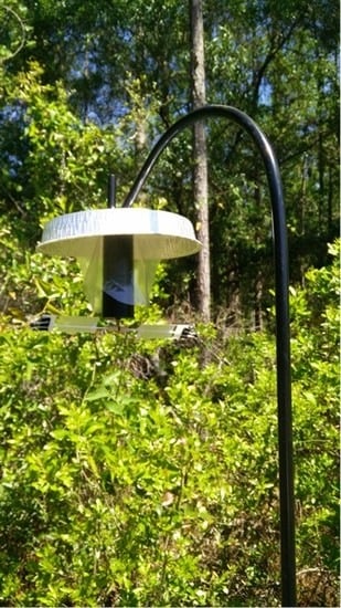
Microscope slides coated with petroleum jelly (Vaseline®, Cheesebrough-Pond’s USA, Inc., Greenwich, CT, USA) were prepared in advance by heating the petroleum jelly in a beaker with deionized water such that the proportion of petroleum jelly to water was 1:1 (v:v). Two slides were attached to each other with one drop of water and dipped into the liquid petroleum jelly so that they were covered about ¾ of their length on one side only. The slides were cooled vertically at room temperature and once cooled were placed into a 50 mL conical tube (back to back) for storage. The traps were assembled using a battery-powered rotating garden motor (In the Breeze® Hanging Display Motor, Item #10025, supplied by Amazon.com) with a straightened metal rod of approximately 25–30 cm in length and 1.3 mm (16-gauge) thickness attached to the bottom portion of the motor to which the coated slides were secured in place with small binder clips (Figure 1A). A small twist tie was attached to the metal rod to hold the slides in horizontal position. The traps were then hung from a standard 2.13 m (84 inch) iron garden hook, such that the slides were positioned at approximately 1.8–1.9 m from the ground. An aluminum dish of 23 cm in diameter and a small plastic bag were used to cover the motor and help keep excess rain and debris from the slides (Figure 1B).

2.2. Spore Trap Deployment

Spore traps were deployed at three locations in north central Florida between 19 May and 6 October 2016. All three locations had trees presenting pitch canker symptoms. The first location was in the Austin Cary Forest in Alachua County. This forest is part of the University of Florida School of Forest Resources and Conservation and consists of mostly loblolly (Pinus taeda L.) and slash pine (P. elliottii Engelm.) stands on poorly drained Ultic Alaquods [30]. This site has an elevation of 50 m, mean precipitation for 2016 of 1068.5 mm, with annual minimum, average, and maximum temperatures of 15.9, 21.8, and 27.8 °C, respectively. This site had a moderate presence of pitch canker, with around 20% of trees showing symptoms of the disease. Trees were approximately eight years of age and about 10 m high. A second set of traps was placed in the Goethe State Forest in Levy County. Traps were placed in a naturally seeded area with predominantly slash pine and some longleaf pine (P. palustris Mill.). Altitude in this area is about 8 m and soils are of Smyrna-fine type (Aeric Haplaquods) [31]. Annual minimum, average, and maximum temperatures at Goethe in 2016 were 14.7, 20.42, and 26.1 °C, respectively. Trees at this site had a variable range of ages, including mostly mature trees but also younger trees (under ten years of age) within the area where traps were placed. Pitch canker symptoms were present and moderately abundant, with approximately 20%–30% incidence. Finally, a third set of traps was placed at a commercially managed slash pine forest located near Lake Butler (Union County, FL) that was having a very high incidence of pitch canker (greater than 80%). Respective minimum, average, and maximum annual mean temperatures at this location in 2016 were 14.6, 20.7, and 26.9 °C and it had a mean precipitation of 1267 mm that year. This area has poorly-drained Mascotte sand type soils [32]. Two sets of three traps were placed at each site near trees showing symptoms of pitch canker disease. Each set consisted of three traps separated by 50–100 m and groups were separated from each other by a minimum of 800 m within a site (Figure 2). The slides in the spore traps were replaced weekly and stored at 4 °C in labeled 50 mL conical tubes until they were processed for DNA extraction.

2.3. DNA Extraction and PCR Optimization

An initial optimization of the DNA extraction protocol was performed to test the efficiency of pre-treating the samples with water or isopropanol to separate the spores from the petroleum jelly. Slides covered with petroleum jelly were inoculated with suspensions of a fixed spore count ranging between 102 and 107 F. circinatum microconidia. A fixed volume of the suspension corresponding to a given spore count was pipetted onto the coated slides and allowed to air-dry overnight. The contents from the surface of the coated middle section (25 × 35 mm) of each slide were scraped off into a 2 mL tube using sterilized toothpicks. The contents were treated with 1 mL isopropanol and 1 mL water and heated at 65 °C for 15 min. The tubes were then vortexed on maximum speed to dislodge the particles from the petroleum jelly and heated again for another 10 min at 65 °C. Samples were again vortexed and centrifuged at 14,000 rpm for 10 min. The petroleum jelly containing the microconidia separated from the aqueous phase and the aqueous phase was discarded. Water was then added to the jelly and samples were heated at 65 °C for 15 min, vortexed 30 s on maximum speed, and then centrifuged at 14,000 rpm for 5 min. The aqueous phase containing the spores was then transferred to a 1.5 mL microcentrifuge tube using a 3 cc syringe with 26 G needle. Spores were then centrifuged at 14,000 rpm for 15 min and the residual water was carefully poured off. DNA extraction was then performed using the CTAB (Cetyl Trimethyl Ammonium Bromide) method [29]. As controls, DNA samples were also extracted from serial dilutions of F. circinatum spore suspensions ranging from 108 to 103 spores. Conventional PCR was carried out using primer pairs CIRC 1L and CIRC 4L and EF1F and EF1R [12,29,33]. PCR products were visualized in 1% agarose gels with SYBR® Green. quantitative PCR was carried out using the protocol described by Dreaden et al. (2012) [29] and the same primer pairs were used for conventional PCR. Samples from spore suspensions of 107 microconidia were serially diluted from 10 to 106 to establish the standard curve. Ct (cycle threshold) values and standard curves for quantitative PCR were obtained directly from the instrument (Eppendorf® Mastercycler using Realplex 2.0 software-Eppendorf, Hauppauge, NY, USA). The qPCR results were considered for further analysis if the standard curve slope was below 0.98 and plate efficiency values were between 0.9 and 1.1.

2.4. Analysis of Environmental Samples Using Quantitative PCR

Once the DNA extraction and PCR protocols were established and the standard curve was optimized, quantitative PCR was performed for all the samples collected. Each sample was included in triplicate and reactions were carried out in 96-well plates. Comparisons among plates were deemed appropriate if the standard curves agreed with the known quantities and if the efficiency values were between 0.9 and 1.1. Plates with efficiency values outside that range were re-done or excluded from analysis. The amount of DNA was estimated based on the standard curve composed of serial microconidia dilutions ranging between 1 ng and 0.01 picograms. Samples were discarded if the standard curve slope was under 0.98 and/or if the plate efficiency was between 0.9 and 1.1. DNA quantity was estimated in picograms and plotted against the dates of sample collection for each site.

3. Results

3.1. Recovery of F. circinatum DNA from Petroleum Jelly Matrix Was Aided by Heat Treatment

Samples were treated by heating at 65 °C, which melted the petroleum jelly, and by adding isopropanol or water, followed by centrifugation to separate the material from the petroleum jelly. Results using slides inoculated with known quantities of spores show that there is a minimum amount that are detected using conventional PCR. In samples treated with isopropanol, slides inoculated with 106 spores or less did not yield any amplification product, whereas slides with 107 spores showed a band of high intensity of 300 bp (Figure 3). Samples treated with water showed that 105 or greater are clearly detected using the F. circinatum specific primers CIRC 1L and CIRC 4L. Increasing intensity agrees with the increasing number of spores in each sample. The results were comparable to those obtained from spore suspensions (Figure 3).

3.2. Environmental Samples of F. circinatum were Detectable with Quantitative PCR Using Species-Specific Primers

Once the DNA extraction protocol was optimized, 432 DNA samples collected from the field sites were amplified using quantitative PCR to estimate the amount of F. circinatum DNA released into the environment every week. Of these samples, 307 (71%) passed the quality control tests (standard curve slope below 0.98 and plate efficiency values between 0.9 and 1.1) to be included in the analysis.
Among the 307 field samples included in the analyses, less than 11% were negative for F. circinatum DNA (Table 1). Because the samples were used in triplicate for quantitative PCR, if all replicates were negative for F. circinatum DNA, then these samples were considered negative. If one or more replicates were positive for F. circinatum DNA, then the sample was assumed positive, as negative results could be due to DNA quantities near the limit of detection. In this study, while optimizing qPCR conditions using serial dilutions of microconidia suspensions, detection became erratic after the 33rd cycle, which corresponded to approximately 105 spores, or 0.1 picograms of DNA, therefore, samples with DNA quantities below that threshold were considered undetected. When samples were categorized by trap within each site, the range of undetected DNA was between 0 and 17.6%, 0 and 11.8, and 0 and 15.8 in Austin Cary, Goethe Forest, and Lake Butler sites, respectively (Table 1). In general, Goethe State Forest showed a more consistent detection of F. circinatum DNA, with undetected samples ranging between 0 and 10.8 percent. In contrast, Austin Cary Forest and Lake Butler sites showed a higher variation of spore detection, with some traps having no undetected DNA and others with undetected DNA up to 17.6% to 15.8%, respectively (Table 1).

3.3. Quantification of F. circinatum DNA from Field Sites Show Low Levels of the Pathogen across Sites, with Some Instances of High Spore Release

Amounts of F. circinatum DNA recovered from field samples ranged from zero (undetected) to a maximum of 159, 412, and 560 picograms in the Austin Cary Forest, Goethe State Forest, and Lake Butler sites, respectively. However median quantities were rather consistent across sites, ranging between 0.76 and 1.76 picograms overall (Table 2). This suggests that overall quantities were generally low, with some instances of high spore release. High standard error values also reflect high variation among samples, depending on the collection dates, suggesting that spores were not uniformly present throughout the growing season.
When samples were broken down by weekly samples, F. circinatum DNA levels were overall low throughout the 2016 season, with most samples under 20 picograms (Figure 4). However, a peak of high F. circinatum abundance was observed at the end of May and the beginning of June in all sites, with Lake Butler showing the highest amounts of detected F. circinatum and the Austin Cary Forest with the lowest levels. F. circinatum quantities peaked early at the Goethe State Forest, the westernmost location, whereas quantities peaked about 2–3 weeks later at both Lake Butler and Austin Cary sites (Figure 4). A smaller peak was observed at the end of July through mid-August in all three sites.

4. Discussion

There are many types of spore traps available for monitoring the presence of fungal spores and other airborne microorganisms in the environment [15,23]. While some commercially available traps, like the Burkhard spore trap [21] or Hi-Vol filter samplers (Thermo Fisher Scientific, Waltham, MA, USA), offer a very detailed monitoring approach, such as estimating air volume passing through the trap or rotating discs that partition spore collections into specific times [21], they are often expensive. This limits the number of traps that can be placed at each site, and subsequently, the geographic range for large-scale monitoring. Others are as simple as a microscope slide coated with petroleum jelly [16], filter paper, or culture plate where spores are passively deposited [12], which may be limited to certain weather conditions. Some have rotating arms, like the rotorod-type traps, but may have limited portability [13,18,19], as they require an electrical outlet or large battery source. The spore traps constructed for this project are low-cost and consist of materials readily available in garden and hardware stores. For under $50 per trap, this method allows placing multiple traps on different sites for long-term monitoring, which increases statistical power when designing more detailed experiments. Although these traps would not accurately measure the amount of air volume passing by the traps, and therefore allow a precise quantification of inoculum per unit of air, they are highly useful for surveying areas for the presence of spores, small particles, or even small insects. The traps were light, easy to assemble, and weather resistant. During the period the traps were in the field, there were several severe storms, including a tropical storm with winds of nearly 50 km/h, as well as heavy rains that passed through the sites where the traps were placed. All traps continued to rotate uninterrupted and sustained very limited damage, which included some cracked slides and torn aluminum covers. Although these traps can function for up to three weeks, the battery for the motor needs replacement if monitoring should extend beyond this period. Quantitative PCR allowed the detection and quantification of F. circinatum in the environment, showing a peak of spore release at the beginning of the summer. While high variation was observed throughout the study, the presence of multiple traps at each site increased the statistical power of the analysis. Our results showed high spore quantities at all three locations between the end of May and beginning of June. However, the fluctuations of F. circinatum we observed throughout the year did not seem to agree with observations for Monterey pine (Pinus radiata D. Don) in California by Schweigkofler et al. [12], where higher spore counts were detected in the fall. A more recent study using high-speed rotating arm spore traps to monitor F. circinatum dispersal on Monterey pine in Spain showed no clear temporal patterns of spore release but suggests correlations with air temperature and leaf wetness [13]. We analyzed public data from weather stations near our sampling sites and from our own stations within the sites and did not find any significant correlations with mean daily temperature or precipitation. However, this does not rule out that other biological and environmental factors, such as air moisture, temperature fluctuations, and isolated precipitation patterns may be playing a role in spore release.
Because these spore traps can be easily made with commonly available parts, the applications for these are considerable. Currently, we deployed 36 traps in an area in Mississippi with a severe outbreak of pine needle cast to assess the causal agent of this outbreak. Real-time PCR markers are being designed by the United States (US) Forest Service for several of the candidate species that were isolated from the area and that could be the cause of this disease. Additionally, some adaptations are under way to help monitor the vectors of rose rosette virus at the University of Tennessee (Mark Windham, personal communication). Variations to the traps can allow monitoring of other organisms, such as small insects, that could be trapped and counted using adhesive material on a slightly larger surface area instead of glass slides. Different adhesive materials or films, like the MicroAmpTM optical adhesive film (ThermoFisher Scientific, Waltham, MA USA), could be used instead of petroleum jelly for projects focused on microscopic visualization of the trapped particles. Finally, these traps could be easily used for citizen science projects and for research in areas with low budgets, like developing countries and rural communities, for basic monitoring of local pathogen dispersal.

5. Conclusions

We built an in-house spore trap to test the presence and estimate the quantity of Fusarium circinatum in the field. Because of their low cost and the availability of the materials, we deployed 18 traps among three sites (six traps per site) from May to October, 2016. Traps were changed on a weekly basis and the presence of F. circinatum DNA was estimated using quantitative PCR. Our results show that qPCR identified F. circinatum in low quantities (under 100 picograms) with a peak of higher abundance around late May and early June. These traps, could be used for early detection of airborne pathogens in situations where a general assessment is needed, and can be modified with different adhesive options for use in microscopy or for small insect trapping. Its low cost could facilitate monitoring experiments in low-budget projects as well as for citizen science experiments.

Author Contributions

Conceptualization, T.Q. and J.S.; Field trials, T.Q., J.H., P.J.; PCR optimization and execution, K.S. (Keumchul Shin), K.S. (Katherine Smith), J.H.; Formal Analysis, T.Q.; Resources, J.S.; Data Curation, T.Q.; Writing—Original Draft Preparation, T.Q.; Writing—Review & Editing, T.Q., J.S., K.S. (Keumchul Shin), K.S. (Katherine Smith); Visualization, T.Q., J.S.; Supervision, J.S., T.Q.; Project Administration, J.S., T.Q.; Funding Acquisition, J.S.

Funding

This research was funded by the University of Florida/Institute of Food and Agricultural Sciences Climate Change Seed Grant Program for 2016–2017, https://research.ifas.ufl.edu/climatechange/. The cost of the special number has been cover by the PINESTRENGTH project.

Acknowledgments

The authors would like to acknowledge that funding for this work was made available through the University of Florida/IFAS Climate Change Seed Grant Program. The authors also wish to thank Boby Cahal, from the Florida Forest Service, Scott Sager, from the University of Florida, and Paul Belonger and Keith Palmer from Weyerhaeuser Co., for their cooperation in deploying the traps at the Goethe State Forest, Austin Cary Forest, and Lake Butler sites. Finally, we would like to thank the two anonymous reviewers for their valuable suggestions that greatly improved this manuscript as well as the support of the European COST (European Cooperation in Science and Technology) Action Grant FP1406 PINESTRENGTH for covering the publication fees of this article.

Conflicts of Interest

The authors declare no conflict of interest.

References

  1. Gordon, T.R. Pitch canker disease of pines. Phytopathology 2006, 96, 657–659. [Google Scholar] [CrossRef] [PubMed]
  2. Dwinell, L.D.; Barrow-Broaddus, J.B.; Kuhlman, E.G. Pitch canker: A disease complex of southern pines. Plant Dis. 1985, 69, 270–276. [Google Scholar] [CrossRef]
  3. Wingfield, M.J.; Hammerbacher, A.; Ganley, R.J.; Steenkamp, E.T.; Gordon, T.R.; Wingfield, B.D.; Coutinho, T.A. Pitch canker caused by Fusarium circinatum—A growing threat to pine plantations and forests worldwide. Australas. Plant Pathol. 2008, 37, 319–334. [Google Scholar] [CrossRef]
  4. Barnard, E.L.; Blakeslee, G.M. Pitch Canker of Slash Pine Seedlings: A New Disease in Forest Tree Nurseries. Plant Dis. 1980, 64, 695–696. [Google Scholar] [CrossRef]
  5. Hepting, G.H.; Roth, E.R. Pitch canker, a new disease of some southern pines. J. For. 1946, 44, 742–744. [Google Scholar]
  6. Coutinho, T.A.; Steenkamp, E.T.; Mongwaketsi, K.; Wilmot, M.; Wingfield, M.J. First outbreak of pitch canker in a South African pine plantation. Australas. Plant Pathol. 2007, 36, 256–261. [Google Scholar] [CrossRef]
  7. Pérez-Sierra, A.; Landeras, E.; León, M.; Berbegal, M.; García-Jiménez, J.; Armengol, J. Characterization of Fusarium circinatum from Pinus spp. in northern Spain. Mycol. Res. 2007, 111, 832–839. [Google Scholar] [CrossRef] [PubMed]
  8. McCain, A.H.; Koehler, C.S.; Tjosvold, S.A. Pitch canker threatens California pines. Calif. Agric. 1987, 41, 22–23. [Google Scholar]
  9. Dick, M.A. Pine pitch canker-the threat to New Zealand. N. Z. For. 1998, 42, 30–34. [Google Scholar]
  10. Kobayashi, T.; Muramoto, M. Pitch canker of Pinus luchuensis, a new disease in Japanese forests. For. Pests 1989, 38, 169–173. [Google Scholar]
  11. Calderon, C.; Ward, E.; Freeman, J.; McCartney, A. Detection of airborne fungal spores sampled by rotating-arm and Hirst-type spore traps using polymerase chain reaction assays. J. Aerosol Sci. 2002, 33, 283–296. [Google Scholar] [CrossRef]
  12. Schweigkofler, W.; O’Donnell, K.; Garbelotto, M. Detection and quantification of airborne conidia of Fusarium circinatum, the causal agent of pine pitch canker, from two California sites by using a real-time PCR approach combined with a simple spore trapping method. Appl. Environ. Microbiol. 2004, 70, 3512–3520. [Google Scholar] [CrossRef] [PubMed]
  13. Dvořák, M.; Janoš, P.; Botella, L.; Rotková, G.; Zas, R. Spore Dispersal Patterns of Fusarium circinatum on an Infested Monterey Pine Forest in North-Western Spain. Forests 2017, 8, 432. [Google Scholar] [CrossRef]
  14. Garbelotto, M.; Smith, T.; Schweigkofler, W. Variation in rates of spore deposition of Fusarium circinatum, the causal agent of pine pitch canker, over a 12-month-period at two locations in Northern California. Phytopathology 2008, 98, 137–143. [Google Scholar] [CrossRef] [PubMed]
  15. Lacey, M.E.; West, J.S. The Air Spora; Lacey, M.E., West, J.S., Eds.; Springer: Boston, MA, USA, 2006. [Google Scholar]
  16. Ostry, M.E.; Nicholls, T.H. A Technique for Trapping Fungal Spores; USDA Forest Service, Ed.; North Central Forest Experiment Station: St. Paul, MN, USA, 1982. [Google Scholar]
  17. Lacey, M.E.; West, J.S. Air Sampling Techniques. In The Air Spora; Lacey, M.E., West, J.S., Eds.; Springer: Boston, MA, USA, 2006; pp. 35–47. [Google Scholar]
  18. Chandelier, A.; Helson, M.; Dvorak, M.; Gischer, F. Detection and quantification of airborne inoculum of Hymenoscyphus pseudoalbidus using real-time PCR assays. Plant Pathol. 2014, 63, 1296–1305. [Google Scholar] [CrossRef]
  19. Rahman, A.; Miles, T.D.; Martin, F.N.; Quesada-Ocampo, L.M. Molecular approaches for biosurveillance of the cucurbit downy mildew pathogen, Pseudoperonospora cubensis. Can. J. Plant Pathol. 2017, 39, 282–296. [Google Scholar] [CrossRef]
  20. Klosterman, S.J.; Anchieta, A.; McRoberts, N.; Koike, S.T.; Subbarao, K.V.; Voglmayr, H.; Choi, Y.-J.; Thines, M.; Martin, F.N. Coupling Spore Traps and Quantitative PCR Assays for Detection of the Downy Mildew Pathogens of Spinach (Peronospora effusa) and Beet (P. schachtii). Phytopathology 2014, 104, 1349–1359. [Google Scholar] [CrossRef] [PubMed]
  21. Hirst, J.M. An automatic volumetric spore trap. Ann. Appl. Biol. 1952, 39, 257–265. [Google Scholar] [CrossRef]
  22. An, H.R.; Mainelis, G.; White, L. Development and calibration of real-time PCR for quantification of airborne microorganisms in air samples. Atmos. Environ. 2006, 40, 7924–7939. [Google Scholar] [CrossRef]
  23. West, J.; Kimber, R. Innovations in air sampling to detect plant pathogens. Ann. Appl. Biol. 2015, 166, 4–17. [Google Scholar] [CrossRef] [PubMed] [Green Version]
  24. Karolewski, Z.; Kaczmarek, J.; Jedryczka, M.; Cools, H.J.; Fraaije, B.A.; Lucas, J.A.; Latunde-dada, A.O. Detection and quantification of airborne inoculum of Pyrenopeziza brassicae in Polish and UK winter oilseed rape crops by real-time PCR assays. Grana 2012, 51, 270–279. [Google Scholar] [CrossRef]
  25. Lang-Yona, N.; Dannemiller, K.; Yamamoto, N.; Burshtein, N.; Peccia, J.; Yarden, O.; Rudich, Y. Annual distribution of allergenic fungal spores in atmospheric particulate matter in the Eastern Mediterranean; a comparative study between ergosterol and quantitative PCR analysis. Atmos. Chem. Phys. 2012, 12, 2681–2690. [Google Scholar] [CrossRef] [Green Version]
  26. Luo, Y.; Ma, Z.; Reyes, H.C.; Morgan, D.; Michailides, T.J. Quantification of airborne spores of Monilinia fructicola in stone fruit orchards of California using real-time PCR. Eur. J. Plant Pathol. 2007, 118, 145–154. [Google Scholar] [CrossRef]
  27. Rogers, S.L.; Atkins, S.D.; West, J.S. Detection and quantification of airborne inoculum of Sclerotinia sclerotiorum using quantitative PCR. Plant Pathol. 2009, 58, 324–331. [Google Scholar] [CrossRef]
  28. Ioos, R.; Fourrier, C.; Iancu, G.; Gordon, T.R. Sensitive detection of Fusarium circinatum in pine seed by combining an enrichment procedure with a real-time polymerase chain reaction using dual-labeled probe chemistry. Phytopathology 2009, 99, 582–590. [Google Scholar] [CrossRef] [PubMed]
  29. Dreaden, T.J.; Smith, J.A.; Barnard, E.L.; Blakeslee, G. Development and evaluation of a real-time PCR seed lot screening method for Fusarium circinatum, causal agent of pitch canker disease. For. Pathol. 2012, 42, 405–411. [Google Scholar] [CrossRef]
  30. Powell, T.L.; Starr, G.; Clark, K.L.; Martin, T.A.; Gholz, H.L. Ecosystem and understory water and energy exchange for a mature, naturally regenerated pine flatwoods forest in north Florida. Can. J. For. Res. 2005, 35, 1568–1580. [Google Scholar] [CrossRef]
  31. Slabaugh, J.D.; Jones, A.O.; Puckett, W.E.; Schuster, J.N. Soil Survey of Levy County, Florida. Available online: https://www.nrcs.usda.gov/Internet/FSE_MANUSCRIPTS/florida/levyFL1996/Levy.pdf (accessed on 20 September 2018).
  32. Dearstyne, D.A.; Leach, D.E.; Sullivan, K.S. Soil Survey of Union County, Florida. Available online: https://www.nrcs.usda.gov/Internet/FSE_MANUSCRIPTS/florida/unionFL1991/Union.pdf (accessed on 20 September 2018).
  33. O’Donnell, K.; Kistler, H.C.; Cigelnik, E.; Ploetz, R.C. Multiple evolutionary origins of the fungus causing Panama disease of banana: Concordant evidence from nuclear and mitochondrial gene genealogies. Proc. Natl. Acad. Sci. USA 1998, 95, 2044–2049. [Google Scholar] [CrossRef] [PubMed] [Green Version]
Figure 1. (A) Diagram of the in-house spore trap with specifications for its construction. (B) Photograph of spore trap in the field.
Forests 09 00586 g001
Figure 2. Map of Florida showing the three locations were spore traps were deployed. Map from: http://www.worldatlas.com/webimage/countrys/namerica/usstates/outline/fl.htm (scale 1:6,000,000).
Forests 09 00586 g002
Figure 3. Agarose gel (1% w/v Tris-EDTA buffer) showing PCR products obtained from spore material recovered from slides covered with petroleum jelly. DNA was extracted by heating the jelly and treating with isopropanol and water. Spore suspensions were used as controls. Lanes 1–6: DNA from jelly-covered slides inoculated with 1 × 10n incremental amounts of 102–107 spores and extracted using isopropanol. Lane 7: 100 bp ladder. Lane 8: Water. Lanes 9–14: DNA from jelly-covered slides inoculated with 1 × 10n incremental amounts of 102–107 spores and extracted using water. Lane 15: TE buffer. Lanes 16–21: DNA from spore suspensions of 1 × 10n spores with incremental amounts of 103–108 spores.
Forests 09 00586 g003
Figure 4. Amount of DNA (in picograms) detected using quantitative PCR from weekly field samples collected between mid-May and October, 2016. Data is shown by site: Austin Cary Forest (AC), Goethe State Forest (GF), and Lake Butler (LB).
Forests 09 00586 g004
Table 1. Number of DNA samples detected and undetected per site and per trap within site across 20 weeks of sampling. The percentage of undetected samples is shown as a percentage of the total samples assayed. AC = Austin Cary Forest; GF = Goethe State Forest: and LB = Lake Butler.
Number of DNA Samples by SiteNumber of DNA Samples by Trap within Site
SiteN * UndetectedN Detected% UndetectedTrap IDN UndetectedN Detected% Undetected
AC119110.8AC121511.8
AC221511.8
AC30170
AC421511.8
AC531417.6
AC621511.8
GF4943.8GF11156.3
GF20170
GF30160
GF41156.3
GF50160
GF621511.8
LB8997.5LB11156.3
LB20170
LB31175.6
LB421611.1
LB51185.3
LB631615.8
* N = total number.
Table 2. Summary of Fusarium circinatum quantities (in picograms) detected from 18 spore traps placed on three sites in north central Florida across 24 weeks.
Amount DNA [Picograms]
SiteTrap IDnMeanStandard ErrorMinMaxMedian
Austin Cary ForestAC1451.50.2607.70.76
AC24611.45.50154.00.84
AC3502.50.65019.30.63
AC4482.20.43012.10.92
AC5509.94.80159.00.93
AC6484.31.6049.90.69
Goethe State ForestGF1431.70.33010.10.93
GF2459.44.10135.00.93
GF3447.02.70.1275.21.06
GF4454.21.2039.60.88
GF5453.71.1031.61.1
GF6501.40.2109.120.52
Lake ButlerLB14731.217.40666.00.78
LB25077.139.70.191392.01.1
LB3499.33.40116.00.82
LB4471.70.49020.40.48
LB5508.22.5077.70.97
LB6496.53.60134.00.88