Next Issue
Volume 16, April
Previous Issue
Volume 15, December


Neurol. Int., Volume 16, Issue 1 (February 2024) – 19 articles

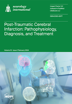
Cover Story (view full-size image): Traumatic brain injury (TBI) remains a leading cause of short and long-term morbidity. For those who require hospital admission, post-traumatic cerebral infarction (PTCI) acutely after TBI is an important but under-recognized complication that is associated with negative clinical outcomes. In this article, we describe the incidence and pathophysiology of PTCI. We then discuss the diagnostic and treatment approaches for the most common etiologies of isolated PTCI, including brain herniation syndromes, cervical artery dissection, venous thrombosis, and post-traumatic vasospasm. We aim to highlight the importance of this condition and future clinical research needs with the goal of improving patient outcomes after TBI. View this paper
  • Issues are regarded as officially published after their release is announced to the table of contents alert mailing list.
  • You may sign up for e-mail alerts to receive table of contents of newly released issues.
  • PDF is the official format for papers published in both, html and pdf forms. To view the papers in pdf format, click on the "PDF Full-text" link, and use the free Adobe Reader to open them.
Order results
Result details
Select all
Export citation of selected articles as:
15 pages, 1378 KiB  
Article
Effectiveness of Switching CGRP Monoclonal Antibodies in Non-Responder Patients in the UAE: A Retrospective Study
by Reem Suliman, Vanessa Santos, Ibrahim Al Qaisi, Batool Aldaher, Ahmed Al Fardan, Hajir Al Barrawy, Yazan Bader, Jonna Lyn Supena, Kathrina Alejandro and Taoufik Alsaadi
Neurol. Int. 2024, 16(1), 274-288; https://doi.org/10.3390/neurolint16010019 - 18 Feb 2024
Viewed by 1799
Abstract
Calcitonin gene-related peptide monoclonal antibodies (CGRP mAbs) have shown promising effectiveness in migraine management compared to other preventative treatment options. Many questions remain regarding switching between antibody classes as a treatment option in patients with migraine headaches. This preliminary retrospective real-world study explored [...] Read more.
Calcitonin gene-related peptide monoclonal antibodies (CGRP mAbs) have shown promising effectiveness in migraine management compared to other preventative treatment options. Many questions remain regarding switching between antibody classes as a treatment option in patients with migraine headaches. This preliminary retrospective real-world study explored the treatment response of patients who switched between CGRP mAb classes due to lack of efficacy or poor tolerability. A total of 53 patients with migraine headache switched between three of the CGRP mAbs types due to lack of efficacy of the original prescribed CGRP mAbs, specifically eptinezumab, erenumab, and galcanezumab. Fremanezumab was not included due to unavailability in the UAE. Galcanezumab and eptinezumab target the CGRP ligand (CGRP/L), while erenumab targets CGRP receptors (CGRP/R). The analysis of efficacy demonstrated that some improvements were seen in both class switch cohorts (CGRP/R to CGRP/L and CGRP/L to CGRP/R). The safety of switching between CGRP classes was well observed, as any adverse events presented before the class switch did not lead to the discontinuation of treatment following the later switch. The findings of this study suggest that switching between different classes of CGRP mAbs is a potentially safe and clinically viable practice that may have some applications for those experiencing side effects on their current CGRP mAb or those witnessing suboptimal response. Full article
Show Figures

Figure 1

11 pages, 1756 KiB  
Case Report
The Effects of Intensive Rehabilitation Combined with Thiamine Treatment on Cognitive Recovery in a Case of Non-Alcoholic Wernicke–Korsakoff Syndrome
by Cinzia Palmirotta, Gilda Turi, Serena Tagliente, Michele Pansini, Stefania De Trane and Gianvito Lagravinese
Neurol. Int. 2024, 16(1), 263-273; https://doi.org/10.3390/neurolint16010018 - 14 Feb 2024
Viewed by 2084
Abstract
Wernicke–Korsakoff Syndrome (WKS) is a severe neurological disorder resulting from thiamine deficiency, commonly associated with alcohol consumption but also stemming from dietary imbalances or other clinical conditions. Cognitive deficits, affecting memory and executive functions, pose a serious concern, with partial recovery often not [...] Read more.
Wernicke–Korsakoff Syndrome (WKS) is a severe neurological disorder resulting from thiamine deficiency, commonly associated with alcohol consumption but also stemming from dietary imbalances or other clinical conditions. Cognitive deficits, affecting memory and executive functions, pose a serious concern, with partial recovery often not complete. A 28-year-old woman underwent surgery for acute necrotizing hemorrhagic pancreatitis, leading to admission for post-acute intensive treatment due to prolonged bed rest syndrome. Clinical examinations revealed sensory–motor neuropathy, denervation in the active phase, mammillary body hyperintensity, and cognitive impairment. The patient exhibited poor orientation, lacked awareness of her clinical condition, and experienced impaired nonverbal memory, practical constructive issues, and planning difficulties—consistent with WKS. The patient received high-dose thiamine (300 mg TDS), coupled with daily physiokinesitherapy and occupational therapy. A final neuropsychological evaluation three months later showed substantial remission of executive and memory difficulties, improved spatial–temporal orientation, and enhanced awareness. The complex case required timely multidisciplinary intervention for accurate diagnosis and effective rehabilitation. The patient experienced rapid clinical improvement and cognitive recovery with high-dose thiamine and physiotherapy. Full article
Show Figures

Graphical abstract

10 pages, 892 KiB  
Article
Safety and Efficacy of Low-Dose Eptifibatide for Tandem Occlusions in Acute Ischemic Stroke
by Paweł Latacz, Tadeusz Popiela, Paweł Brzegowy, Bartłomiej Lasocha, Krzysztof Kwiecień and Marian Simka
Neurol. Int. 2024, 16(1), 253-262; https://doi.org/10.3390/neurolint16010017 - 9 Feb 2024
Viewed by 1527
Abstract
Objectives: The optimal treatment strategy for ischemic stroke in patients presenting with tandem occlusions of the internal carotid artery remains controversial. Several studies have demonstrated better clinical outcomes after eptifibatide, which is a short half-life antiplatelet agent. This retrospective analysis focused on the [...] Read more.
Objectives: The optimal treatment strategy for ischemic stroke in patients presenting with tandem occlusions of the internal carotid artery remains controversial. Several studies have demonstrated better clinical outcomes after eptifibatide, which is a short half-life antiplatelet agent. This retrospective analysis focused on the safety and efficacy of low-dose eptifibatide administration in stroke patients with tandem lesions. Methods: We evaluated the results of endovascular treatment in 148 stroke patients with tandem lesions. Patients in whom balloon angioplasty alone resulted in satisfactory cerebral flow did not receive eptifibatide (33 patients); others received this drug together with stent implantation (115 patients). Eptifibatide was given as an intravenous bolus of 180 μg/kg and then in a modified low dose of 1 μg/kg/min for 24 hours. Results: There were no statistically significant differences between both groups regarding 30-day mortality, frequency of thrombotic events, or hemorrhagic complications. An analysis of clinical status at 30-day follow-up revealed that the administration of eptifibatide was associated with a statistically significant better outcome: a higher rate of either no neurological symptoms or only mild symptoms (4 NIHSS points maximally). Conclusions: The administration of eptifibatide in stroke patients presenting with tandem lesions is relatively safe. Moreover, treatment with this drug can improve clinical outcomes in these challenging patients. Full article
(This article belongs to the Special Issue Treatment Strategy and Mechanism of Acute Ischemic Stroke)
Show Figures

Graphical abstract

14 pages, 3952 KiB  
Article
Mapping Motor Neuroplasticity after Successful Surgical Brachial Plexus Reconstruction Using Navigated Transcranial Magnetic Stimulation (nTMS)
by Gregor Durner, Ina Ulrich, Alexandra Gerst, Ralf Becker, Christian Rainer Wirtz, Gregor Antoniadis, Maria Teresa Pedro and Andrej Pala
Neurol. Int. 2024, 16(1), 239-252; https://doi.org/10.3390/neurolint16010016 - 1 Feb 2024
Viewed by 1097
Abstract
Brachial plexus reconstruction (BPR) consists of the complex surgical restoration of nerve structures. To further understand the underlying motor cortex changes and evaluate neuroplasticity after a successful surgery, we performed a navigated transcranial magnetic stimulation (nTMS) study mapping the postoperative motor representation of [...] Read more.
Brachial plexus reconstruction (BPR) consists of the complex surgical restoration of nerve structures. To further understand the underlying motor cortex changes and evaluate neuroplasticity after a successful surgery, we performed a navigated transcranial magnetic stimulation (nTMS) study mapping the postoperative motor representation of the formerly plegic arm. We conducted a prospective nTMS study mapping the musculocutaneous nerve as a representative, prominent target of BPR including a patient (n = 8) and a control group (n = 10). Measurements like resting motor threshold (RMT), cortical motor area location, and size were taken. Mathematical analysis was performed using MATLAB 2022, statistical analysis was performed using SPSS 26, and nTMS mapping was performed using the Nexstim NBS 5.1 system. Mapping was feasible in seven out of eight patients. Median RMT on the affected hemisphere was 41% compared to 50% on the unaffected hemisphere and they were 37% and 36% on the left and right hemispheres of the control group. The motor area location showed a relocation of bicep brachii representation at the middle precentral gyrus of the corresponding contralateral hemisphere. Motor area size was increased compared to the control group and the patient’s unaffected, ipsilateral hemisphere. Understanding cortical reorganization is important for potential future treatments like therapeutic nTMS. The issue of motor neuroplasticity in patients with brachial plexus lesions is worth exploring in further studies. Full article
Show Figures

Figure 1

13 pages, 1640 KiB  
Case Report
Missense Variants in COL4A1/2 Are Associated with Cerebral Aneurysms: A Case Report and Literature Review
by Masahiro Uemura, Natsuki Tanaka, Shoichiro Ando, Takehiko Yanagihara and Osamu Onodera
Neurol. Int. 2024, 16(1), 226-238; https://doi.org/10.3390/neurolint16010015 - 1 Feb 2024
Viewed by 1713
Abstract
Background: Although cerebral aneurysm (CA) is a defining complication of COL4A1/2-related vasculopathy, the specific factors influencing its onset remain uncertain. This study aimed to identify and analyze these factors. Methods: We described a family presenting with a novel variant of the COL4A1 [...] Read more.
Background: Although cerebral aneurysm (CA) is a defining complication of COL4A1/2-related vasculopathy, the specific factors influencing its onset remain uncertain. This study aimed to identify and analyze these factors. Methods: We described a family presenting with a novel variant of the COL4A1 gene complicated with CA. Concurrently, an exhaustive review of previously documented patients with COL4A1/2-related vasculopathy was conducted by sourcing data from PubMed, Web of Science, Google Scholar, and Ichushi databases. We compared the variant types and locations between patients with CA (positive group) and those without CA (negative group). Results: This study included 53 COL4A1/2 variants from 76 patients. Except for one start codon variant, all the identified variants in CA were missense variants. Otherwise, CA was not associated with other clinical manifestations, such as small-vessel disease or other large-vessel abnormalities. A higher frequency of missense variants (95.5% vs. 58.1%, p = 0.0035) was identified in the CA-positive group. Conclusions: CA development appears to necessitate qualitative alterations in COL4A1/2, and the underlying mechanism seems independent of small-vessel disease or other large-vessel anomalies. Our findings suggest that a meticulous evaluation of CA is necessary when missense variants in COL4A1/2 are identified. Full article
Show Figures

Figure 1

16 pages, 769 KiB  
Review
Processing Speed and Attentional Shift/Mental Flexibility in Patients with Stroke: A Comprehensive Review on the Trail Making Test in Stroke Studies
by Anna Tsiakiri, Foteini Christidi, Dimitrios Tsiptsios, Pinelopi Vlotinou, Sofia Kitmeridou, Paschalina Bebeletsi, Christos Kokkotis, Aspasia Serdari, Konstantinos Tsamakis, Nikolaos Aggelousis and Konstantinos Vadikolias
Neurol. Int. 2024, 16(1), 210-225; https://doi.org/10.3390/neurolint16010014 - 23 Jan 2024
Viewed by 2523
Abstract
The Trail Making Test (TMT) is one of the most commonly administered tests in clinical and research neuropsychological settings. The two parts of the test (part A (TMT-A) and part B (TMT-B)) enable the evaluation of visuoperceptual tracking and processing speed (TMT-A), as [...] Read more.
The Trail Making Test (TMT) is one of the most commonly administered tests in clinical and research neuropsychological settings. The two parts of the test (part A (TMT-A) and part B (TMT-B)) enable the evaluation of visuoperceptual tracking and processing speed (TMT-A), as well as divided attention, set-shifting and cognitive flexibility (TMT-B). The main cognitive processes that are assessed using TMT, i.e., processing speed, divided attention, and cognitive flexibility, are often affected in patients with stroke. Considering the wide use of TMT in research and clinical settings since its introduction in neuropsychological practice, the purpose of our review was to provide a comprehensive overview of the use of TMT in stroke patients. We present the most representative studies assessing processing speed and attentional shift/mental flexibility in stroke settings using TMT and applying scoring methods relying on conventional TMT scores (e.g., time-to-complete part A and part B), as well as derived measures (e.g., TMT-(B-A) difference score, TMT-(B/A) ratio score, errors in part A and part B). We summarize the cognitive processes commonly associated with TMT performance in stroke patients (e.g., executive functions), lesion characteristics and neuroanatomical underpinning of TMT performance post-stroke, the association between TMT performance and patients’ instrumental activities of daily living, motor difficulties, speech difficulties, and mood statue, as well as their driving ability. We also highlight how TMT can serve as an objective marker of post-stroke cognitive recovery following the implementation of interventions. Our comprehensive review underscores that the TMT stands as an invaluable asset in the stroke assessment toolkit, contributing nuanced insights into diverse cognitive, functional, and emotional dimensions. As research progresses, continued exploration of the TMT potential across these domains is encouraged, fostering a deeper comprehension of post-stroke dynamics and enhancing patient-centered care across hospitals, rehabilitation centers, research institutions, and community health settings. Its integration into both research and clinical practice reaffirms TMT status as an indispensable instrument in stroke-related evaluations, enabling holistic insights that extend beyond traditional neurological assessments. Full article
(This article belongs to the Special Issue Emerging Issues in Vascular Cognitive Impairment)
Show Figures

Graphical abstract

8 pages, 3956 KiB  
Brief Report
Association between Brain White Matter Lesions and Disease Activity in HAM/TSP Patients
by Keiko Tamaki, Shinji Ouma, Nobutaka Takahashi, Shinsuke Fujioka and Yoshio Tsuboi
Neurol. Int. 2024, 16(1), 202-209; https://doi.org/10.3390/neurolint16010013 - 22 Jan 2024
Viewed by 1098
Abstract
Human T-cell leukemia virus type 1-associated myelopathy/tropical spastic paraparesis (HAM/TSP) patients may have brain white matter (WM) lesions, but the association of these lesions with disease activity is poorly understood. We retrospectively evaluated the brain WM lesions of 22 HAM/TSP patients (male 4: [...] Read more.
Human T-cell leukemia virus type 1-associated myelopathy/tropical spastic paraparesis (HAM/TSP) patients may have brain white matter (WM) lesions, but the association of these lesions with disease activity is poorly understood. We retrospectively evaluated the brain WM lesions of 22 HAM/TSP patients (male 4: female 18) including 5 rapid progressors, 16 slow progressors, and 1 very slow progressor. The severity of WM brain lesions on axial Fluid Attenuated Inversion Recovery images was evaluated utilizing the Fazekas scale, cerebrospinal fluid biomarkers, and proviral load in peripheral blood mononuclear cells. Imaging and biological data were compared at the first visit and a subsequent visit more than 4 years later. Patients with comorbidities including adult T-cell leukemia–lymphoma and cerebrovascular disease were excluded. The results revealed that brain WM lesions in the rapid progressors group were more pronounced than those in slow progressors. In patients with HAM/TSP, severe and persistent inflammation of the spinal cord may cause brain WM lesions. Full article
Show Figures

Graphical abstract

13 pages, 1479 KiB  
Review
A Comprehensive Review on the Role of Resting-State Functional Magnetic Resonance Imaging in Predicting Post-Stroke Motor and Sensory Outcomes
by Foteini Christidi, Ilias Orgianelis, Ermis Merkouris, Christos Koutsokostas, Dimitrios Tsiptsios, Efstratios Karavasilis, Evlampia A. Psatha, Anna Tsiakiri, Aspasia Serdari, Nikolaos Aggelousis and Konstantinos Vadikolias
Neurol. Int. 2024, 16(1), 189-201; https://doi.org/10.3390/neurolint16010012 - 19 Jan 2024
Viewed by 2210
Abstract
Stroke is a major leading cause of chronic disability, often affecting patients’ motor and sensory functions. Functional magnetic resonance imaging (fMRI) is the most commonly used method of functional neuroimaging, and it allows for the non-invasive study of brain activity. The time-dependent coactivation [...] Read more.
Stroke is a major leading cause of chronic disability, often affecting patients’ motor and sensory functions. Functional magnetic resonance imaging (fMRI) is the most commonly used method of functional neuroimaging, and it allows for the non-invasive study of brain activity. The time-dependent coactivation of different brain regions at rest is described as resting-state activation. As a non-invasive task-independent functional neuroimaging approach, resting-state fMRI (rs-fMRI) may provide therapeutically useful information on both the focal vascular lesion and the connectivity-based reorganization and subsequent functional recovery in stroke patients. Considering the role of a prompt and accurate prognosis in stroke survivors along with the potential of rs-fMRI in identifying patterns of neuroplasticity in different post-stroke phases, this review provides a comprehensive overview of the latest literature regarding the role of rs-fMRI in stroke prognosis in terms of motor and sensory outcomes. Our comprehensive review suggests that with the advancement of MRI acquisition and data analysis methods, rs-fMRI emerges as a promising tool to study the motor and sensory outcomes in stroke patients and evaluate the effects of different interventions. Full article
(This article belongs to the Special Issue Treatment Strategy and Mechanism of Acute Ischemic Stroke)
Show Figures

Figure 1

3 pages, 161 KiB  
Conference Report
The Sixth Brainstorming Research Assembly for Young Neuroscientists (BraYn), Naples, Italy, 27–29 September 2023
by Giovanni Ferrara
Neurol. Int. 2024, 16(1), 186-188; https://doi.org/10.3390/neurolint16010011 - 19 Jan 2024
Viewed by 1257
Abstract
The BraYn association aims to bolster young neuroscientists’ research endeavors through collaborative support, fundraising assistance, and events promoting knowledge exchange and collaboration across Europe. Central to its mission is the annual BraYn conference, tailored for PhD students, postdocs, junior PIs, neurologists, and clinicians. [...] Read more.
The BraYn association aims to bolster young neuroscientists’ research endeavors through collaborative support, fundraising assistance, and events promoting knowledge exchange and collaboration across Europe. Central to its mission is the annual BraYn conference, tailored for PhD students, postdocs, junior PIs, neurologists, and clinicians. This gathering champions cooperation, offering talks by key figures, educational workshops, and opportunities for attendees to present their work, compete for grants, and engage in international scientific experiences. The conference, established in 2018, has grown substantially in attendance and industry support and was adapted during the pandemic with virtual editions. The last sixth edition in Naples (27–29 September 2023) attracted over 300 delegates, focusing on peer-to-peer discussions, interdisciplinary collaboration, and interaction with renowned speakers, solidifying its place as a flagship event for Europe’s budding neuroscience researchers. Full article
24 pages, 2676 KiB  
Case Report
Extra-Axial Cavernous Angioma: A Case Report and Review of the Literature
by Shakiba Hassanzadeh, Linlin Gao, Anthony M. Alvarado, Paul J. Camarata, Nelli S. Lakis and Mohammad Haeri
Neurol. Int. 2024, 16(1), 162-185; https://doi.org/10.3390/neurolint16010010 - 12 Jan 2024
Cited by 1 | Viewed by 1923
Abstract
Cavernous angiomas (CAs) are benign vascular malformations predominantly seen in the brain parenchyma and therefore referred to as intra-axial. Extra-axial dural-based cavernous angiomas, on the other hand, are rare vascular lesions found outside of the brain parenchyma. They occur in the middle fossa [...] Read more.
Cavernous angiomas (CAs) are benign vascular malformations predominantly seen in the brain parenchyma and therefore referred to as intra-axial. Extra-axial dural-based cavernous angiomas, on the other hand, are rare vascular lesions found outside of the brain parenchyma. They occur in the middle fossa and may be easily misdiagnosed as meningiomas due to their extra-axial location. In addition, CAs that are located outside the middle fossa, such as in the convexity, have a better prognosis since they are more surgically accessible. Surgical resection is the main treatment of choice in CAs. However, other options, such as embolization and radiotherapy, may also be considered therapeutic choices or additive treatment options. The pathogenesis of CA and the involvement of other factors (genetics or environmental factors) are still unknown and require further investigation. We are presenting a young man who presented for evaluation of seizure-like events without any family history of neurologic conditions. The physical examination was unremarkable except for a slightly antalgic gait. Imaging studies showed an extra-axial left tentorial mass suggestive of a meningioma, hemangiopericytoma, or other extra-axial lesions. The lesion was resected where its vascular nature was mentioned initially, and the histology proved the diagnosis of cavernous angioma. Here we give an overview of the known pathogenesis, causes, clinical features, and diagnostic and therapeutic options in CA. Better knowledge about CA, its causes, clinical features, and treatment options would help clinicians in early diagnosis and patient management. Full article
Show Figures

Figure 1

23 pages, 858 KiB  
Systematic Review
Pulmonary Function Tests Post-Stroke. Correlation between Lung Function, Severity of Stroke, and Improvement after Respiratory Muscle Training
by Fotios Drakopanagiotakis, Konstantinos Bonelis, Paschalis Steiropoulos, Dimitrios Tsiptsios, Anastasia Sousanidou, Foteini Christidi, Aimilios Gkantzios, Aspasia Serdari, Styliani Voutidou, Chrysoula-Maria Takou, Christos Kokkotis, Nikolaos Aggelousis and Konstantinos Vadikolias
Neurol. Int. 2024, 16(1), 139-161; https://doi.org/10.3390/neurolint16010009 - 11 Jan 2024
Viewed by 2268
Abstract
Stroke is a significant cause of mortality and chronic morbidity caused by cardiovascular disease. Respiratory muscles can be affected in stroke survivors, leading to stroke complications, such as respiratory infections. Respiratory function can be assessed using pulmonary function tests (PFTs). Data regarding PFTs [...] Read more.
Stroke is a significant cause of mortality and chronic morbidity caused by cardiovascular disease. Respiratory muscles can be affected in stroke survivors, leading to stroke complications, such as respiratory infections. Respiratory function can be assessed using pulmonary function tests (PFTs). Data regarding PFTs in stroke survivors are limited. We reviewed the correlation between PFTs and stroke severity or degree of disability. Furthermore, we reviewed the PFT change in stroke patients undergoing a respiratory muscle training program. We searched PubMed until September 2023 using inclusion and exclusion criteria in order to identify studies reporting PFTs post-stroke and their change after a respiratory muscle training program. Outcomes included lung function parameters (FEV1, FVC, PEF, MIP and MEP) were measured in acute or chronic stroke survivors. We identified 22 studies of stroke patients, who had undergone PFTs and 24 randomised controlled trials in stroke patients having PFTs after respiratory muscle training. The number of patients included was limited and studies were characterised by great heterogeneity regarding the studied population and the applied intervention. In general, PFTs were significantly reduced compared to healthy controls and predicted normal values and associated with stroke severity. Furthermore, we found that respiratory muscle training was associated with significant improvement in various PFT parameters and functional stroke parameters. PFTs are associated with stroke severity and are improved after respiratory muscle training. Full article
(This article belongs to the Special Issue Treatment Strategy and Mechanism of Acute Ischemic Stroke)
Show Figures

Graphical abstract

13 pages, 1894 KiB  
Article
Contingent Negative Variation in the Evaluation of Neurocognitive Disorders Due to Possible Alzheimer’s Disease
by Arquímedes Montoya-Pedrón, Carmen María Ocaña Montoya, Jorge Esteban Santos Toural, Tania Acosta Lee, Miguel Enrique Sánchez-Hechavarría, Erislandis López-Galán and Gustavo Alejandro Muñoz-Bustos
Neurol. Int. 2024, 16(1), 126-138; https://doi.org/10.3390/neurolint16010008 - 11 Jan 2024
Viewed by 1368
Abstract
The usefulness of Contingent Negative Variation (CNV) potential as a biomarker of neurocognitive disorders due to possible Alzheimer’s disease, is based on its possible physiological correlates. However, its application in the diagnostic evaluation of these disorders is still incipient. The aim of this [...] Read more.
The usefulness of Contingent Negative Variation (CNV) potential as a biomarker of neurocognitive disorders due to possible Alzheimer’s disease, is based on its possible physiological correlates. However, its application in the diagnostic evaluation of these disorders is still incipient. The aim of this study is to characterize the patterns of cognitive processing of information in the domain of nonspecific global attention, by recording potential CNV in a group of patients with neurocognitive disorders due to possible Alzheimer’s disease. An experimental study of cases and controls was carried out. The sample included 39 patients classified according to DSM-5 with a neurocognitive disorder subtype possibly due Alzheimer’s disease, and a Control Group of 53 subjects with normal cognitive functions. CNV potential was registered using standard protocol. The analysis of variance obtained significant differences in mean values and confidence intervals of total CNV amplitude between the three study groups. The late CNV segment amplitudes makes it possible to discriminate between the level of mild and major dysfunction in the group of patients. The CNV total amplitudes of potential allows for effective discrimination between normal cognitive functioning and neurocognitive disorders due to possible Alzheimer’s disease. Full article
(This article belongs to the Topic Translational Advances in Neurodegenerative Dementias)
Show Figures

Figure 1

13 pages, 2306 KiB  
Article
Variability of the Cervical Branch Depending on the Facial Nerve Branching Pattern and Anthropometric Type of the Head
by Angela Babuci, Victor Palarie, Ilia Catereniuc, Zinovia Zorina, Sergiu Visnevschi, Diana Heimes, Sofia Lehtman and Peer W. Kämmerer
Neurol. Int. 2024, 16(1), 113-125; https://doi.org/10.3390/neurolint16010007 - 4 Jan 2024
Cited by 2 | Viewed by 3057
Abstract
(1) Background: Considering that the specialty literature supplies only general data about the variability of the cervical branch of the facial nerve, this study aimed to determine this branch’s variation and individual peculiarities depending on the nerve branching pattern and anthropometric type of [...] Read more.
(1) Background: Considering that the specialty literature supplies only general data about the variability of the cervical branch of the facial nerve, this study aimed to determine this branch’s variation and individual peculiarities depending on the nerve branching pattern and anthropometric type of the head. (2) Methods: The study was conducted on 75 hemifaces of adult formalized cadavers. Ahead of anatomical dissection, each head was measured to establish the anthropometric type, according to Franco and colleagues. The branching patterns were then distributed according to the Davis classification. (3) Results: The number of cervical branches (CB) of the facial nerve varied from one to five branches, with the following rate: 1 CB (61.3%), 2 CB (28%), 3 CB (6.7%), 4 CB (2.7%), and 5 CB (1.3%). Seven branching patterns of the facial nerve were revealed: Type I in 18.7%, Type II in 14.7%, Type III in 20%, Type IV in 14.6%, Type V in 5.3%, Type VI in 18.7%, and Type NI in 8% (bizarre types). According to the branching pattern, the mean numbers of the cervical branches were as follows: Type I—1.6 ± 1.02; Type II—1.4 ± 0.50; Type III—1.4 ± 0.50; Type IV—1.4 ± 0.67; Type V—2.0 ± 1.41; Type VI—1.8 ± 1.12; and Type-NI—1.8 ± 0.75; p = 0.599. According to the anthropometric type of the head, the mean number of CB in the mesocephalic type (MCT) was 1.5 ± 0.82, in the dolichocephalic type (DCT), 1.7 ± 0.87, and in the brachycephalic type, (BCT) 1.8 ± 1.04; p = 0.668. (4) Conclusions: The cervical branch of the facial nerve varies depending on the facial nerve branching pattern and the anthropometric type of the head. The highest degree of variation was characteristic of BCT and Type V and the lowest, of MCT and Types II, III, and IV. Full article
Show Figures

Figure 1

18 pages, 3063 KiB  
Review
Post-Traumatic Cerebral Infarction: A Narrative Review of Pathophysiology, Diagnosis, and Treatment
by Roy A. Poblete, Charlotte Zhong, Anish Patel, Grace Kuo, Philip Y. Sun, Jiayu Xiao, Zhaoyang Fan, Nerses Sanossian, Amytis Towfighi and Patrick D. Lyden
Neurol. Int. 2024, 16(1), 95-112; https://doi.org/10.3390/neurolint16010006 - 4 Jan 2024
Cited by 1 | Viewed by 4752
Abstract
Traumatic brain injury (TBI) is a common diagnosis requiring acute hospitalization. Long-term, TBI is a significant source of health and socioeconomic impact in the United States and globally. The goal of clinicians who manage TBI is to prevent secondary brain injury. In this [...] Read more.
Traumatic brain injury (TBI) is a common diagnosis requiring acute hospitalization. Long-term, TBI is a significant source of health and socioeconomic impact in the United States and globally. The goal of clinicians who manage TBI is to prevent secondary brain injury. In this population, post-traumatic cerebral infarction (PTCI) acutely after TBI is an important but under-recognized complication that is associated with negative functional outcomes. In this comprehensive review, we describe the incidence and pathophysiology of PTCI. We then discuss the diagnostic and treatment approaches for the most common etiologies of isolated PTCI, including brain herniation syndromes, cervical artery dissection, venous thrombosis, and post-traumatic vasospasm. In addition to these mechanisms, hypercoagulability and microcirculatory failure can also exacerbate ischemia. We aim to highlight the importance of this condition and future clinical research needs with the goal of improving patient outcomes after TBI. Full article
Show Figures

Figure 1

21 pages, 3362 KiB  
Review
The Management of Intracranial Aneurysms: Current Trends and Future Directions
by Aviraj S. Deshmukh, Stefano M. Priola, Aris H. Katsanos, Gianluca Scalia, Aderaldo Costa Alves, Abhilekh Srivastava and Christine Hawkes
Neurol. Int. 2024, 16(1), 74-94; https://doi.org/10.3390/neurolint16010005 - 3 Jan 2024
Cited by 5 | Viewed by 3856
Abstract
Intracranial aneurysms represent a major global health burden. Rupture of an intracranial aneurysm is a catastrophic event. Without access to treatment, the fatality rate is 50% in the first 30 days. Over the last three decades, treatment approaches for intracranial aneurysms have changed [...] Read more.
Intracranial aneurysms represent a major global health burden. Rupture of an intracranial aneurysm is a catastrophic event. Without access to treatment, the fatality rate is 50% in the first 30 days. Over the last three decades, treatment approaches for intracranial aneurysms have changed dramatically. There have been improvements in the medical management of aneurysmal subarachnoid haemorrhage, and there has been an evolution of treatment strategies. Endovascular therapy is now the mainstay of the treatment of ruptured intracranial aneurysms based on robust randomised controlled trial data. There is now an expansion of treatment indications for unruptured intracranial aneurysms to prevent rupture with both microsurgical clipping and endovascular treatment. Both microsurgical and endovascular treatment modalities have evolved, in particular with the introduction of innovative endovascular treatment options including flow diversion and intra-saccular flow disruption. These novel therapies allow clinicians to treat more complex and previously untreatable aneurysms. We aim to review the evolution of treatment strategies for intracranial aneurysms over time, and discuss emerging technologies that could further improve treatment safety and functional outcomes for patients with an intracranial aneurysm. Full article
(This article belongs to the Special Issue Global Burden of Neurological Disorder)
Show Figures

Figure 1

12 pages, 726 KiB  
Article
Longitudinal Neuropsychological Assessment of Symptomatic Edema after Subthalamic Nucleus Deep Brain Stimulation Surgery: A Case Series Study
by Silvia De Ieso, Giulia Di Rauso, Francesco Cavallieri, Daniela Beltrami, Alessandro Marti, Manuela Napoli, Rosario Pascarella, Alberto Feletti, Valentina Fioravanti, Giulia Toschi, Vittorio Rispoli, Francesca Antonelli, Annette Puzzolante, Giacomo Pavesi, Federico Gasparini and Franco Valzania
Neurol. Int. 2024, 16(1), 62-73; https://doi.org/10.3390/neurolint16010004 - 28 Dec 2023
Viewed by 1304
Abstract
Severe non-infectious or non-haemorrhagic brain edema surrounding the electrode represents a rare complication of subthalamic nucleus deep brain stimulation (STN-DBS) surgery. The aim of this study is to report three patients with advanced Parkinson’s Disease (PD) who developed symptomatic brain edema after STN-DBS [...] Read more.
Severe non-infectious or non-haemorrhagic brain edema surrounding the electrode represents a rare complication of subthalamic nucleus deep brain stimulation (STN-DBS) surgery. The aim of this study is to report three patients with advanced Parkinson’s Disease (PD) who developed symptomatic brain edema after STN-DBS surgery treated with intravenous steroids with a specific profile of reversible cognitive alterations. Patients were both assessed with a comprehensive neuropsychological battery including attention, memory, visuo-spatial and executive tasks. They were also briefly assessed for emotional and behavioural alterations, and for possible limitations in the activities of daily living. Normative data for an Italian population were available for all neuropsychological tests. The patients were firstly assessed before the surgery (baseline) as soon as they became symptomatic for the post-surgery edema and a few more times in follow-up up to ten months. In all patients we observed the resolution of cognitive deficits within six months after surgery with the corresponding reabsorption of edema at brain CT scans. The appearance of post-DBS edema is a fairly frequent and clinically benign event. However, in some rare cases it can be very marked and lead to important clinical—albeit transient—disturbances. These events can compromise, at least from a psychological point of view, the delicate path of patients who undergo DBS and can prolong the post-operative hospital stay. In this setting it could be helpful to perform a brain CT scan in 2–3 days with the aim of detecting the early appearance of edema and treating it before it can constitute a relevant clinical problem. Full article
Show Figures

Figure 1

29 pages, 10286 KiB  
Article
Investigating the Protective Effects of a Citrus Flavonoid on the Retardation Morphogenesis of the Oligodendroglia-like Cell Line by Rnd2 Knockdown
by Shoya Fukatsu, Yuki Miyamoto, Yu Oka, Maki Ishibashi, Remina Shirai, Yuki Ishida, Shin Endo, Hironori Katoh and Junji Yamauchi
Neurol. Int. 2024, 16(1), 33-61; https://doi.org/10.3390/neurolint16010003 - 26 Dec 2023
Cited by 3 | Viewed by 1381
Abstract
Recent discoveries suggest links between abnormalities in cell morphogenesis in the brain and the functional deficiency of molecules controlling signal transduction in glial cells such as oligodendroglia. Rnd2 is one such molecule and one of the Rho family monomeric GTP-binding proteins. Despite the [...] Read more.
Recent discoveries suggest links between abnormalities in cell morphogenesis in the brain and the functional deficiency of molecules controlling signal transduction in glial cells such as oligodendroglia. Rnd2 is one such molecule and one of the Rho family monomeric GTP-binding proteins. Despite the currently known functions of Rnd2, its precise roles as it relates to cell morphogenesis and disease state remain to be elucidated. First, we showed that signaling through the loss of function of the rnd2 gene affected the regulation of oligodendroglial cell-like morphological differentiation using the FBD-102b cell line, which is often utilized as a differentiation model. The knockdown of Rnd2 using the clustered regularly interspaced palindromic repeats (CRISPR)/CasRx system or RNA interference was shown to slow morphological differentiation. Second, the knockdown of Prag1 or Fyn kinase, a signaling molecule acting downstream of Rnd2, slowed differentiation. Rnd2 or Prag1 knockdown also decreased Fyn phosphorylation, which is critical for its activation and for oligodendroglial cell differentiation and myelination. Of note, hesperetin, a citrus flavonoid with protective effects on oligodendroglial cells and neurons, can recover differentiation states induced by the knockdown of Rnd2/Prag1/Fyn. Here, we showed that signaling through Rnd2/Prag1/Fyn is involved in the regulation of oligodendroglial cell-like morphological differentiation. The effects of knocking down the signaling cascade molecule can be recovered by hesperetin, highlighting an important molecular structure involved in morphological differentiation. Full article
Show Figures

Graphical abstract

13 pages, 1131 KiB  
Systematic Review
Phantosmia in Parkinson’s Disease: A Systematic Review of the Phenomenology of Olfactory Hallucinations
by Tommaso Ercoli, Caterina Francesca Bagella, Claudia Frau, Elisa Ruiu, Sabrine Othmani, Giansalvo Gusinu, Carla Masala, Leonardo Antonio Sechi, Paolo Solla and Giovanni Defazio
Neurol. Int. 2024, 16(1), 20-32; https://doi.org/10.3390/neurolint16010002 - 22 Dec 2023
Cited by 1 | Viewed by 2101
Abstract
Olfactory dysfunction is a prevalent non-motor symptom in Parkinson’s disease (PD), affecting approximately 65–90% of subjects. PD patients may also report odor perception in the absence of any external source, often referred to as olfactory hallucinations (OHs) or phantosmia. This study aims to [...] Read more.
Olfactory dysfunction is a prevalent non-motor symptom in Parkinson’s disease (PD), affecting approximately 65–90% of subjects. PD patients may also report odor perception in the absence of any external source, often referred to as olfactory hallucinations (OHs) or phantosmia. This study aims to explore the current understanding of OHs in PD and offer a comprehensive overview of their prevalence and characteristics. We conducted a systematic search of the literature published on PubMed from inception to July 2023 regarding OHs in PD, following PRISMA guidelines. From the 2875 studies identified through database searching, 29 studies fulfilled the necessary criteria and underwent data extraction. The frequency of OHs in PD patients varies widely, ranging from 0.5% to 18.2%, with female prevalence ranging from 36% to 75% of the patients. Olfactory experiences may vary widely, ranging from pleasant scents to unpleasant odors. Several studies have indicated the concurrent presence of other types of hallucinations alongside phantosmia, especially visual and auditory hallucinations. OHs in PD are a type of hallucination that has been largely overlooked. To gain a deeper understanding of OHs in PD patients, the next crucial step should involve the development and validation of a dedicated questionnaire. Full article
Show Figures

Figure 1

19 pages, 3303 KiB  
Systematic Review
Planimetric and Volumetric Brainstem MRI Markers in Progressive Supranuclear Palsy, Multiple System Atrophy, and Corticobasal Syndrome. A Systematic Review and Meta-Analysis
by Maria-Evgenia Brinia, Ioanna Kapsali, Nikolaos Giagkou and Vasilios C. Constantinides
Neurol. Int. 2024, 16(1), 1-19; https://doi.org/10.3390/neurolint16010001 - 19 Dec 2023
Cited by 2 | Viewed by 1370
Abstract
Background: Various MRI markers—including midbrain and pons areas (Marea, Parea) and volumes (Mvol, Pvol), ratios (M/Parea, M/Pvol), and composite markers (magnetic resonance imaging Parkinsonism Indices 1,2; MRPI 1,2)—have been proposed as [...] Read more.
Background: Various MRI markers—including midbrain and pons areas (Marea, Parea) and volumes (Mvol, Pvol), ratios (M/Parea, M/Pvol), and composite markers (magnetic resonance imaging Parkinsonism Indices 1,2; MRPI 1,2)—have been proposed as imaging markers of Richardson’s syndrome (RS) and multiple system atrophy–Parkinsonism (MSA-P). A systematic review/meta-analysis of relevant studies aiming to compare the diagnostic accuracy of these imaging markers is lacking. Methods: Pubmed and Scopus were searched for studies with >10 patients (RS, MSA-P or CBS) and >10 controls with data on Marea, Parea, Mvol, Pvol, M/Parea, M/Pvol, MRPI 1, and MRPI 2. Cohen’s d, as a measure of effect size, was calculated for all markers in RS, MSA-P, and CBS. Results: Twenty-five studies on RS, five studies on MSA-P, and four studies on CBS were included. Midbrain area provided the greatest effect size for differentiating RS from controls (Cohen’s d = −3.10; p < 0.001), followed by M/Parea and MRPI 1. MSA-P had decreased midbrain and pontine areas. Included studies exhibited high heterogeneity, whereas publication bias was low. Conclusions: Midbrain area is the optimal MRI marker for RS, and pons area is optimal for MSA-P. M/Parea and MRPIs produce smaller effect sizes for differentiating RS from controls. Full article
Show Figures

Figure 1

Previous Issue
Next Issue
Back to TopTop