Article

Ingested Microplastics Can Act as Microbial Vectors of Ichthyofauna

by
Abdulhusein Jawdhari
1,2,3,
György Deák
2,
Dan Florin Mihăilescu
1,
Nicolai Crăciun
2,4,
Andrea Cristina Staicu
4,
Ioana Stanca
5,6,
Derniza Cozorici
6,7,
Sergiu Fendrihan
2,6,
Cristian-Emilian Pop
1,2,6,* and
Maria Mernea
1
1
Department of Anatomy, Animal Physiology and Biophysics, Faculty of Biology, University of Bucharest, 91–95 Splaiul Independenței Str., 050095 Bucharest, Romania
2
National Institute for Research and Development in Environmental Protection, 294 Splaiul Independenței Str., 060031 Bucharest, Romania
3
Department of Animal Production Techniques, Almussib Technical Instated, Al-Furat Al-Awsat Technical University, Babylon-Najaf Street, Najaf 54003, Iraq
4
Zoology Section, Department of Biochemistry and Molecular Biology, Faculty of Biology, University of Bucharest, 91–95 Splaiul Independenței Str., 050095 Bucharest, Romania
5
Department of Marine Biology, Faculty of Science and Engineering, University of Groningen, Nijenborgh 4, 9747 AG Groningen, The Netherlands
6
Non-Governmental Research Organization Biologic, 14 Schitului Str., 032044 Bucharest, Romania
7
Advanced Polymer Materials Group, National University of Science and Technology Politehnica Bucharest, 1–7 Gh. Polizu Street, 011061 Bucharest, Romania
*
Author to whom correspondence should be addressed.
Microbiol. Res. 2024, 15(2), 614-625; https://doi.org/10.3390/microbiolres15020040
Submission received: 2 April 2024 / Revised: 19 April 2024 / Accepted: 22 April 2024 / Published: 23 April 2024
(This article belongs to the Special Issue Zoonotic Bacteria: Infection, Pathogenesis and Drugs)

Abstract

:
Microplastics (plastic particles < 5 mm) are ubiquitous pollutants that have the ability to carry microbiota, including pathogens. Microbial adhesion is usually a sign of pathogenicity; thus, we investigated the adherent microbiota found on 4 mm nylon strips, which were ingested and excreted by wild fish specimens. Retention times were recorded and the polymer analysis of the excreted samples was performed, which showed no signs of degradation, nor did their controls, represented by the nylon strips submerged in the same water tanks. Both the ingested samples and controls presented pathogens in large quantities. Following Matrix-Assisted Laser Desorption/Ionization Time-Of-Flight identification, the dominant genus was represented by Aeromonas, revealing the fact that nylon microplastics can serve as undegradable physical carriers for this pathogen, among others, in the aquatic environment.

1. Introduction

Microplastics pollution has become ubiquitous, so much so that these particles are now being unintentionally accumulated in both animals [1,2] and humans [3]. With the rise in microplastic contamination, concerns have also risen regarding the potential health risks of such exposure. While most recent scientific reports focused on microplastics’ (MPs’) toxicity in terms of teratogenic effects [4], as well as their accumulation in organs such as the placenta [5], lungs [6] or the intestinal tissues [7], including microplastic-related toxicity represented by endocrine-disrupting effects [8,9,10], fewer scientific releases reported MPs as potential microbial carriers, especially in the case of MPs that have lower density than water, thus being concentrated in the neustonic layer [11].
The most frequent MP pollution occurs in the aquatic environment, due to anthropogenic factors such as industrial activities and littering [12,13]. Nylon MPs are some of the most prevalent contaminants in the aquatic environment, which result from lost or discarded fishing gear, mainly represented by plastic monofilament lines and nylon nets [14,15], but also from everyday consumer products and improper waste management [16,17]. Once the aquatic environment has been contaminated, MPs scale up the food chain as they are mistakenly ingested as food by aquatic fauna or via trophic transfer [18,19]. Fish are the main consumers of MPs, with emerging evidence suggesting that MPs accumulate in the gastrointestinal tract of various fish species [20,21]. Sequeira et al. [22] reported that predatory fish species, in particular, present higher MP accumulation compared to omnivorous species; in a study that involved 198 species examined across 24 countries, MPs were detected in 60% of wild-caught fish and 14% of aquaculture fish [22]. Indeed, predators are hypothesized to ingest more MPs than other species [23]. However, another study reported omnivorous fish are more prone to such events [24], while other studies [25,26,27] report microplastics to directly and indirectly affect biotic and food web interactions, revealing a much more complicated dynamic.
Once ingested, MPs can block the gastrointestinal tract, resulting in physical damage [28] or the induction of fake satiation [29], resulting in the starvation or even potential death of the animal [30,31]. Furthermore, MPs’ ingestion can induce intestinal dysbiosis in both humans [30] and animals [32].
When MPs are excreted, they may carry along a bacterial load represented by the hosts’ intestinal microbiota. Although several reports have stated the interaction of MPs with the intestinal microbiota in terms of its toxicological effects [33] and polymer degradation [34], the colonization of MPs by bacteria, especially pathogens, should not be overlooked. In a marine study, Zettler et al., 2010 [35] coined the term “plastisphere”, which represents the diverse microbial community inhabiting plastic particles. MPs provide a hydrophobic surface that supports the formation of microbial biofilms [36,37], with some inhabitants of the plastisphere being opportunistic pathogens, such as members of the Vibrio genus, strains of which are capable of reproducible biofilm formation on multiple types of polymers [38].
Members of the plastisphere are known to be released in the water column when MPs reach a new environment [39]; furthermore, it was recorded that all surfaces of MPs in the marine environment are rapidly colonized by bacteria [40], among which, fish pathogens such as Aeromonas have a particularly high affinity [41]. It is also worth mentioning that the biohazard aspect of MPs extends beyond natural environments, with MPs providing a stable and protective habitat for diverse wastewater bacteria, including pathogenic and antibiotic-resistant species [36,42].
Although evidence has been built up during the past decade, MPs have only recently started to be in the spotlight as pathogens carriers [43], mainly due to the fact that pollution with such agents is growing, and pathogens may use these durable fragments to hitchhike long distances in water [44,45]. Therefore, the purpose of this study was to evaluate, under controlled conditions, to what extent nylon MPs are colonized by bacteria, especially by fish and waterborne pathogens, and to also evaluate if the digestion process affects the structure of the polymer.

2. Materials and Methods

2.1. Specimens and Exposure

Wild specimens of Oreochromis niloticus (Nile tilapia) weighing ≈400 g each, were grouped in groups of five in three water tanks of 300 L, under similar conditions (Supplementary Table S1), and deprived of food for one week prior to the exposure. Water was aerated via filtered (Midisart 0.2 μm PTFE filter, Sartorius, NY, USA) air pumping; temperature, pH, and oxygen saturation were monitored daily using a portable multiparameter probe (HQ4300, Hach, Loveland, CO, USA). “Aller Futura” feed (Aller Aqua, Christiansfeld, Denmark) was grinded and reformed into pellets, with each pellet containing one strip of thermo-resistant nylon 6 (known as polycaprolactam or polyamide 6) [46], measuring 2 mm in width and 4 mm in length. Nylon 6 is an odorless and solid polymer with the molecular formula (C6H11NO)n, capable of withstanding temperatures up to 215 °C [47,48]. The nylon-containing feed pellets were left to dry at room temperature for 24 h and then autoclaved (121 °C, 15 psi, 20 min). Control was represented by the same nylon-containing feed pellets, which were inserted in the water tanks in sieved recipients that prevented their consumption, yet allowed contact with the aquatic environment.
Each group received and fully ingested 50 nylon-containing feed pellets, weighing ≈10 g. After their ingestion, every 24 h, each group received 10 g of sterilized non-nylon feed pellets to facilitate bowel movement and the excretion of the nylon strips over the course of one week. The nylon strips were recovered immediately after excretion by individual collection from the water tanks with the use of a large serological pipette attached to a pipette pump (Hirschman pipetus, Hirschman, Germany) and stored at 4 °C prior to analysis.
The statistical comparison of results obtained in the three groups was performed using an ANOVA test. The number of nylon strips were mediated per day over the three groups and standard deviations were calculated. The overall differences between the numbers of nylon strips recovered after different time periods was addressed by the ANOVA test. A pairwise comparison between the strips excreted at various times was performed using t-tests.
No specimens were harmed during the experiment, and all actions were performed in conformity with the Guide for the Use and Care of Laboratory Animals recommendations [49] regarding reductions in animal suffering.

2.2. Microbiota Recovery

The excreted nylon strips of the groups, as well as their respective controls, were tapped dry using sterile paper and then bathed through three successive sterile water baths to separate non-adherent microbiota.
In order to recover the adherent microbiota, the dried nylon strips were inserted in liquid Amies media (Rmbio, Missoula, MT, USA), shaken, and incubated for 2 h at room temperature prior to plate inoculations. Wet feces samples were also prelevated per group, diluted in liquid Amies media (100 mg/mL), and inoculated on the same growth media as nylon recovered samples, with the use of a 10 µL sterile loop.
All nylon recovered samples were spread at 100 µL on Chapman agar, 5% blood agar, bile aesculin agar, Salmonella–Shigella agar, MacConkey sorbitol agar, DCL agar, Yersinina agar, and xylose lysine deoxycholate agar plates (MLT, Arad, Romania), with the use of a Drigalski spatula and a Petri dish turntable (Schuett-Biotec, Göttingen, Germany). The media plates were incubated at 23 °C for 36 h, and the developed colonies were counted under a manual colony counter (Schuett-biotec, Göttingen, Germany) and then isolated on fresh media plates. Following a macroscopic observation of the colonies and their morphology at 48 h post-incubation at 23 °C, the isolates were used for taxonomic identification analysis.

2.3. Taxonomic Identification

Final taxonomic identification was performed with a Matrix-Assisted Laser Desorption/Ionization Time-of-Flight (MALDI-TOF) mass spectrometer (Bruker MALDI Biotyper, Bruker Daltonics, Bremen, Germany); the data acquisition was performed using MBT Compass IVD software (Server Version: 4.1.80 PYTH) coupled with the Species/Entry List MBT IVD Library (Revision J, Bruker Daltonics, Bremen, Germany). To perform the assay, MSP 96 target polished steel plate spots (Bruker Daltonics, Bremen, Germany) were loaded with isolated colonies, and on each spot, α-Cyano-4-hydroxycinnamic acid (Bruker Daltonics, Bremen, Germany) matrix was added, along with the stock solution (acetonitrile, trifluoracetic acid, and distilled water).
MALDI-TOF technology identifies the distinctive proteomic signature of a microorganism and compares its characteristic patterns with a reference database to establish the taxonomic identity. The technology has been considered rapid and cost effective for microbial identification in both clinical and research purposes [50]. Despite its limitations (such as the requirement for an extensive database), the technology is favorable as culture conditions that could significantly impact microbial physiology and protein expression profiles [51] do not affect microbial identification by MALDI-TOF [52,53].

2.4. Polymer Analysis

2.4.1. Differential Scanning Calorimetry Analysis

To investigate the thermal behavior of the excreted nylon strips versus controls, a Differential Scanning Calorimetry (DSC) analysis was conducted using a Netzsch 204 F1 Phoenix instrument (Netzsch Gerätebau GmbH, Selb, Germany). The samples, weighing between 3.5 and 4 mg, were placed in individual aluminum pans and positioned on the sample platform of the DSC instrument. The experiments were carried out in a controlled nitrogen environment with a purity of 99.99%. The samples were heated from 0 to 250 °C at a heating rate of 5 °C/min, with a continuous nitrogen flow rate of 20 mL/min. Two complete heating–cooling cycles were performed for each sample, and the resulting thermograms were analyzed using Proteus analysis software (version 4.8.5).

2.4.2. Fourier-Transform Infrared Spectroscopy

Fourier-Transform Infrared Spectroscopy (FTIR) was performed on the negative control (NC) represented by a pristine nylon sample, the positive controls (PCs) represented by nylon were kept under water, and the samples (S) represented by nylon pieces that transited the digestive system of fish were recovered from the water tanks. Measurements were performed on three nylon strips corresponding to each condition (NC, PC, and S) using a Bruker Tensor 27 spectrometer (Bruker Optik GmbH, Ettlingen, Germany) with an attenuated total reflection (ATR) module. The analyzed nylon strips were tightly pressed on the ATR crystal in order to perform the measurement. The absorbance of samples was recorded in the 4000–400 cm−1 range with a resolution of 4 cm−1 during 1 min. For an easy comparison, spectra were normalized by 0 to 1. Spectra acquisition and analysis were performed with OPUS software version 7.2 (Bruker Optik GmbH, Ettlingen, Germany).
Carbonyl indices (ICO) were calculated based on the measured spectra as the ratio between the maximum absorbance of the carbonyl group found at ~1634 cm−1 and the maximum absorbance of the C-H band at ~684 cm−1 [54]. The triplicate measurements performed in each condition (CN, CP, and S) were used to calculate mean indices and their standard deviations.

3. Results

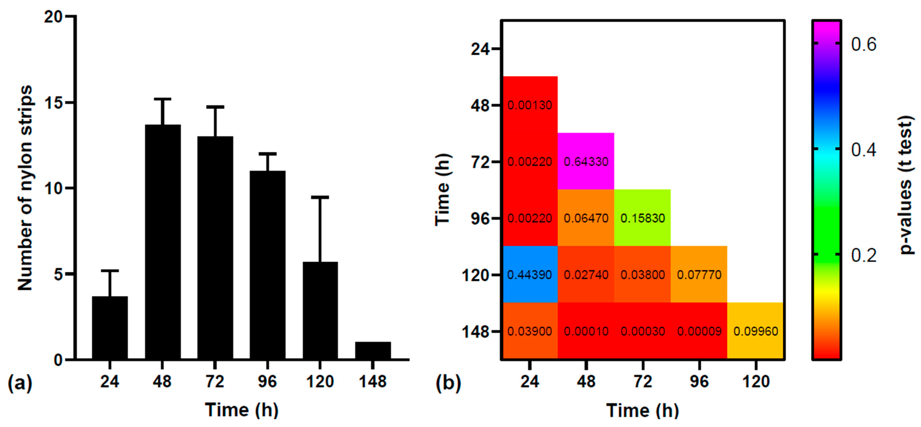
3.1. Statistical Analysis of MPs Excretion

The mean numbers of nylon strips that were recovered from the three groups of fish are shown in Figure 1a. From the 150 nylon strips that were fed to the three groups of fish (50/group), only 6 strips were not recovered: 3 from the first group, 1 from the second group, and 2 from the third group. The differences between the three groups of fish were not statistically significant (the ANOVA test F value was 0.0053 and p-value was 0.9946), showing the similar excretion rate of the nylon strips. When addressing the differences between the numbers of nylon strips recovered in time, the ANOVA test showed that there were significant differences between these datasets (the ANOVA test F value was 21.81 and p-value was <0.0001). By performing pairwise t-tests (Figure 1b), we identified significant differences between the following numbers of strips: (i) those recovered after 24 h and those recovered after 48, 72, 96, and 148 h; (ii) those recovered after 48 h and those recovered after 120 and 148 h; (iii) those recovered after 72 h and those recovered after 120 and 148 h; and (iv) those recovered after 96 h and those recovered after 148 h. If we consider the number of strips recovered after 24 h (3.66 ± 1.53) as a reference, we observe that the number of recovered strips increases after 48 h (13.66 ± 1.53), 72 h (13.00 ± 1.73), and 96 h (11.00 ± 1.00). The numbers of strips excreted at 24 h and 120 h (5.66 ± 3.78) are similar (not statistically significant, p-value = ~0.444 > 0.05). The number of strips recovered after 148 h (1.00 ± 0.00) is the smallest and is statistically different from the numbers of strips recovered after all time intervals except at 120 h.

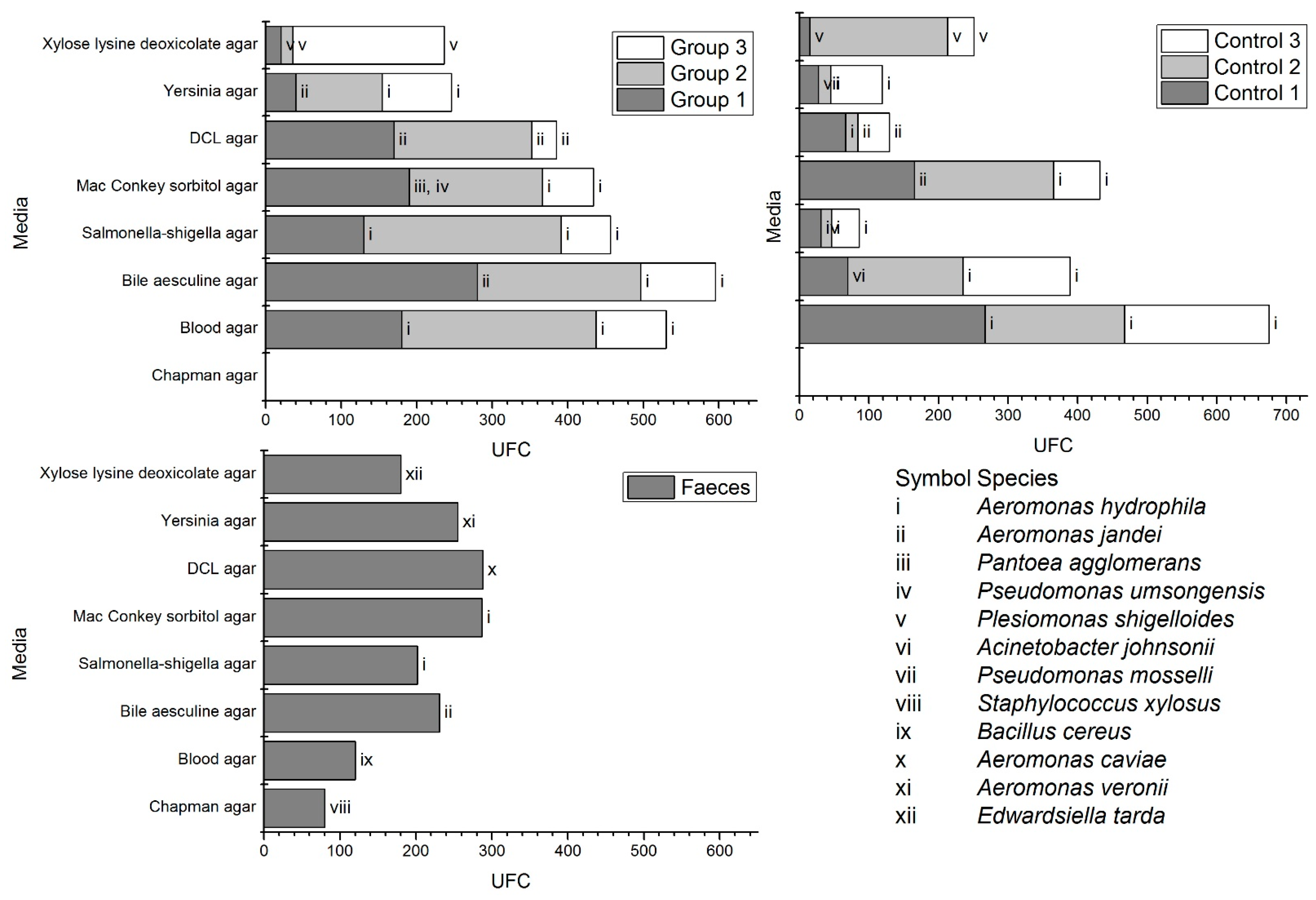
3.2. Microbial Diversity

Aeromonas jandaei and Aeromonas hydrophila were the predominant species found on all samples and controls. Pantoea agglomerans and Pseudomonas umsongensis were detected only on group 1 samples and grew strictly on MacConkey sorbitol agar plates. Plesiomonas shigelloides was detected on both group 1 samples and the group 1 control and grew on xylose lysine deoxicolat. Compared to group 1 samples, control 1 was differentiated by presenting Acinetobacter johnsonii colonies. No species grew on Chapman agar for all groups and their controls, yet such growth was present from feces samples. The wet feces samples revealed Staphylococcus xylosus on Chapman agar, Bacillus cereus on blood agar, A. jandaei on bile aesculine agar, A. hydrophila on both Salmonella–Shigella and MacConkey sorbitol agar plates, Aeromonas caviae only on DCS agar, Aeromonas veronii on Yersinia agar, and interestingly, Edwardsiella tarda on xylose lysine deoxicholat agar (Figure 2). The detected species as well as the CFU/mL of the initial inoculation are present in Supplementary Table S2.

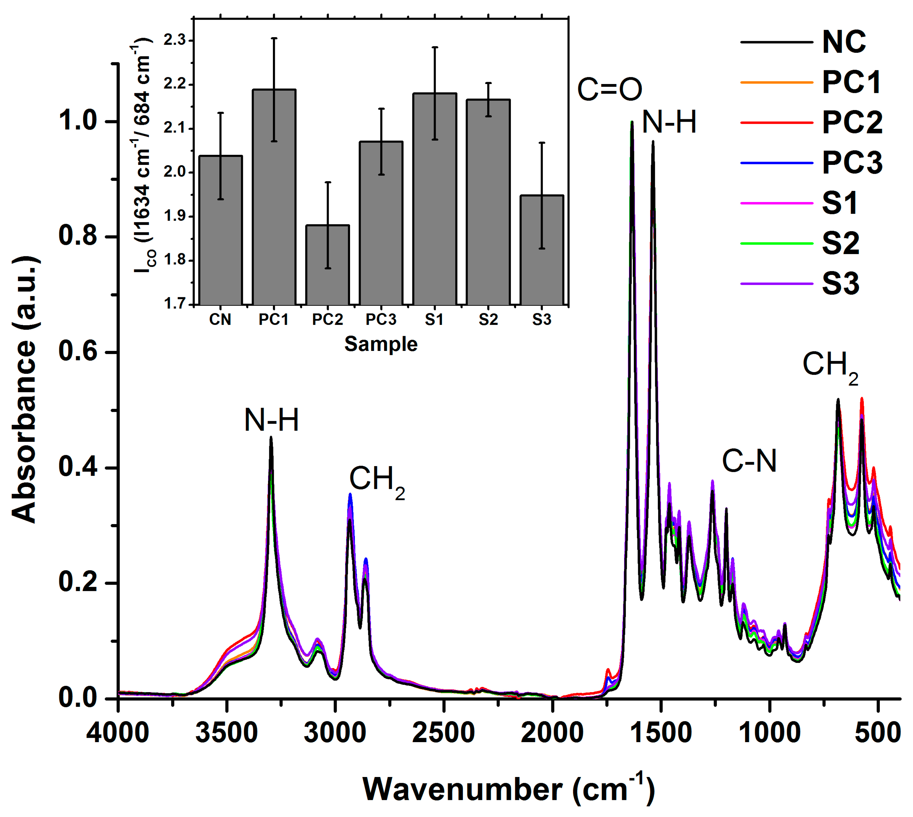
3.3. Polymer Structural Integrity

In the DSC analysis, the first cooling and second heating cycles revealed no discernible differences among the nylon samples excreted by the fish, their respective controls, and the standard nylon. The phase transition was recorded at 214 °C during the first cooling cycle, corresponding to the crystallization of the nylon polymer as it cooled down from its melted state. This process involves the reorganization of polymer chains into a more ordered, crystalline structure. The second heating cycle indicated a melting temperature of around 220 °C for all samples and their controls. Only some slight changes were observed, associated with the sample’s foil shape, which, due to its nature of having a thin and irregular form, possessed the potential to introduce fluctuations in both heat transfer and surface effects during the course of DSC analysis [55].
The FTIR spectra recorded on the samples are presented in Figure 3. The spectrum of the negative control is in good agreement with the FTIR spectra of nylon 6 that were previously published [56,57,58]. Its main absorption peaks are located at 3296 cm−1 (N-H stretching), 2933 cm−1 and 2866 cm−1 (C-H stretching), 1634 cm−1 (C=O stretching), 1537 cm−1 (deformation of N-H), 1199 cm1 (stretching of C-N), and 684 cm−1 (CH2 group deformation). The peak assignment was performed according to [57]. In what concerns the positive controls and samples, the spectra present the same peaks as the negative control, without the occurrence of new peaks. This shows that no major transformations occurred in the structure of nylon in these samples.
To assess the degradation of nylon strips, we used the measured spectra to calculate the carbonyl index (ICO). These are based on the ratio between the height of the carbonyl absorption band (~1634 cm−1) that is expected to increase if the samples are oxidized and the height of one CH2 absorption band (like the one at ~684 cm−1) that is expected to remain constant regardless of oxidation [54]. The results presented in the insert in Figure 2 show that mean ICO values vary over the considered conditions. Pairwise t-tests conducted on the negative control versus positive controls and samples returned p-values that exceeded the confidence interval of 0.05. This shows that no statistically significant differences are seen in the oxidation states of the nylon strips that underwent a type of treatment relative to the pristine nylon strips.
Altogether, the results indicate that the polymer structure remained unchanged despite the digestion and bacterial and aquatic environment exposure.

4. Discussion

The present study investigated the excretion rate of nylon MPs with a focus on their colonization by adherent bacteria, as well as polymer integrity. The microbial capability to adhere is a generally recognized pathogenicity factor [59], of current interest in regard to MP pollution. A recent report [60] indicated that microorganism–microplastic interaction can lead to changes in the physical, structural, and functional characteristics of the microorganisms, raising concerns in the clinical sector, especially due to the fact that MPs can serve as carriers of antibiotic-resistant bacteria [61]. Our findings show that nylon MPs are robust carriers for an abundant microbial load, especially from the Aeromonas genus, as previous works revealed the presence of such pathogens on various different types of other polymers [41,62,63]. The excretion dynamics of nylon MPs by O. niloticus were examined over a one-week period, with the excretion rates varying significantly over time. The majority of nylon strips were not excreted within the first day but after 48 and 72 h, with a gradual decrease observed thereafter, results that correlate with reports in the literature [64]. This indicates the retention or accumulation of MPs within the gastrointestinal tract of fish, which merits further investigation; furthermore, it also strengthens the hypothesis that MPs and the attached microbiota can be carried and spread by fish for long distances in natural environments (i.e., during migration).
The analysis of the microbiota recovered from excreted nylon strips as well as controls revealed the presence of various pathogens, mainly A. jandaei and A. hydrophila, as predominant species across all samples, as well as P. shigelloides.
The prevalence of A. hydrophila and A. jandei was the highest on bile aesculine agar, although from a clinical point of view, such was expected on blood media [65] and/or MacConkey and xylose lysine deoxycholate agar [66]. This event is in correlation with Arcos et al. [67] who stated that there is a clear difference in medium selectivity depending on the sample origin in regard to A. hydrophila. However, thus can be doubted, and further research should be performed for the present context.
Additionally, P. agglomerans and P. umsongensis were detected only in excreted samples. P. umsongensis has been shown to degrade polyaromatic hydrocarbons and petroleum [68]; a link between this species and nylon polymers should be further investigated before any suppositions are to be made.
P. shigelloides (formerly known as Aeromonas shigelloides) [69] was detected in all of the exposed groups as well as in all controls. It should be stated that the pathogen is commonly found together with Aeromonadaceae family members [70], especially in intestinal infections [71,72,73]. P. shigelloides is identified as one of the main pathogens in various fish and presents zoonotic significance, with the main exposure risk being via the consumption of contaminated seafood [74]. The prevalence of P. shigelloides enteritis varies considerably, with higher rates reported from Southeast Asia and Africa and lower numbers from North America and Europe [75]; thus, its adherence on microplastics can be considered of high public health concern.
DSC and FTIR analyses were conducted to assess the structural integrity of nylon MPs following exposure to fish digestion and the aquatic environment. To the best of our knowledge, this is the first study that aimed to evaluate polymers’ structural integrity after their digestion by fish. The DSC results revealed no discernible differences in the thermal behavior of excreted nylon MPs compared to controls and the reference sample, indicating the preservation of polymers’ structures. Similarly, FTIR spectra demonstrated insignificant alterations in the chemical composition of nylon MPs, further supporting the resilience of polymer integrity.
Nylon MPs’ colonization by pathogens is of concern regarding their potential transmission through MP-contaminated aquatic environments. Given the widespread distribution of microplastics in aquatic ecosystems, and the attachment of biofouling microorganisms that can cause buoyant microplastics to sink [76,77], there is a heightened risk of microbial dissemination for the above-stated pathogens in terms of health hazards for both aquatic organisms and humans.

5. Conclusions

The results of this study enhance the findings that MPs can be colonized by diverse bacterial species and raise concerns regarding the transmission of pathogens, especially Aeromonas, through contaminated MPs. With its limitations, the present study only evaluated the cultivable aerobic/facultative aerobic microbiota; the high-throughput sequencing of 16S rDNA followed by qRT-PCR to confirm the actual number of species should be performed in future studies for a more comprehensive observation. It should also be highlighted that the digestion process did not affect the structure of the polymers, nor did its complete excretion took place on the first day; therefore, MPs and associated microbiota can be carried over long distances by fish. With the widespread distribution of microplastics in aquatic habitats, there is a pressing need for comprehensive risk assessment and mitigation strategies to safeguard both public and environmental health.
In regard to such, this study contributes to the understanding of the complex interactions between ichthyofauna, microplastics, and microbial communities, strengthening the urgent need for interdisciplinary research and collaborative efforts to address the challenges raised by microplastic pollution on aquatic ecosystems and, consequently, on human health.

Supplementary Materials

The following supporting information can be downloaded at: https://www.mdpi.com/article/10.3390/microbiolres15020040/s1, Table S1: Supplementary Table S1; Table S2: Supplementary Table S2.

Author Contributions

Conceptualization, A.J. and C.-E.P.; methodology, A.J., C.-E.P., M.M. and D.C.; software, D.F.M.; validation, G.D., N.C. and A.C.S.; formal analysis, S.F.; investigation, A.J. and I.S.; resources, C.-E.P.; data curation, G.D., D.F.M. and S.F.; writing—original draft preparation, A.J. and I.S.; writing—review and editing, C.-E.P. and M.M.; visualization, D.F.M.; supervision, G.D. and D.F.M.; project administration, C.-E.P.; funding acquisition, C.-E.P. All authors have read and agreed to the published version of the manuscript.

Funding

This research was funded by Non-Governmental Research Organization Biologic, Bucharest, Romania.

Institutional Review Board Statement

This research was approved by University of Bucharest Ethics Committee of Research, decision no. 42/07.07.2021.

Informed Consent Statement

Not applicable.

Data Availability Statement

The data presented in this study are available on request from the corresponding author.

Acknowledgments

The authors would like to thank Hanganu Dorin for providing the Oreochromis niloticus specimens.

Conflicts of Interest

The authors declare no conflicts of interest.

References

  1. de Vries, A.N.; Govoni, D.; Árnason, S.H.; Carlsson, P. Microplastic ingestion by fish: Body size, condition factor and gut fullness are not related to the amount of plastics consumed. Mar. Pollut. Bull. 2020, 151, 110827. [Google Scholar] [CrossRef] [PubMed]
  2. Li, B.; Liang, W.; Liu, Q.-X.; Fu, S.; Ma, C.; Chen, Q.; Su, L.; Craig, N.J.; Shi, H. Fish Ingest Microplastics Unintentionally. Environ. Sci. Technol. 2021, 55, 10471–10479. [Google Scholar] [CrossRef] [PubMed]
  3. Senathirajah, K.; Attwood, S.; Bhagwat, G.; Carbery, M.; Wilson, S.; Palanisami, T. Estimation of the mass of microplastics ingested—A pivotal first step towards human health risk assessment. J. Hazard. Mater. 2021, 404, 124004. [Google Scholar] [CrossRef] [PubMed]
  4. Hofstede, L.T.; Vasse, G.F.; Melgert, B.N. Microplastics: A threat for developing and repairing organs? Camb. Prism. Plast. 2023, 1, e19. [Google Scholar] [CrossRef]
  5. Weingrill, R.B.; Lee, M.-J.; Benny, P.; Riel, J.; Saiki, K.; Garcia, J.; Oliveira, L.F.A.d.M.; Fonseca, E.J.d.S.; Souza, S.T.d.; D’Amato, F.d.O.S.; et al. Temporal trends in microplastic accumulation in placentas from pregnancies in Hawaii. Environ. Int. 2023, 180, 108220. [Google Scholar] [CrossRef] [PubMed]
  6. Amato-Lourenço, L.F.; Carvalho-Oliveira, R.; Júnior, G.R.; Dos Santos Galvão, L.; Ando, R.A.; Mauad, T. Presence of airborne microplastics in human lung tissue. J. Hazard Mater. 2021, 416, 126124. [Google Scholar] [CrossRef] [PubMed]
  7. Ibrahim, Y.S.; Tuan Anuar, S.; Azmi, A.A.; Wan Mohd Khalik, W.M.A.; Lehata, S.; Hamzah, S.R.; Ismail, D.; Ma, Z.F.; Dzulkarnaen, A.; Zakaria, Z.; et al. Detection of microplastics in human colectomy specimens. JGH Open 2021, 5, 116–121. [Google Scholar] [CrossRef] [PubMed]
  8. Ullah, S.; Ahmad, S.; Guo, X.; Ullah, S.; Ullah, S.; Nabi, G.; Wanghe, K. A review of the endocrine disrupting effects of micro and nano plastic and their associated chemicals in mammals. Front. Endocrinol. 2022, 13, 1084236. [Google Scholar] [CrossRef] [PubMed]
  9. EndocrineSociety. Plastics Pose Threat to Human Health. Available online: https://www.endocrine.org/news-and-advocacy/news-room/2020/plastics-pose-threat-to-human-health (accessed on 8 March 2024).
  10. Pop, C.-E.; Miu, B.A.; Németh, D.; Wolff, R.; Mihăilescu, D.F.; Avramescu, S.M.; Mernea, M. Bisphenol A analysis and quantification inconsistencies via HPLC-UV: A systematic review with technical notes. Discov. Appl. Sci. 2024, 6, 171. [Google Scholar] [CrossRef]
  11. Stabnikova, O.; Stabnikov, V.; Marinin, A.; Klavins, M.; Klavins, L.; Vaseashta, A. Microbial Life on the Surface of Microplastics in Natural Waters. Appl. Sci. 2021, 11, 11692. [Google Scholar] [CrossRef]
  12. D’Avignon, G.; Gregory-Eaves, I.; Ricciardi, A. Microplastics in lakes and rivers: An issue of emerging significance to limnology. Environ. Rev. 2022, 30, 228–244. [Google Scholar] [CrossRef]
  13. Issac, M.N.; Kandasubramanian, B. Effect of microplastics in water and aquatic systems. Environ. Sci. Pollut. Res. 2021, 28, 19544–19562. [Google Scholar] [CrossRef] [PubMed]
  14. Osman, A.I.; Hosny, M.; Eltaweil, A.S.; Omar, S.; Elgarahy, A.M.; Farghali, M.; Yap, P.-S.; Wu, Y.-S.; Nagandran, S.; Batumalaie, K.; et al. Microplastic sources, formation, toxicity and remediation: A review. Environ. Chem. Lett. 2023, 21, 2129–2169. [Google Scholar] [CrossRef] [PubMed]
  15. Naji, A.; Esmaili, Z.; Khan, F.R. Plastic debris and microplastics along the beaches of the Strait of Hormuz, Persian Gulf. Mar. Pollut. Bull. 2017, 114, 1057–1062. [Google Scholar] [CrossRef] [PubMed]
  16. Sobhani, Z.; Lei, Y.; Tang, Y.; Wu, L.; Zhang, X.; Naidu, R.; Megharaj, M.; Fang, C. Microplastics generated when opening plastic packaging. Sci. Rep. 2020, 10, 4841. [Google Scholar] [CrossRef]
  17. Hernandez, L.M.; Xu, E.G.; Larsson, H.C.E.; Tahara, R.; Maisuria, V.B.; Tufenkji, N. Plastic Teabags Release Billions of Microparticles and Nanoparticles into Tea. Environ. Sci. Technol. 2019, 53, 12300–12310. [Google Scholar] [CrossRef] [PubMed]
  18. Nair, H.T.; Perumal, S. Trophic Transfer and Accumulation of Microplastics in Freshwater Ecosystem: Risk to Food Security and Human Health. Int. J. Ecol. 2022, 2022, 1234078. [Google Scholar] [CrossRef]
  19. Garrido Gamarro, E.; Ryder, J.; Elvevoll, E.O.; Olsen, R.L. Microplastics in fish and shellfish–a threat to seafood safety? J. Aquat. Food Prod. Technol. 2020, 29, 417–425. [Google Scholar] [CrossRef]
  20. Rochman, C.M.; Tahir, A.; Williams, S.L.; Baxa, D.V.; Lam, R.; Miller, J.T.; Teh, F.-C.; Werorilangi, S.; Teh, S.J. Anthropogenic debris in seafood: Plastic debris and fibers from textiles in fish and bivalves sold for human consumption. Sci. Rep. 2015, 5, 1–10. [Google Scholar] [CrossRef] [PubMed]
  21. Walkinshaw, C.; Lindeque, P.K.; Thompson, R.; Tolhurst, T.; Cole, M. Microplastics and seafood: Lower trophic organisms at highest risk of contamination. Ecotoxicol. Environ. Saf. 2020, 190, 110066. [Google Scholar] [CrossRef] [PubMed]
  22. Sequeira, I.F.; Prata, J.C.; da Costa, J.P.; Duarte, A.C.; Rocha-Santos, T. Worldwide contamination of fish with microplastics: A brief global overview. Mar. Pollut. Bull. 2020, 160, 111681. [Google Scholar] [CrossRef] [PubMed]
  23. Justino, A.K.S.; Ferreira, G.V.B.; Fauvelle, V.; Schmidt, N.; Lenoble, V.; Pelage, L.; Martins, K.; Travassos, P.; Lucena-Frédou, F. From prey to predators: Evidence of microplastic trophic transfer in tuna and large pelagic species in the southwestern Tropical Atlantic. Environ. Pollut. 2023, 327, 121532. [Google Scholar] [CrossRef] [PubMed]
  24. Mizraji, R.; Ahrendt, C.; Perez-Venegas, D.; Vargas, J.; Pulgar, J.; Aldana, M.; Patricio Ojeda, F.; Duarte, C.; Galbán-Malagón, C. Is the feeding type related with the content of microplastics in intertidal fish gut? Mar. Pollut. Bull. 2017, 116, 498–500. [Google Scholar] [CrossRef] [PubMed]
  25. Malinowski, C.R.; Searle, C.L.; Schaber, J.; Höök, T.O. Microplastics impact simple aquatic food web dynamics through reduced zooplankton feeding and potentially releasing algae from consumer control. Sci. Total Environ. 2023, 904, 166691. [Google Scholar] [CrossRef] [PubMed]
  26. Clere, I.K.; Ahmmed, F.; Remoto, P.I.I.I.J.G.; Fraser-Miller, S.J.; Gordon, K.C.; Komyakova, V.; Allan, B.J.M. Quantification and characterization of microplastics in commercial fish from southern New Zealand. Mar. Pollut. Bull. 2022, 184, 114121. [Google Scholar] [CrossRef] [PubMed]
  27. Wieczorek, A.M.; Morrison, L.; Croot, P.L.; Allcock, A.L.; MacLoughlin, E.; Savard, O.; Brownlow, H.; Doyle, T.K. Frequency of Microplastics in Mesopelagic Fishes from the Northwest Atlantic. Front. Mar. Sci. 2018, 5, 39. [Google Scholar] [CrossRef]
  28. Gregory, M.R. Environmental implications of plastic debris in marine settings—Entanglement, ingestion, smothering, hangers-on, hitch-hiking and alien invasions. Philos. Trans. R. Soc. B Biol. Sci. 2009, 364, 2013–2025. [Google Scholar] [CrossRef] [PubMed]
  29. Uy, C.A.; Johnson, D.W. Effects of microplastics on the feeding rates of larvae of a coastal fish: Direct consumption, trophic transfer, and effects on growth and survival. Mar. Biol. 2022, 169, 27. [Google Scholar] [CrossRef] [PubMed]
  30. Unuofin, J.O.; Igwaran, A. Microplastics in seafood: Implications for food security, safety, and human health. J. Sea Res. 2023, 194, 102410. [Google Scholar] [CrossRef]
  31. Carpenter, E.J.; Anderson, S.J.; Harvey, G.R.; Miklas, H.P.; Peck, B.B. Polystyrene spherules in coastal waters. Science 1972, 178, 749–750. [Google Scholar] [CrossRef] [PubMed]
  32. Souza-Silva, T.G.d.; Oliveira, I.A.; Silva, G.G.d.; Giusti, F.C.V.; Novaes, R.D.; Paula, H.A.d.A. Impact of microplastics on the intestinal microbiota: A systematic review of preclinical evidence. Life Sci. 2022, 294, 120366. [Google Scholar] [CrossRef] [PubMed]
  33. Parsaeimehr, A.; Miller, C.M.; Ozbay, G. Microplastics and their interactions with microbiota. Heliyon 2023, 9, e15104. [Google Scholar] [CrossRef] [PubMed]
  34. Tamargo, A.; Molinero, N.; Reinosa, J.J.; Alcolea-Rodriguez, V.; Portela, R.; Bañares, M.A.; Fernández, J.F.; Moreno-Arribas, M.V. PET microplastics affect human gut microbiota communities during simulated gastrointestinal digestion, first evidence of plausible polymer biodegradation during human digestion. Sci. Rep. 2022, 12, 528. [Google Scholar] [CrossRef] [PubMed]
  35. Zettler, E.R.; Mincer, T.J.; Amaral-Zettler, L.A. Life in the “plastisphere”: Microbial communities on plastic marine debris. Environ. Sci. Technol. 2013, 47, 7137–7146. [Google Scholar] [CrossRef] [PubMed]
  36. Marathe, N.P.; Bank, M.S. The Microplastic-Antibiotic Resistance Connection. In Microplastic in the Environment: Pattern and Process; Bank, M.S., Ed.; Springer International Publishing: Cham, Switzerland, 2022; pp. 311–322. [Google Scholar]
  37. Gaylarde, C.C.; de Almeida, M.P.; Neves, C.V.; Neto, J.A.B.; da Fonseca, E.M. The Importance of Biofilms on Microplastic Particles in Their Sinking Behavior and the Transfer of Invasive Organisms between Ecosystems. Micro 2023, 3, 320–337. [Google Scholar] [CrossRef]
  38. Mincer, T.J.; Bos, R.P.; Zettler, E.R.; Zhao, S.; Asbun, A.A.; Orsi, W.D.; Guzzetta, V.S.; Amaral-Zettler, L.A. Sargasso Sea Vibrio bacteria: Underexplored potential pathovars in a perturbed habitat. Water Res. 2023, 242, 120033. [Google Scholar] [CrossRef]
  39. Nikolopoulou, I.; Piperagkas, O.; Moschos, S.; Karayanni, H. Bacteria Release from Microplastics into New Aquatic Environments. Diversity 2023, 15, 115. [Google Scholar] [CrossRef]
  40. Harrison, J.P.; Schratzberger, M.; Sapp, M.; Osborn, A.M. Rapid bacterial colonization of low-density polyethylene microplastics in coastal sediment microcosms. BMC Microbiol. 2014, 14, 232. [Google Scholar] [CrossRef] [PubMed]
  41. Viršek, M.K.; Lovšin, M.N.; Koren, Š.; Kržan, A.; Peterlin, M. Microplastics as a vector for the transport of the bacterial fish pathogen species Aeromonas salmonicida. Mar. Pollut. Bull. 2017, 125, 301–309. [Google Scholar] [CrossRef]
  42. Kruglova, A.; Muñoz-Palazón, B.; Gonzalez-Martinez, A.; Mikola, A.; Vahala, R.; Talvitie, J. The dangerous transporters: A study of microplastic-associated bacteria passing through municipal wastewater treatment. Environ. Pollut. 2022, 314, 120316. [Google Scholar] [CrossRef]
  43. Beans, C. Are microplastics spreading infectious disease? Proc. Natl. Acad. Sci. USA 2023, 120, e2311253120. [Google Scholar] [CrossRef] [PubMed]
  44. Zhao, H.; Hong, X.; Chai, J.; Wan, B.; Zhao, K.; Han, C.; Zhang, W.; Huan, H. Interaction between Microplastics and Pathogens in Subsurface System: What We Know So Far. Water 2024, 16, 499. [Google Scholar] [CrossRef]
  45. Zhong, H.; Wu, M.; Sonne, C.; Lam, S.S.; Kwong, R.W.M.; Jiang, Y.; Zhao, X.; Sun, X.; Zhang, X.; Li, C.; et al. The hidden risk of microplastic-associated pathogens in aquatic environments. Eco-Environ. Health 2023, 2, 142–151. [Google Scholar] [CrossRef] [PubMed]
  46. Esangbedo, M.O.; Abifarin, J.K. Determination and managerial implications of machine conditions for high-grade industrial polycaprolactam (nylon 6). Sci. Rep. 2023, 13, 10779. [Google Scholar] [CrossRef] [PubMed]
  47. Information, N.C.f.B. PubChem Compound Summary for, Nylon 6. Available online: https://pubchem.ncbi.nlm.nih.gov/compound/Nylon-6 (accessed on 21 March 2024).
  48. Society, A.C. Nylon 6. Available online: https://commonchemistry.cas.org/detail?cas_rn=25038-54-4 (accessed on 21 March 2024).
  49. Close, B.; Banister, K.; Baumans, V.; Bernoth, E.-M.; Bromage, N.; Bunyan, J.; Erhardt, W.; Flecknell, P.; Gregory, N.; Hackbarth, H. Recommendations for euthanasia of experimental animals: Part 2. Lab. Anim. 1997, 31, 1–32. [Google Scholar] [CrossRef] [PubMed]
  50. Singhal, N.; Kumar, M.; Kanaujia, P.K.; Virdi, J.S. MALDI-TOF mass spectrometry: An emerging technology for microbial identification and diagnosis. Front. Microbiol. 2015, 6, 791. [Google Scholar] [CrossRef] [PubMed]
  51. Welker, M. Proteomics for routine identification of microorganisms. Proteomics 2011, 11, 3143–3153. [Google Scholar] [CrossRef]
  52. Valentine, N.; Wunschel, S.; Wunschel, D.; Petersen, C.; Wahl, K. Effect of culture conditions on microorganism identification by matrix-assisted laser desorption ionization mass spectrometry. Appl. Environ. Microbiol. 2005, 71, 58–64. [Google Scholar] [CrossRef]
  53. Carbonnelle, E.; Beretti, J.L.; Cottyn, S.; Quesne, G.; Berche, P.; Nassif, X.; Ferroni, A. Rapid identification of Staphylococci isolated in clinical microbiology laboratories by matrix-assisted laser desorption ionization-time of flight mass spectrometry. J. Clin. Microbiol. 2007, 45, 2156–2161. [Google Scholar] [CrossRef] [PubMed]
  54. Benítez, A.; Sánchez, J.J.; Arnal, M.L.; Müller, A.J.; Rodríguez, O.; Morales, G. Abiotic degradation of LDPE and LLDPE formulated with a pro-oxidant additive. Polym. Degrad. Stab. 2013, 98, 490–501. [Google Scholar] [CrossRef]
  55. UserCom. Information for users of Mettler Toledo thermal analysis systems. 2000; 11.
  56. Bhullar, S.K.; Rana, D.; Ozsel, B.K.; Orhan, M.; Jun, M.B.G.; Buttar, H.S.; Ostrovidov, S.; Ramalingam, M. Development of Silver-Based Bactericidal Composite Nanofibers by Airbrushing. J. Nanosci. Nanotechnol. 2018, 18, 2951–2955. [Google Scholar] [CrossRef] [PubMed]
  57. Rodríguez, M.; Vázquez-Vélez, E.; Martinez, H.; Torres, A. Superficial Surface Treatment using Atmospheric Plasma on Recycled Nylon 6,6. J. Nucl. Phys. Mater. Sci. Radiat. Appl. 2021, 8, 191–196. [Google Scholar] [CrossRef]
  58. Polyamide (Nylon 6). Available online: https://spectra.chem.ut.ee/textile-fibres/polyamide/ (accessed on 9 March 2024).
  59. Pakbin, B.; Brück, W.M.; Rossen, J.W.A. Virulence Factors of Enteric Pathogenic Escherichia coli: A Review. Int. J. Mol. Sci. 2021, 22, 9922. [Google Scholar] [CrossRef] [PubMed]
  60. Yang, W.; Li, Y.; Boraschi, D. Association between Microorganisms and Microplastics: How Does It Change the Host-Pathogen Interaction and Subsequent Immune Response? Int. J. Mol. Sci. 2023, 24, 4065. [Google Scholar] [CrossRef] [PubMed]
  61. Pham, D.N.; Clark, L.; Li, M. Microplastics as hubs enriching antibiotic-resistant bacteria and pathogens in municipal activated sludge. J. Hazard. Mater. Lett. 2021, 2, 100014. [Google Scholar] [CrossRef]
  62. Cholewińska, P.; Moniuszko, H.; Wojnarowski, K.; Pokorny, P.; Szeligowska, N.; Dobicki, W.; Polechoński, R.; Górniak, W. The Occurrence of Microplastics and the Formation of Biofilms by Pathogenic and Opportunistic Bacteria as Threats in Aquaculture. Int. J. Environ. Res. Public Health 2022, 19, 8137. [Google Scholar] [CrossRef] [PubMed]
  63. Rohrbach, S.; Gkoutselis, G.; Hink, L.; Weig, A.R.; Obst, M.; Diekmann, A.; Ho, A.; Rambold, G.; Horn, M.A. Microplastic polymer properties as deterministic factors driving terrestrial plastisphere microbiome assembly and succession in the field. Environ. Microbiol. 2023, 25, 2681–2697. [Google Scholar] [CrossRef] [PubMed]
  64. Sun, T.; Ji, C.; Li, F.; Shan, X.; Wu, H. The legacy effect of microplastics on aquatic animals in the depuration phase: Kinetic characteristics and recovery potential. Environ. Int. 2022, 168, 107467. [Google Scholar] [CrossRef] [PubMed]
  65. Gillespie, S.H. 17—Examination of faeces for bacterial pathogens. In Medical Microbiology Illustrated; Gillespie, S.H., Ed.; Butterworth-Heinemann: Oxford, UK, 1994; pp. 192–210. [Google Scholar]
  66. Desmond, E.; Janda, J.M. Growth of Aeromonas species on enteric agars. J. Clin. Microbiol. 1986, 23, 1065–1067. [Google Scholar] [CrossRef]
  67. Arcos, M.L.; Vicente, A.d.; Moriñigo, M.A.; Romero, P.; Borrego, J.J. Evaluation of several selective media for recovery of Aeromonas hydrophila from polluted waters. Appl. Environ. Microbiol. 1988, 54, 2786–2792. [Google Scholar] [CrossRef]
  68. Furmanczyk, E.M.; Kaminski, M.A.; Dziembowski, A.; Lipinski, L.; Sobczak, A. Draft Genome Sequence of the Type Strain Pseudomonas umsongensis DSM 16611. Genome Announc. 2017, 5, 39. [Google Scholar] [CrossRef]
  69. Ellner, P.D.; McCarthy, L.R. Aeromonas shigelloides Bacteremia: A Case Report. Am. J. Clin. Pathol. 1973, 59, 216–218. [Google Scholar] [CrossRef]
  70. Pereira, C.S.; Amorim, S.D.; Santos, A.F.; Siciliano, S.; Moreno, I.B.; Ott, P.H.; Rodrigues Ddos, P. Plesiomonas shigelloides and Aeromonadaceae family pathogens isolated from marine mammals of Southern and Southeastern Brazilian coast. Braz. J. Microbiol. 2008, 39, 749–755. [Google Scholar] [CrossRef]
  71. Holmberg, S.D.; Farmer, J.J., III. Aeromonas hydrophila and Plesiomonas shigelloides as Causes of Intestinal Infections. Rev. Infect. Dis. 1984, 6, 633–639. [Google Scholar] [CrossRef]
  72. Alabi, S.A.; Odugbemi, T. Occurrence of Aeromonas species and Plesiomonas shigelloides in patients with and without diarrhoea in Lagos, Nigeria. J. Med. Microbiol. 1990, 32, 45–48. [Google Scholar] [CrossRef]
  73. Pitarangsi, C.; Echeverria, P.; Whitmire, R.; Tirapat, C.; Formal, S.; Dammin, G.J.; Tingtalapong, M. Enteropathogenicity of Aeromonas hydrophila and Plesiomonas shigelloides: Prevalence among individuals with and without diarrhea in Thailand. Infect. Immun. 1982, 35, 666–673. [Google Scholar] [CrossRef]
  74. Duman, M.; García Valdés, E.; Ay, H.; Altun, S.; Saticioglu, I.B. Description of a Novel Fish Pathogen, Plesiomonas shigelloides subsp. oncorhynchi, Isolated from Rainbow Trout (Oncorhynchus mykiss): First Genome Analysis and Comparative Genomics. Fishes 2023, 8, 179. [Google Scholar] [CrossRef]
  75. Janda, J.M.; Abbott, S.L.; McIver, C.J. Plesiomonas shigelloides Revisited. Clin. Microbiol. Rev. 2016, 29, 349–374. [Google Scholar] [CrossRef]
  76. Cole, M.; Lindeque, P.; Halsband, C.; Galloway, T.S. Microplastics as contaminants in the marine environment: A review. Mar. Pollut. Bull. 2011, 62, 2588–2597. [Google Scholar] [CrossRef]
  77. Sulaiman, B.; Woodward, J.C.; Shiels, H.A. Riverine microplastics and their interaction with freshwater fish. Water Biol. Secur. 2023, 2, 100192. [Google Scholar] [CrossRef]
Figure 1. (a) Mean values and standard deviations calculated for the numbers of nylon strips recovered over considered time ranges from the three groups of fish. (b) p-values obtained from t-tests used to carry out a pairwise comparison between the numbers of nylon strips recovered from the three groups of fish at different time intervals. The p-values are labeled on the figure. The values associated with red and dark orange are statistically significant (p-values < 0.05).
Microbiolres 15 00040 g001
Figure 2. Identified species and their respective CFU/mL, represented per type of growth media used.
Microbiolres 15 00040 g002
Figure 3. FTIR spectra of the negative control (NC), positive controls (PC1, PC2, and PC3), and samples (S1, S2, S3). The functional groups that give rise to the main absorption bands are labeled on the figure. The variation of carbonyl indices (ICOs) over the samples is represented in the insert.
Microbiolres 15 00040 g003
Disclaimer/Publisher’s Note: The statements, opinions and data contained in all publications are solely those of the individual author(s) and contributor(s) and not of MDPI and/or the editor(s). MDPI and/or the editor(s) disclaim responsibility for any injury to people or property resulting from any ideas, methods, instructions or products referred to in the content.

