Article

Towards an Integrative Understanding of tRNA Aminoacylation–Diet–Host–Gut Microbiome Interactions in Neurodegeneration

1
Expert Biomed, Inc., 11933 SW 271st TER Homestead, Miami Dade, FL 33032-3305, USA
2
Stop Alzheimers Corp., Miami Dade, FL 33032, USA
3
Nova Southeastern University, 3301 College Ave, Fort Lauderdale, FL 33314, USA
4
University of Texas at San Antonio, 1 UTSA Circle, San Antonio, TX 78249, USA
*
Author to whom correspondence should be addressed.
Nutrients 2018, 10(4), 410; https://doi.org/10.3390/nu10040410
Submission received: 14 February 2018 / Revised: 19 March 2018 / Accepted: 22 March 2018 / Published: 26 March 2018
(This article belongs to the Special Issue Brain Aging and Gut-Brain Axis)

Abstract

:
Transgenic mice used for Alzheimer’s disease (AD) preclinical experiments do not recapitulate the human disease. In our models, the dietary tryptophan metabolite tryptamine produced by human gut microbiome induces tryptophanyl-tRNA synthetase (TrpRS) deficiency with consequent neurodegeneration in cells and mice. Dietary supplements, antibiotics and certain drugs increase tryptamine content in vivo. TrpRS catalyzes tryptophan attachment to tRNAtrp at initial step of protein biosynthesis. Tryptamine that easily crosses the blood–brain barrier induces vasculopathies, neurodegeneration and cell death via TrpRS competitive inhibition. TrpRS inhibitor tryptophanol produced by gut microbiome also induces neurodegeneration. TrpRS inhibition by tryptamine and its metabolites preventing tryptophan incorporation into proteins lead to protein biosynthesis impairment. Tryptophan, a least amino acid in food and proteins that cannot be synthesized by humans competes with frequent amino acids for the transport from blood to brain. Tryptophan is a vulnerable amino acid, which can be easily lost to protein biosynthesis. Some proteins marking neurodegenerative pathology, such as tau lack tryptophan. TrpRS exists in cytoplasmic (WARS) and mitochondrial (WARS2) forms. Pathogenic gene variants of both forms cause TrpRS deficiency with consequent intellectual and motor disabilities in humans. The diminished tryptophan-dependent protein biosynthesis in AD patients is a proof of our model-based disease concept.

Graphical Abstract

1. Introduction

Alzheimer’s disease (AD) clinical trials have been a tremendous investment loss. Efforts to combat neurodegeneration associated with AD are hindered by a lack of animal models recapitulating disease. The main reason is that the transgenic mice used for AD preclinical experiments clearly do not recapitulate the etiology of human disease. The failed clinical trials indicate that a cause of AD is still unknown. Animal models can certainly be useful to find out more about the biological bases of AD and develop efficient pharmacological treatments. The ethologically-based mouse models of AD would be extremely useful for new preclinical trials. The neuronal loss in brain is a main characteristic of AD. The neurofibrillary tangles (NFT) containing tau protein along with amyloid β (Aβ) plaques are the pathological features of AD brain. Although the relationship between the primary structure and function of the AD hallmarks tau protein and amyloid precursor protein (APP) has been relatively well studied, there are, to our knowledge, no structure-function analyses of biosynthesis for these proteins and other proteins implicated in neurodegeneration. Critics of the “amyloid hypothesis”, which posits that the AD is triggered by a build-up of Aβ in the brain, say the results of the failed clinical trials are evidence of its weakness [1]. Here we explore how Aβ and tau could become overrepresented in brain of AD and related diseases. In this report, we analyze results supporting the alternative hypothesis of “impaired protein biosynthesis causing neurodegeneration”. We used the basic local alignment search tool (BLAST) of the National Center for Biotechnology Information (NCBI) to find and summarize the evidence emphasizing the critical role of tryptophan content in proteins for the disease development. Here, we analyze the alterations in short peptide profiles [2] as an early indication of neurodegeneration in humans, which support our experimental cell and animal models of neurodegeneration induced by the natural inhibitors of protein biosynthesis [3,4,5]. These competitive inhibitors of a key enzyme of protein biosynthesis tryptophanyl-tRNA synthetase (TrpRS) are tryptophan metabolites tryptamine and tryptophanol [6,7] that can be produced by human and mammalian gut microbiota [8,9]. The tryptamine producing staphylococci were present in 60% of the human stool samples [10]. These bacteria possess the sadA gene, an aromatic amino acid decarboxylase that catalyzes conversion of tryptophan to tryptamine. The tryptamine production under aerobic conditions varied in different Staphylococcal strains from 71 ± 6 to 231 ± 10 mg/L. Tryptamine increases the adherence and internalization of staphylococci into human cells. The cytotoxic effect of tryptamine, but not of other trace amines tyramine and phenylethylamine, was demonstrated in human cells [10]. In colonocytes—the epithelial cells of colon—tryptamine is a selective modulator of the aryl hydrocarbon receptor (AhR), a ligand-activated transcription factor [11]. Investigation of the urine metabolic profiles showed significant increases in the concentrations of microbial tryptamine in probiotic supplemented mice [12]. Tryptamine produced also at the range 445 to 1157 mg/L by yeast strains isolated from the cow’s cheese in Spain [13]. The fungal pathogen Fusarium of bread wheat [14] and chickpea [15] produce tryptamine. The fungal microbiota (human gut mycobiome) was studied in 16 faecal samples from healthy humans with a vegetarian diet. Fungi were detected in each sample while Fusarium was the most abundant genus [16]. Tryptamine is increased under sporulating conditions in wheat pathogenic fungus Stagonospora nodorum [17]. The gut microbiome is altered in AD patients [18,19]. The gut microbiome of AD participants has decreased microbial diversity and is compositionally distinct from control age- and sex-matched individuals [19]. Our study revealed the gut bacterial sequence specific for AD patients [18]. This sequence belongs to the gene encoding Na(+)-transporting NADH: Ubiquinone reductase (NQR) while both the NQR substrate ubiquinone and Tryptophan are the products of Shikimate pathway. Both tryptamine and tryptophanol prevent the formation of adenylyl-enzyme [6], the intermediate (synonym adenylate-enzyme) in the reaction of tRNA aminoacylation [20]. In this report, we revisited our data to analyze them together with other related publications to shed light on different aspects of AD modelling. This approach prompts us to suggest the model-based disease concept that (1) the tryptophan metabolite tryptamine present in different dietary products in different geographical locations [21,22,23,24,25], produced by human gut microbes [10], inhibit the host protein biosynthesis via the TrpRS deficiency that leads to neurodegeneration and cell death in human organs [4]; (2) the microbial production of tryptamine can be modified by human diet and dietary supplements that can cause significant increase (six-fold) in excreted tryptamine [26]; (3) prescription drugs—antidepressants ( inhibitors of monoamine oxidase (MAO catabolizing tryptamine to indole-3-acetic acid (IAA)) potentiate the tryptamine activities causing seizures and death in animals [27]; (4) usage of antibiotics increases the tryptamine production in animals [9,28]; and (5) the levels of tryptamine (~10 mg/kg) is higher than those of tryptophan (~1 mg/kg) in transgenic potato tubers expressing tryptophan decarboxylase [29]. TrpRS activities can also be altered in vivo because (i) TrpRS is the interferon-inducible protein [30]; (ii) TrpRS is a secreted dietary protein that can be consumed with a cow milk [31,32,33]; (iii) TrpRS is a human autoantigen and anti-TrpRS autoantibodies bind both human and bovine TrpRS [34]; (iv) TrpRS expression is modulated by hypoxia [35]. This report also includes our experimental data on (1) TrpRS in AD vasculopathies revealed with the monoclonal antibodies (mAb) to TrpRS and (2) tryptamine-induced vasculopathies in the mouse model.

2. Materials and Methods

2.1. BLAST for Short Peptides Altered in Mild Cognitive Impairment and Alzheimer’s disease

We used BLAST for analysis of selected peptides to reveal the functions of proteins possessing tripeptides altered at the different stages of neurodegeneration, both mild and severe forms [2]. To this end, we designed the peptide sequences of 4 or 5 amino acids that included an altered tripeptide of interest as a search strategy in BLAST. The Tables S1–S4 summarize the results of the conducted search. The analysis of the altered Trp-containing peptides is included in the Tables S1 and S2, while the altered peptides lacking Trp are in the Tables S3 and S4.

2.2. Search for Tryptophan-Free Proteins

The Trp content in the selected proteins and polypeptides with a link or putative link to neurodegeneration was verified using Standard Protein BLAST of the NCBI databases and “Find” function of Microsoft Word to search for Trp (W) in the protein sequence of interest.

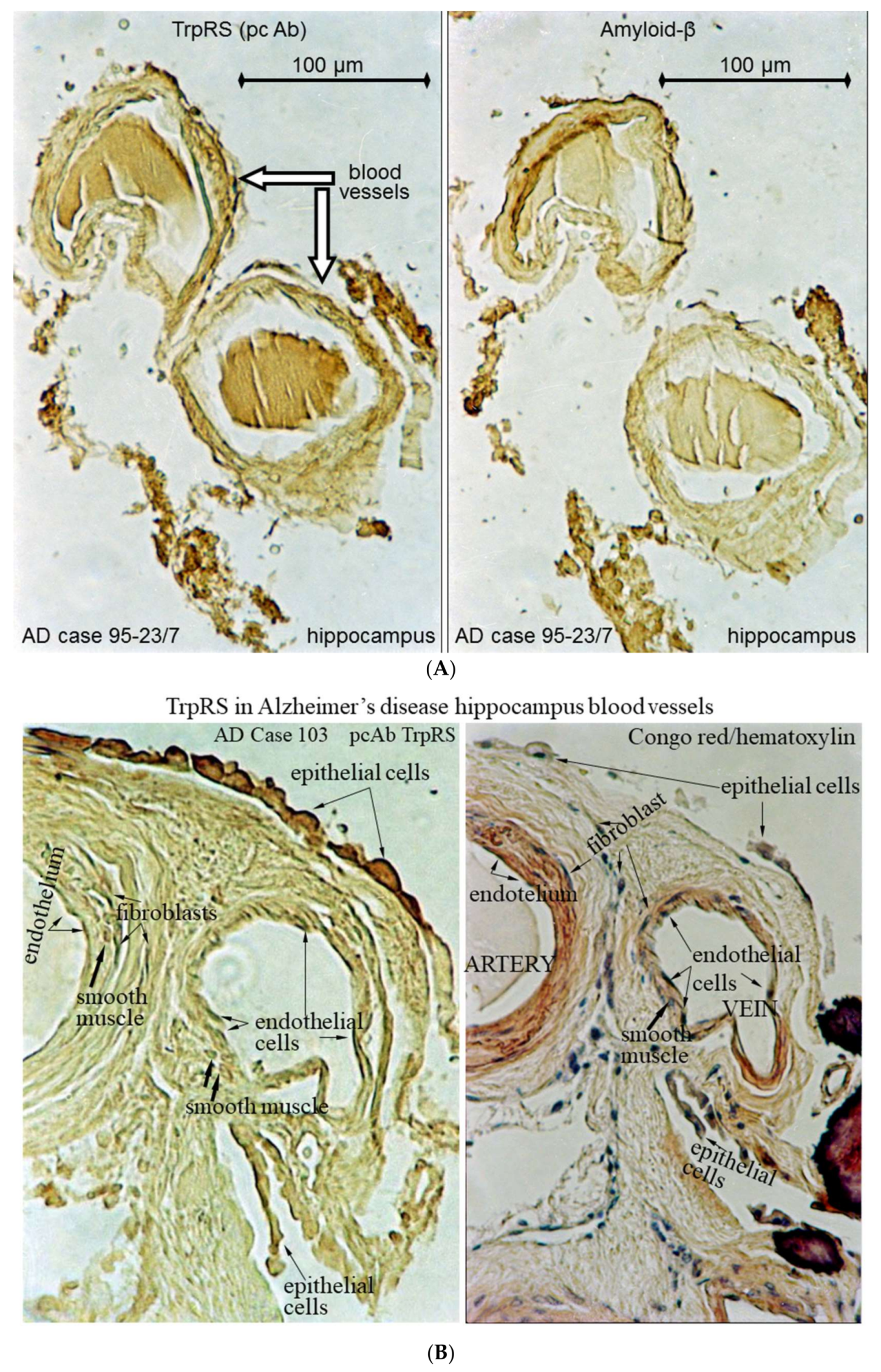
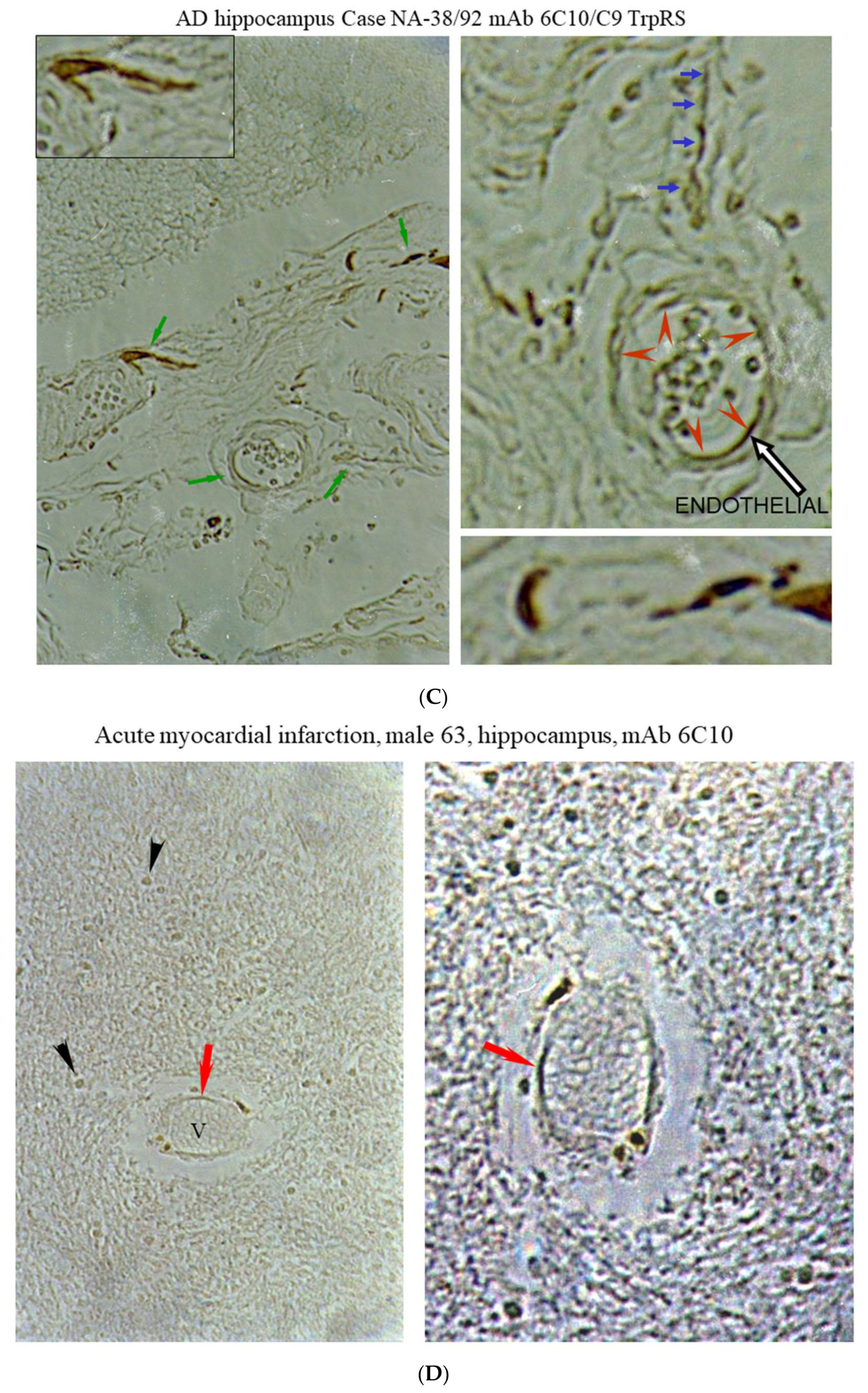
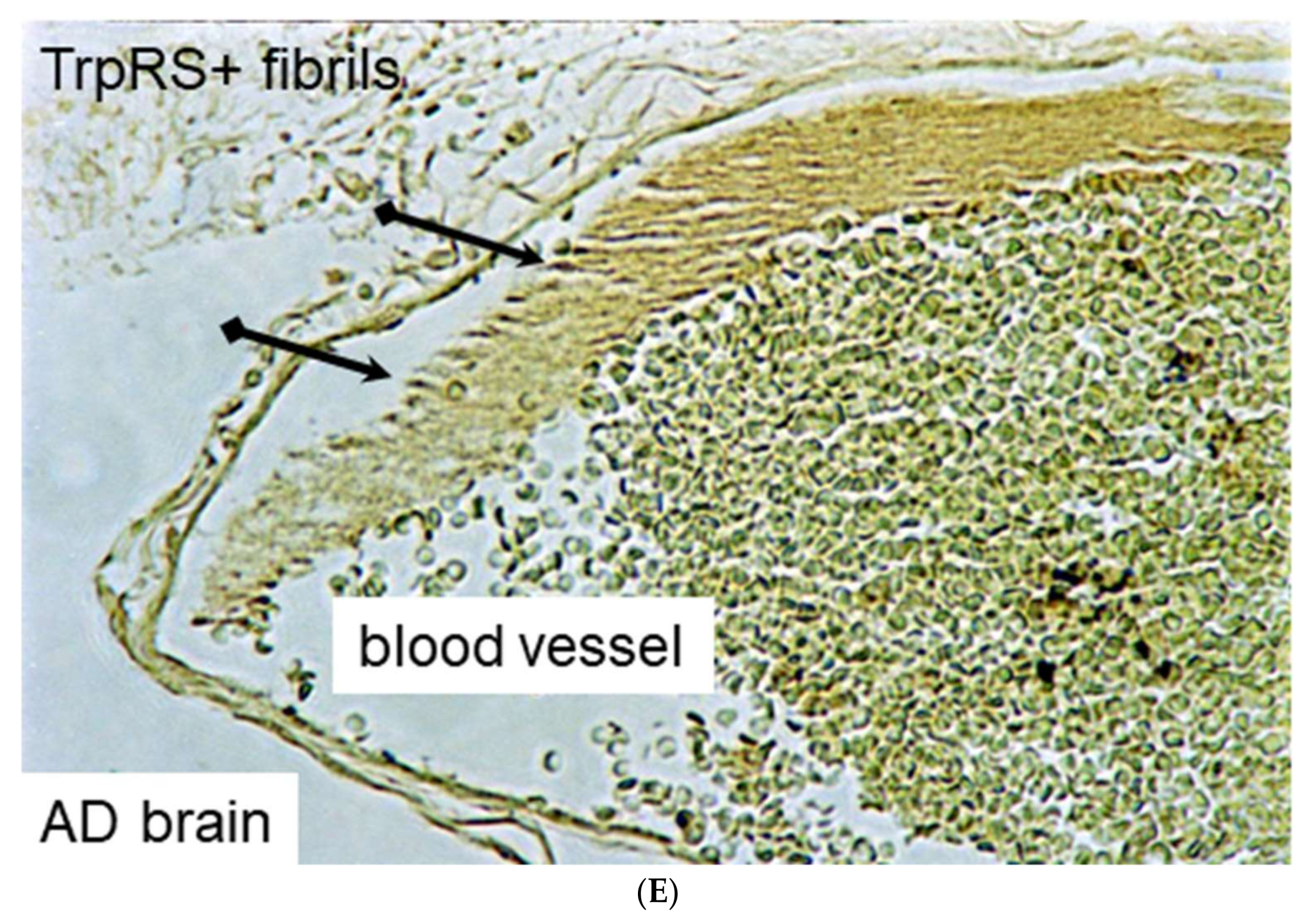
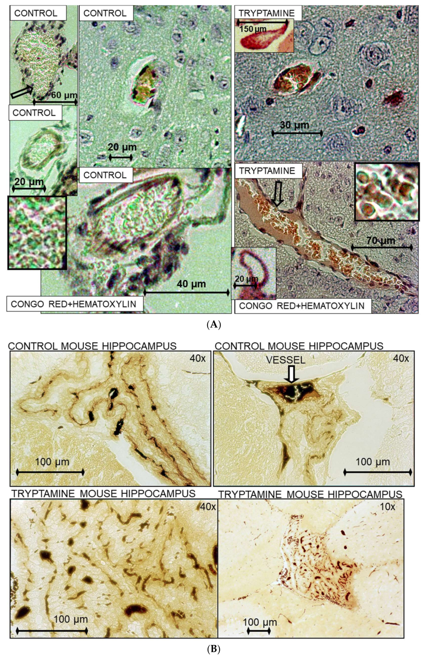
2.3. Histochemical Analysis of Vasculopathies

The treatment of mice with tryptamine at the concentrations that inhibit TrpRS, histochemical analysis and immunostaining of brain serial sections of AD, controls and mice were conducted as previously described; the monoclonal antibodies (mAb) and polyclonal antibodies (pcAb) to TrpRS are characterized and mapped [3,36].

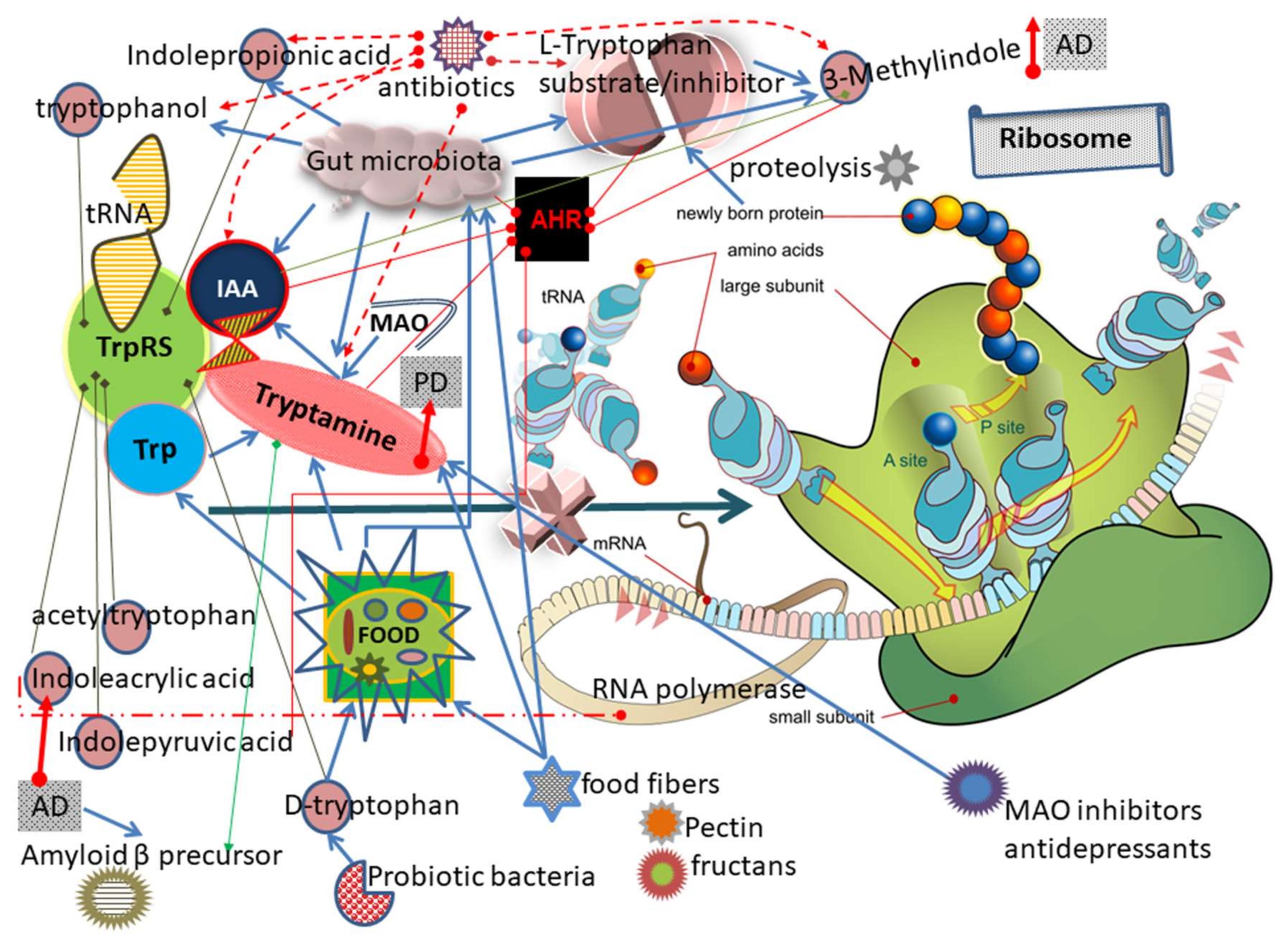
3. Results and Discussion

3.1. TrpRS Inhibition and Aggregation Can Occur Physiologically

TrpRS is responsible for the presence of Trp in proteins. Here, we evaluate the TrpRS inhibition by natural compounds in vitro and in vivo. In the initial step of protein biosynthesis, TrpRS catalyzes the attachment of Trp to its cognate tRNAtrp with formation of tryptophanyl tRNAtrp. We demonstrated the substrate inhibition (50–90%) of human TrpRS by Trp at physiological concentrations 10–50 µM [3]. Trp is an essential amino acid with Km for purified bovine pancreatic TrpRS 1.4 ± 0.2 × 10⁻⁷ M in Trp-dependent ATP–PPi exchange reaction [37]. The Michaelis constant Km is the substrate, such as tryptophan concentration at which the reaction rate is half of Vmax, denoting a dose of a naturally occurring agent such as tryptophan that is within the range of concentrations or potencies that would occur naturally. The 10–50 µM physiological range refers to the naturally accurred Tryptophan, which includes its participation in a number of activities as a precursor, substrate and a product. Tryptophan as well as tryptamine occurs in the human body as the free and bound forms. A portion of the free tryptophan is involved in the de novo protein biosynthesis. The estimated tryptophan/tryptamine ratio does not reflect the exact in vivo status and depends on the methods of extraction and determination. The TrpRS substrate inhibition by Trp seems plausible because the Trp substrate inhibition was also demonstrated for other human enzymes implicated in tryptophan metabolism, such as Indoleamine 2,3-Dioxygenase [38,39] and Tryptophan Hydroxylase [40]. Moreover, Trp inhibits an enzyme specified by the tryptophan operon: anthranilate synthetase in bacteria [41] and plant [42]. Another substrate of the enzyme, tRNA can also inhibit mammalian TrpRS at the 2.9 µM Trp concentration [43]. The formation of fibrillary tangles of filaments resembling those in AD brain was detected in non-neuronal kidney cells [44], neuronal human cells and mouse brain [3] following treatment with TrpRS inhibitors (Table 1) tryptamine and tryptophanol [6,7] at the range of concentrations that can be available physiologically with the human dietary products [21] and excreted by healthy humans [8]. The levels of soluble and insoluble TrpRS forms and activity were altered following tryptamine and tryptophanol treatment in non-neuronal and neuronal cells [3,44,45].
The aggregated TrpRS was revealed in the tryptamine-treated cells and in Alzheimer’s postmortem brain [36]. TrpRS is prone to self-aggregation [47] and fibril formation [36] that cause enzyme inactivation. TrpRS self-aggregates in vitro under the enzyme concentrations higher than 10–15 µM or ~1 mg/mL [48]. TrpRS can be overexpressed in vivo intracellularly and extracellularly since this enzyme is the interferon-inducible protein [30] that is rapidly secreted upon pathogen infection [49].

3.2. Mechanism of TrpRS Inhibition by Tryptamine and Tryptophanol

Tryptamine is a competitive inhibitor of bovine pancreatic TrpRS [50]. Incubation of Trp (1.8 mM) and [β-1802]ATP with purified mammalian TrpRS in the presence of Mg2+ for 21 h at 37 °C led, as expected, to complete positional isotope exchange. Incubation of [β-1802]ATP and Mg2+ and TrpRS in the absence of added Trp for 21 h under the same experimental conditions also led to complete positional isotope exchange, and this could be attributed to endogenous Trp present in the enzyme preparation. Tryptamine and tryptophanol are potent competitive inhibitors of l-Trp for TrpRS, the Ki for tryptamine being 6.0 × 10⁻⁷ M (Appendix A). Incubation of the enzyme with 6 mM tryptamine , ATP and Mg2+ or 6 mM dl-tryptophanol, ATP and Mg2+ for 21 h at 37 °C gave no evidence of positional isotope exchange in the recovered ATP. These results show that an adenylyl-enzyme—the intermediate in the reaction of tRNA aminoacylation—is not formed between MgATP and TrpRS in the presence of tryptamine or tryptophanol [6]. Binding of Trp, tryptamine and ATP to TrpRS was studied by equilibrium dialysis experiments. There are two binding sites per mole of enzyme both for Trp and for tryptamine. The dissociation constant Ks for Trp is 0.95 μM and for tryptamine is 1.8 μM [48]. No cooperativity between the TrpRS subunits for the binding of the amino acid or of the competitive inhibitor is evidenced by equilibrium dialysis experiments. Therefore, TrpRS is roughly two-fold more tightly bound to Trp than to tryptamine. It was demonstrated that [14C]tryptophan bound at the tryptophanyl-adenylate active site of the bovine pancreatic TrpRS was completely removed from the enzyme by incubation with an excess of [3H]tryptamine [51].

3.3. Tryptamine and Tryptophanol Cytotoxicity and Bioavailability

Tryptamine induces cytotoxicity/neurotoxicity, neurofibrillary tangles, autophagic, axonal and mitochondrial pathologies and amyloidosis [3,5,52]. These characteristics are similar to those reveled in AD. Tryptophanol that also induced formation of fibrillary tangles in kidney cells is less toxic than tryptamine [44]. As a biogenic amine, tryptamine is enzymatically produced by decarboxylation of Trp. Tryptamine is catabolized to indole-3-acetic acid (IAA) by monoamine oxidase (MAO). Therefore, bioavailability of tryptamine depends on the levels and enzymatic activities of decarboxylase and MAO enzymes. The pathway for tryptophanol remains unknown. In our studies, mice had a diet with roughly two milligrams of total tryptophan per day (~0.2 mg of free tryptophan). Tryptamine that was injected intraperitoneally (each injection of 200 μg of tryptamine for 2.5 weeks was administered for every second day) induced neurodegeneration with the behavioral changes in mice [3].

3.4. Tryptamine and Tryptophanol Increases Following Antibiotic Treatment

Profiling study of urinary and fecal metabolites in Wistar rats exposed to a broad spectrum β-lactam antibiotic, Imipenem/cilastatin sodium demonstrated significant increase of tryptamine (up to four-fold in feces, 13-fold in urine) and tryptophanol (>51-fold in feces) along with Trp alterations in a time-dependent manner [9].
Early antibiotic intervention with in-feed antibiotics (olaquindox, oxytetracycline calcium and kitasamycin) from the postnatal day 7 to day 42 of pigs increased tryptamine concentration in the feces [28].

3.5. Dietary Exposure of Human Population to Tryptamine

In a recent report, the risk assessment of dietary exposure to tryptamine was analyzed for the Austrian population [24]. For fresh/cooked fish, preserved fish, cheese, raw sausage, condiments, sauerkraut and fermented tofu, maximum tolerable levels of tryptamine were 1650; 3200; 2840; 4800, 14120; 1740; and 2400 mg/kg, respectively. This assessment did not take into account the combined effects of simultaneously ingested biogenic amines, and increased susceptibility to tryptamine, e.g., due to reduced MAO activity, did not estimate prolonged effects of tryptamine consumption and did not consider the tryptamine content in individuals. Tryptamine was substantially higher (4.43-fold) in vaginal tract of smokers compared to non-smokers [53]. Smoking is a significant risk factor for AD [54].

3.6. Concentration-Dependent Tryptamine Effects in Animal Experiments: Seizures, Death

For comparison, we provide here the tryptamine concentrations used in the experiments with animals. In the mouse model of neurodegeneration, we observed convulsions following intravenous (i.v.) injections of one milligram tryptamine per mouse of ~25 g or ~40 mg/kg [3,4,5]. Tryptamine was utilized as an inducer of seizures in laboratory animals (i.v. administration of tryptamine at 20 mg/kg to rats) for the anti-seizure drug development by the pharmaceutical companies [55]. The potentiation of tryptamine toxicity on mice was studied with and without prior administration of MAO inhibitors, which are currently in use as antidepressants [27]. The LD50 for tryptamine hydrochloride injected subcutaneously in mice was found to be ~500 mg/kg, while death occurred without any marked symptoms of central stimulation. After subcutaneous administration of a MAO inhibitors nialamide or phenelzine, the LD50 of tryptamine was reduced from 500 to 85 mg/kg. Furthermore, the mice showed pronounced symptoms of central stimulation, including tremor, convulsions, and marked agitation [27]. Chronic administration of monoamine oxidase inhibitor antidepressant drugs (daily doses: tranylcypromine.HCl, 0.5 and 1.0 mg kg−1; phenelzine sulfate, 5 and 10 mg kg−1, each for 28 days; clorgyline.HCl, 1.0 mg kg−1; (−)-deprenyl.HCl, 1.0 mg kg−1, each for 14 days) resulted in decreases in [3H]tryptamine binding site density in brain cortical membranes from male Sprague–Dawley rats [56]. These results can indicate that unlabeled endogenous tryptamine is accumulated in brain following administration of the antidepressant drugs. Tryptamine easily crosses blood-brain barrier [57]. Tryptamine half-life depends on the activities of MAOs in the different body compartments. The tryptamine half-life in human or animal tissues of different organs including brain reflects tryptamine degradation by MAO-A, MAO-B [57] and diamine oxidase [58] during postmortem and extraction periods. In the author’s previous study of the radiolabeled tryptophan and tryptamine in the kidney cultured cells, the uptake for tryptamine exceeds the tryptophan uptake at 2.1–2.4-fold in both original and tryptamine resistant cells during at least two hours [4]. The half-life for TrpRS was determined to be less than two hours in human HeLa cells [59]. Despite the short half-life (1.6 min in the rat spinal cord [60]), the concentration of tryptamine can be high within a short period of time as a result of its high turnover [57]. Tryptamine is a precursor of the hallucinogen N,N-Dimethyltryptamine (DMT), an endogenous sigma-1 receptor regulator [61]. Administration of DMT in combination with MAO inhibitor can induce mania and psychosis [62]. A method of in vivo tryptamine monitoring in the brain and other organs does not currently exist.

3.7. Tryptamine in Healthy Human Population and in Diseases

The tryptamine production is determined to be varied in human gut of healthy subjects (n = 68) with overage tryptamine content in stool samples of 2.00 ± 1.24 µmol/g dry matter (~2.00 ± 1.24 mM) [8]. Quantitative abundance metabolomics profiles for human fecal metabolites include tryptamine with ~100-fold maximum difference among individuals from Oklahoma [63]. The free tryptophan content in normal human plasma of 5.5 μM (approx 1.3 mg/L) in males and 5.9 μM in females was similar in younger and older groups [64]. Metabolomics revealed the elevated urinary tryptamine excretion (3.3-fold) in Parkinson’s disease (PD) [65]. The author suggests here that the reason for the tryptamine elevation is the tryptamine-induced neurodegeneration in PD. High tryptamine levels (2.78 µg/g) detected in cataractous lenses [66] correlates with 1.43-fold increased risk of developing AD by older people with cataract [67] and 26% increased hazard of PD in cataract patients [68]. After an oral dose of normal human feces in the germ-free mice, the feces become the major route of the tryptamine elimination [69].

3.8. Tryptamine Upregulates Transcription of Genes Including Gene Encoding Aβ Precursor

The 22 transcripts were upregulated in mouse Hepa cells by tryptamine treatment at 50 μM for 6 h [70]. In this study, the 10 of 22 transcripts were up-regulated at 2 to 38.24-fold including the gene (4.10-fold) encoding Aβ precursor-like protein 2 (Aplp2), which has been implicated in AD pathogenesis [70]. The genes encoding Cytochrome P4501a2 (Cyp1a2) activated via aryl hydrocarbon receptor (AHR); and apoptotic protease activating factor (Apaf1) were upregulated by tryptamine at 38.24-fold and 2.23-fold, respectively. This study links our model of the tryptamine-induced neurodegeneration with Aβ hypothesis of AD.

3.9. The Diet Additives Increase Tryptamine Content in Animals

In the distal colon contents (dcc) of porcine neonates, the bacterial tryptamine was higher with formula diet (tryptamine at ~75 µg/mg of dcc) relative to sow diet (tryptamine at ~45 µg/mg of dcc) [71]. The diet fiber additives (4%) fructooligosaccharides (FOS, fructan) and pectin increased roughly six-fold the fecal tryptamine (1.17 µmol/g, cellulose control, 4% of diet) in adult cats [26]. In dogs, FOS (0.5% of diet) increased the fecal tryptamine (2.11 µmol/g versus 1.53 µmol/g in control) [72]. A 2.5-fold increase was observed in the tryptamine fecal concentrations following feeding fructan inulin (3 g/kg of body weight per day) to normal ponies [73]. In a rumen fluid of the ruminant animal goat, the metabolomics revealed that a high grain feeding (50% maize) increased the levels of tryptamine to 284.08 µM compared to control (145 µM tryptamine) with no maize feeding [74].
Note that the diet additives increasing tryptamine levels in animals inulin and oligofructose are the regular components in the diets of Americans [75] and pectin, which is an abundant polysaccharide in the human diet is metabolized by gut bacteria [76].

3.10. Other Trp and Tryptamine Metabolites Can Inhibit TrpRS

In this section, we discuss the possibility that other Trp metabolites, including products of tryptamine degradation can be implicated in neurodegeneration and other diseases via TrpRS inhibition. Table 1 displays the TrpRS inhibitory analysis of different Trp metabolites.

3.10.1. IAA

The product of tryptamine degradation IAA (synonyms: beta-Indolylacetic acid, indolylacetic acid) is also a TrpRS inhibitor [46] although less potent than tryptamine. IAA is a uremic toxin derived from Trp fermentation by gut microbiota; it accumulates in patients with chronic kidney disease on haemodialysis [77]. IAA upregulates the eight genes regulated by the transcription factor aryl hydrocarbon receptor (AHR) [78]. The reduction in tryptophan metabolism, but the activation of IAA and AHR were revealed in the gut microbiota of individuals with inflammatory bowel disease [79]. Note, tryptamine forms the two-fold chiral helixes [80] with IAA in vitro. IAA is a plant hormone of the auxin class. IAA production is widespread among environmental bacteria that inhabit soils, waters, but also plant and animal hosts. IAA was detected in mammalian feces [9]. IAA was measured in human plasma of 15 healthy individuals at 0.61–3.32 µmol/L [81]. In the recent study, IAA and Trp were detected in feces of healthy subjects at ~2–12 nmol/g and ~100 nmol/g respectively [79]. Effect of eight antibiotics on the formation of IAA from Trp by ruminal microorganisms was studied: seven of eight antibiotics increased IAA [82]. We conclude that the product of tryptamine degradation by MAO can be also involved in protein inhibition in vivo.

3.10.2. Indolepyruvic Acid

TrpRS inhibitor, indolepyruvic acid (Table 1) is the product of catalysis of Trp by Trp transaminase, the first step in the IAA biosynthetic pathway, and is the substrate for indolepyruvate decarboxylase. Indole-3-pyruvic acid is the AHR protagonist [83].

3.10.3. IPA

TrpRS inhibitor indole-3-propionic acid (IPA, synonyms: 3-(3-Indolyl)propanoic acid, 3-Indolepropionic acid, β-(3-Indolyl)propionic acid; β-Indole-3-propionic acid) is a plant hormone with numerous cell growth functions. The plasma metabolomics revealed effects of antibiotic treatment on both IAA and IPA in rats [84]. Production of IPA was shown to be completely dependent on the presence of gut microflora and can be established by colonization with the bacterium Clostridium sporogenes [85]. Higher IPA is associated with reduced low-grade inflammation [86].

3.10.4. d-Tryptophan

TrpRS potent inhibitor d-tryptophan was recently identified in probiotic bacteria [87]. d-tryptophan and d-phenylalanine exhibited toxic effects on E. coli growth [88]. Transport of d-tryptophan occurs via the amino acid transporter expressed predominantly in the distal intestine [89]. The diet contains both processing-induced and naturally formed d-amino acids [90,91].

3.10.5. Indoleacrylic Acid (IAcrA)

In metabolomics study, IAcrA is increased two-fold while Trp decreased ~1.2-fold in plasma of AD versus cognitively normal (CN) [2]. A plant growth hormone IAcrA is an inhibitor of TrpRS [92,93] and tryptophan synthetase [94] and a marker of cardiotoxicity [95]. Commensal Peptostreptococcus species produce the Trp metabolite IAcrA [96]. IAcrA upregulates the gene expression of Trp operon in E. coli [97]. IAcrA has two effects: it prevents the Trp repressor from acting and it inhibits the charging of tRNATrp by TrpRS. The TrpRS inhibition by IAcrA [93], which is increased in AD plasma [2], may be sufficiently great to cause a significant decrease in the availability of charged tRNATrp. The lowered concentrations of charged tRNATrp may restrict ribosome movement at tryptophan codons, thereby uncoupling transcription and translation.

3.10.6. 3-Methylindole (3MI)

Trp decreased 1.229-fold and its product, the pulmonary toxin 3MI, increased 1.469-fold in the plasma of AD versus mild cognitive impairment (MCI) [2]. The 3MI (skatole) is produced from IAA by mixed populations of pig fecal bacteria [98] and also by Clostridium drakei and Clostridium scatologenes [99]. 3MI presents in the intestine of humans and pigs, causes acute pulmonary edema and emphysema [100] and induces the expression of AHR [101]. The olfactory impairment was correlated with olfactory neuronal populations in mice treated with 3MI [102]. The 27 compounds were screened for their ability to reduce the conversion of Trp to 3MI. Some of them reduced 3MI production by more than 80%. At least part of the inhibition occurs at the level of Trp conversion to IAA [103]. 3MI is a putative TrpRS inhibitor as the analog of its substrate.

3.11. Acetyltryptophan in Macular Degeneration

In patients with neovascular age-related macular degeneration (NVAMD), the increased levels of small peptides were detected in the plasma. This could be the result of excessive proteosomal activity, abnormalities in removal of peptides by peptidases or altered function of peptide transporters. Additional features in NVAMD are modified amino acids acetylphenylalanine and acetyltryptophan. The human plasma acetyltryptophan is higher and detected in elevated tripeptides in patients with NVAMD versus similarly-aged controls [104]. The acetyltryptophan is a TrpRS inhibitor [46] (Table 1). Therefore, we suggest that the increased level of acetyltryptophan-containing tripeptides could be the result of excessive degradation of abnormal proteins, containing modified Trp while elevated free acetyltryptophan may inhibit the TrpRS-dependent protein biosynthesis. Aspergillus nidulans reveals the non-ribosomal peptide biosynthetic pathway to acetylate Trp [105]. Acetyltryptophan and tryptophan were 2.4-fold and 1.7-fold, respectively higher in germ-free compared to conventional mice [85].

3.12. Di- and Tripeptides in AD, Mild Cognitive Impairment (MCI) and Cognitively Normal (CN)

In Table 2, we summarize the data on the levels of di- and tripeptides in plasma and cerebrospinal fluid (CSF) from the metabolomics study conducted by the Mayo clinic [2].
In plasma and CSF of patients with AD and MCI (Table 2), the levels of di- and tripeptides are altered compared to CN. We suggest that some of these short peptides can serve as markers: Glu-Ser is increased in AD and MCI plasma and decreased in AD and MCI CSF; Trp-Gly-Phe is decreased in AD/CN and AD/MCI plasma; Met-Trp-Gln (MWQ) is decreased in AD/CN and MCI/CN plasma; Pro-Lys-Pro is decreased in AD/CN and AD/MCI in CSF. We selected human proteins containing tripeptides altered in AD and/or MCI compared to CN by using NCBI database and amino acid sequences designed for tetra- or penta-peptides as a search tool such as KMWQ or SMWQY (Tables S1–S4). Using this search approach we revealed that human cytoskeleton protein axonemal dynein contains 11 tripeptides (Table S1) that are decreased in AD and/or MCI including two tripeptides MWQ. The Aβ oligomers are enriched in axons and interact with dynein motors. This interaction interferes with the coupling of the dynein motor with its adaptor SNAPIN [106], which is a Trp-free protein (Table 3).
The increase in peptide levels could be the result of excessive proteosomal activity, abnormalities in removal of peptides by peptidases or altered function of peptide transporters. The decrease in peptide levels could be the result of tRNA aminoacylation blockage (Figure 1). The Figure 1 summarizes the data on TrpRS inhibition, tryptamine and Trp metabolites produced by gut microbiota as well as effects of the diet.

3.13. Link of Trp Frequency in Proteins to AD and Related Disorders

In the previous report, the author analyzed the frequency of Trp in proteins implicated in AD and related disorders [4]. Here we update and summarize the data on Trp-free proteins (Table 3). The analysis of Trp frequency and Trp position in proteins can shed light on a role of protein biosynthesis in protein abnormalities revealed in AD and related disorders. The average protein contains ~1.3% Trp (UGG nuclear codon). Some human proteins contain little or no Trp. Insufficient amount of aminoacylated tryptophanyl-tRNATrp for translating Trp-containing peptides can cause protein deficiency while Trp-free proteins might appear as overproduced in comparison with the reduced proteins containing Trp. Here is the example of such a scenario: one of 13 COX subunits, a human COX subunit VI-c of 7 kDa (75 aa) contains no tryptophan (Swiss-Prot: P09669.2). The protein level of this subunit is higher than a level of citrate synthase (9 Trp) in temporal cortex of AD; occipital cortex of Spinocerebellar Ataxia Type I; cerebellar cortex, frontal cortex and occipital cortex of Friedreich’s Ataxia than in control, whereas other COX subunits that contain Trp are decreased in neurodegenerative diseases [107]. The tryptophanyl-tRNA deficiency developed by using Trp metabolites [3] correlates with accumulation of Trp-free proteins in AD brain (Table 3). It was hypothesized [4] that under shortage of tryptophanyl-tRNATrp, the proteins and polypeptides containing no Trp are relatively overrepresented in organs and/or fluids of patients in comparison with underrepresented deficient proteins containing Trp. In such a scenario, the Trp-free proteins become easily accessible for detection and isolation from organs and bodily fluids of patients. Some Trp-free proteins such as tau protein and synuclein are able to aggregate and form insoluble fibrils. The author focused mainly on the sequences of human proteins in the analysis of Trp content. The author suggests that the accumulation of tryptophan-free proteins such as tau protein is not a cause of disease but a consequence of TrpRS inhibition in disease.

3.14. Ribosomal Frameshifting and Bypassing

If there is deficient tryptophanyl-tRNA, the ribosome will be arrested at the Trp codon before it gets to the stop signal (Figure 1). However, there are adaptation mechanisms that help a paused ribosome in cells. Ribosomal frameshifting, particularly +1 frameshifts, can occur as a translational error. Certain codons, if they become ‘hungry’ through aminoacyl tRNA limitation, can frameshift in error to generate a nonsense product [108]. The potential role of ribosomal frameshifting in generating aberrant APP under neurodegeneration is suggested. Tryptamine treatment leading in tryptophanyl-tRNAtrp limitation can stimulate frameshifting of ribosomal translation and consequent generation of aberrant proteins. The Aβ1–42 polypeptide is a product of carboxy-terminal proteolysis (Asp672–Ala713) of full-length APP770 (Swiss-Prot: P05067). The alternatively spliced APP mini (278 aa) carboxyl form (GenBank: BAH12049), contains single Trp. The shifty APP278 mRNA sequence (5′CCCGUGAAUGGAGAGUUCAGCCUGGACGAUCUCCAGCCGUGGCAUUCUUUUGGG) includes the (−1) stop codon UGA in the (−1) frame and potential shifty site GA_GAG 24 nt 5′ of the Trp codon UGG. A single Trp of mini APP278 corresponds to Trp621 of the full-length APP770. Single Trp of APP278 can be removed by frameshifting to cause degradation of the unstable mutant protein into Trp-free Aβ (Table 3).

3.15. Trp Levels Decreased in the Human Bodily Fluids of Diseased Individuals

Total Trp levels were found to be diminished in plasma and CSF of multiple sclerosis, motor neuron disease and ataxia [109]. Protein-bound plasma Trp was lower in epileptic patients compared to normal volunteers [110]. In the metabolomics conducted by Mayo clinic, the levels of aminoacylated tRNA was altered in plasma and CSF of AD and MCI [2]. The enzymatic activity of TrpRS can be reduced as a result of inhibition by d and l tryptophan and its metabolites (Figure 1), or hypoxia-induced downregulation of the TrpRS gene expression [35], or TrpRS self-aggregation [36]. The reduction in tRNAtrp aminoacylation ultimately leads to the inhibition and blockage of protein biosynthesis (Table 2).

3.16. TrpRS Inhibition Leads to Both Translation and Transcription Impairments

In Salmonella typhimurium, the IAcrA usage to de-repress the trp operon results in a decline in RNA polymerase electron microscopic density observed on promoter-distal portions of cloned trp operons. This may be attributable to premature transcription termination accompanying translation inhibition due to the IAcrA interference with normal TrpRS activity [93]. In other words, electron microscopy directly demonstrates the decrease in RNA polymerase induced by TrpRS inhibitor. Therefore, TrpRS inhibition leads to both translation and transcription impairments. Using BLAST analysis we revealed that human DNA-directed RNA polymerase II can be decreased in AD since this enzyme contains tripeptide Pro-Lys-Pro (PKP) decreased in AD CSF; tripeptides Ile-Ser-Lys (ISK) and Ala-Thr-Pro (ATP) decreased in MCI plasma (Table S4). Our data indicate that the protein biosynthesis impairment induced by TrpRS inhibition causes neuronal death and abnormal protein conformation intracellularly and extracellularly in the brain of tryptamine-treated mice [3]. This model mimics closely AD and related neurodegenerative disorders.

3.17. Mutations in Genes Encoding Cytoplasmic (WARS) and Mitochondrial (WARS2) TrpRS in Humans

The experimental models of TrpRS deficiency are supported by recent discoveries of mutations in gene encoding mitochondrial TrpRS (WARS2) that implicate in intellectual disability, delayed myelination, seizures, whereas de novo synthesis of proteins inside mitochondria was reduced in the patient’s fibroblasts [111,112,113]. In a report by Musante et al., the p.Trp13Gly variant was found to significantly reduce the level of WARS2 protein in the mitochondrial fraction. Burke et al. reported biallelic mutations in WARS2 with reduced level of WARS2 protein that cause the levodopa-responsive infantile-onset parkinsonism [114]. In the distal hereditary motor neuropathy (DHMN), a heterozygous mutation was identified in the cytoplasmic TrpRS gene (WARS) that co-segregates with the neuropathy in the family. This mutation has a dominant negative effect on aminoacylation activity of TrpRS, which subsequently compromised protein synthesis and reduced cell viability [115]. DHMN characterized by degeneration and loss of motor neuron cells in the anterior horn of the spinal cord and subsequent muscle atrophy. Mutations in genes encoding other members of aminoacyl-tRNA synthetase family (prolyl-tRNA synthetase and asparaginyl-tRNA synthetase) are also related to infantile-onset neurodegenerative disorder [116].

3.18. Bacterial Toxic Analog of Amino Acid Alanine Causes Neurodegeneration in Monkey

The chronic dietary exposure of vervets (Chlorocebus sabaeus) to a cyanobacterial toxin present in the traditional Chamorro diet, beta-N-methylamino-l-alanine (BMAA), triggers the formation of both NFT and Aβ deposits similar in structure and density to those found in brain tissues of Chamorros who died with Guamanian amyotrophic lateral sclerosis/parkinsonism dementia complex [117]. Although BMAA is a putative inhibitor of aminoacyl-tRNA synthetase, the inhibitory activity for BMAA is unknown.

3.19. TrpRS, Trp and Tryptamine in Vascular Dysfunctions and Pathological Changes

Cerebral amyloid angiopathy is common in AD and may contribute to dementia and cerebral hemorrhage. In human gut metagenome study, the aminoacyl-tRNA biosynthesis was altered in symptomatic atherosclerosis [118]. Vascular dementia (VaD) is the most common cause of dementia in the elderly, second to AD. The VaD cause or main risk factors are hypertension, diabetes mellitus, atherosclerosis, coronary artery disease [119]. In spinal cord injury in rats, application of physiological concentrations of Trp, tyrosine or phenylalanine (10–100 μM) induced tonic local constrictions of capillaries leading to a decrease in capillary diameter, which should severely impair blood flow. The Trp-induced constrictions produced changes in vessel morphology and widespread displacement of red blood cells. Tryptamine produced from Trp by aromatic amino acid decarboxylase induces similar vasoconstriction [120]. Tryptamine increases blood pressure by vasoconstriction and implicates in cardiovascular pathologies, including hypertension, migraine and myocardial infarction [121,122,123,124]. It has been shown that tryptamine (1–5 mg/kg, i.v.) decreases the resistance of the cerebral blood vessels and lowers the velocity of the blood flow and pO2 in the brain tissues of cats [125]. Here, we demonstrate the TrpRS depositions in the cerebral blood vessels of AD patients (Figure 2) and effects of tryptamine on cerebral blood vessels in mice (Figure 3).
In AD human brain, TrpRS was mainly visualized in association with congophilic angiopathy, as blood clot-like depositions inside the vessels and in the endothelial, epithelial and fibroblast cells (Figure 2A,B). The strong immunostaining of endothelial cells periphery with anti-TrpRS monoclonal antibody (mAb) can be an indicator of intensive TrpRS secretion (Figure 2C). The significant immunostaining of the endothelial cells with anti-TrpRS mAb was revealed in some blood vessels of hippocampus in acute myocardial infarction (Figure 2D). We did not reveal such vessel immunostaining in the brain serial sections immunostained with the secondary antibodies. The microscopy visualizes the fibrils immunostained with anti-TrpRS antibodies inside the AD cerebral blood vessel (Figure 2E). We show that tryptamine causes vasculopathies in mouse hippocampus including congophilic angiopathy (Figure 3A) and angiogenesis (Figure 3B). It was reported earlier that in the AD hippocampus, the ongoing angiogenesis results in increased vascular density compared with controls [126]. To our knowledge, this is the first study that demonstrates the vasculopathies induced by tryptamine in the hippocampus. Human trace amine receptors (including TAAR1, TAAR2, TAAR5, TAAR6, TAAR8, TAAR9) are expressed in the brain and play significant physiological and neuropathological roles by activation of trace amines including tryptamine [127].

4. Conclusions

Our analysis reveals that the tryptophan-containing tri-peptides diminished in the mild and severe AD compared to the normally cognitive individuals. This report demonstrates for the first time that the dietary product tryptamine induces vasculopathies in the hippocampus of mouse brain at the concentrations that inhibit TrpRS, while TrpRS is associated with vasculopathies in AD brain. The mutations in genes encoding cytoplasmic (WARS) and mitochondrial (WARS2) TrpRS enzymes cause the TrpRS deficiency, intellectual disability and Parkinsonism. Tryptamine producing microbes including bacteria and fungi present in most humans and in food. Tryptamine production in the human microbiome can be increased by a number of factors including antibiotics, dietary supplements and probiotics. Altogether, the data presented here indicate that the inhibition of TrpRS by tryptamine and other Trp metabolites cause the protein biosynthesis impairment with consequent neurodegeneration.

Lethal Tryptamine Drug Combinations.

Most antidepressants—the MAO inhibitors raise tryptamine. When levels get too high, serious complications can occur including hypertension and seizures. In severe cases, this can even lead to death.

Supplementary Materials

The following are available online at https://www.mdpi.com/2072-6643/10/4/410/s1, Table S1: Proteins containing Met-Trp-Gln (MWQ) decreased in AD/CN and MCI/CN plasma, Table S2: Proteins containing Trp-Gly-Phe (WGF) decreased in AD/CN and AD/MCI plasma, Table S3: Proteins containing Pro-Lys-Pro (PKP) decreased in AD/CN and AD/MCI in CSF, Table S4: Human RNA polymerase subunits contain tripeptides decreased in AD or MCI.

Acknowledgments

This work was supported by Art Medicus PLLC, Yonkers, NY, USA.

Author Contributions

E.L.P. conceived and designed the experiments; E.L.P. performed the experiments; E.L.P. and G.P. analyzed the data; E.L.P. contributed reagents/materials/analysis tools including anti-TrpRS antibodies; E.L.P. wrote the paper.

Conflicts of Interest

The authors declare no conflict of interest.

Appendix A

Dissociation constant (Kdis) for tryptamine in the tryptophan-dependent ATP-[32P] pyrophosphate exchange catalyzed by bovine pancreatic TrpRS after covalent binding of 1 mole of R-Trp-tRNA to the enzyme (one site of modified TrpRS was occupied after the binding) is 13 ± 4 × 10⁻⁷ M versus tryptamine Kdis 3.6 ± 0.6 × 10⁻⁷ M for the native enzyme [128]. The different affinities of the two subunits of bovine pancreatic TrpRS for tryptophan were observed by Graves et al. [129] and Mazat et al. [130]. Taking an initial symmetrical dimeric TrpRS the two dissociation constants for tryptophan at pH 8 and 25 °C are respectively K1 = 2.0 ± 0.5 µM and K2 = 10 ± 4 µM [129]. They are respectively K1 = 1 ± 0.25 µM and K2 = 20 ± 8 µM if one considers a sequenced binding of the two tryptophan molecules. The tryptamine Ki for L-tryptophan and the fluorinated substrate L-6-fluoro-tryptophan [131] were determined in ATP-[32P] pyrophosphate exchange catalyzed by bovine pancreatic TrpRS to be 3.2 ± 0.6 × 10⁻⁷ M and 1.7 ± 0.4 × 10⁻⁷ M, respectively [132].

References

  1. Abbott, A.; Dolgin, E. Failed alzheimer’s trial does not kill leading theory of disease. Nature 2016, 540, 15–16. [Google Scholar] [CrossRef] [PubMed]
  2. Trushina, E.; Dutta, T.; Persson, X.M.; Mielke, M.M.; Petersen, R.C. Identification of altered metabolic pathways in plasma and CSF in mild cognitive impairment and alzheimer’s disease using metabolomics. PLoS ONE 2013, 8, e63644. [Google Scholar]
  3. Paley, E.L.; Denisova, G.; Sokolova, O.; Posternak, N.; Wang, X.; Brownell, A.L. Tryptamine induces tryptophanyl-tRNA synthetase-mediated neurodegeneration with neurofibrillary tangles in human cell and mouse models. Neuromolecular Med. 2007, 9, 55–82. [Google Scholar] [CrossRef]
  4. Paley, E.L. Tryptamine-induced tryptophanyl-tRNAtrp deficiency in neurodifferentiation and neurodegeneration interplay: Progenitor activation with neurite growth terminated in alzheimer’s disease neuronal vesicularization and fragmentation. J. Alzheimers Dis. 2011, 26, 263–298. [Google Scholar] [PubMed]
  5. Paley, E.L.; Perry, G.; Sokolova, O. Tryptamine induces axonopathy and mitochondriopathy mimicking neurodegenerative diseases via tryptophanyl-tRNA deficiency. Curr. Alzheimer Res. 2013, 10, 987–1004. [Google Scholar] [CrossRef] [PubMed]
  6. Lowe, G.; Tansley, G. An investigation of the mechanism of activation of tryptophan by tryptophanyl-tRNA synthetase from beef pancreas. Eur. J. Biochem. 1984, 138, 597–602. [Google Scholar] [CrossRef] [PubMed]
  7. Favorova, O.O.; Khochkina, L.L.; Shaigo, M.; Parin, A.V.; Khil’ko, S.H.; Prasolov, V.S.; Kiselev, L.L. Truptophanyl tRNA synthetase. Isolation and characterization of the 2 enzyme forms. Mol. Biol. 1974, 8, 729–740. [Google Scholar] [PubMed]
  8. Swanson, K.S.; Grieshop, C.M.; Flickinger, E.A.; Bauer, L.L.; Wolf, B.W.; Chow, J.; Garleb, K.A.; Williams, J.A.; Fahey, G.C., Jr. Fructooligosaccharides and Lactobacillus acidophilus modify bowel function and protein catabolites excreted by healthy humans. J. Nutr. 2002, 132, 3042–3050. [Google Scholar] [CrossRef] [PubMed]
  9. Zheng, X.; Xie, G.; Zhao, A.; Zhao, L.; Yao, C.; Chiu, N.H.; Zhou, Z.; Bao, Y.; Jia, W.; Nicholson, J.K.; et al. The footprints of gut microbial-mammalian co-metabolism. J. Proteome Res. 2011, 10, 5512–5522. [Google Scholar] [CrossRef] [PubMed]
  10. Luqman, A.; Nega, M.; Nguyen, M.T.; Ebner, P.; Gotz, F. Sada-expressing staphylococci in the human gut show increased cell adherence and internalization. Cell Rep. 2018, 22, 535–545. [Google Scholar] [CrossRef] [PubMed]
  11. Cheng, Y.; Jin, U.H.; Allred, C.D.; Jayaraman, A.; Chapkin, R.S.; Safe, S. Aryl hydrocarbon receptor activity of tryptophan metabolites in young adult mouse colonocytes. Drug Metab. Dispos. 2015, 43, 1536–1543. [Google Scholar] [CrossRef] [PubMed]
  12. Martin, F.P.; Wang, Y.; Sprenger, N.; Yap, I.K.; Lundstedt, T.; Lek, P.; Rezzi, S.; Ramadan, Z.; van Bladeren, P.; Fay, L.B.; et al. Probiotic modulation of symbiotic gut microbial-host metabolic interactions in a humanized microbiome mouse model. Mol. Syst. Biol. 2008, 4, 157. [Google Scholar] [CrossRef] [PubMed]
  13. Atanassova, M.R.; Fernandez-Otero, C.; Rodriguez-Alonso, P.; Fernandez-No, I.C.; Garabal, J.I.; Centeno, J.A. Characterization of yeasts isolated from artisanal short-ripened cows’ cheeses produced in Galicia (NW Spain). Food Microbiol. 2016, 53, 172–181. [Google Scholar] [CrossRef] [PubMed]
  14. Powell, J.J.; Carere, J.; Fitzgerald, T.L.; Stiller, J.; Covarelli, L.; Xu, Q.; Gubler, F.; Colgrave, M.L.; Gardiner, D.M.; Manners, J.M.; et al. The Fusarium crown rot pathogen Fusarium pseudograminearum triggers a suite of transcriptional and metabolic changes in bread wheat (Triticum aestivum L.). Ann. Bot. 2017, 119, 853–867. [Google Scholar] [PubMed]
  15. Kulkarni, G.B.; Sanjeevkumar, S.; Kirankumar, B.; Santoshkumar, M.; Karegoudar, T.B. Indole-3-acetic acid biosynthesis in fusarium delphinoides strain GPK, a causal agent of wilt in chickpea. Appl. Biochem. Biotechnol. 2013, 169, 1292–1305. [Google Scholar] [CrossRef] [PubMed]
  16. Suhr, M.J.; Banjara, N.; Hallen-Adams, H.E. Sequence-based methods for detecting and evaluating the human gut mycobiome. Lett. Appl. Microbiol. 2016, 62, 209–215. [Google Scholar] [CrossRef] [PubMed]
  17. Gummer, J.P.; Trengove, R.D.; Oliver, R.P.; Solomon, P.S. Dissecting the role of G-protein signalling in primary metabolism in the wheat pathogen stagonospora nodorum. Microbiology 2013, 159, 1972–1985. [Google Scholar] [CrossRef] [PubMed]
  18. Paley, E.L.; Merkulova-Rainon, T.; Faynboym, A.; Shestopalov, V.I.; Aksenoff, I. Geographical distribution and diversity of gut microbial NADH: Ubiquinone oxidoreductase sequence associated with alzheimer’s disease. J. Alzheimers Dis. 2018, 61, 1531–1540. [Google Scholar] [CrossRef] [PubMed]
  19. Vogt, N.M.; Kerby, R.L.; Dill-McFarland, K.A.; Harding, S.J.; Merluzzi, A.P.; Johnson, S.C.; Carlsson, C.M.; Asthana, S.; Zetterberg, H.; Blennow, K.; et al. Gut microbiome alterations in alzheimer’s disease. Sci. Rep. 2017, 7, 13537. [Google Scholar] [CrossRef] [PubMed]
  20. Lagerkvist, U.; Akesson, B.; Branden, R. Aminoacyl adenylate, a normal intermediate or a dead end in aminoacylation of transfer ribonucleic acid. J. Biol. Chem. 1977, 252, 1002–1006. [Google Scholar] [PubMed]
  21. Shalaby, A.R. Changes in biogenic amines in mature and germinating legume seeds and their behavior during cooking. Die Nahr. 2000, 44, 23–27. [Google Scholar] [CrossRef]
  22. Ly, D.; Kang, K.; Choi, J.Y.; Ishihara, A.; Back, K.; Lee, S.G. HPLC analysis of serotonin, tryptamine, tyramine, and the hydroxycinnamic acid amides of serotonin and tyramine in food vegetables. J. Med. Food 2008, 11, 385–389. [Google Scholar] [CrossRef] [PubMed]
  23. Yang, J.; Ding, X.; Qin, Y.; Zeng, Y. Safety assessment of the biogenic amines in fermented soya beans and fermented bean curd. J. Agric. Food. Chem. 2014, 62, 7947–7954. [Google Scholar] [CrossRef] [PubMed]
  24. Wust, N.; Rauscher-Gabernig, E.; Steinwider, J.; Bauer, F.; Paulsen, P. Risk assessment of dietary exposure to tryptamine for the Austrian population. Food Addit. Contam. Part A Chem. Anal. Control Expo. Risk Assess. 2017, 34, 404–420. [Google Scholar] [CrossRef] [PubMed]
  25. Renes, E.; Diezhandino, I.; Fernandez, D.; Ferrazza, R.E.; Tornadijo, M.E.; Fresno, J.M. Effect of autochthonous starter cultures on the biogenic amine content of ewe’s milk cheese throughout ripening. Food Microbiol. 2014, 44, 271–277. [Google Scholar] [CrossRef] [PubMed]
  26. Barry, K.A.; Wojcicki, B.J.; Middelbos, I.S.; Vester, B.M.; Swanson, K.S.; Fahey, G.C., Jr. Dietary cellulose, fructooligosaccharides, and pectin modify fecal protein catabolites and microbial populations in adult cats. J. Anim. Sci. 2010, 88, 2978–2987. [Google Scholar] [CrossRef] [PubMed]
  27. Maxwell, D.R.; Gray, W.R.; Taylor, E.M. Relative activity of some inhibitors of mono-amine oxidase in potentiating the action of tryptamine in vitro and in vivo. Br. J. Pharmacol. Chemother. 1961, 17, 310–320. [Google Scholar] [CrossRef] [PubMed]
  28. Yu, M.; Zhang, C.; Yang, Y.; Mu, C.; Su, Y.; Yu, K.; Zhu, W. Long-term effects of early antibiotic intervention on blood parameters, apparent nutrient digestibility, and fecal microbial fermentation profile in pigs with different dietary protein levels. J. Anim. Sci. Biotechnol. 2017, 8, 60. [Google Scholar] [CrossRef] [PubMed]
  29. Yao, K.; De Luca, V.; Brisson, N. Creation of a metabolic sink for tryptophan alters the phenylpropanoid pathway and the susceptibility of potato to phytophthora infestans. Plant Cell 1995, 7, 1787–1799. [Google Scholar] [CrossRef] [PubMed]
  30. Rubin, B.Y.; Anderson, S.L.; Xing, L.; Powell, R.J.; Tate, W.P. Interferon induces tryptophanyl-tRNA synthetase expression in human fibroblasts. J. Biol. Chem. 1991, 266, 24245–24248. [Google Scholar] [PubMed]
  31. Bianchi, L.; Puglia, M.; Landi, C.; Matteoni, S.; Perini, D.; Armini, A.; Verani, M.; Trombetta, C.; Soldani, P.; Roncada, P.; et al. Solubilization methods and reference 2-de map of cow milk fat globules. J. Proteomics 2009, 72, 853–864. [Google Scholar] [CrossRef] [PubMed]
  32. Nissen, A.; Bendixen, E.; Ingvartsen, K.L.; Rontved, C.M. Expanding the bovine milk proteome through extensive fractionation. J. Dairy Sci. 2013, 96, 7854–7866. [Google Scholar] [CrossRef] [PubMed]
  33. D’Alessandro, A.; Zolla, L.; Scaloni, A. The bovine milk proteome: Cherishing, nourishing and fostering molecular complexity. An interactomics and functional overview. Mol. Biosyst. 2011, 7, 579–597. [Google Scholar] [CrossRef] [PubMed]
  34. Paley, E.L.; Alexandrova, N.; Smelansky, L. Tryptophanyl-tRNA synthetase as a human autoantigen. Immunol. Lett. 1995, 48, 201–207. [Google Scholar] [CrossRef]
  35. Paley, E.L.; Paley, D.E.; Merkulova-Rainon, T.; Subbarayan, P.R. Hypoxia signature of splice forms of tryptophanyl-tRNA synthetase marks pancreatic cancer cells with distinct metastatic abilities. Pancreas 2011, 40, 1043–1056. [Google Scholar] [CrossRef] [PubMed]
  36. Paley, E.L.; Smelyanski, L.; Malinovskii, V.; Subbarayan, P.R.; Berdichevsky, Y.; Posternak, N.; Gershoni, J.M.; Sokolova, O.; Denisova, G. Mapping and molecular characterization of novel monoclonal antibodies to conformational epitopes on NH2 and COOH termini of mammalian tryptophanyl-tRNA synthetase reveal link of the epitopes to aggregation and Alzheimer’s disease. Mol. Immunol. 2007, 44, 541–557. [Google Scholar] [CrossRef] [PubMed]
  37. Nevinsky, G.A.; Favorova, O.O.; Lavrik, O.I.; Petrova, T.D.; Kochkina, L.L.; Savchenko, T.I. Fluorinated tryptophans as substrates and inhibitors of the ATP-[32P] PPi exchange reaction catalysed by tryptophanyl tRNA synthetase. FEBS Lett. 1974, 43, 135–138. [Google Scholar] [CrossRef]
  38. Efimov, I.; Basran, J.; Sun, X.; Chauhan, N.; Chapman, S.K.; Mowat, C.G.; Raven, E.L. The mechanism of substrate inhibition in human indoleamine 2,3-dioxygenase. J. Am. Chem. Soc. 2012, 134, 3034–3041. [Google Scholar] [CrossRef] [PubMed]
  39. Nienhaus, K.; Nienhaus, G.U. Different mechanisms of catalytic complex formation in two l-tryptophan processing dioxygenases. Front. Mol. Biosci. 2017, 4, 94. [Google Scholar] [CrossRef] [PubMed]
  40. Tidemand, K.D.; Peters, G.H.; Harris, P.; Stensgaard, E.; Christensen, H.E.M. Isoform-specific substrate inhibition mechanism of human tryptophan hydroxylase. Biochemistry 2017, 56, 6155–6164. [Google Scholar] [CrossRef] [PubMed]
  41. Ito, J.; Cox, E.C.; Yanofsky, C. Anthranilate synthetase, an enzyme specified by the tryptophan operon of escherichia coli: Purification and characterization of component I. J. Bacteriol. 1969, 97, 725–733. [Google Scholar] [PubMed]
  42. Singh, M.; Widholm, J.M. Study of a corn (Zea Mays L.) mutant (blue fluorescent-1) which accumulates anthranilic acid and its beta-glucoside. Biochem. Genet. 1975, 13, 357–367. [Google Scholar] [CrossRef] [PubMed]
  43. Malygin, E.G.; Zinoviev, V.V.; Fasiolo, F.; Kisselev, L.L.; Kochkina, L.L.; Achverdyan, V.Z. Interaction of aminoacyl-tRNA synthetases and tRNA: Positive and negative cooperativity of their active centres. Mol. Biol. Rep 1976, 2, 445–454. [Google Scholar] [CrossRef] [PubMed]
  44. Paley, E.L.; Baranov, V.N.; Alexandrova, N.M.; Kisselev, L.L. Tryptophanyl-tRNA synthetase in cell lines resistant to tryptophan analogs. Exp. Cell Res. 1991, 195, 66–78. [Google Scholar] [CrossRef]
  45. Paley, E.L. Chaperon-like activation of serum-inducible tryptophanyl-tRNA synthetase phosphorylation through refolding as a tool for analysis of clinical samples. Transl. Oncol. 2011, 4, 377–389. [Google Scholar] [CrossRef] [PubMed]
  46. Kisselev, L.L.; Favorova, O.O.; Kovaleva, G.K. Tryptophanyl-tRNA synthetase from beef pancreas. Methods Enzymol. 1979, 59, 234–257. [Google Scholar] [PubMed]
  47. Tuzikov, F.V.; Tuzikova, N.A.; Vavilin, V.I.; Zinov’ev, V.V.; Malygin, E.G.; Favorova, O.O.; Zargarova, T.A.; Sudomoina, M.A.; Kiselev, L.L. Aggregation of tryptophanyl-tRNA synthetase depending on temperature. Study by a low-angle scatter X-ray method. Mol. Biol. 1991, 25, 740–751. [Google Scholar]
  48. Iborra, F.; Dorizzi, M.; Labouesse, J. Tryptophanyl-transfer ribonucleic-acid synthetase from beef pancreas. Ligand binding and dissociation equilibrium between the active dimeric and inactive monomeric structures. Eur. J. Biochem. 1973, 39, 275–282. [Google Scholar] [CrossRef] [PubMed]
  49. Ahn, Y.H.; Park, S.; Choi, J.J.; Park, B.K.; Rhee, K.H.; Kang, E.; Ahn, S.; Lee, C.H.; Lee, J.S.; Inn, K.S.; et al. Secreted tryptophanyl-tRNA synthetase as a primary defence system against infection. Nat. Microbiol. 2016, 2, 16191. [Google Scholar] [CrossRef] [PubMed]
  50. Sharon, N.; Lipmann, F. Reactivity of analogs with pancreatic tryptophan-activating enzyme. Arch. Biochem. Biophys. 1957, 69, 219–227. [Google Scholar] [CrossRef]
  51. Dorizzi, M.; Labouesse, B.; Labouesse, J. Isolation and stoichiometry of beef pancreas tryptophanyl-tRNA synthetase complexes with tryptophan and tryptophanyladenylate. Eur. J. Biochem. 1971, 19, 563–572. [Google Scholar] [CrossRef] [PubMed]
  52. Herrera, F.; Martin, V.; Carrera, P.; Garcia-Santos, G.; Rodriguez-Blanco, J.; Rodriguez, C.; Antolin, I. Tryptamine induces cell death with ultrastructural features of autophagy in neurons and glia: Possible relevance for neurodegenerative disorders. Anat. Rec. A Discov. Mol. Cell. Evol. Biol. 2006, 288, 1026–1030. [Google Scholar] [CrossRef] [PubMed]
  53. Nelson, T.M.; Borgogna, J.C.; Michalek, R.D.; Roberts, D.W.; Rath, J.M.; Glover, E.D.; Ravel, J.; Shardell, M.D.; Yeoman, C.J.; Brotman, R.M. Cigarette smoking is associated with an altered vaginal tract metabolomic profile. Sci. Rep. 2018, 8, 852. [Google Scholar] [CrossRef] [PubMed]
  54. Cataldo, J.K.; Prochaska, J.J.; Glantz, S.A. Cigarette smoking is a risk factor for Alzheimer’s disease: An analysis controlling for tobacco industry affiliation. J. Alzheimers Dis. 2010, 19, 465–480. [Google Scholar] [CrossRef] [PubMed]
  55. Okuyama, S.; Chaki, S.; Yoshikawa, R.; Suzuki, Y.; Ogawa, S.; Imagawa, Y.; Kawashima, N.; Ikeda, Y.; Kumagai, T.; Nakazato, A.; et al. In vitro and in vivo characterization of the dopamine D4 receptor, serotonin 5-HT2A receptor and alpha-1 adrenoceptor antagonist (R)-(+)-2-Amino-4-(4-Fluorophenyl)-5-[1-[4-(4-Fluorophenyl)-4-Oxobutyl]Pyrrolidin-3-yl]Thiazole (NRA0045). J. Pharmacol. Exp. Ther. 1997, 282, 56–63. [Google Scholar] [PubMed]
  56. Mousseau, D.D.; McManus, D.J.; Baker, G.B.; Juorio, A.V.; Dewhurst, W.G.; Greenshaw, A.J. Effects of age and of chronic antidepressant treatment on [3H]tryptamine and [3H]dihydroalprenolol binding to rat cortical membranes. Cell. Mol. Neurobiol. 1993, 13, 3–13. [Google Scholar] [CrossRef] [PubMed]
  57. Mousseau, D.D. Tryptamine: A metabolite of tryptophan implicated in various neuropsychiatric disorders. Metab. Brain Dis. 1993, 8, 1–44. [Google Scholar] [CrossRef] [PubMed]
  58. Samonina-Kosicka, J.; Kanska, M. Mechanistic studies of reactions catalysed by diamine oxidase using isotope effects. Isotopes Environ. Health Stud. 2013, 49, 357–364. [Google Scholar] [CrossRef] [PubMed]
  59. Paley, E.L. Tryptamine-mediated stabilization of tryptophanyl-tRNA synthetase in human cervical carcinoma cell line. Cancer Lett. 1999, 137, 1–7. [Google Scholar] [CrossRef]
  60. Juorio, A.V.; Durden, D.A. The distribution and turnover of tryptamine in the brain and spinal cord. Neurochem. Res. 1984, 9, 1283–1293. [Google Scholar] [CrossRef] [PubMed]
  61. Fontanilla, D.; Johannessen, M.; Hajipour, A.R.; Cozzi, N.V.; Jackson, M.B.; Ruoho, A.E. The hallucinogen N,N-dimethyltryptamine (DMT) is an endogenous sigma-1 receptor regulator. Science 2009, 323, 934–937. [Google Scholar] [CrossRef] [PubMed]
  62. Brown, T.; Shao, W.; Ayub, S.; Chong, D.; Cornelius, C. A physician’s attempt to self-medicate bipolar depression with N,N-dimethyltryptamine (DMT). J. Psychoact. Drugs 2017, 49, 294–296. [Google Scholar] [CrossRef] [PubMed]
  63. Sankaranarayanan, K.; Ozga, A.T.; Warinner, C.; Tito, R.Y.; Obregon-Tito, A.J.; Xu, J.; Gaffney, P.M.; Jervis, L.L.; Cox, D.; Stephens, L.; et al. Gut microbiome diversity among cheyenne and arapaho individuals from Western Oklahoma. Curr. Biol. 2015, 25, 3161–3169. [Google Scholar] [CrossRef] [PubMed]
  64. Demling, J.; Langer, K.; Mehr, M.Q. Age dependence of large neutral amino acid levels in plasma. Focus on tryptophan. Adv. Exp. Med. Biol. 1996, 398, 579–582. [Google Scholar] [PubMed]
  65. Luan, H.; Liu, L.F.; Meng, N.; Tang, Z.; Chua, K.K.; Chen, L.L.; Song, J.X.; Mok, V.C.; Xie, L.X.; Li, M.; et al. LC-MS-based urinary metabolite signatures in idiopathic Parkinson’s disease. J. Proteome Res. 2015, 14, 467–478. [Google Scholar] [CrossRef] [PubMed]
  66. Candia, O.A.; Alvarez, L.J.; Lanzetta, P.A.; Cook, P. Tryptamine in the vertebrate lens. Biochim. Biophys. Acta 1983, 762, 232–240. [Google Scholar] [CrossRef]
  67. Lai, S.W.; Lin, C.L.; Liao, K.F. Cataract may be a non-memory feature of Alzheimer’s disease in older people. Eur. J. Epidemiol. 2014, 29, 405–409. [Google Scholar] [CrossRef] [PubMed]
  68. Lai, S.W.; Lin, C.L.; Liao, K.F.; Chang-Ou, K.C. Increased risk of parkinson’s disease in cataract patients: A population-based cohort study. Parkinsonism Relat. Disord. 2015, 21, 68–71. [Google Scholar] [CrossRef] [PubMed]
  69. Marcobal, A.; Kashyap, P.C.; Nelson, T.A.; Aronov, P.A.; Donia, M.S.; Spormann, A.; Fischbach, M.A.; Sonnenburg, J.L. A metabolomic view of how the human gut microbiota impacts the host metabolome using humanized and gnotobiotic mice. ISME J. 2013, 7, 1933–1943. [Google Scholar] [CrossRef] [PubMed]
  70. Vikstrom Bergander, L.; Cai, W.; Klocke, B.; Seifert, M.; Pongratz, I. Tryptamine serves as a proligand of the AHR transcriptional pathway whose activation is dependent of monoamine oxidases. Mol. Endocrinol. 2012, 26, 1542–1551. [Google Scholar] [CrossRef] [PubMed]
  71. Saraf, M.K.; Piccolo, B.D.; Bowlin, A.K.; Mercer, K.E.; LeRoith, T.; Chintapalli, S.V.; Shankar, K.; Badger, T.M.; Yeruva, L. Formula diet driven microbiota shifts tryptophan metabolism from serotonin to tryptamine in neonatal porcine colon. Microbiome 2017, 5, 77. [Google Scholar] [CrossRef] [PubMed]
  72. Swanson, K.S.; Grieshop, C.M.; Flickinger, E.A.; Bauer, L.L.; Healy, H.P.; Dawson, K.A.; Merchen, N.R.; Fahey, G.C., Jr. Supplemental fructooligosaccharides and mannanoligosaccharides influence immune function, ileal and total tract nutrient digestibilities, microbial populations and concentrations of protein catabolites in the large bowel of dogs. J. Nutr. 2002, 132, 980–989. [Google Scholar] [CrossRef] [PubMed]
  73. Crawford, C.; Sepulveda, M.F.; Elliott, J.; Harris, P.A.; Bailey, S.R. Dietary fructan carbohydrate increases amine production in the equine large intestine: Implications for pasture-associated laminitis. J. Anim. Sci. 2007, 85, 2949–2958. [Google Scholar] [CrossRef] [PubMed]
  74. Mao, S.Y.; Huo, W.J.; Zhu, W.Y. Microbiome-metabolome analysis reveals unhealthy alterations in the composition and metabolism of ruminal microbiota with increasing dietary grain in a goat model. Environ. Microbiol. 2016, 18, 525–541. [Google Scholar] [CrossRef] [PubMed]
  75. Moshfegh, A.J.; Friday, J.E.; Goldman, J.P.; Ahuja, J.K. Presence of inulin and oligofructose in the diets of Americans. J. Nutr. 1999, 129, 1407S–1411S. [Google Scholar] [CrossRef] [PubMed]
  76. Ndeh, D.; Rogowski, A.; Cartmell, A.; Luis, A.S.; Basle, A.; Gray, J.; Venditto, I.; Briggs, J.; Zhang, X.; Labourel, A.; et al. Complex pectin metabolism by gut bacteria reveals novel catalytic functions. Nature 2017, 544, 65–70. [Google Scholar] [CrossRef] [PubMed]
  77. Brito, J.S.; Borges, N.A.; Esgalhado, M.; Magliano, D.C.; Soulage, C.O.; Mafra, D. Aryl hydrocarbon receptor activation in chronic kidney disease: Role of uremic toxins. Nephron 2017, 137, 1–7. [Google Scholar] [CrossRef] [PubMed]
  78. Gondouin, B.; Cerini, C.; Dou, L.; Sallee, M.; Duval-Sabatier, A.; Pletinck, A.; Calaf, R.; Lacroix, R.; Jourde-Chiche, N.; Poitevin, S.; et al. Indolic uremic solutes increase tissue factor production in endothelial cells by the aryl hydrocarbon receptor pathway. Kidney Int. 2013, 84, 733–744. [Google Scholar] [CrossRef] [PubMed]
  79. Lamas, B.; Richard, M.L.; Leducq, V.; Pham, H.P.; Michel, M.L.; Da Costa, G.; Bridonneau, C.; Jegou, S.; Hoffmann, T.W.; Natividad, J.M.; et al. CARD9 impacts colitis by altering gut microbiota metabolism of tryptophan into aryl hydrocarbon receptor ligands. Nat. Med. 2016, 22, 598–605. [Google Scholar] [CrossRef] [PubMed]
  80. Koshima, H.; Honke, S. Chiral bimolecular crystallization of tryptamine and achiral carboxylic acids. J. Org. Chem. 1999, 64, 790–793. [Google Scholar] [CrossRef] [PubMed]
  81. Martinez, E.; Artigas, F.; Sunol, C.; Tusell, J.M.; Gelpi, E. Liquid-chromatographic determination of indole-3-acetic acid and 5-hydroxyindole-3-acetic acid in human plasma. Clin. Chem. 1983, 29, 1354–1357. [Google Scholar] [PubMed]
  82. Yokoyama, M.T.; Carlson, J.R. Dissimilation of tryptophan and related indolic compounds by ruminal microorganisms in vitro. Appl. Microbiol. 1974, 27, 540–548. [Google Scholar] [PubMed]
  83. Nguyen, L.P.; Hsu, E.L.; Chowdhury, G.; Dostalek, M.; Guengerich, F.P.; Bradfield, C.A. D-amino acid oxidase generates agonists of the aryl hydrocarbon receptor from D-tryptophan. Chem. Res. Toxicol. 2009, 22, 1897–1904. [Google Scholar] [CrossRef] [PubMed]
  84. Behr, C.; Kamp, H.; Fabian, E.; Krennrich, G.; Mellert, W.; Peter, E.; Strauss, V.; Walk, T.; Rietjens, I.M.; van Ravenzwaay, B. Gut microbiome-related metabolic changes in plasma of antibiotic-treated rats. Arch. Toxicol. 2017, 91, 3439–3454. [Google Scholar] [CrossRef] [PubMed]
  85. Wikoff, W.R.; Anfora, A.T.; Liu, J.; Schultz, P.G.; Lesley, S.A.; Peters, E.C.; Siuzdak, G. Metabolomics analysis reveals large effects of gut microflora on mammalian blood metabolites. Proc. Natl. Acad. Sci. USA 2009, 106, 3698–3703. [Google Scholar] [CrossRef] [PubMed]
  86. de Mello, V.D.; Paananen, J.; Lindstrom, J.; Lankinen, M.A.; Shi, L.; Kuusisto, J.; Pihlajamaki, J.; Auriola, S.; Lehtonen, M.; Rolandsson, O.; et al. Indolepropionic acid and novel lipid metabolites are associated with a lower risk of type 2 diabetes in the finnish diabetes prevention study. Sci. Rep. 2017, 7, 46337. [Google Scholar] [CrossRef] [PubMed]
  87. Kepert, I.; Fonseca, J.; Muller, C.; Milger, K.; Hochwind, K.; Kostric, M.; Fedoseeva, M.; Ohnmacht, C.; Dehmel, S.; Nathan, P.; et al. D-tryptophan from probiotic bacteria influences the gut microbiome and allergic airway disease. J. Allergy Clin. Immunol. 2017, 139, 1525–1535. [Google Scholar] [CrossRef] [PubMed]
  88. Soutourina, J.; Plateau, P.; Blanquet, S. Metabolism of d-aminoacyl-tRNAs in escherichia coli and saccharomyces cerevisiae cells. J. Biol. Chem. 2000, 275, 32535–32542. [Google Scholar] [CrossRef] [PubMed]
  89. Hatanaka, T.; Huang, W.; Nakanishi, T.; Bridges, C.C.; Smith, S.B.; Prasad, P.D.; Ganapathy, M.E.; Ganapathy, V. Transport of D-serine via the amino acid transporter ATB0,+ expressed in the colon. Biochem. Biophys. Res. Commun. 2002, 291, 291–295. [Google Scholar] [CrossRef] [PubMed]
  90. Friedman, M. Chemistry, nutrition, and microbiology of D-amino acids. J. Agric. Food Chem. 1999, 47, 3457–3479. [Google Scholar] [CrossRef] [PubMed]
  91. Genchi, G. An overview on D-amino acids. Amino Acids 2017, 49, 1521–1533. [Google Scholar] [CrossRef] [PubMed]
  92. Berka, R.M.; Cui, X.; Yanofsky, C. Genomewide transcriptional changes associated with genetic alterations and nutritional supplementation affecting tryptophan metabolism in bacillus subtilis. Proc. Natl. Acad. Sci. USA 2003, 100, 5682–5687. [Google Scholar] [CrossRef] [PubMed]
  93. French, S.; Martin, K.; Patterson, T.; Bauerle, R.; Miller, O.L., Jr. Electron microscopic visualization of trp operon expression in Salmonella typhimurium. Proc. Natl. Acad. Sci. USA 1985, 82, 4638–4642. [Google Scholar] [CrossRef] [PubMed]
  94. Matchett, W.H. Inhibition of tryptophan synthetase by indoleacrylic acid. J. Bacteriol. 1972, 110, 146–154. [Google Scholar] [PubMed]
  95. Li, A.; Guo, X.; Xie, J.; Liu, X.; Zhang, Z.; Li, Y.; Zhang, Y. Validation of biomarkers in cardiotoxicity induced by periplocin on neonatal rat cardiomyocytes using UPLC-Q-TOF/MS combined with a support vector machine. J. Pharm. Biomed. Anal. 2016, 123, 179–185. [Google Scholar] [CrossRef] [PubMed]
  96. Wlodarska, M.; Luo, C.; Kolde, R.; d’Hennezel, E.; Annand, J.W.; Heim, C.E.; Krastel, P.; Schmitt, E.K.; Omar, A.S.; Creasey, E.A.; et al. Indoleacrylic acid produced by commensal peptostreptococcus species suppresses inflammation. Cell Host Microbe 2017, 22, 25–37.e26. [Google Scholar] [CrossRef] [PubMed]
  97. Khodursky, A.B.; Peter, B.J.; Cozzarelli, N.R.; Botstein, D.; Brown, P.O.; Yanofsky, C. DNA microarray analysis of gene expression in response to physiological and genetic changes that affect tryptophan metabolism in escherichia coli. Proc. Natl. Acad. Sci. USA 2000, 97, 12170–12175. [Google Scholar] [CrossRef] [PubMed]
  98. Jensen, M.T.; Cox, R.P.; Jensen, B.B. 3-methylindole (skatole) and indole production by mixed populations of pig fecal bacteria. Appl. Environ. Microbiol. 1995, 61, 3180–3184. [Google Scholar] [PubMed]
  99. Whitehead, T.R.; Price, N.P.; Drake, H.L.; Cotta, M.A. Catabolic pathway for the production of skatole and indoleacetic acid by the acetogen clostridium drakei, clostridium scatologenes, and swine manure. Appl. Environ. Microbiol. 2008, 74, 1950–1953. [Google Scholar] [CrossRef] [PubMed]
  100. Bray, T.M.; Emmerson, K.S. Putative mechanisms of toxicity of 3-methylindole: From free radical to pneumotoxicosis. Annu. Rev. Pharmacol. Toxicol. 1994, 34, 91–115. [Google Scholar] [CrossRef] [PubMed]
  101. Rasmussen, M.K.; Balaguer, P.; Ekstrand, B.; Daujat-Chavanieu, M.; Gerbal-Chaloin, S. Skatole (3-methylindole) is a partial aryl hydrocarbon receptor agonist and induces CYP1A1/2 and CYP1B1 expression in primary human hepatocytes. PLoS ONE 2016, 11, e0154629. [Google Scholar] [CrossRef] [PubMed]
  102. Kim, J.W.; Hong, S.L.; Lee, C.H.; Jeon, E.H.; Choi, A.R. Relationship between olfactory function and olfactory neuronal population in C57Bl6 mice injected intraperitoneally with 3-methylindole. Otolaryngol. Head Neck Surg. 2010, 143, 837–842. [Google Scholar] [CrossRef] [PubMed]
  103. Hammond, A.C.; Carlson, J.R. Inhibition of ruminal degradation of l-tryptophan to 3-methylindole, in vitro. J. Anim. Sci. 1980, 51, 207–214. [Google Scholar] [CrossRef] [PubMed]
  104. Osborn, M.P.; Park, Y.; Parks, M.B.; Burgess, L.G.; Uppal, K.; Lee, K.; Jones, D.P.; Brantley, M.A., Jr. Metabolome-wide association study of neovascular age-related macular degeneration. PLoS ONE 2013, 8, e72737. [Google Scholar] [CrossRef] [PubMed]
  105. Sung, C.T.; Chang, S.L.; Entwistle, R.; Ahn, G.; Lin, T.S.; Petrova, V.; Yeh, H.H.; Praseuth, M.B.; Chiang, Y.M.; Oakley, B.R.; et al. Overexpression of a three-gene conidial pigment biosynthetic pathway in aspergillus nidulans reveals the first nrps known to acetylate tryptophan. Fungal Genet. Biol. 2017, 101, 1–6. [Google Scholar] [CrossRef] [PubMed]
  106. Tammineni, P.; Cai, Q. Defective retrograde transport impairs autophagic clearance in Alzheimer disease neurons. Autophagy 2017, 13, 982–984. [Google Scholar] [CrossRef] [PubMed]
  107. Kish, S.J.; Mastrogiacomo, F.; Guttman, M.; Furukawa, Y.; Taanman, J.W.; Dozic, S.; Pandolfo, M.; Lamarche, J.; DiStefano, L.; Chang, L.J. Decreased brain protein levels of cytochrome oxidase subunits in Alzheimer’s disease and in hereditary spinocerebellar ataxia disorders: A nonspecific change? J. Neurochem. 1999, 72, 700–707. [Google Scholar] [CrossRef] [PubMed]
  108. Buchan, J.R.; Stansfield, I. Halting a cellular production line: Responses to ribosomal pausing during translation. Biol. Cell 2007, 99, 475–487. [Google Scholar] [CrossRef] [PubMed]
  109. Monaco, F.; Fumero, S.; Mondino, A.; Mutani, R. Plasma and cerebrospinal fluid tryptophan in multiple sclerosis and degenerative diseases. J. Neurol. Neurosurg. Psychiatry 1979, 42, 640–641. [Google Scholar] [CrossRef] [PubMed]
  110. Pratt, J.A.; Jenner, P.; Johnson, A.L.; Shorvon, S.D.; Reynolds, E.H. Anticonvulsant drugs alter plasma tryptophan concentrations in epileptic patients: Implications for antiepileptic action and mental function. J. Neurol. Neurosurg. Psychiatry 1984, 47, 1131–1133. [Google Scholar] [CrossRef] [PubMed]
  111. Theisen, B.E.; Rumyantseva, A.; Cohen, J.S.; Alcaraz, W.A.; Shinde, D.N.; Tang, S.; Srivastava, S.; Pevsner, J.; Trifunovic, A.; Fatemi, A. Deficiency of WARS2, encoding mitochondrial tryptophanyl tRNA synthetase, causes severe infantile onset leukoencephalopathy. Am. J. Med. Genet. A 2017, 173, 2505–2510. [Google Scholar] [CrossRef] [PubMed]
  112. Musante, L.; Puttmann, L.; Kahrizi, K.; Garshasbi, M.; Hu, H.; Stehr, H.; Lipkowitz, B.; Otto, S.; Jensen, L.R.; Tzschach, A.; et al. Mutations of the aminoacyl-tRNA-synthetases SARS and WARS2 are implicated in the etiology of autosomal recessive intellectual disability. Hum. Mutat. 2017, 38, 621–636. [Google Scholar] [CrossRef] [PubMed]
  113. Wortmann, S.B.; Timal, S.; Venselaar, H.; Wintjes, L.T.; Kopajtich, R.; Feichtinger, R.G.; Onnekink, C.; Muhlmeister, M.; Brandt, U.; Smeitink, J.A.; et al. Biallelic variants in WARS2 encoding mitochondrial tryptophanyl-tRNA synthase in six individuals with mitochondrial encephalopathy. Hum. Mutat. 2017, 38, 1786–1795. [Google Scholar] [CrossRef] [PubMed]
  114. Burke, E.A.; Frucht, S.J.; Thompson, K.; Wolfe, L.A.; Yokoyama, T.; Bertoni, M.; Huang, Y.; Sincan, M.; Adams, D.R.; Taylor, R.W.; et al. Biallelic mutations in mitochondrial tryptophanyl-tRNA synthetase cause Levodopa-rresponsive infantile-onset Parkinsonism. Clin. Genet. 2017, 93, 712–718. [Google Scholar] [CrossRef] [PubMed]
  115. Tsai, P.C.; Soong, B.W.; Mademan, I.; Huang, Y.H.; Liu, C.R.; Hsiao, C.T.; Wu, H.T.; Liu, T.T.; Liu, Y.T.; Tseng, Y.T.; et al. A recurrent WARS mutation is a novel cause of autosomal dominant distal hereditary motor neuropathy. Brain 2017, 140, 1252–1266. [Google Scholar] [CrossRef] [PubMed]
  116. Mizuguchi, T.; Nakashima, M.; Kato, M.; Yamada, K.; Okanishi, T.; Ekhilevitch, N.; Mandel, H.; Eran, A.; Toyono, M.; Sawaishi, Y.; et al. PARS2 and NARS2 mutations in infantile-onset neurodegenerative disorder. J. Hum. Genet. 2017, 62, 525–529. [Google Scholar] [CrossRef] [PubMed]
  117. Cox, P.A.; Davis, D.A.; Mash, D.C.; Metcalf, J.S.; Banack, S.A. Dietary exposure to an environmental toxin triggers neurofibrillary tangles and amyloid deposits in the brain. Proc. Biol. Sci. 2016, 283, 20152397. [Google Scholar] [CrossRef] [PubMed]
  118. Karlsson, F.H.; Fak, F.; Nookaew, I.; Tremaroli, V.; Fagerberg, B.; Petranovic, D.; Backhed, F.; Nielsen, J. Symptomatic atherosclerosis is associated with an altered gut metagenome. Nat. Commun. 2012, 3, 1245. [Google Scholar] [CrossRef] [PubMed]
  119. McVeigh, C.; Passmore, P. Vascular dementia: Prevention and treatment. Clin. Interv. Aging 2006, 1, 229–235. [Google Scholar] [CrossRef] [PubMed]
  120. Li, Y.; Lucas-Osma, A.M.; Black, S.; Bandet, M.V.; Stephens, M.J.; Vavrek, R.; Sanelli, L.; Fenrich, K.K.; Di Narzo, A.F.; Dracheva, S.; et al. Pericytes impair capillary blood flow and motor function after chronic spinal cord injury. Nat. Med. 2017, 23, 733–741. [Google Scholar] [CrossRef] [PubMed]
  121. Anwar, M.A.; Ford, W.R.; Broadley, K.J.; Herbert, A.A. Vasoconstrictor and vasodilator responses to tryptamine of rat-isolated perfused mesentery: Comparison with tyramine and beta-phenylethylamine. Br. J. Pharmacol. 2012, 165, 2191–2202. [Google Scholar] [CrossRef] [PubMed]
  122. Anwar, M.A.; Ford, W.R.; Herbert, A.A.; Broadley, K.J. Signal transduction and modulating pathways in tryptamine-evoked vasopressor responses of the rat isolated perfused mesenteric bed. Vascul. Pharmacol. 2013, 58, 140–149. [Google Scholar] [CrossRef] [PubMed]
  123. Broadley, K.J. The vascular effects of trace amines and amphetamines. Pharmacol. Ther. 2010, 125, 363–375. [Google Scholar] [CrossRef] [PubMed]
  124. Repetto, S.; Ambrosetti, P. Changes of urinary tryptamine in angina pectoris. Minerva Med. 1980, 71, 1203. [Google Scholar] [PubMed]
  125. Mashkovskii, M.D.; Lanskii, V.P. The effect of precursors and various analogs of serotonin on the cerebral circulation. Biull. Eksp. Biol. Med. 1968, 66, 58–61. [Google Scholar] [CrossRef] [PubMed]
  126. Desai, B.S.; Schneider, J.A.; Li, J.L.; Carvey, P.M.; Hendey, B. Evidence of angiogenic vessels in Alzheimer’s disease. J. Neural. Transm. 2009, 116, 587–597. [Google Scholar] [CrossRef] [PubMed]
  127. Khan, M.Z.; Nawaz, W. The emerging roles of human trace amines and human trace amine-associated receptors (hTAARs) in central nervous system. Biomed. Pharmacother. 2016, 83, 439–449. [Google Scholar] [CrossRef] [PubMed]
  128. Zinoviev, V.V.; Rubtsova, N.G.; Lavrik, O.I.; Malygin, E.G.; Akhverdyan, V.Z.; Favorova, O.O.; Kisselev, L.L. Comparison of the atp-[32p]pyrophosphate exchange reactions catalysed by native (two-site) and chemically modified (one-site) tryptophanyl-trna synthetase. FEBS Lett. 1977, 82, 130–134. [Google Scholar] [CrossRef]
  129. Graves, P.V.; Mazat, J.P.; Juguelin, H.; Labouesse, J.; Labouesse, B. Anticooperative binding of l-tryptophan to tryptophanyl-tRNA synthetase from beef pancreas. Study at equilibrium by dialysis and changes in spectroscopic properties. Eur. J. Biochem. 1979, 96, 509–518. [Google Scholar] [CrossRef] [PubMed]
  130. Mazat, J.P.; Merle, M.; Graves, P.V.; Merault, G.; Gandar, J.C.; Labouesse, B. Kinetic anticooperativity in pre-steady-state formation of tryptophanyl adenylate by tryptophanyl-tRNA synthetase from beef pancreas. A consequence of the tryptophan anticooperative binding. Eur. J. Biochem. 1982, 128, 389–398. [Google Scholar] [CrossRef] [PubMed]
  131. Nevinsky, G.A.; Favorova, O.O.; Lavrik, O.I.; Petrova, T.D.; Kochkina, L.L.; Savchenko, T.I. Fluorinated tryptophans as substrates and inhibitors of the ATP--(32p)ppi exchange reaction catalysed by tryptophanyl tRNA synthetase. FEBS Lett. 1974, 43, 135–138. [Google Scholar] [CrossRef]
  132. Favorova, O.O.; Kochkina, L.L.; Meldrajs, J.A.; Kisselev, L.L.; Zinoviev, V.V. Kinetic parameters of tryptophan: tRNA ligase catalyzed ATP-(32p) pyrophosphate exchange as an approach to extimation of the order of substrate binding. FEBS Lett. 1975, 56, 322–326. [Google Scholar] [CrossRef]
Figure 1. Scheme of inhibition and blockage of protein biosynthesis due to the replacement of Trp from TrpRS with Trp metabolites, the substrate-like inhibitors of TrpRS (abbreviations and explanations are in the text).
Nutrients 10 00410 g001
Figure 2. TrpRS is associated with AD vasculopathies. (A) The images demonstrate a partial co-localization of TrpRS (left panel) and Aβ (right panel) in the serial sections immunostained with anti-TrpRS antibodies (Ab) or Ab to Aβ. The clot-like TrpRS deposition is visible inside the vessel. The Aβ deposition is visualized in a neighboring section; (B) The blood vessels in the serial sections stained with Congo red/hematoxylin (right panel) or anti-TrpRS Ab (left panel); (C) Immunostaining of TrpRS in the blood vessel endothelial cells of AD hippocampus with mAb 6C10; (D) Immunostaining of TrpRS in the blood vessel endothelial cells of acute myocardial infarction hippocampus with mAb 6C10; (E) Microscopy of AD cerebral blood vessel immunostained with anti-TrpRS Ab. Arrows show the TrpRS immunostaining associated with fibrils inside the blood vessel.
Nutrients 10 00410 g002aNutrients 10 00410 g002bNutrients 10 00410 g002c
Figure 3. Vasculopathies induced by tryptamine in the hippocampal area of mouse brain. (A) The congophilic cerebral angiopathy in blood vessels of tryptamine-treated mouse; (B) The Gallyas silver staining shows the evidence of tryptamine-induced angiogenesis.
Nutrients 10 00410 g003
Table 1. Reversible substrate-like inhibitors of bovine Tryptophanyl tRNA Synthetase (TrpRS) [46].
InhibitorKi (M)
5,7-Difluorotryptophan2 × 10−5 ± 0.5 a
4,5,6,7-Tetrafluorotryptophan1.2 × 10−5 ± 0.3 a
d-Tryptophan5 × 10−5 a
Tryptamine6 × 10−5 a,#
β-Indolylacetic acid9 × 10−3 b
β-Indolylpropionic acid8.5 × 10−3 b
β-Indolylpyruvic acid5 × 10−4 b
N-Formyl-l-tryptophan4.6 × 10−4 b
N-Acetyl-l-tryptophan2.5 × 10−4 b
Adenine1.8 × 10−2 a
Adenosine3.1 × 10−3 a
a In the reaction of ATP[32P]pyrophosphate exchange; b In the reaction of tRNAtrp charging; # Note, the Km value of purified bovine pancreatic TrpRS for tryptophan in ATP-[32P]pyrophosphate exchange (0.9 × 10⁻⁷ M) is six times lower than Ki for tryptamine (6.0 × 10⁻⁷ M) as reported [7,44]. In other studies, the tryptophan Km for purified bovine pancreatic TrpRS is 1.4 ± 0.2 × 10⁻⁷ M [37] and 1.14 × 10⁻⁶ M for bovine kidney TrpRS in the cell extract [44] in the reaction of ATP-[32P]pyrophosphate exchange.
Table 2. The di- and tripeptides altered in plasma and CSF of AD and MCI.
AD/CN
PLASMA
AD/MCI
PLASMA
AD/CN
CSF
AD/MCI
CSF
MCI/CN
PLASMA
MCI/CN
CSF
Increase
Met His Lys *Gly HisPro Pro $Tyr Pro §Ala Leu ##Pro Pro $
Val Ser LysArg Asn GlnTyr Tyr ThrAsn Gly SerTrp Ala Ile ©Gln Pro Lys
Phe Ala Arg Phe Val Val
Met Glu Cys Thr Ser Gln
Thr Ser Gln Glu Ser #
Glu Ser #
Val Gly
Decrease
Cys Tyr CysTrp Gly Phe ©!Ala Phe ArgPro Lys Pro **Ile Ser LysLeu Leu Ala
Trp Gly Phe ©!Ala Leu ##Pro Lys Pro ** Asp Asn GluAsn Gln Gln
Ser Asp Gly Thr Gly Leu Glu GlnAla Met Lys
Met Trp Gln ©! Asp Glu Met Trp Gln ©!Glu Ser #
Met His Lys * Glu Ser # Ala Thr ProAla Ala Asp
Cys Cys Tyr Tyr Pro §
Arg Cys Cys
Met Ala His
AD—Alzheimer’s disease; MCI—mild cognitive impairment; CN—cognitively normal; CSF—cerebrospinal fluid (the data compiled based on Supplemental Tables S1–S10 of Trushina et al., PLOS, 2013, Mayo clinic study, metabolomics data [2]. Trp is a rarest (~1.3%) amino acid, Ser is the most frequent (8.1%) amino acid in proteins. Black letters—increase, white letters—decrease, © Trp-peptides; ! decrease in Trp possessing tripeptides; *, #, $, §, ##, ** repeat.
Table 3. Human Trp-free proteins and polypeptides.
Protein/PeptideAmino AcidsFunctionDatabase ID
COX subunit VI-c75electron transferSwiss-Prot: P09669.2
tau protein758microtubule-associatedSwiss-Prot: P10636
islet amyloid peptide89pro-amylin glycemicGenBank: AAA52281
beta-amyloid peptide40not understoodSwiss-Prot: P86906.1
prion protein108controversialPDB: 1I4M_A
alpha-synuclein140not understoodGenBank: NP_000336
beta-synuclein134unknownGenBank:NP_001001502
gamma-synuclein 127unknownGenBank: AAL05870
collagen, type XXV645AD plaque componentGenBank:EAX06240
ubiquitin156regulationGenBank: CAA44911
S100B92regulationGenBank: CAG46920
histone H2A130chromatin structureGenBank: CAA58539
histone H3136chromatin structureGenBank: CAB02546
neurofilament medium540cytoskeletonGenBank:NP_001099011
myelin basic protein160myelinationGenBank: NP_001020263
arrestin409signal trunsductionGenBank: CAA77577
TATA box binding338transcription factorGenBank: AAI09054
calcitonin141hormoneGenBank: NP_001029124
thyroid hormone138stimulating hormoneGenBank: AAH69298
glycoprotein hormones116hormoneGenBank: NP_000726
oxytocin125hormoneGenBank: AAI01844
arginine vasopressin164hormoneGenBank: AAI26197
prothymosin alpha111immunityGenBank NP_001092755
snapin136synaptic transmissionGenBank: AAD11417.1
Interleukin-9144cytokineGenBank: AAH66284.1
interleukin-18189Increased in ADNCBI: NP_001230140.1
epidermal growth factor71Increased in ADGenBank: CAA34902.2
interleukin-2 isoform X1131lymphokineNCBI: XP_016863666.1
C-X-C motif chemokine 10 precursor98Cytokine, elevated in ADNCBI: NP_001556.2
NADH dehydrogenase (ubiquinone) flavoprotein 3108mitochondrial isoform b
Renal carcinoma antigen NY-REN-4
NCBI: NP_001001503.1
NADH dehydrogenase (ubiquinone) flavoprotein 3473mitochondrial isoform a precursorNCBI: NP_066553.3
NADH dehydrogenase (ubiquinone) iron-sulfur124mitochondrial precursor protein 6NCBI: NP_004544.1
NADH dehydrogenase119NADHDH2GenBank: AAP97198.1
NADH dehydrogenase210Human gut metagenomGenBank: EKC78685.1
NADH dehydrogenase167human gut metagenomGenBank: EKC44884.1
syntaxin259synaptic vesicles REN31PIR: G01485
syntaxin-2 isoform 3277synaptic vesiclesNCBI: NP_001337978
syntaxin-3 isoform 1289synaptic vesiclesNP_004168.1
GTPase HRas189regulating cell divisionSwiss-Prot: P01112.1

