Nanoplasmon–Semiconductor Hybrid for Interface Catalysis


1
College of Sciences, Liaoning Shihua University, Fushun 113001, China
2
College of Electronic Science and Technology, Shenzhen University, Shenzhen 518060, China
3
College of Chemistry, Liaoning University, Shenyang 110036, China
4
School of Mathematics and Physics, Beijing Key Laboratory for Magneto-Photoelectrical Composite and Interface Science, University of Science and Technology Beijing, Beijing 100083, China
*
Author to whom correspondence should be addressed.
†
Contributed Equally.
Catalysts 2018, 8(10), 429; https://doi.org/10.3390/catal8100429
Submission received: 8 September 2018
/
Revised: 25 September 2018
/
Accepted: 28 September 2018
/
Published: 29 September 2018
(This article belongs to the Section Nanostructured Catalysts)
Abstract
:We firstly, in this review, introduce the optical properties of plasmonic metals, and then focus on introducing the unique optical properties of the noble metal–metal-oxide hybrid system by revealing the physical mechanism of plasmon–exciton interaction, which was confirmed by theoretical calculations and experimental investigations. With this noble metal–metal-oxide hybrid system, plasmonic nanostructure–semiconductor exciton coupling interactions for interface catalysis has been analyzed in detail. This review can provide a deeper understanding of the physical mechanism of exciton–plasmon interactions in surface catalysis reactions.
1. Introduction
Surface plasmons (SPs) are coherent collective electrons oscillating along the interface where the signs of the real part of the dielectric function are different in the two sides [1]. Specifically, localized surface plasmons (LSPs) have been broadly applied in the fields of surface plasmon resonance sensors [2], surface-enhanced Raman scattering (SERS) [3], tip-enhanced Raman scattering (TERS) [4], and plasmonic photodetector [5].
Nowadays, the novel applications of SPs in surface catalysis, such as photocatalysis, have been extensively investigated [6,7,8,9,10,11,12,13,14,15,16,17,18]. To use solar energy in photocatalytic applications more efficiently, understanding the internal mechanism of SPs is of paramount importance. In most cases, with a properly designed nanostructure that is usually efficient in light-trapping [19,20], localized surface plasmon resonance (LSPR) can occur where the confined free electrons oscillate with the same frequency as the incident radiation and lead to a highly and intense localized electromagnetic field. Based on this phenomenon, SERS has been broadly studied, where the Raman signals can be enhanced over a large frequency range. After light absorption and LSPR excitation, the accumulated energy is transferred to electrons in the conduction band. Highly energetic electrons generated from plasmon decay are named as “hot electrons”, and these are a critical part in driving the surface catalysis; thus, not only is the energy to overcome the reaction barrier produced but also electrons for reduction catalysis are obtained. Further, by using propagating surface plasmon polaritons (PSPPs), the damages caused by direct incident lasers can also be avoided. Hence, plasmon-driven surface catalysis has several outstanding merits, e.g., the extremely high surface sensitivity and improved catalytic efficiency [21]. However, according to the catalysis dynamics investigated by ultrafast transient absorption spectroscopy, the lifetime of the hot electrons is relatively short and this is the major challenge for plasmon-induced hot-electron-transfer catalysis. Thermo-plasmonics has important implications for surface catalysis and chemical processes [22].
Metal oxides have emerged as potential candidates for solving these problems. For example, monolayer MoS2, a two-dimensional material with a direct band gap of 1.8 eV [23,24], has a wide range of electronic, mechanical, thermal, optical, and chemical properties that have attracted a significant amount of attention [25]. Monolayer MoS2 has a high transparency (>92%) in the visible light region, large surface-to-bulk ratio, quantum confinement effects, and high potential for promoting surface catalysis [23]. Furthermore, an MoS2 monolayer can efficiently protect plasmonic metals (usually Ag) from rapid oxidation.
However, low yield of hot electrons and the large band gaps limit the applications of metal-oxides in catalysis. However, TiO2 in particular has been attracting much interest in the photocatalytic field as an outstanding electron-accepting metal-oxide. The conduction band of TiO2 has a high density of states. Because of its merits, TiO2 has the ability to permit fast electron injection. Extensive studies have investigated the hybrid systems consisting of Au or Ag NPs with TiO2 [26,27,28,29,30,31,32]. According to transient absorption spectroscopy [33,34,35], TiO2 has many outstanding optical properties that make it superior to dye-sensitized semiconductors, such as SnO2, ZnO [36,37], and In2O3. Many studies attempted to improve its photocatalytic efficiency [38,39,40], and the catalysis dynamics of TiO2 photocatalytic reactivity have been investigated extensively [41,42,43,44,45].
Moreover, its thermal stability, photostability, low cost, and harmlessness make TiO2 a highly robust competitor. However, TiO2 has some drawbacks, including the large band gap of 3.3 eV, which limits the photo-absorption in the UV region of the solar spectroscopy [46,47,48,49]. Another crucial obstacle is the charge-charge recombination in metal-oxides, whose rate should be reduced to improve the catalysis efficiency [47,50]. The recombination results in an overall loss of the charge carriers before they reach the surface, and can be addressed by maximizing photon absorption with the help of plasmonic nanostructures [51].
The results of experiments in recent years can be seen from the Table 1.
2. Mechanisms
Cavity quantum electrodynamics (QED) is the proper mechanical description to study the quantum interactions between light and matter inside a microcavity. Taking an excited isolated atom for example, there is no effective mechanism, which leads to electron decay due to the two orthogonal eigenstates (ground and excited levels). However, Purcell discovered that the spontaneous emission is not only related to the emitter but also depends on environments, which can be confirmed by the case in which an atom is inside the cavity with perfectly reflecting walls. Based on this theory, we can consider the emitter and its environment as a whole system.
Although the connection between plasmonic nanostructures and a semiconductor forms a metal–semiconductor Schottky junction [20], combining the merits of noble metals and metal-oxides is a promising way to optimize photocatalytic devices by addressing several limitations, and to further improve the surface catalytic efficiency. The lifetime of hot electrons is obviously prolonged from the femtosecond scale to the picosecond scale, which is vital in driving the surface catalysis confirmed by Ding and coworkers with the fabricated graphene-Ag NPs system [52].
On the other hand, the band gaps of metal oxides are decreased and the density of states (DOS) is adjusted for improving the efficiency of hybrid structures. The localized SPR effect induced by plasmonic nanostructures can increase photon absorption due to the confined field enhancement. Understanding the internal mechanism and tenability of these hybrid systems, still experimentally and theoretically challenging subjects, is important for further investigations of the dynamics and various applications.
When excitons of metal-oxides strongly couple to the localized SPs, the novel hybridized energy states form plexcitons (also exciton-polaritons) as a type of polariton. Several studies have reported the optical advanced properties of plexcitonic nanostructures [53,54,55,56,57]. Importantly, the hybrid systems already have many applications such as in chemical sensors, pH meters [58], light harvesting [59], and optically active devices [60,61].
Hence, the mechanism of plasmon–exciton coupling interactions with the semi-classical theory and quantum theory will be introduced for deeper understanding of the unique properties of noble metal–metal-oxide hybrid systems. Further, the merits of the hybrid system will be confirmed experimentally and theoretically, by analyzing the ultrafast transient absorption and plasmon–exciton co-driven catalysis.
2.1. Free-Space Spontaneous Emission
To illustrate the basic physical mechanism underlying spontaneous emission, we first consider the electric dipole interaction between the single two-level system and the single mode of the electromagnetic field. The two-level system, which is formally analogous to a spin-1/2 system with two possible states, can be conveniently described by the Hamiltonian:
where and are the energies of the excited and the ground levels, respectively. The wave function of the two-level atom can be described as , where and are the probability amplitudes of finding the atom in states and which represent the upper and lower level states of the atom, respectively.
In the absence of any interaction, an atom initially in its excited state will remain there at all times. Transitions between the eigenstates of the atom result from the coupling of the atom to some other system. In the case of dipole coupling to a single electromagnetic field mode of frequency , the total atom-field system can be described by the Jaynes–Cummings Hamiltonian:
where and the annihilation and creation operators and obey the boson commutation relation . In the dipole and rotating wave approximation, the interaction Hamiltonian () between this field mode and the two-level atom can be described as follows:
where is the dipole moment operator of the transition, and is the electric field operator related to the polarization of the field mode. Hence, the total atom-field system in Equation (2) can be re-expressed as follows:
where is the atomic transition frequency, and is half the so-called vacuum Rabi frequency. The vacuum Rabi frequency normally depends on the location of the atom. The interaction is physically transparent: the atom can either absorb a photon and undergo a transition towards the excited state, or it can emit a photon when undergoing a transition from the excited to the ground state. Within the framework based on the Jaynes–Cummings model, the excitation number in the atom-field system is conserved. On the resonance (), when the frequency of single electromagnetic field mode equals the frequency of atom transition, the time-dependent states of atom-field system are as follows:
Therefore, the probability for the atom to be in its ground electronic state is as follows:
The equation can simply demonstrate the form of spontaneous emission: the quantized field (fluctuation) in the vacuum induces transitions between the two states. Further, the puzzling oscillatory behavior of at the “vacuum Rabi frequency” can also be presented by the simple model, which results from the periodic exchange between the atom and cavity.
Based on Fermi’s golden rule, the transition rate of corresponding radiation from an excited state towards the lower energy level can be calculated as follows:
In the equation, represents the transition decay rate from the initial (excited) state to the final state . and are the electric dipole and vacuum-field operators, respectively. represents the final photonic density of states, and the decay rate of the excited atomic state population in the free space can be represented as follows:
based on , where is the radiative lifetime and [62,63,64,65,66].
2.2. Spontaneous Emission in Cavities
The Hamiltonian of a system that consists of an atom coupling to a single mode field can be described as . The atom-cavity mode system has two dissipative processes: to the free space electromagnetic field background or to the outside world due to the mirror losses and diffraction. Further, the master equation of the atom-cavity mode can be described as follows:
where and . According to the master Equation (8), the evolution of the state can be presented as follows:
where the state can be introduced as .
The coupling between LSPs and excitons results from the coherent dipole-dipole interaction [67]. Based on the brief introduction of basic concepts, we move forward to distinguishing the different regimes. The interactions could be divided into two different kinds (in the weak or strong coupling regime) by the lifetime of the LSPs and excitons. When the system is in the weak coupling regime, the lifetime of plasmonic resonance is relatively short compared with the spontaneous decay rate of the isolated emitter. In contrast, when the lifetime of LSPR is much longer, then it is in the limit of strong coupling.
In other ways, regimes can also be determined by comparing the three typical constants.
- The dipole coupling constant between the atom and the cavity mode ;
- The decay rate of the mode ;
- The rate of spontaneous emission into electromagnetic field modes .
When and overwhelm the Hamiltonian dipole interaction represented by (), the system is in the weak coupling regime. In the limit regime, the role of the vacuum field is considered as a perturbation to the emitter, where the cavity and emitter can be separately treated. In contrast, the system is in the strong coupling regime if . In this limit regime, the emitter and cavity can only be treated as a system, and can only be descripted with quantum electrodynamics treatment.
2.2.1. Weak Coupling
The interaction between matter and light in the weak coupling regime, is not stronger than that outside the system, for example, the fast relaxation of LSPs [67,68]. LSPs can modify the absorption cross-section due to plasmonic nanostructures. The final density of states, for instance, is maximum at the plasmon resonance wavelength when the emitter is near metal NPs. When the cavity has only one mode of frequency , can be described as follows:
which represents the density of states and can be influenced by the quality factor of cavity . Further, according to Equation (11), it is obvious to find the local density of states (LDOS) maximum at [69]. In addition, the decay rate of an emitter placed within a plasmonic cavity can be calculated as follows:
2.2.2. Strong Coupling
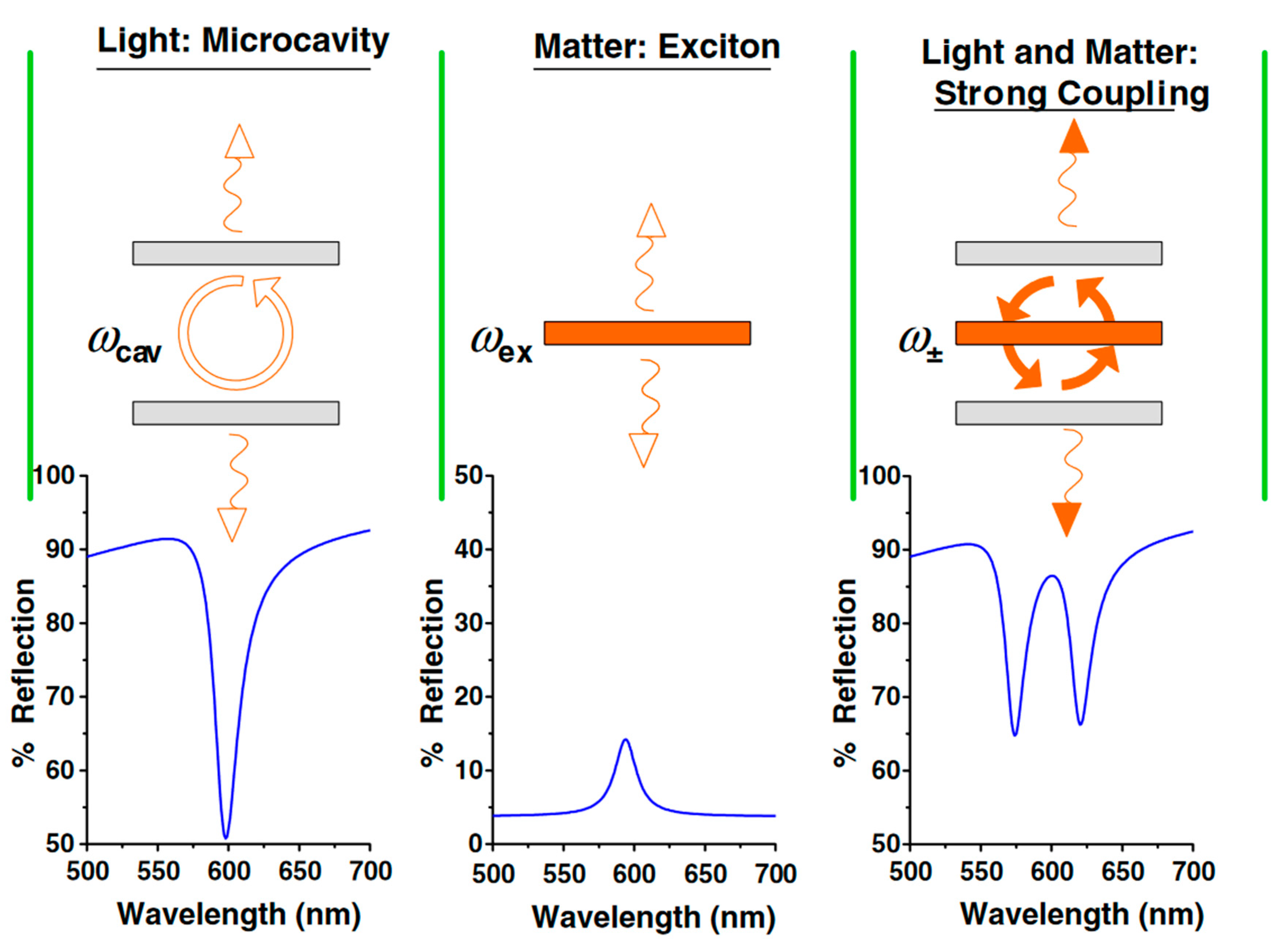
The new quasiparticle (plexciton) in the strong coupling regime is formed with distinct properties possessed by neither original particle. The coherent coupling interaction between the atom and the cavity mode is so strong that when the photon emitted into the cavity is similar to be reabsorbed before escape (the light and matter exchange energy periodically), and results in two new mixed states separated energetically (named as Rabi splitting) [68,73,74]. Detailed information can be seen from Figure 1.
The perturbative analysis of coupling between the cavity mode and atom ceases to be justified. General solution for arbitrary , , and is of the form
where
The constants and can be determined from initial conditions and , respectively. In the strong coupling regime with , and , it can be adjusted to
According to Equation (16), the evolution of the upper state population can be described, as well as the split at the vacuum Rabi frequency.
Based on the Jaynes–Cummings Hamiltonian, the system only has two states: and . When the atom-field interaction is considered, the eigenenergies can be described as follows:
Here is the n-photon generalized Rabi frequency:
The energies of the state and state cross at , but the atom-field interaction removes this degeneracy, causing the dressed states and to repel each other, or anticross. In other word, the anticrossing corresponds to the resonance condition (a central peak at and two sidebands at ).
2.3. Femtosecond Absorption
Hot electrons can be generated from plasmon decay in plasmonic nanostructures, which can be treated as efficient light-trapping components and can significantly improve the photocatalytic efficiency of traditional semiconductor devices. The hybrid system of a noble metal and a metal oxide was once considered to have two separate components and electron transfer was considered to be difficult because the individual electrons were not able to get sufficient energy to overcome the Schottky barrier [76]. However, the electron transfer from gold to the TiO2 electrode had been proved by the generated photocurrent under excitation of the plasmon band [77,78], which is relevant because the wide bandgap of TiO2 limits the generation of photocurrents. The generated hot electrons will go through three steps: generation, injection, and regeneration. The reverse electron transfer from TiO2 to gold, as the inverse process, is also proved in core-shell Ag-TiO2 NPs [79]. Based on the investigations, the energy needed to overcome the reaction barrier in hybrid systems is relatively smaller than the bandgap of TiO2. Hence, bringing the plasmonic nanostructures in contact with a semiconductor is a promising way to develop photocatalytic devices.
The electromagnetic decay takes places on a femtosecond timescale in plasmonic nanostructures, either radiatively through non-radiatively by transferring the energy to hot electrons or re-emitted photons [80,81,82,83,84].
To further reveal the internal mechanism of the prolonged lifetime of hot electrons in plasmon–exciton coupling interactions, the pump-probe ultrafast transient absorption spectroscopy (UTAS) is usually applied to investigate the timescale of the hot electron transfer process. Among the hot electrons transfer processes, the fast-electron injection into the metal oxide before recombination is crucial for improving the conversion efficiency. Direct evidence of electron transfer from Au NPs to TiO2 can be obtained. Furube and coworkers revealed the internal mechanism of ultrafast plasmon-induced electron transfer from 10 nm Au NPs to TiO2 NPs with femtosecond transient absorption spectroscopy [15,85,86], and revealed that the hot electron generation and injection were completed within 50 fs [42,45,87,88,89,90,91,92,93,94,95,96,97]. When the plasmons of gold NPs are excited, the electrons with a non-Fermi distribution undergo relaxation through the re-emission of photons or carrier multiplication within 100 fs due to the electron–electron interaction [52,98,99], electron–phonon interaction at the timescale of 1–10 ps, and phonon–phonon interactions around 100 ps [100,101]. Hence, researchers concluded that the hot electron injection resulted from electron–electron interaction.
The N3/TiO2 system, whose carrier injection efficiency is nearly 100%, is used for the comparison of the electron transfer yield [102]. According to the transient absorption intensity ratio between Au/TiO2 and N3/TiO2 (~100% injection efficiency), the electron injection yield in Au/TiO2 was evaluated to be about 40% under 550 nm excitation.
In addition, the Au/ZrO2 system is set for monitoring the response of excited Au nanodots as a control group, where no electron is able to transfer from Au nanodots to ZrO2, since the conduction band edge of ZrO2 is located 0.9 V above that of TiO2 [102]. The observed transient absorption of Au/TiO2 can be attributed to the electron transfer into TiO2, because there is no transient absorption for Au/ZrO2 with a probe laser of 3500 nm. Further, the electron transfer was completed within 240 fs, as shown in Figure 2. The timescale of the regeneration process was also investigated by Tian and coworkers [103,104].
Similarly, an Ag NP–graphene hybrid has also been investigated widely as an appropriate candidate. With the unique properties of graphene, the graphene–Ag nanostructure hybrid systems have been widely applied in investigating plasmon–exciton co-driven surface catalysis, whose mechanism was not clearly elucidated. Ding and coworkers fabricated the graphene-Ag nanowire hybrid system (Figure 3a) to reveal the dynamic process of plasmon–exciton coupling interaction with UTAS, as shown in Figure 3b–e.
The fitted curve in Figure 3c indicates that the lifetime of plasmonic hot electrons is about 3.2 ± 0.8 ps, which is obviously prolonged compared with the situation for isolated Ag NWs (150 fs). Furthermore, graphene cannot only prolong lifetime of the hot electrons dramatically, but also results in the significant accumulations of the hot electrons.
Moreover, the mechanism of exciton–plasmon coupling can also be investigated based on hybrid system of MoS2-Ag NP, according to the transmission spectra (Figure 4) and ultrafast absorption spectroscopy (Figure 4) results with a pump laser of 400 nm. It is revealed that the enhancement factors of excitonic states of MoS2 are different; the excitonic state (637 nm) is enhanced significantly with plasmon–exciton coupling interaction, instead of the B excitonic state (595 nm). By comparing the lifetimes of the two excitonic states of MoS2 (Figure 4), we find that the plasmon–exciton coupling interaction has a strong impact on the lifetime of excitonic states. According to the fitted transient absorption spectra of MoS2-Ag NP (size of 21 nm) in Figure 4e–h, the lifetime of electron-electron interaction in the hybrid system is enhanced by about 8 times than that of MoS2 alone. As for the electron–photon interaction, the lifetime is also increased significantly for excitonic states A and B. Hence, it is confirmed that the exciton–plasmon interaction improves the efficiency and probability of catalysis due to the long lifetime of carriers.
3. Applications on Photocatalysis
Since 2010, plasmonic hot electrons have been found to be critical in catalysis monitored by the SERS and TERS [12,106,107]. However, based on the investigations, the efficiency of surface plasmon-to-hot electron conversion is considered to be lower than 1% [108]. Hence, several studies attempted to achieve the goal of increasing the efficiency of plasmon-driven surface catalysis.
Before describing the specific application of the hybrid system, we experimentally confirm that the plasmon–exciton coupling degree can be well manipulated, for example, by changing the size of Ag NPs in the Ag NP–TiO2 film hybrids.
Ding first synthesized a nanosized TiO2 film on the quartz, with the thickness of ~208 nm and the absorption peak was centered at 524 nm. Above the TiO2 film, Ag NPs with different sizes were synthesized, as shown in Figure 5. The optimal parameters of components can be studied by UV-Visible absorption spectroscopy, and the ultrafast transfer process of plasmonic hot electron from Ag NPs into TiO2 film can be investigated by UTAS.
When the Ag NPs are generated under UV irradiation, in addition to the large size, the absorption intensity gradually increases and the strong absorbance peak is red shifted. The strong plasmon–exciton coupling interaction is formed only when the surface plasmon resonance peak of Ag NPs overlaps with the absorption peak of the TiO2 film (524 nm).
According to the in situ real-time UV-Visible absorbance spectra of hybrid systems (Figure 5), the absorption intensity increases gradually when the UV irradiation time increases from 2 min to 15 min; further, the SPR peak of Ag NPs at 15 min is around 532 nm, while the growth is halted at 30 min and decreases when the time is up to 60 min.
According to the absorption peaks of the Ag NP–TiO2 film where the Ag NPs were fabricated within 2 min (Figure 6a), there are two ultrafast absorption peaks around 532 nm and 475 nm. Focusing on the case of 532 nm, the electron-electron interaction is about 2 ps while the phonon- electron interaction can approach 71 ps, as shown in Figure 6b. According to the Ag NP size dependent, the ultrafast process of exciton–plasmon interaction of TiO2 film-Ag NP hybrids in Figure 6c, when the size of Ag NPs increases, the intensity of absorption spectrum become stronger while the lifetime decreases. The shorter lifetime represents the stronger exciton–plasmon interaction in Ag NP–TiO2 film hybrids. We can conclude that the degree of exciton–plasmon interaction can be well manipulated.
Figure 7a proves again that the superposition between the absorption peak of Ag NPs and TiO2 film can help in monitoring the degree of the coupling interaction, due to the strongest SERS intensity of catalysis of Ag NP–TiO2 film at 15 min.
The efficiency of the oxidation catalysis can be monitored by the ratio of the intensity at 1437 and 1071 cm−1, where the former is attributed to the Ag mode of DMAB and the latter is attributed to the A1 mode of PATP. The excitation wavelength-dependent oxidation catalysis illustrates that, based on the match between the excitation laser wavelength and SPR peak of Ag NPs, we can obtain the highest yield of the product excited on 532 nm due to the plasmon–exciton coupling interaction. Further, the oxidation catalysis is efficient and stable on the Ag NP–TiO2 film hybrids under different environments, including atmospheric, aqueous, and icy environments [110].
Thus, based on UV–Visible absorption spectroscopy, ultrafast transient absorption spectroscopy, and SERS, the plasmon–exciton coupling interaction in the Ag NP–TiO2 film can be investigated in detail. To obtain the maximum catalytic activity for oxidation catalysis, the degree of coupling interaction can be adjusted by changing the size of Ag NPs and monitoring by the superposition of the SPR peak of Ag NPs and the absorption peak of the TiO2 film.
Yang synthesized monolayer MoS2–Ag NP hybrids where the size of Ag NPs was monitored for shifting LSPR peak to match the exciton energy of monolayer MoS2 and, hence, to further monitor the degree of plasmon–exciton coupling interaction.
As shown in Figure 8, when the size increases, the LSPR peak of Ag NPs is red-shifted, as is the absorption peak of the hybrid system attributed to exciton–plasmon interaction. When they are coupled, LSPR can significantly enhance the excitation rate of the monolayer MoS2 exciton through EM, and the generated collective states result in stronger optical absorption than the individual components.
On the monolayer MoS2 substrate, there is no obvious phenomenon regarding occurring catalysis. With a low-intensity laser, the probability of the reduction catalysis on the Ag NPs substrate is much lower than on the MoS2–Ag NP hybrid system. Further, the advantages of the exciton–plasmon coupling can be confirmed by investigating the Ag NP size-dependent exciton–plasmon co-driven surface catalysis, as shown in Figure 9. With the help of the strongest exciton–plasmon interaction near 532 nm, and highest probability and efficiency of reduction catalysis can be achieved.
In Figure 10, comparing the ratio between the Raman intensities of reactant (1338 cm−1) and product (1432 cm−1), we can conclude that the probability of the reduction catalysis for 4NBT adsorbed on Ag NPs directly is much lower than that on the MoS2–Ag NP hybrid substrate, which supports the aforementioned conclusion.
Also, the physical mechanism of the exciton–plasmon interaction for catalysis has been interpreted theoretically, which revealed ultrafast charge transfer between plasmons and excitons in the hybrid system [112].
4. Conclusions
We reviewed the principle of plasmonic nanostructure–semiconductor exciton interaction and the applications of plasmonic nanostructure–semiconductor exciton interaction for chemical reactions. The exciton–plasmon interaction has greater potential for surface catalytic reactions than plasmon alone. This review can promote a better understanding of the physical mechanism of exciton–plasmon coupling for surface catalysis.
Author Contributions
Investigation, J.W., X.M.; Resources, J.W.; Data Curation, N.F., Y.S.; Writing-Original Draft Preparation, J.W., N.F.; Writing-Review & Editing, J.W., X.M.; Supervision, J.W. and X.M.; Project Administration, J.W., X.M.; Funding Acquisition, J.W.
Funding
This work was supported by the National Nature Science Foundation of China (Grant No. 91436102, 11374353, 11474141), Fundamental Research Funds for the Central Universities and talent scientific research fund of LSHU (No. 6008).
Conflicts of Interest
The authors declare no conflict of interest.
References
- Ritchie, R.H. Plasma Losses by Fast Electrons in Thin Films. Phys. Rev. 1957, 106, 874–881. [Google Scholar] [CrossRef]
- Brockman, J.M.; Nelson, B.P.; Corn, R.M. Surface Plasmon Resonance Imaging Measurements of Ultrathin Organic Films. Annu. Rev. Phys. Chem. 2000, 51, 41–63. [Google Scholar] [CrossRef] [PubMed]
- Lin, W.; Cao, Y.; Wang, P.; Sun, M. Unified Treatment for Plasmon–Exciton Co-driven Reduction and Oxidation Reactions. Langmuir 2017, 33, 12102–12107. [Google Scholar] [CrossRef] [PubMed]
- Zhang, Z.L.; Sheng, S.X.; Wang, R.M.; Sun, M.T. Tip-Enhanced Raman Spectroscopy. Anal. Chem. 2016, 88, 9328–9346. [Google Scholar] [CrossRef] [PubMed]
- Viti, L.; Hu, J.; Coquillat, D.; Politano, A.; Knap, W.; Vitiello, M.S. Efficient Terahertz detection in black-phosphorus nano-transistors with selective and controllable plasma-wave, bolometric and thermoelectric response. Sci. Rep. 2016, 6, 20474. [Google Scholar] [CrossRef] [PubMed] [Green Version]
- Ueno, K.; Juodkazis, S.; Shibuya, T.; Yokota, Y.; Mizeikis, V.; Sasaki, K.; Misawa, H. Nanoparticle Plasmon-Assisted Two-Photon Polymerization Induced by Incoherent Excitation Source. J. Am. Chem. Soc. 2008, 130, 6928–6929. [Google Scholar] [CrossRef] [PubMed]
- Hubert, C.; Rumyantseva, A.; Lerondel, G.; Grand, J.; Kostcheev, S.; Billot, L.; Vial, A.; Bachelot, R.; Royer, P.; Chang, S.H.; et al. Near-Field Photochemical Imaging of Noble Metal Nanostructures. Nano Lett. 2005, 5, 615–619. [Google Scholar] [CrossRef] [PubMed]
- Lin, W.; Cao, E.; Zhang, L.; Xu, X.; Song, Y.; Liang, W.; Sun, M. Electrically enhanced hot hole driven oxidation catalysis at the interface of a plasmon-exciton hybrid. Nanoscale 2018, 10, 5482–5488. [Google Scholar] [CrossRef] [PubMed]
- Deeb, C.; Ecoffet, C.; Bachelot, R.; Plain, J.; Bouhelier, A.; Soppera, O. Plasmon-based free-radical photopolymerization: Effect of diffusion on nanolithography processes. J. Am. Chem. Soc. 2011, 133, 10535–10542. [Google Scholar] [CrossRef] [PubMed]
- Sun, M.; Zhang, Z.; Zheng, H.; Xu, H. In-situ plasmon-driven chemical reactions revealed by high vacuum tip-enhanced Raman spectroscopy. Sci. Rep. 2012, 2, 647. [Google Scholar] [CrossRef] [PubMed]
- Chen, C.J.; Osgood, R.M. Direct Observation of the Local-Field-Enhanced Surface Photochemical Reactions. Phys. Rev. Lett. 1983, 50, 1705–1708. [Google Scholar] [CrossRef]
- Fang, Y.; Li, Y.; Xu, H.; Sun, M. Ascertaining p,p’-dimercaptoazobenzene produced from p-aminothiophenol by selective catalytic coupling reaction on silver nanoparticles. Langmuir 2010, 26, 7737–7746. [Google Scholar] [CrossRef] [PubMed]
- Canpean, V.; Iosin, M.; Astilean, S. Disentangling SERS signals from two molecular species: A new evidence for the production of p,p′-dimercaptoazobenzene by catalytic coupling reaction of p-aminothiophenol on metallic nanostructures. Chem. Phys. Lett. 2010, 500, 277–282. [Google Scholar] [CrossRef]
- Sun, M.; Fang, Y.; Zhang, Z.; Xu, H. Activated vibrational modes and Fermi resonance in tip-enhanced Raman spectroscopy. Phys. Rev. E Stat. Nonlinear Soft Matter Phys. 2013, 87, 020401. [Google Scholar] [CrossRef] [PubMed]
- Furube, A.; Du, L.; Hara, K.; Katoh, R.; Tachiya, M. Ultrafast Plasmon-Induced Electron Transfer from Gold Nanodots into TiO2 Nanoparticles. J. Am. Chem. Soc. 2007, 129, 14852–14853. [Google Scholar] [CrossRef] [PubMed]
- Gabudean, A.M.; Biro, D.; Astilean, S. Localized surface plasmon resonance (LSPR) and surface-enhanced Raman scattering (SERS) studies of 4-aminothiophenol adsorption on gold nanorods. J. Mol. Struct. 2011, 993, 420–424. [Google Scholar] [CrossRef]
- Christopher, P.; Xin, H.; Linic, S. Visible-light-enhanced catalytic oxidation reactions on plasmonic silver nanostructures. Nat. Chem. 2011, 3, 467. [Google Scholar] [CrossRef] [PubMed]
- Cao, E.; Guo, X.; Zhang, L.; Shi, Y.; Lin, W.; Liu, X.; Fang, Y.; Zhou, L.; Sun, Y.; Song, Y. Electrooptical Synergy on Plasmon–Exciton-Codriven Surface Reduction Reactions. Adv. Mater. Interfaces 2017, 4, 1700869. [Google Scholar] [CrossRef]
- Tian, Y.; Tatsuma, T. Mechanisms and Applications of Plasmon-Induced Charge Separation at TiO2 Films Loaded with Gold Nanoparticles. J. Am. Chem. Soc. 2005, 127, 7632–7637. [Google Scholar] [CrossRef] [PubMed]
- Sun, M.; Zhang, Z.; Wang, P.; Li, Q.; Ma, F.; Xu, H. Remotely excited Raman optical activity using chiral plasmon propagation in Ag nanowires. Light Sci. Appl. 2013, 2, e112. [Google Scholar] [CrossRef]
- Sun, M.; Xu, H. A novel application of plasmonics: Plasmon-driven surface-catalyzed reactions. Small 2012, 8, 2777–2786. [Google Scholar] [CrossRef] [PubMed]
- Politano, A.; Argurio, P.; Di, P.G.; Sanna, V.; Cupolillo, A.; Chakraborty, S.; Arafat, H.A.; Curcio, E. Photothermal Membrane Distillation for Seawater Desalination. Adv. Mater. 2017, 29, 1603504. [Google Scholar] [CrossRef] [PubMed]
- Wang, Q.H.; Kalantar-Zadeh, K.; Kis, A.; Coleman, J.N.; Strano, M.S. Electronics and optoelectronics of two-dimensional transition metal dichalcogenides. Nat. Nanotechnol. 2012, 7, 699–712. [Google Scholar] [CrossRef] [PubMed] [Green Version]
- Kuc, A.; Zibouche, N.; Heine, T. Influence of quantum confinement on the electronic structure of the transition metal sulfide TS2. Phys. Rev. B 2011, 83, 2237–2249. [Google Scholar] [CrossRef]
- Wilson, J.A.; Yoffe, A.D. The transition metal dichalcogenides discussion and interpretation of the observed optical, electrical and structural properties. Adv. Phys. 1969, 18, 193–335. [Google Scholar] [CrossRef]
- Lana-Villarreal, T.; Gómez, R. Tuning the photoelectrochemistry of nanoporous anatase electrodes by modification with gold nanoparticles: Development of cathodic photocurrents. Chem. Phys. Lett. 2005, 414, 489–494. [Google Scholar] [CrossRef]
- Kowalska, E.; Mahaney, O.O.; Abe, R.; Ohtani, B. Visible-light-induced photocatalysis through surface plasmon excitation of gold on titania surfaces. Phys. Chem. Chem. Phys. 2010, 12, 2344–2355. [Google Scholar] [CrossRef] [PubMed]
- Toyoda, T.; Tsugawa, S.; Shen, Q. Photoacoustic spectra of Au quantum dots adsorbed on nanostructured TiO2 electrodes together with the photoelectrochemical current characteristics. J. Appl. Phys. 2009, 105, 034314. [Google Scholar] [CrossRef]
- Kowalska, E.; Abe, R.; Ohtani, B. Visible light-induced photocatalytic reaction of gold-modified titanium(IV) oxide particles: Action spectrum analysis. Chem. Commun. 2009, 241–243. [Google Scholar] [CrossRef] [PubMed]
- Gong, D.; Weng, C.J.H.; Tang, Y.; Tay, Q.; Lai, Y.; Highfield, J.G.; Chen, Z. Silver decorated titanate/titania nanostructures for efficient solar driven photocatalysis. J. Solid State Chem. 2012, 189, 117–122. [Google Scholar] [CrossRef]
- Tanaka, A.; Ogino, A.; Iwaki, M.; Hashimoto, K.; Ohnuma, A.; Amano, F.; Ohtani, B.; Kominami, H. Gold-titanium(IV) oxide plasmonic photocatalysts prepared by a colloid-photodeposition method: Correlation between physical properties and photocatalytic activities. Langmuir 2012, 28, 13105–13111. [Google Scholar] [CrossRef] [PubMed]
- Ingram, D.B.; Christopher, P.; Bauer, J.L.; Linic, S. Predictive Model for the Design of Plasmonic Metal/Semiconductor Composite Photocatalysts. ACS Catal. 2011, 1, 1441–1447. [Google Scholar] [CrossRef]
- Fujishima, A.; Honda, K. Electrochemical Photolysis of Water at a Semiconductor Electrode. Nature 1972, 238, 37–38. [Google Scholar] [CrossRef] [PubMed]
- Fujishima, A.; Rao, T.N.; Tryk, D.A. Titanium dioxide photocatalysis. J. Photochem. Photobiol. C Photochem. Rev. 2000, 1, 1–21. [Google Scholar] [CrossRef]
- Nazeeruddin, M.K.; Kay, A.; Rodicio, I.; Humphrybaker, R.; Mueller, E.; Liska, P.; Vlachopoulos, N.; Graetzel, M. Conversion of Light to Electricity by cis-XzBis(2,2’-bipyridyl-4,4’-dicarboxylate)ruthenium(II) Charge-Transfer Sensitizers (X = C1−, Br−, I−, CN−, and SCN−) on Nanocrystalline Ti02 Electrodes. J. Am. Chem. Soc. 1993, 115, 6382–6390. [Google Scholar] [CrossRef]
- Chen, Z.H.; Tang, Y.B.; Liu, C.P.; Leung, Y.H.; Yuan, G.D.; Chen, L.M.; Wang, Y.Q.; Bello, I.; Zapien, J.A.; Zhang, W.J. Vertically Aligned ZnO Nanorod Arrays Sentisized with Gold Nanoparticles for Schottky Barrier Photovoltaic Cells. J. Phys. Chem. C 2009, 113, 13433–13437. [Google Scholar] [CrossRef]
- Liu, W.; Lin, W.; Zhao, H.; Wang, P.; Sun, M. The nature of plasmon-exciton codriven surface catalytic reaction. J. Raman Spectrosc. 2018, 49, 383–387. [Google Scholar] [CrossRef]
- Kamat, P.V. Photochemistry on Nonreactive and Reactive (Semiconductor) Surfaces. Chem. Rev. 1993, 93, 267–300. [Google Scholar] [CrossRef]
- Hoffmann, M.R.; Martin, S.T.; Choi, W.; Bahnemannt, D.W. Environmental Applications of Semiconductor Photocatalysis. Chem. Rev. 1995, 95, 69–96. [Google Scholar] [CrossRef]
- Linsebigler, A.L.; Lu, G.; Yates, J.T. Photocatalysis on TiOn Surfaces: Principles, Mechanisms, and Selected Results. Chem. Rev. 1995, 95, 735–758. [Google Scholar] [CrossRef]
- Vinodgopal, K.; Bedja, I.; Hotchandani, S.; Kamat, P.V. A Photocatalytic Approach for the Reductive Decolorization of Textile Azo Dyes in Colloidal Semiconductor Suspensions. Langmuir 1994, 10, 1767–1771. [Google Scholar] [CrossRef]
- Bahnemann, D.; Henglein, A.; Lilie, J.; Spanhel, L. Flash Photolysis Observation of the Absorption Spectra of Trapped Positive Holes and Electrons in Colloidal TiO2. J. Phys. Chem. 1984, 88, 709–711. [Google Scholar] [CrossRef]
- Gaoa, R.; Safranyc, A.; Rabania, J. Fundamental reactions in TiO2 nanocrystallite aqueous solutions studied by pulse radiolysis. Radiat. Phys. Chem. 2002, 65, 599–609. [Google Scholar] [CrossRef]
- Colombo, D.P., Jr.; Roussel, K.A.; Saeh, J.; Skinner, D.E.; Cavaleri, J.J.; Bowman, R.M. Femtosecond study of the intensity dependence of electron-hole dynamics in TiO2 nanoclusters. Chem. Phys. Lett. 1995, 232, 207–214. [Google Scholar] [CrossRef]
- Ramakrishna, G.; Ghosh, H.N. Optical and Photochemical Properties of Sodium Dodecylbenzenesulfonate (DBS)-Capped TiO2 Nanoparticles Dispersed in Nonaqueous Solvents. Langmuir 2003, 19, 505–508. [Google Scholar] [CrossRef]
- Christopher, P.; Ingram, D.B.; Linic, S. Enhancing Photochemical Activity of Semiconductor Nanoparticles with Optically Active Ag Nanostructures: Photochemistry Mediated by Ag Surface Plasmons. J. Phys. Chem. C 2010, 114, 9173–9177. [Google Scholar] [CrossRef]
- Lin, W.; Xu, X.; Quan, J.; Sun, M. Propagating surface plasmon polaritons for remote excitation surface-enhanced Raman scattering spectroscopy. Appl. Spectrosc. Rev. 2018, 53, 771–782. [Google Scholar] [CrossRef]
- Wang, J.; Tafen, D.N.; Lewis, J.P.; Hong, Z.; Manivannan, A.; Zhi, M.; Li, M.; Wu, N. Origin of Photocatalytic Activity of Nitrogen-Doped TiO2 Nanobelts. J. Am. Chem. Soc. 2009, 131, 12290–12297. [Google Scholar] [CrossRef] [PubMed]
- Ohno, T.; Sarukawa, K.; Tokieda, K.; Matsumura, M. Morphology of a TiO2 Photocatalyst (Degussa, P-25) Consisting of Anatase and Rutile Crystalline Phases. J. Catal. 2001, 203, 82–86. [Google Scholar] [CrossRef]
- Fujishima, A.; Zhang, X.; Tryk, D. TiO2 photocatalysis and related surface phenomena. Surf. Sci. Rep. 2008, 63, 515–582. [Google Scholar] [CrossRef]
- Zhu, K.; Neale, N.R.; Miedaner, A.; Frank, A.J. Enhanced Charge-Collection Efficiencies and Light Scattering in Dye-Sensitized Solar Cells Using Oriented TiO2 Nanotubes Arrays. Nano Lett. 2007, 7, 69–74. [Google Scholar] [CrossRef] [PubMed]
- Ding, Q.; Shi, Y.; Chen, M.; Li, H.; Yang, X.; Qu, Y.; Liang, W.; Sun, M. Ultrafast Dynamics of Plasmon-Exciton Interaction of Ag Nanowire- Graphene Hybrids for Surface Catalytic Reactions. Sci. Rep. 2016, 6, 32724. [Google Scholar] [CrossRef] [PubMed] [Green Version]
- Fofang, N.T.; Grady, N.K.; Fan, Z.; Govorov, A.O.; Halas, N.J. Plexciton dynamics: Exciton-plasmon coupling in a J-aggregate-Au nanoshell complex provides a mechanism for nonlinearity. Nano Lett. 2011, 11, 1556–1560. [Google Scholar] [CrossRef] [PubMed]
- Wiederrecht, G.P.; Wurtz, G.A.; Hranisavljevic, J. Coherent Coupling of Molecular Excitons to Electronic Polarizations of Noble Metal Nanoparticles. Nano Lett. 2004, 4, 2121–2125. [Google Scholar] [CrossRef]
- Wurtz, G.A.; Evans, P.R.; Hendren, W.; Atkinson, R.; Dickson, W.; Pollard, R.J.; Zayats, A.V.; Harrison, W.; Bower, C. Molecular Plasmonics with Tunable Exciton−Plasmon Coupling Strength in J-Aggregate Hybridized Au Nanorod Assemblies. Nano Lett. 2007, 7, 1297–1303. [Google Scholar] [CrossRef] [PubMed]
- Govorov, A.O.; Bryant, G.W.; Zhang, W.; Skeini, T.; Lee, J.; Kotov, N.A.; Slocik, J.M.; Naik, R.R. Exciton−Plasmon Interaction and Hybrid Excitons in Semiconductor−Metal Nanoparticle Assemblies. Nano Lett. 2006, 6, 984–994. [Google Scholar] [CrossRef]
- Bellessa, J.; Symonds, C.; Vynck, K.; Beaur, L.; Brioude, A.; Lemaitre, A. Giant Rabi splitting in metal/semiconductor nanohybrids. Superlattices Microstruct. 2011, 49, 209–216. [Google Scholar] [CrossRef]
- Bishnoi, S.W.; Rozell, C.J.; Levin, C.S.; Gheith, M.K.; Johnson, B.R.; Johnson, D.H.; Halas, N.J. All-Optical Nanoscale pH Meter. Nano Lett. 2006, 6, 1687–1692. [Google Scholar] [CrossRef] [PubMed]
- Govorov, A.O.; Carmeli, I. Hybrid Structures Composed of Photosynthetic System and Metal Nanoparticles: Plasmon Enhancement Effect. Nano Lett. 2007, 7, 620–625. [Google Scholar] [CrossRef] [PubMed]
- Wang, J.; Xu, X.; Mu, X.; Ma, F.; Sun, M. Magnetics and spintronics on two-dimensional composite materials of graphene/hexagonal boron nitride. Mater. Today Phys. 2017, 3, 93–117. [Google Scholar] [CrossRef]
- Artuso, R.D.; Bryant, G.W. Optical Response of Strongly Coupled Quantum Dot-Metal Nanoparticle Systems: Double Peaked Fano Structure and Bistability. Nano Lett. 2008, 8, 2106–2111. [Google Scholar] [CrossRef] [PubMed]
- Purcell, E.M. Confined Electrons and Photons: New Physics and Applications; Burstein, E., Weisbuch, C., Eds.; Springer: New York, NY, USA, 1995; p. 839. [Google Scholar]
- Bharadwaj, P.; Deutsch, B.; Novotny, L. Optical Antennas. Adv. Opt. Photonics 2009, 1, 438–483. [Google Scholar] [CrossRef]
- Henkel, C.; Sandoghdar, V. Single-molecule spectroscopy near structured dielectrics. Opt. Commun. 1998, 158, 250–262. [Google Scholar] [CrossRef] [Green Version]
- Kuhn, H. Classical Aspects of Energy Transfer in Molecular Systems. J. Chem. Phys. 1970, 53, 101–108. [Google Scholar] [CrossRef]
- Yeung, M.S.; Gustafson, T.K. Spontaneous emission near an absorbing dielectric surface. Phys. Rev. A 1996, 54, 5227–5242. [Google Scholar] [CrossRef] [PubMed]
- Giannini, V.; Fernandez-Dominguez, A.I.; Heck, S.C.; Maier, S.A. Plasmonic nanoantennas: Fundamentals and their use in controlling the radiative properties of nanoemitters. Chem. Rev. 2011, 111, 3888–3912. [Google Scholar] [CrossRef] [PubMed]
- Savasta, S.; Saija, R.; Ridolfo, A.; Di, S.O.; Denti, P.; Borghese, F. Nanopolaritons: Vacuum Rabi Splitting with a Single Quantum Dot in the Center ofa Dimer Nanoantenna. ACS Nano 2010, 4, 6369–6376. [Google Scholar] [CrossRef] [PubMed]
- Berman, P.R. Cavity Quantum Electrodynamics; Academic Press, Inc.: Boston, MA, USA, 1994. [Google Scholar]
- Vahala, K.J. Optical microcavities. Nature 2003, 424, 839–846. [Google Scholar] [CrossRef] [PubMed]
- Giannini, V.; Sánchez-Gil, J.A.; Muskens, O.L.; Rivas, J.G. Electrodynamic calculations of spontaneous emission coupled to metal nanostructures of arbitrary shape: Nanoantenna-enhanced fluorescence. J. Opt. Soc. Am. B 2009, 26, 1569–1577. [Google Scholar] [CrossRef]
- Maier, S.A. Plasmonics: Fundamentals and Applications; Springer: Berlin/Heidelberg, Germany, 2007. [Google Scholar]
- Manjavacas, A.; Garcia de Abajo, F.J.; Nordlander, P. Quantum plexcitonics: Strongly interacting plasmons and excitons. Nano Lett. 2011, 11, 2318–2323. [Google Scholar] [CrossRef] [PubMed]
- Schlather, A.E.; Large, N.; Urban, A.S.; Nordlander, P.; Halas, N.J. Near-field mediated plexcitonic coupling and giant Rabi splitting in individual metallic dimers. Nano Lett. 2013, 13, 3281–3286. [Google Scholar] [CrossRef] [PubMed]
- Tischler, J.R.; Bradley, M.S.; Zhang, Q.; Atay, T.; Nurmikko, A.; Bulović, V. Solid state cavity QED: Strong coupling in organic thin films. Org. Electron. 2007, 8, 94–113. [Google Scholar] [CrossRef]
- McFarland, E.W.; Tang, J. Aphotovoltaicdevicestructure based on internal electron emission. Nature 2003, 421, 616–618. [Google Scholar] [CrossRef] [PubMed]
- Fang, Y.; Zhang, Z.; Sun, M. High vacuum tip-enhanced Raman spectroscope based on a scanning tunneling microscope. Rev. Sci. Instrum. 2016, 87, 033104. [Google Scholar] [CrossRef] [PubMed]
- Zhao, G.; Kozuka, H.; Yoko, T. Sol-gel preparation and photoelectrochemical properties ofTiO2 containing Au andAg metal particles. Thin Solid Films 1996, 277, 147–154. [Google Scholar] [CrossRef]
- Li, H.; Bian, Z.; Zhu, J.; Huo, Y.; Li, H.; Lu, Y. Mesoporous Au/TiO2 nanocomposites with enhanced photocatalytic activity. J. Am. Chem. Soc. 2007, 129, 4538–4539. [Google Scholar] [CrossRef] [PubMed]
- Sönnichsen, C.; Raschke, G.; Plessen, G.V.; Feldmann, J.; Wilson, O.; Mulvaney, P.; Franzl, T.; Wilk, T. Drastic reduction of plasmon damping in gold nanorods. Phys. Rev. Lett. 2002, 88, 077402. [Google Scholar] [CrossRef] [PubMed]
- Lehmann, J.; Merschdorf, M.; Pfeiffer, W.; Thon, A.; Voll, S.; Gerber, G. Surface Plasmon Dynamics in Silver Nanoparticles Studied by Femtosecond Time-Resolved Photoemission. Phys. Rev. Lett. 2000, 85, 2921–2924. [Google Scholar] [CrossRef] [PubMed]
- Hofmann, J.; Steinmann, W. Plasma Resonance in the Photoemission of Silver. Phys. Stat. Sol. 1968, 30, K53–K56. [Google Scholar] [CrossRef]
- Endriz, J.G.; Spicer, W.E. Surface-Plasmon-One-Electron Decay and its Observation in Photoemission. Phys. Rev. Lett. 1970, 24, 64–68. [Google Scholar] [CrossRef]
- Ding, Q.; Chen, M.; Fang, Y.; Zhang, Z.; Sun, M. Plasmon-Driven Diazo Coupling Reactions of p-Nitroaniline via −NH2 or −NO2 in Atmosphere Environment. J. Phys. Chem. C 2017, 121, 5225–5231. [Google Scholar] [CrossRef]
- Wang, J.; Lin, W.; Xu, X.; Ma, F.; Sun, M. Plasmon-Exciton Coupling Interaction for Surface Catalytic Reactions. Chem. Rec. 2018, 18, 481–490. [Google Scholar] [CrossRef] [PubMed]
- Du, L.; Furube, A.; Yamamoto, K.; Hara, K.; Katoh, R.; Tachiya, M. Plasmon-Induced Charge Separation and Recombination Dynamics in Gold-TiO2 Nanoparticle Systems: Dependence on TiO2 Particle Size. J. Phys. Chem. C 2009, 113, 6454–6462. [Google Scholar] [CrossRef]
- Duonghong, D.; Ramsden, J.; Gratzel, M. Dynamics of Interfacial Electron-Transfer Processes in Colloidal Semiconductor Systems. J. Am. Chem. Soc. 1982, 104, 2977–2985. [Google Scholar] [CrossRef]
- Arbour, C.; Sharma, D.K.; Langford, C.H. Picosecond Flash Spectroscopy of TiO2, Colloids with Adsorbed Dyes. J. Phys. Chem. 1990, 94, 331–335. [Google Scholar] [CrossRef]
- Rothenberger, G.; Moser, J.; Gratzel, M.; Serpone, N.; Sharma, D.K. Charge Carrier Trapping and Recombination Dynamics in Small Semiconductor Particles. J. Am. Chem. Soc. 1985, 107, 8054–8059. [Google Scholar] [CrossRef]
- Kolle, U.; Moser, J.; Gratzel, M. Dynamics of Interfacial Charge-Transfer Reactions in Semiconductor Dispersions. Reduction of Cobaltoceniumdicarboxylate in Colloidal TiO2. Inorg. Chem. 1985, 24, 2253–2258. [Google Scholar] [CrossRef]
- Arbour, C.; Sharma, D.K.; Langford, C.H. Kinetics of electron build-up in TiO2 colloids probed by hole scavenging after picosecond excitation. Chem. Commun. 1987, 917–918. [Google Scholar] [CrossRef]
- Bahnemann, D.; Henglein, A.; Spanhel, L. Detection of the intermediates of colloidal TiO2-catalysed photoreactions. Faraday Discuss. Chem. Soc. 1984, 78, 151–163. [Google Scholar] [CrossRef]
- Bahnemann, D.W.; Hilgendorff, M.; Memming, R. Charge Carrier Dynamics at TiO2 Particles: Reactivity of Free and Trapped Holes. J. Phys. Chem. B 1997, 101, 4265–4275. [Google Scholar] [CrossRef]
- Li, R.; Zhang, Y.; Xu, X.; Zhou, Y.; Chen, M.; Sun, M. Optical characterizations of two-dimensional materials using nonlinear optical microscopies of CARS, TPEF, and SHG. Nanophotonics 2018, 7, 873–881. [Google Scholar] [CrossRef] [Green Version]
- Dimitrijevic, N.M.; Saponjic, Z.V.; Bartels, D.M.; Thurnauer, M.C.; And, D.M.T.; Rajh, T. Revealing the Nature of Trapping Sites in Nanocrystalline Titanium Dioxide by Selective Surface Modification. J. Phys. Chem. B 2003, 107, 7368–7375. [Google Scholar] [CrossRef]
- Rabani, J.; Yamashita, K.; Ushida, K.; Stark, J.; Kira, A. Fundamental Reactions in Illuminated Titanium Dioxide Nanocrystallite Layers Studied by Pulsed Laser. J. Phys. Chem. B 1998, 102, 1689–1695. [Google Scholar] [CrossRef]
- Asahi, T.; Furube, A.; Masuhara, H. Direct measurement of picosecond interfacial electron transfer from photoexcited TiO2 powder to an adsorbed molecule in the opaque suspension. Chem. Phys. Lett. 1997, 275, 234–238. [Google Scholar] [CrossRef]
- Brongersma, M.L.; Halas, N.J.; Nordlander, P. Plasmon-induced hot carrier science and technology. Nat. Nanotechnol. 2015, 10, 25–34. [Google Scholar] [CrossRef] [PubMed]
- Harutyunyan, H.; Martinson, A.B.; Rosenmann, D.; Khorashad, L.K.; Besteiro, L.V.; Govorov, A.O.; Wiederrecht, G.P. Anomalous ultrafast dynamics of hot plasmonic electrons in nanostructures with hot spots. Nat. Nanotechnol. 2015, 10, 770–774. [Google Scholar] [CrossRef] [PubMed]
- Link, S.; El-Sayed, M.A. Spectral Properties and Relaxation Dynamics of Surface Plasmon Electronic Oscillations in Gold and Silver Nanodots and Nanorods. J. Phys. Chem. B 1999, 103, 8410–8426. [Google Scholar] [CrossRef]
- Link, S.; El-Sayed, M.A. Shape and size dependence of radiative, non-radiative and photothermal properties of gold nanocrystals. Int. Rev. Phys. Chem. 2000, 19, 409–453. [Google Scholar] [CrossRef]
- Katoh, R.; Furube, A.; Yoshihara, T.; Hara, K.; Fujihashi, G.; Takano, S.; Murata, S.; Arakawa, H.; Tachiya, M. Efficiencies of Electron Injection from Excited N3 Dye into Nanocrystalline Semiconductor (ZrO2, TiO2, ZnO, Nb2O5, SnO2, In2O3) Films. J. Phys. Chem. B 2004, 108, 4818–4822. [Google Scholar] [CrossRef]
- Tian, Y.; Wang, X.; Zhang, D.; Shi, X.; Wang, S. Effects of electron donors on the performance of plasmon-induced photovoltaic cell. J. Photochem. Photobiol. A Chem. 2008, 199, 224–229. [Google Scholar] [CrossRef]
- Wang, J.; Ma, F.; Liang, W.; Sun, M. Electrical properties and applications of graphene, hexagonal boron nitride (h-BN), and graphene/h-BN heterostructures. Mater. Today Phys. 2017, 2, 6–34. [Google Scholar] [CrossRef]
- Lin, W.; Shi, Y.; Yang, X.; Li, J.; Cao, E.; Xu, X.; Pullerits, T.; Liang, W.; Sun, M. Physical Mechanism on Exciton-Plasmon Coupling Revealed by Femtosecond Pump-Probe Transient Absorption Spectroscopy. Mater. Today Phys. 2017, 3, 33–40. [Google Scholar] [CrossRef]
- Quan, J.; Cao, E.; Mu, X.; Sun, M. Surface catalytic reaction driven by plasmonic waveguide. Appl. Mater. Today 2018, 11, 50–56. [Google Scholar] [CrossRef]
- Zhang, Z.; Xu, P.; Yang, X.; Liang, W.; Sun, M. Surface plasmon-driven photocatalysis in ambient, aqueous and high-vacuum monitored by SERS and TERS. J. Photochem. Photobiol. C Photochem. Rev. 2016, 27, 100–112. [Google Scholar] [CrossRef]
- Mubeen, S.; Lee, J.; Singh, N.; Krämer, S.; Stucky, G.D.; Moskovits, M. An autonomous photosynthetic device in which all charge carriers derive from surface plasmons. Nat. Nanotechnol. 2013, 8, 247–251. [Google Scholar] [CrossRef] [PubMed]
- Ding, Q.; Li, R.; Chen, M.; Sun, M. Ag nanoparticles-TiO2 film hybrid for plasmon-exciton co-driven surface catalytic reactions. Appl. Mater. Today 2017, 9, 251–258. [Google Scholar] [CrossRef]
- Lin, W.; Ren, X.; Cui, L.; Sun, M. Electro-optical tuning of plasmon-driven double reduction interface catalysis. Appl. Mater. Today 2018, 11, 189–192. [Google Scholar] [CrossRef]
- Yang, X.; Yu, H.; Guo, X.; Ding, Q.; Pullerits, T.; Wang, R.; Zhang, G.; Liang, W.; Sun, M. Plasmon-exciton coupling of monolayer MoS2 -Ag nanoparticles hybrids for surface catalytic reaction. Mater. Today Energy 2017, 5, 72–78. [Google Scholar] [CrossRef]
- Xu, X.; Shi, Y.; Liu, X.; Sun, M. Femtosecond dynamics of monolayer MoS2 -Ag nanoparticles hybrid probed at 532 nm. Chem. Phys. Lett. 2018, 692, 208–213. [Google Scholar] [CrossRef]
Figure 1.
The mechanism of strong coupling between light and matter revealed by reflectance spectra [75].
Figure 1.
The mechanism of strong coupling between light and matter revealed by reflectance spectra [75].

Figure 2.
Transient absorption of N3/TiO2, Au/TiO2 and Au/ZrO2 at 3500 nm [15].
Figure 2.
Transient absorption of N3/TiO2, Au/TiO2 and Au/ZrO2 at 3500 nm [15].

Figure 3.
(a) The SEM image of Ag NW–graphene hybrid system. (b) The corresponding ultrafast pump-probe transient absorption spectroscopy excited by 400 nm, and (c) fitted at 532 nm. (d) The corresponding ultrafast pump-probe transient absorption spectroscopy excited in NIR region, and (e) fitted at 1103 nm [52].

Figure 4.
The ultrafast transient absorption spectra of (a) monolayer MoS2, and (b–d) monolayer MoS2-Ag NPs hybrid system where sizes of Ag NPs are 6.1, 14.5 and 21 nm, respectively. The transient absorption spectra of (e,f) monolayer MoS2 fitted at excitonic state A and B respectively, and (g,h) monolayer MoS2-Ag NPs hybrid fitted at excitonic state A and B respectively, where the size of Ag NPs is 21 nm [105].


Figure 5.
The SEM images of Ag NPs synthesized on TiO2 film under different UV irradiation for (a) 2, (b) 5, (c) 15, (d) 30, and (e) 60 min. (f) The corresponding in situ real-time UV–vis absorbance spectra of 5(a–e), respectively [109].
Figure 5.
The SEM images of Ag NPs synthesized on TiO2 film under different UV irradiation for (a) 2, (b) 5, (c) 15, (d) 30, and (e) 60 min. (f) The corresponding in situ real-time UV–vis absorbance spectra of 5(a–e), respectively [109].

Figure 6.
(a) The 3D ultrafast transient absorption spectrum of AgNPs-TiO2 film which is synthesized within 2 min, and (b) fitted 532.7 nm. (c) The ultrafast transient absorption spectra at 532.7 nm for AgNPs-TiO2 film hybrids with different synthesized time [109].
Figure 6.
(a) The 3D ultrafast transient absorption spectrum of AgNPs-TiO2 film which is synthesized within 2 min, and (b) fitted 532.7 nm. (c) The ultrafast transient absorption spectra at 532.7 nm for AgNPs-TiO2 film hybrids with different synthesized time [109].


Figure 7.
(a) The SERS spectra of plasmon-driven oxidation catalysis on AgNPs-TiO2 film with different UV irradiation time. (b) Laser wavelength-dependent SERS spectra on 15 min AgNPs-TiO2 film hybrid. (c) Relative ratio between intensities at 1437 cm−1 and 1071 cm−1, with different excitation wavelengths [109].

Figure 8.
(a) The transmission spectra of Ag NPs, monolayer MoS2 and MoS2-Ag NPs hybrids, (b) the transmission spectra of Ag NPs with different diameters, (c) the transmission spectra of MoS2-Ag NPs hybrids with different sizes of Ag NPs; (d) The absorbances for hybrids with different sizes Ag NPs at 532 nm [111].

Figure 9.
(a–c) The SEM images of MoS2-Ag NPs with different sizes of Ag NPs, and (d–f) laser power dependent SERS spectra of the MoS2-Ag NPs hybrids that corresponds to 9(a–c) [111].
Figure 9.
(a–c) The SEM images of MoS2-Ag NPs with different sizes of Ag NPs, and (d–f) laser power dependent SERS spectra of the MoS2-Ag NPs hybrids that corresponds to 9(a–c) [111].

Figure 10.
(a) The SEM of the Ag substrate, where the right regime is covered by MoS2, and the corresponding surface catalytic reactions in both regimes [111], (b) is the SERS spectra.
Figure 10.
(a) The SEM of the Ag substrate, where the right regime is covered by MoS2, and the corresponding surface catalytic reactions in both regimes [111], (b) is the SERS spectra.


Table 1.
The table demonstrates results of plasmon–exciton coupling for catalysis.
| The Coupling of Plasmon-Exciton for Surface Catalysiss | ||||
|---|---|---|---|---|
| Materials | Graphene + Metal | TiO2 + Metal | MoS2 + Metal | |
| Date | ||||
| 2015 | Plasmon + graphene for catalysis | plasmon + TiO2 | ||
| 2016 | Ultrafast Plasmon-Exciton Interaction of Ag Nanowire-Graphene | Ultrafast Plasmon on Ag/TiO2 | ||
| 2017 | Plasmon–Exciton-Co-driven catalysis Reactions | Ag nanoparticles-TiO2 film hybrid for catalysis | MoS2-Ag nanoparticles hybrids for catalysis | |
| 2018 | Electrically enhanced hot hole driven oxidation catalysis | Femtosecond dynamics of MoS2-Ag nanoparticles hybrid | ||
© 2018 by the authors. Licensee MDPI, Basel, Switzerland. This article is an open access article distributed under the terms and conditions of the Creative Commons Attribution (CC BY) license (http://creativecommons.org/licenses/by/4.0/).
Share and Cite
MDPI and ACS Style
Wang, J.; Feng, N.; Sun, Y.; Mu, X. Nanoplasmon–Semiconductor Hybrid for Interface Catalysis. Catalysts 2018, 8, 429. https://doi.org/10.3390/catal8100429
AMA Style
Wang J, Feng N, Sun Y, Mu X. Nanoplasmon–Semiconductor Hybrid for Interface Catalysis. Catalysts. 2018; 8(10):429. https://doi.org/10.3390/catal8100429
Chicago/Turabian StyleWang, Jingang, Naixing Feng, Ying Sun, and Xijiao Mu. 2018. "Nanoplasmon–Semiconductor Hybrid for Interface Catalysis" Catalysts 8, no. 10: 429. https://doi.org/10.3390/catal8100429
Note that from the first issue of 2016, this journal uses article numbers instead of page numbers. See further details here.