

Preparation of Thiadiazole Modified UiO-68-CdS Composites for RhB Degradation under Visible Light Irradiation
1
Guangxi Key Laboratory of Sericulture Ecology and Applied Intelligent Technology, Hechi University, Hechi 546300, China
2
MOE Key Laboratory of Bioinorganic and Synthetic Chemistry, School of Chemistry, Sun Yat-sen University, Guangzhou 510275, China
*
Author to whom correspondence should be addressed.
Crystals 2023, 13(5), 785; https://doi.org/10.3390/cryst13050785
Submission received: 2 April 2023
/
Revised: 2 May 2023
/
Accepted: 6 May 2023
/
Published: 8 May 2023
(This article belongs to the Special Issue Novel Nanomaterials for Catalytic and Biological Applications (Volume II))
Abstract
:In this paper, the photosensitive Zr-MOF material Thiadiazole-modified UiO-68 (UiO-68N2S) was used to prepare CdS@UiO-68N2S composites by MOF post-reaction. The chemical composition is characterized using PXRD, FT-IR, XPS, SEM, and TGA. Rhodamine B was used as the model dye for photocatalytic degradation to evaluate the performance of CdS@UiO-68N2S under visible light irradiation. Experimental results show that the degradation rate of a 25 mg/L RhB solution (10 mL) reached 94% with 10 mg CdS@UiO-68N2S as a photocatalyst under blue light irradiation in 13 h at room temperature. The mechanism study revealed that O2•− is the reactive oxygen species for the degradation of Rhodamine B. Recycle experiments showed that CdS@UiO-68N2S can be reused for three rounds without a significant reduction of its catalytic function.
1. Introduction
The degradation of organic pollutants in wastewater by using a semiconductor as a photocatalyst is an efficient method for wastewater treatment [1]. The direct use of visible light to drive catalytic degradation requires an ambient condition with less secondary pollution [2]. Since it has proven to be an effective and green approach, a great deal of research effort has focused on designing efficient photocatalysts, such as titanium dioxide (TiO2) [3], cadmium sulfide (CdS) [4], and bismuth trioxide (Bi2O3), which have been widely studied [5]. CdS is a representative semiconductor with an energy band gap of 2.42 eV, and its excellent photoelectric properties have been widely used [6]. Nevertheless, there are still some problems that limit the use of pure CdS particles, including the tendency for aggregation resulting in a reduction of the surface area [7], the recombination of photogenerated electron-hole pairs [8], and photo corrosion during the photoreaction [9]. Many attempts have been made to improve the activity and stability of CdS, such as embedding CdS particles into polymeric matrices [10], synthesizing CdS quantum dots [11,12], and combining them with other components, such as noble metals [13,14], semiconductors [15,16], and carbon materials [17].
Metal–Organic Frameworks (MOFs) are self-assembled from metal ions and organic linkers, which have physical and chemical properties such as high surface area, structural adaptability, and flexibility [18,19,20,21,22]. MOFs seem to be excellent carriers for CdS particles; it was found that many MOFs, including ZIF−67 [23], MIL−101 [8], UiO−66(NH2) [24], MIL−53(Fe) [25], MIL−125(Ti) [26], MIL−68(Fe) [27], etc., can utilize their nanosize cavity or specific functional group to anchor the Cd(II) and further give birth to the CdS aggregation in their highly ordered cavity, which helps in affecting its catalytic activity through the interplaying of MOFs and CdS [28,29]. This strategy is now opening up a new avenue for enhancing the light-induced electron transfer between CdS and MOF and improving the efficiency of photocatalysis [30,31].
Zr-MOF possesses a Zr6 cluster group as a secondary building unit (SBU), and the Zr6 connection with multi-topic aromatic acid affords the Zr-MOF stable porous coordination networks under a wide range of conditions, making it a potential candidate for nanoparticle carrier [32]. UiO−68N2S is analogous to UiO−68 [33], and its framework is obtained by linking the thiadiazole-modified tritylene-dicarboxylic acid (H2BTDB = 4,7-dicarboxylic acid phenyl-2,1,3-benzothiadiazole) with Zr6 nodes (Figure 1) [34,35]. H2BTDB is a type of linker for photo-sensitive MOF construction [36], and recent research showed that UiO-68N2S can produce a fluorescence quenching effect toward foreign guest molecules such as aniline and its derivatives; the chemical environment of the thiadiazole group is altered by the hydrogen bonding interaction with amino groups, then generating electron leaps in other pathways [37]. These findings suggest that the thiadiazole group is a good photosensitive group when responding to external stimuli [38]. In addition to the aforementioned photosensitive properties, the thiadiazole group of BTDB2- in UiO−68N2S contains S and N atoms, which are good binding sites toward Cd(II) ions selectively [39]. Herein, we report for the first time on coupling UiO−68N2S with CdS through the sequential incorporation of Cd2+ and S2- into UiO−68N2S (Figure 1). Evaluation of the visible-light photoactivity of CdS@UiO−68N2S through RhB degradation shows improved activity over CdS and UiO−68N2S and good recyclability.
2. Materials and Methods
2.1. Materials and General Methods
The organic ligand, reagents, and solvents were commercially available and were used as received without further purification. Powder X-ray diffraction (PXRD) patterns were recorded on a Rigaku Miniflflex600 diffractometer (Rigaku, Tokyo, Japan) with a scanning angle of 3–60° and a scan rate of 5°/min. IR spectra were recorded in the range of 4000–450 cm−1 on a Nicolet 6700 FT-IR spectrometer (Thermo Fisher Scientific, Waltham, American) using the KBr disc technique. Scanning electron microscope (SEM) micrographs were obtained using a Phenom™ Pro instrument (Phenom-World, Eindhoven, Netherland) with a 5 kV accelerating voltage. Thermogravimetric analysis (TGA) for the polycrystalline sample was performed on a SEIKO EXSTAR6000 (SEIKO, Kyoto, Japan) under an N2 atmosphere in the temperature range of 30–800 °C at a heating rate of 10 °C min−1. The X-ray photoelectron spectroscopy (XPS) measurements were recorded on AXIS SUPRA spectrometer (Shimadzu, Kyoto, Japan) using Al Kα radiation. Inductively coupled plasma mass spectrometry (ICP-MS) analysis of the digested sample was performed on an Agilent 7700X ICP-MS (Agilent technology, Santa Clara, American). UV-Vis spectra were recorded on an Agilent 8453 (Agilent technology, Santa Clara, American) UV-Vis for the RhB degradation as well as the ultrasonically dispersed samples of the catalyst.
2.2. Synthesis of the Complexes
Synthesis of UiO-68N2S. H2BTDB ligand (0.0244 g, 0.065 mmol) was immersed in DMF (N,N-dimethylformamide) containing 6 mL in reaction tubes, and 100 µL of trifluoroacetic acid (TFA) was added. The solution was stirred magnetically and heated in an oil bath at 120 °C until the ligand was dissolved. Then, ZrOCl2·8H2O (0.021 g, 0.065 mmol) and 100 mg (0.82 mmol) of benzoic acid were added. The reaction tube was placed in the oven at 120 °C for 3 days. The obtained yellow precipitates were washed with DMF (10 mL × 3) and then anhydrous methanol (10 mL × 3) at room temperature. The supernatant was removed by centrifugation, and the sample was collected and dried at 50 °C to obtain the final product of UiO-68N2S.
Synthesis of CdS@UiO-68N2S. UiO−68N2S (0.03 g, 0.011 mmol) was immersed in a solution containing 0.5 g (1.62 mmol) of Cd(NO3)2·4H2O in 15 mL MeOH and stirred magnetically in an oil bath at 50 °C for 12 h. The obtained precipitate was then washed with anhydrous methanol (10 mL × 3), centrifuged, and the supernatant removed. The precipitate was immersed in 6 mL of MeOH, and 0.5 mL of ammonium sulfide solution was added dropwise under magnetic stirring in an oil bath at 50 °C for 12 h. The mixture was processed by centrifugation, the supernatant was removed, and the resulting solid was washed with anhydrous methanol (10 mL × 3) and dried at room temperature to obtain a sample of UiO−68N2S loaded with CdS (CdS@UiO−68N2S).
Synthesis of CdS. Cd(NO3)2·4H2O (1.0 g, 3.24 mmol) was dissolved in 50 mL of MeOH in a beaker, and then a solution of ammonium sulfide (1 mL, 14.68 mol/L) was added dropwise under stirring at room temperature; the precipitates were washed with MeOH (10 mL × 3) and dried at 50 °C to obtain the sample of CdS.
Catalytic reactions. The photocatalyst of CdS@UiO-68N2S (0.01 g, 0.0027 mmol) was placed in 10 mL of RhB solution at a concentration of 0.025 mg/mL and magnetically stirred under a light-proof environment for about 2 h until the adsorption–desorption equilibrium. The photocatalytic performance experiments were carried out at room temperature with a blue light-emitting diode (LED) lamp (λ = 480 nm, 80 W) as the light source, and the absorbance of the RhB solution was measured at a certain time interval after the start of light exposure to calculate the degradation rate. The photocatalytic activity is reflected by the plots of c/co vs. t (time), where co is the initial RhB concentration and c is the remaining RhB concentration at time t.
Catalyst recovery and reuse. The photocatalyst CdS@UiO-68N2S was recovered by centrifugation after the photocatalytic reaction, washed repeatedly using methanol, and dried. The photocatalytic experiments were repeated under the same conditions.
3. Results and Discussion
3.1. Characterization
PXRD patterns had been recorded for samples to confirm the crystalline phase of UiO–68N2S and CdS@UiO–68N2S composites. Figure 2a shows the PXRD patterns of the UiO–68N2S, where the diffraction peaks at 2θ of 4.6, 5.3, 7.5, 8.9, and 9.5° correspond to the diffractions of the (111), (200), (220), (311), and (222) planes of UiO−68 [33], suggesting the as-prepared UiO−68N2S is isostructural to UiO-68. Slight diffraction peaks at 2θ of 24.9, 25.9, 27.6, 43.6, 47.4, and 51.6° were also found in accordance with the diffractions of the (100), (002), (101), (102), (2-10), and (103) crystal planes of CdS (JCPDF 41-1049), suggesting a possible low CdS content in the CdS@UiO–68N2S sample. The IR spectra of the CdS and CdS@UiO–68N2S were recorded in the wavelength range of 4000–400 cm−1 using KBr compacts. Figure 2b shows the IR spectra of CdS@UiO−68N2S, where the typical Cd–S bond vibrations at 1012 cm−1, 845 cm−1, and 627 cm−1 were found [23,40], further indicating the successful loading of CdS in UiO−68N2S.
To probe the elemental composition and its relative content in CdS@UiO−68N2S, we carried out X-ray photoelectron spectroscopy (XPS), as shown in Figure 3. The XPS pattern has strong absorption at an electron binding energy of 183 eV, corresponding to the absorption of 3d electrons of element Zr (Figure 3a) [41], strong absorption at an electron binding energy of 165 eV, corresponding to the absorption of 2p electrons of element S (Figure 3b), and absorption at an electron binding energy of 405 eV, corresponding to the absorption of 3d electrons of element Cd (Figure 3c) [42]. The ratio of Zr/S/Cd of CdS@UiO-68N2S given by XPS analysis was calculated to be 6.0/4.3/0.11. ICP-MS element analysis was conducted on the digested sample of CdS@UiO−68N2S, which reveals a precise Zr/Cd ratio of 6/0.21. Using this ratio, the chemical formula of CdS@UiO−68N2S can be deduced by combination with the chemical formula of UiO−68 as [Zr6O4(OH)4)(BTDB)6(CdS)0.21]. It should be noted that the ratio of Zr/S/Cd deduced from the XPS is slightly lower than the theoretical value of Zr/S/Cd = 6.0/6.2/0.21 in CdS@UiO-68N2S, and this difference is expected to arise from the fact that the signal response of the XPS is mainly derived from the elements on the surface of the sample.
For further analyzing the guest composition of CdS@UiO-68N2S, the thermogravimetric analysis (TGA) was conducted as shown in Figure 4, where the first mass loss of 20 wt% in the temperature range of 25–250 °C was witnessed for CdS@UiO-68N2S, which can be attributed to MeOH incorporated during the solvent exchange and CdS incorporation processes. Compared with guest content of 40 wt% in pristine UiO−68N2S samples, the reduction in guest content is believed to result from the incorporation of CdS, which reduces the space for guest molecules in UiO−68N2S. After the decomposition of the linker at around 500 °C, the TG curve goes into a mass plateau with the rest mass of 25 wt% and 18.5 wt% for CdS@UiO−68N2S and UiO−68N2S, respectively. The increase in the rest mass of CdS@UiO−68N2S is believed to have arisen from the residue of CdO in addition to ZrO2 (ca. 21 wt%). The TG data of CdS@UiO−68N2S helps in deducing the possible composition of CdS@UiO−68N2S as [Zr6O4(OH)4)(BTDB)6](CdS)0.21(MeOH)23.
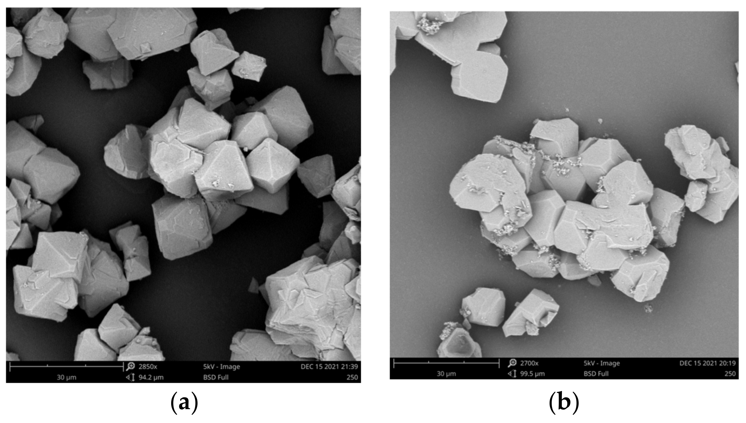
Scanning electron microscopy (SEM) was used to characterize the morphology of the samples. UiO68−N2S presents an octahedron morphology (Figure 5), which is similar to that reported for UiO−68 previously [33]. As for CdS@UiO−68N2S, the morphology did not change significantly after the CdS loading experiment. According to the XPS elemental analysis, the small particle found on the surface of the octahedron mostly came from the partial octahedron that crashed during the mechanical string in the complex preparation.
The optical absorption properties of UiO−68N2S, CdS, and CdS@UiO−68N2S samples were examined by UV-Vis spectrometer after the ultrasonic dispersion in MeOH, and the results are presented in Figure 6. It can be seen that UiO-68N2S has a significant absorption effect in the ultraviolet region of 292 nm and 394 nm [37], and CdS has a broad absorption band in the region of less than 540 nm. After loading CdS into UiO−68N2S, the composite shows a better absorption effect in the visible light region around 428 nm. Therefore, the incorporation of CdS made the absorption peak of UiO-68N2S red-shifted, which obviously broadens the light response range and improves the utilization of visible light. According to the relation Eg = 1240/λ [43], the band gap energies of CdS, UiO−68N2S, and CdS@UiO−68N2S can be calculated to be 2.3, 3.1, and 2.9 eV, respectively.
3.2. Catalytic Experiments
The photocatalytic activity of the CdS@UiO68N2S catalyst was estimated by means of the oxidation of RhB as a model substrate. The reaction conditions were conducted in the presence of air and CdS@UiO−68N2S (10 mg) as photocatalysts in a water solution of RhB (25 mg/L, 10 mL) under blue light LED irradiation at room temperature; the degradation of RhB was monitored by the absorbance change at 551 nm through UV-Vis spectrometry (Figure 7a). To our delight, the photocatalytic reactions went smoothly, affording an RhB degradation rate of 94% within 13 h (Figure 7b). To further reveal the kinetics of photocatalytic degradation, the degradation of RhB dye could be applied to a pseudo-first-order kinetic reaction (ln(co/c) = kt) due to R2 > 0.9 [44] (Figure S1), where k is the rate constant. The liner fit gave a k value of 0.1636 h−1 (or 0.0027 min−1) (Figure 7c).
To gain further insight into the photocatalytic mechanism, photo-oxidation of RhB was carried out under different conditions. The control experiments revealed that the photocatalyst was required for the RhB degradation (entries 1 and 2, Table 1). It is important to clarify the photocatalytic ability of the individual components in the CdS@UiO−68N2S. Under the same condition, UiO-68N2S and CdS showed moderate degradation of 39% and 6%, respectively (entries 3 and 4, Table 1). The result revealed a better photocatalytic ability through the incorporation of CdS into UiO−68N2S. It is also important to clarify what kind of active species are involved in photocatalytic RhB degradation. Generally, two main active intermediates, such as ·OH and O2•−, have been proposed for the photocatalytic degradation of RhB [25]. The experiment revealed that only 4% of RhB was degraded when 0.37 mmol of benzoquinone was added to the reaction solution as a scavenger of O2•−, ref. [45], indicating that significant suppression of the RhB degradation occurred (entry 5, Table 1) and that O2•− may be a key participant in the reaction. In contrast, the equal dosage of tert-butanol as a scavenger of ·OH [46] in the reaction system resulted in only slight suppression of the RhB degradation (entry 6, Table 1).
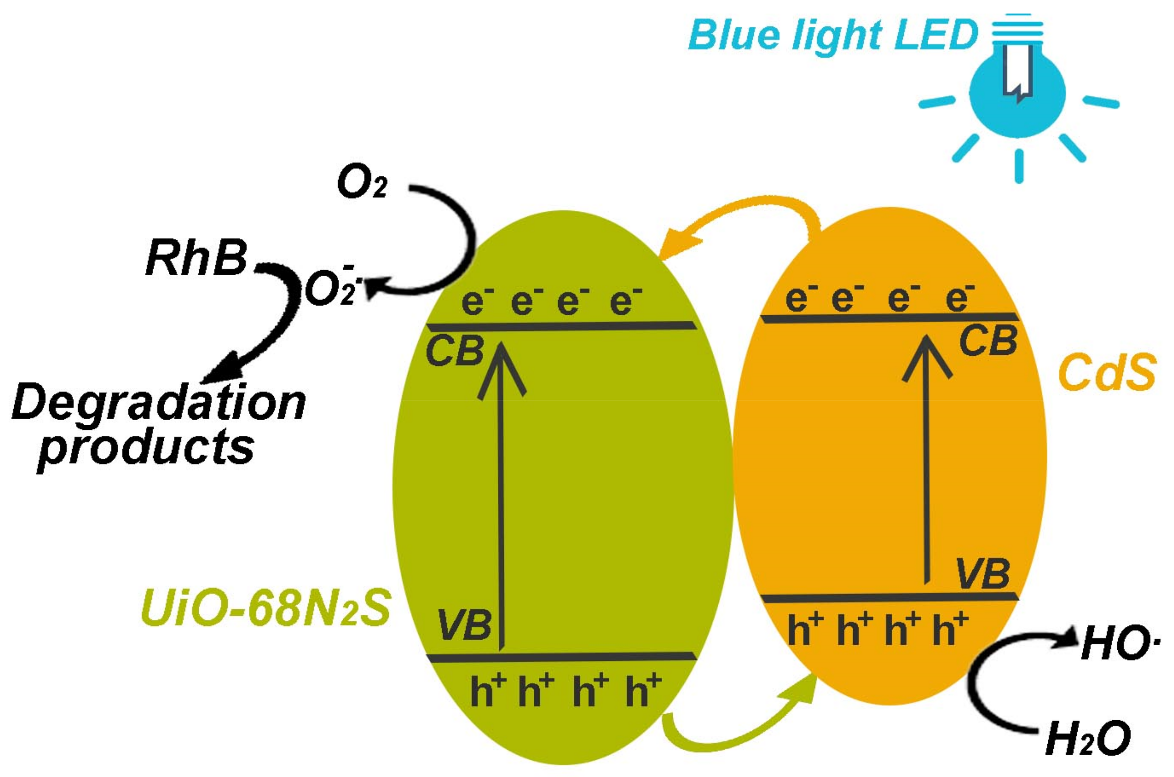
Based on the band gap analysis and the controlled experiment, a possible mechanism of charge transfer or reaction mechanism of the photocatalysis system is proposed in Figure 8. As seen first, under visible light irradiation, due to the appropriate band structures, both CdS and UiO−68N2S can be excited to generate electrons in the CB, leaving holes (h+) in the VB. The photogenerated electrons can be effectively transferred from the CB of CdS to that of UiO−68N2S, while the holes remain in the CdS particles [27]. The photogenerated electrons transferred to the surface of the composite could react with the adsorbed dissolved oxygen to produce free radicals, such as O2•−, resulting in the degradation of RhB [47]. At the same time, the remaining h+ would transfer from the VB of UiO−68N2S to that of CdS and then be combined with H2O to produce active ·OH radicals, which can directly oxidize RhB to some extent. According to the UV-Vis spectra of the RhB degradation process, a continuous reduction of the absorption peak of RhB without new peaks appeared, suggesting that the RhB decomposes into small molecules and finally CO2 and H2O [48]. The major reaction steps in RhB photocatalytic degradation are summarized by the following equations:
CdS@UiO-68N2S + hv → CdS (h+)/UiO-68N2S (e−)
e− + O2 → O2•−
h+ + H2O → ·OH + H+
O2•− (·OH) + RhB → several steps → CO2 + H2O
The performance of CdS@UiO−68N2S in photo RhB degradation is compared with the recently reported photosensitive MOFs or CdS−MOFs complex, including Ag/AgCl@CFNMT [44], 1.5−CdS/MIL−53 [48], BiOBr@Bi−MOF [49], Cu(II)−based MOF [50], and CdS/g−C3N4/MIL−125(Ti) [51]. Their activity toward RhB photocatalytic degradation is summarized in Table 2. In all these cases, the conjugation of semiconductor particles with MOFs showed enhanced catalytic performance over the single components. In addition to the photocatalyst, the process of RhB photocatalytic degradation is also highly dependent on the power of the light source and the homogeneous state of the oxygen source. In this work, CdS@UiO−68N2S shows moderate photocatalytic ability with a first-order kinetic constant of 0.0027 min−1. Given the slight difference in dosage and the same oxygen source of air, the power of the LED light would take responsibility for the photocatalytic ability of CdS@UiO−68N2S. In another aspect, compared to the widely used xenon lamp, the LED light used in our case suggests that dye photodegradation for wastewater treatment can be performed in a more convenient and economical way.
3.3. Photocatalytic Recycle Experiments
The recovery and reuse of a heterogeneous catalyst are of great importance from the perspective of wastewater treatment. The high photocatalytic RhB degradation of CdS@UiO–68N2S under visible light encouraged us to examine its recyclability. After each cycle, CdS@UiO−68N2S was recovered by centrifugation and washed with H2O three times. The ICP-MS analysis of the reaction solution showed that only negligible amounts of Zr(IV) and Cd(II) were detected, indicating the reaction was a heterogeneous process. As depicted in Figure 9a, when the recovered catalyst CdS@UiO−68N2S was subsequently used for another three successive cycles, the RhB degradation was found to be just slightly reduced to 84%, which was believed to result from the mass lost during the centrifugation. PXRD patterns of the reused catalyst showed that the structural integrity of the CdS@UiO−68N2S framework was still maintained after the first and third recycle reactions (Figure 9b), indicating that the CdS@UiO−68N2S framework was robust and stable under the reaction conditions.
4. Conclusions
In the work, the CdS@UiO−68N2S complex was successfully constructed through the incorporation of CdS into the cavities of UiO−68N2S via sequential adsorption of Cd2+ and S2−, and the conjugation of CdS and UiO−68N2S with thiadiazole groups helped in improving the visible light absorption. The photocatalytic activity of CdS@UiO−68N2S was evaluated by the degradation of dye RhB in aqueous solutions under blue light LED irradiation and ambient temperature, which revealed that up to 94% RhB degradation could be achieved under the conditions of 1.0 g/L catalyst dosage and 25 mg/L initial RhB concentration. The photocatalytic activity of CdS@UiO−68N2S was better than that of the individual components, suggesting efficient charge separation via the conjugation. Besides, the CdS@UiO−68N2S could be reused for four cycles without a significant decrease in activity, revealing its potential application as a green catalyst for environmental use.
Supplementary Materials
Supplementary data in the following supporting information can be downloaded at: https://www.mdpi.com/article/10.3390/cryst13050785/s1. Figure S1: Kinetic analysis for the RhB photo degradation with CdS@UiO68-N2S; Figures S2–S6: Photocatalytic degradation of RhB under varied conditions.
Author Contributions
Conceptualization, L.-Q.W. and H.-F.L.; methodology, J.-B.W. and F.Y.; validation, Z.-W.L.; formal analysis, J.-B.W. and F.Y.; investigation, L.-Q.W.; resources, Z.-W.L.; data curation, J.-B.W. and F.Y.; writing—original draft preparation, J.-B.W. and F.Y.; writing—review and editing, L.-Q.W. and H.-F.L.; visualization, L.-Q.W.; supervision, L.-Q.W. and H.-F.L.; project administration, L.-Q.W. and H.-F.L.; funding acquisition, Z.-W.L., L.-Q.W. and H.-F.L. All authors have read and agreed to the published version of the manuscript.
Funding
This research was supported by the Guangxi Natural Science Foundation under Grant No. 2022GXNSFAA035477, the High-level Talents Scientific Research Start-up Project of Hechi University under Grant No. 2021GCC018 and No. 2021GCC020, as well as the Natural Science Foundation of Guangdong Province under Grant No. 2022A1515010051.
Data Availability Statement
Not applicable.
Conflicts of Interest
The authors declare no conflict of interest.
References
- Forgacs, E.; Cserhati, T.; Oros, G. Removal of synthetic dyes from wastewaters: A review. Environ. Int. 2004, 30, 953–971. [Google Scholar] [CrossRef] [PubMed]
- Chen, C.; Ma, W.; Zhao, J. Semiconductor-mediated photo degradation of pollutants under visible-light irradiation. Chem. Soc. Rev. 2010, 39, 4206–4219. [Google Scholar] [CrossRef] [PubMed]
- Basavarajappa, P.S.; Patil, S.B.; Ganganagappa, N.; Reddy, K.R.; Raghu, A.V.; Reddy, C.V. Recent progress in metal-doped TiO2, non-metal doped/codoped TiO2 and TiO2 nanostructured hybrids for enhanced photocatalysis. Int. J. Hydrogen Energy 2020, 45, 7764–7778. [Google Scholar] [CrossRef]
- Ma, L.L.; Sun, H.Z.; Zhang, Y.G.; Lin, Y.L.; Li, J.L.; Wang, E.K.; Yu, Y.; Tan, M.; Wang, J.B. Preparation, characterization and photocatalytic properties of CdS nanoparticles dotted on the surface of carbon nanotubes. Nanotechnology 2008, 19, 8575–8581. [Google Scholar] [CrossRef] [PubMed]
- Liu, X.W.; Cao, H.Q.; Yin, J.F. Generation and photocatalytic activities of Bi@Bi2O3 microspheres. Nano Res. 2011, 4, 470–482. [Google Scholar] [CrossRef]
- Zhang, A.Y.; Wang, W.K.; Pei, D.N.; Yu, H.Q. Degradation of refractory pollutants under solar light irradiation by a robust and self-protected ZnO/CdS/TiO2 hybrid photo catalyst. Water Res. 2016, 92, 78–86. [Google Scholar] [CrossRef]
- Wang, C.; Cao, M.; Wang, P.; Ao, Y. Preparation, characterization of CdS-deposited graphene-carbon nanotubes hybrid photocatalysts with enhanced photocatalytic activity. Mater. Lett. 2013, 108, 336–339. [Google Scholar] [CrossRef]
- Wang, Y.J.; Zhang, Y.N.; Jiang, Z.Q.; Jiang, G.Y.; Zhao, Z.; Wu, Q.H.; Liu, Y.; Xu, Q.; Duan, A.J.; Xu, C.M. Controlled fabrication and enhanced visible-light photocatalytic hydrogen production of Au@CdS/MIL-101 heterostructure. Appl. Catal. B 2016, 185, 307–314. [Google Scholar] [CrossRef]
- Meissner, D.; Memming, R.; Kastening, B. Photoelectrochemistry of cadmium sulfide. Reanalysis of photocorrosion and flat-band potential. J. Phys. Chem. 1988, 92, 3476–3483. [Google Scholar] [CrossRef]
- Persano, L.; Camposeo, A.; Di Benedetto, F.; Stabile, R.; Laera, A.M.; Piscopiello, E.; Tapfer, L.; Pisignano, D. CdS–Polymer Nanocomposites and Light-Emitting Fibers by In Situ Electron-Beam Synthesis and Lithography. Adv. Mater. 2012, 24, 5320–5326. [Google Scholar] [CrossRef]
- Hoffman, A.; Mills, G.; Yee, H.; Hoffmann, M.R. Q-sized cadmium sulfide: Synthesis, characterization, and efficiency of photo initiation of polymerization of several vinylic monomers. J. Phys. Chem. 1992, 96, 5546–5552. [Google Scholar] [CrossRef]
- Harris, R.D.; Homan, S.B.; Kodaimati, M.; He, C.; Nepomnyashchii, A.B.; Swenson, N.K.; Lian, S.; Calzada, R.; Weiss, E.A. Electronic Processes within Quantum Dot-Molecule Complexes. Chem. Rev. 2016, 116, 12865–12919. [Google Scholar] [CrossRef] [PubMed]
- Majeed, I.; Nadeem, M.A.; Al-Oufi, M.; Nadeem, M.A.; Waterhouse, G.I.N.; Badshah, A.; Metson, J.B.; Idriss, H. On the role of metal particle size and surface coverage for photo-catalytic hydrogen production: A case study of the Au/CdS system. Appl. Catal. B 2016, 182, 266–276. [Google Scholar] [CrossRef]
- Feng, J.; An, C.; Dai, L.; Liu, J.; Wei, G.; Bai, S.; Zhang, J.; Xiong, Y. Long-term production of H2 over Pt/CdS nanoplates under sunlight illumination. Chem. Eng. J. 2016, 283, 351–357. [Google Scholar] [CrossRef]
- Zheng, N.-C.; Ouyang, T.; Chen, Y.; Wang, Z.; Chen, D.-Y.; Liu, Z.-Q. Ultrathin CdS shell-sensitized hollow S-doped CeO2 spheres for efficient visible-light photocatalysis. Catal. Sci. Technol. 2019, 9, 1357–1364. [Google Scholar] [CrossRef]
- Zhang, P.; Liu, Y.; Tian, B.; Luo, Y.; Zhang, J. Synthesis of core-shell structured CdS@CeO2 and CdS@TiO2 composites and comparison of their photocatalytic activities for the selective oxidation of benzyl alcohol to benzaldehyde. Catal. Today 2017, 281, 181–188. [Google Scholar] [CrossRef]
- Fang, S.S.; Sun, M.Y.; Zhou, Y.W.; Liang, Q.; Li, Z.Y.; Xu, S. Solvothermal synthesis of CdS QDs/MWCNTs nano composites with high efficient photo catalytic activity under visible light irradiation. J. Alloys Compd. 2016, 656, 771–776. [Google Scholar] [CrossRef]
- Zhou, H.C.; Long, J.R.; Yaghi, O.M. Introduction to metal-organic frameworks. Chem. Rev. 2012, 112, 673–674. [Google Scholar] [CrossRef]
- Cai, G.; Yan, P.; Zhang, L.; Zhou, H.-C.; Jiang, H.-L. Metal–Organic Framework-Based Hierarchically Porous Materials: Synthesis and Applications. Chem. Rev. 2021, 121, 12278–12326. [Google Scholar] [CrossRef]
- Liang, W.; Wied, P.; Carraro, F.; Sumby, C.J.; Nidetzky, B.; Tsung, C.-K.; Falcaro, P.; Doonan, C.J. Metal–Organic Framework-Based Enzyme Biocomposites. Chem. Rev. 2021, 121, 1077–1129. [Google Scholar] [CrossRef]
- Zhang, W.-X.; Liao, P.-Q.; Lin, R.-B.; Wei, Y.-S.; Zeng, M.-H.; Chen, X.-M. Metal Cluster-based Functional Porous Coordination Polymers. Coord. Chem. Rev. 2015, 263, 293–294. [Google Scholar] [CrossRef]
- Yin, Z.; Zhou, Y.-L.; Zeng, M.-H.; Kurmoo, M. The Concept of Mixed Organic Ligands in Metal-Organic Frameworks: Design, Tuning and Functions. Dalton Trans. 2015, 44, 5258–5275. [Google Scholar] [CrossRef] [PubMed]
- Peng, H.-J.; Zhu, L.; Wang, Y.-L.; Chao, H.-Y.; Jiang, L.; Qiao, Z.-P. CdS/ZIF-67 nanocomposites with enhanced performance for visible light CO2 photo reduction. Inorg. Chem. Comm. 2020, 117, 107943. [Google Scholar] [CrossRef]
- Shen, L.; Liang, S.; Wu, W.; Liang, R.; Wu, L. CdS-decorated UiO-66(NH2) nano composites fabricated by a facile photo deposition process: An efficient and stable visible-light-driven photocatalyst for selective oxidation of alcohols. J. Mater. Chem. A 2013, 1, 11473–11482. [Google Scholar] [CrossRef]
- Chaturvedi, G.; Kaur, A.; Kansal, S.K. CdS-Decorated MIL-53(Fe) Microrods with Enhanced Visible Light Photocatalytic Performance for the Degradation of Ketorolac Tromethamine and Mechanism Insight. J. Phys. Chem. C 2019, 123, 16857–16867. [Google Scholar] [CrossRef]
- Zhang, R.; Li, G.; Zhang, Y. Photochemical synthesis of CdS-MIL-125(Ti) with enhanced visible light photocatalytic performance for the selective oxidation of benzyl alcohol to benzaldehyde. Photochem. Photobiol. Sci. 2017, 16, 996. [Google Scholar] [CrossRef]
- Liang, R.; Jing, F.; Yan, G.; Wu, L. Synthesis of CdS-decorated MIL-68(Fe) nanocomposites: Efficient andstable visible light photocatalysts for the selective reduction of 4-nitroaniline to p-phenylenediamine in water. Appl. Catal. B-Environ. 2017, 218, 452–459. [Google Scholar] [CrossRef]
- Jing, C.; Zhang, Y.; Zheng, J.; Ge, S.; Lin, J.; Pan, D.; Naik, N.; Guo, Z. In-situ constructing visible light CdS/Cd-MOF photo catalyst with enhanced photo degradation of methylene blue. Particuology 2022, 69, 111–122. [Google Scholar] [CrossRef]
- Li, S.-R.; Ren, F.-D.; Wang, L.; Chen, Y.-Z. Photocatalytic cascade reactions and dye degradation over CdS–metal–organic framework hybrids. RSC Adv. 2021, 11, 35326. [Google Scholar] [CrossRef] [PubMed]
- Yang, Q.; Xu, Q.; Jiang, H.-L. Metal–organic frameworks meet metal nanoparticles: Synergistic effect for enhanced catalysis. Chem. Soc. Rev. 2017, 46, 4774–4808. [Google Scholar] [CrossRef]
- Dutta, S.; Lee, I.S. Metal-organic framework based catalytic nanoreactors: Synthetic challenges and applications. Mater. Chem. Front. 2021, 5, 3986–4021. [Google Scholar] [CrossRef]
- Bai, Y.; Dou, Y.; Xie, L.-H.; Rutledge, W.; Li, J.-R.; Zhou, H.-C. Zr-based metal-organic frameworks: Design, synthesis, structure, and applications. Chem. Soc. Rev. 2016, 45, 2327–2367. [Google Scholar] [CrossRef] [PubMed]
- Cavka, J.H.; Jakobsen, S.; Olsbye, U.; Guillou, N.; Lamberti, C.; Bordiga, S.; Lillerud, K.P. A New Zirconium Inorganic Building Brick Forming Metal Organic Frameworks with Exceptional Stability. J. Am. Chem. Soc. 2008, 130, 13850–13851. [Google Scholar] [CrossRef] [PubMed]
- Goswami, S.; Miller, C.E.; Logsdon, J.L.; Buru, C.T.; Wu, Y.-L.; Bowman, D.N.; Islamoglu, T.; Asiri, A.M.; Cramer, C.J.; Wasielewski, M.R.; et al. Atomistic Approach toward Selective Photocatalytic Oxidation of a Mustard-Gas Simulant: A Case Study with Heavy-Chalcogen-Containing PCN-57 Analogues. ACS Appl. Mater. Interfaces 2017, 9, 19535–19540. [Google Scholar] [CrossRef] [PubMed]
- Jia, J.; Gutiérrez-Arzaluz, L.; Shekhah, O.; Alsadun, N.; Czaban-Jóźwiak, J.; Zhou, S.; Bakr, O.M.; Mohammed, O.F.; Eddaoudi, M. Access to Highly Efficient Energy Transfer in Metal–Organic Frameworks via Mixed Linkers Approach. J. Am. Chem. Soc. 2020, 142, 8580–8584. [Google Scholar] [CrossRef] [PubMed]
- Wu, S.; Ren, D.; Zhou, K.; Xia, H.-L.; Liu, X.-Y.; Wang, X.; Li, J. Linker Engineering toward Full-Color Emission of UiO-68 Type Metal–Organic Frameworks. J. Am. Chem. Soc. 2021, 143, 10547–10552. [Google Scholar] [CrossRef] [PubMed]
- Mallick, A.; El-Zohry, A.M.; Shekhah, O.; Yin, J.; Jia, J.; Aggarwal, H.; Emwas, A.-H.; Mohammed, O.F.; Eddaoudi, M. Unprecedented Ultralow Detection Limit of Amines using a Thiadiazole-Functionalized Zr(IV)-Based Metal–Organic Framework. J. Am. Chem. Soc. 2019, 141, 7245–7249. [Google Scholar] [CrossRef]
- Mercuri, G.; Giambastiani, G.; Rossin, A. Thiazole- and Thiadiazole-Based Metal–Organic Frameworks and Coordination Polymers for Luminescent Applications. Inorganics 2019, 7, 144. [Google Scholar] [CrossRef]
- Wei, N.; Zhang, Y.-R.; Han, Z.-B. Thiadiazole-functional porous metal–organic framework as luminescent probe for Cd2+. CrystEngComm 2013, 15, 8883–8886. [Google Scholar] [CrossRef]
- Ge, L.; Zuo, F.; Liu, J.K.; Ma, Q.; Wang, C.; Sun, D.Z.; Bartels, L.; Feng, P.Y. Synthesis and efficient visible light photocatalytic hydrogen evolution of polymeric g-C3N4 coupled with CdS quantum dots. J. Phys. Chem. C 2012, 116, 13708–13714. [Google Scholar] [CrossRef]
- Wei, L.-Q.; Ye, B.-H. Cyclometalated Ir–Zr Metal–Organic Frameworks as Recyclable Visible-Light Photocatalysts for Sulfide Oxidation into Sulfoxide in Water. ACS Appl. Mater. Interfaces 2019, 11, 41448–41457. [Google Scholar] [CrossRef] [PubMed]
- Low, J.; Dai, B.; Tong, T.; Jiang, C.; Yu, J. In Situ Irradiated X-Ray Photoelectron Spectroscopy Investigation on a Direct Z-Scheme TiO2/CdS Composite Film Photocatalyst. Adv. Mater. 2018, 31, 1802981. [Google Scholar] [CrossRef] [PubMed]
- Long, J.; Wang, S.; Ding, Z.; Wang, S.; Zhou, Y.; Huang, L.; Wang, X. Amine-functionalized zirconium metal-organic framework as efficient visible-light photo catalyst for aerobic organic transformations. Chem. Commun. 2012, 48, 11656–11658. [Google Scholar] [CrossRef] [PubMed]
- Mahmoodi, N.M.; Taghizadeh, A.; Taghizadeh, M.; Abdi, J. In situ deposition of Ag/AgCl on the surface of magnetic metal-organic framework nanocomposite and its application for the visible-light photocatalytic degradation of Rhodamine dye. J. Hazard. Mate. 2019, 378, 120741. [Google Scholar] [CrossRef] [PubMed]
- Bonesi, S.M.; Manet, I.; Freccero, M.; Fagnoni, M.; Albini, A. Photosensitized Oxidation of Sulfides: Discriminating between the Singlet-Oxygen Mechanism and Electron Transfer Involving Superoxide Anion or Molecular Oxygen. Chem.-Eur. J. 2006, 12, 4844–4857. [Google Scholar] [CrossRef]
- Xia, Q.S.; Yu, X.D.; Zhao, H.M.; Wang, S.P.; Wang, H.; Guo, Z.F.; Xing, H.Z. Syntheses of Novel Lanthanide Metal–Organic Frameworks for Highly Efficient Visible-Light-Driven Dye Degradation. J. Am. Chem. Soc. 2017, 17, 4189–4195. [Google Scholar] [CrossRef]
- Xu, J.; Hu, C.; Xi, Y.; Wan, B.; Zhang, C.; Zhang, Y. Synthesis and visible light photocatalytic activity of β-AgVO3 nanowires. Solid State Sci. 2012, 14, 535–539. [Google Scholar] [CrossRef]
- Hu, L.; Deng, G.; Lu, W.; Pang, S.; Hu, X. Deposition of CdS nanoparticles on MIL-53(Fe) metal-organic framework with enhanced photocatalytic degradation of RhB under visible light irradiation. Appl. Surf. Sci. 2017, 410, 401–413. [Google Scholar] [CrossRef]
- Xu, M.-L.; Jiang, X.-J.; Li, J.-R.; Wang, F.-J.; Li, K.; Cheng, X. Self-Assembly of a 3D Hollow BiOBr@Bi-MOF Heterostructure with Enhanced Photocatalytic Degradation of Dyes. ACS Appl. Mater. Interfaces 2021, 13, 56171–56180. [Google Scholar] [CrossRef]
- Jin, J.-C.; Wang, J.; Guo, J.; Yan, M.-H.; Wang, J.; Srivastava, D.; Kumar, A.; Sakiyama, H.; Muddassir, M.; Pan, Y. A 3D rare cubane-like tetramer Cu(II)-based MOF with 4-fold dia topology as an efficient photocatalyst for dye degradation. Colloids Surf. A Physicochem. Eng. Asp. 2023, 656, 130475. [Google Scholar] [CrossRef]
- Chen, Y.; Zhai, B.; Liang, Y.; Li, Y.; Li, J. Preparation of CdS/ g-C3N4/ MOF composite with enhanced visible-light photocatalytic activity for dye degradation. J. Solid State Chem. 2019, 274, 32–39. [Google Scholar] [CrossRef]
Figure 1.
Construction and the pore structure of UiO-68N2S, as well as the strategy of loading CdS particles.
Figure 1.
Construction and the pore structure of UiO-68N2S, as well as the strategy of loading CdS particles.

Figure 2.
PXRD patterns (a) and IR spectra (b) for CdS@UiO-68N2S and CdS.

Figure 3.
XPS spectra of elements in CdS@UiO-68N2S: Zr (a), S (b), and Cd (c).

Figure 4.
Thermogravimetric analysis of UiO-68N2S and CdS@UiO-68N2S.

Figure 5.
SEM images of (a) UiO-68N2S and CdS@UiO-68N2S (b).

Figure 6.
UV-Vis spectra of UiO-68N2S, CdS, and CdS@UiO-68N2S.

Figure 7.
Time−dependent UV−Vis spectra of RhB phtotodegradation via CdS@UiO−68N2S (a), the dynamic process of the RhB concentration change (b), and the photodegradation kinetics (c).
Figure 7.
Time−dependent UV−Vis spectra of RhB phtotodegradation via CdS@UiO−68N2S (a), the dynamic process of the RhB concentration change (b), and the photodegradation kinetics (c).

Figure 8.
Proposed mechanism of RhB photocatalytic degradation in the presence of CdS@UiO-68N2S and blue-light LED irradiation.
Figure 8.
Proposed mechanism of RhB photocatalytic degradation in the presence of CdS@UiO-68N2S and blue-light LED irradiation.

Figure 9.
Recycling experiments for photo-degradation of RhB by CdS@UiO-68N2S (a), PXRD patterns for the pristine and recovered samples of CdS@UiO-68N2S after the first and third recycle uses (b).
Figure 9.
Recycling experiments for photo-degradation of RhB by CdS@UiO-68N2S (a), PXRD patterns for the pristine and recovered samples of CdS@UiO-68N2S after the first and third recycle uses (b).


Table 1.
Photocatalytic degradation of RhB under different conditions a.
| Entry | Catalyst | Additive | Light | Degradation |
|---|---|---|---|---|
| 1 | CdS@UiO-68N2S | N.D. | Blue light | 94% |
| 2 | N.D. | N.D. | Blue light | 1% |
| 3 | UiO-68N2S | N.D. | Blue light | 39% |
| 4 | CdS b | N.D. | Blue light | 6% |
| 5 | CdS@UiO-68N2S | Benoquinone c | Blue light | 4% |
| 6 | CdS@UiO-68N2S | tert-butanol d | Blue light | 86% |
a Condition: RhB solution (25 mg/L, 10 mL), UiO-68N2S (0.01 g, 0.011 mmol), CdS@UiO-68N2S (0.01 g, 0.0027 mmol), light (blue LED light: wavelength = 480 nm, power = 80 W), R.T. (30 °C), reaction time: 13 h. b CdS (0.01 g). c Benoquinone (0.04 g, 0.37 mmol). d tert-butanol (0.37 mmol) (Figures S2–S6 show the original UV-Vis spectra for entries 2–6, respectively).
Table 2.
Comparison of RhB photodegradation performance of MOF-based catalysts.
| Catalyst | Catalyst Dosage | RhB Concentration | Light Source | Oxygen Source | Catalytic Activity | k a/min−1 |
|---|---|---|---|---|---|---|
| Ag/AgCl@CFNMT b [44] | 0.08 g/L | 20 mg/L | Visible–light LED (100 W) | H2O2 (20 uL) | 98.8% (10 min) | 0.103 |
| 1.5-CdS/MIL53 [48] | 1.0 g/L | 10 mg/L | Xenon lamp (500 W) | Air | 86% (1.5 h) | 0.0158 |
| BiOBr@Bi-MOF [49] | 0.6 g/L | 20 mg/L | Xenon lamp (300 W) | Air | 99.4% (60 min) | 0.07009 |
| Cu(II)-based MOF [50] | 30 mg Cat. for 40 ppm RhB | UV light | Air | 80% (100 min) | 0.01358 | |
| CdS/g-C3N4/MIL125(Ti) [51] | N.D. | N.D. | Xenon lamp (300 W, λ > 420 nm) | Air | 90.2% (90 min) | 0.0414 |
| CdS@UiO-68N2S [this work] | 1.0 g/L | 25 mg/L | Blue light LED (80 W, λ = 480 nm) | Air | 94% (14 h) | 0.0027 |
a First-order kinetic constant, b CFNMT = CoFe2O4/NH2-MIL-125(Ti).
Disclaimer/Publisher’s Note: The statements, opinions and data contained in all publications are solely those of the individual author(s) and contributor(s) and not of MDPI and/or the editor(s). MDPI and/or the editor(s) disclaim responsibility for any injury to people or property resulting from any ideas, methods, instructions or products referred to in the content. |
© 2023 by the authors. Licensee MDPI, Basel, Switzerland. This article is an open access article distributed under the terms and conditions of the Creative Commons Attribution (CC BY) license (https://creativecommons.org/licenses/by/4.0/).
Share and Cite
MDPI and ACS Style
Wei, L.-Q.; Wei, J.-B.; Yang, F.; Li, Z.-W.; Lai, H.-F. Preparation of Thiadiazole Modified UiO-68-CdS Composites for RhB Degradation under Visible Light Irradiation. Crystals 2023, 13, 785. https://doi.org/10.3390/cryst13050785
AMA Style
Wei L-Q, Wei J-B, Yang F, Li Z-W, Lai H-F. Preparation of Thiadiazole Modified UiO-68-CdS Composites for RhB Degradation under Visible Light Irradiation. Crystals. 2023; 13(5):785. https://doi.org/10.3390/cryst13050785
Chicago/Turabian StyleWei, Lian-Qiang, Jiu-Bin Wei, Fei Yang, Zhi-Wei Li, and Hong-Fang Lai. 2023. "Preparation of Thiadiazole Modified UiO-68-CdS Composites for RhB Degradation under Visible Light Irradiation" Crystals 13, no. 5: 785. https://doi.org/10.3390/cryst13050785
Note that from the first issue of 2016, this journal uses article numbers instead of page numbers. See further details here.