
Analysis and Modification of a Colorimetric Nanosensor for Rapid Detection of Escherichia coli in Water

1
Civil and Environmental Engineering Department, California Polytechnic State University, San Luis Obispo, CA 93407, USA
2
Biological Sciences Department, California Polytechnic State University, San Luis Obispo, CA 93407, USA
3
Materials Engineering Department, California Polytechnic State University, San Luis Obispo, CA 93407, USA
*
Author to whom correspondence should be addressed.
Crystals 2024, 14(4), 386; https://doi.org/10.3390/cryst14040386
Submission received: 6 April 2024
/
Revised: 17 April 2024
/
Accepted: 17 April 2024
/
Published: 21 April 2024
(This article belongs to the Special Issue Micro and Nano Optics for Advanced Sensing Technology)
Abstract
:This research analyzed the mechanisms of work and modified a colorimetric nanosensor to make it more cost-effective for the detection of Escherichia coli (E. coli) in water. The base nanosensors modified herein rely on a competitive binding detection mechanism, where positively charged gold nanoparticles coated with polyethyleneimine (PEI-AuNPs) preferably bind to negatively charged E. coli in the presence of β-galactosidase (-Gal) enzymes and chlorophenol red β-d-galactopyranosides (CPRG). The positive surface charge of the nanoparticle, rather than nanoparticle composition or type of chemical coating on its surface, was hypothesized herein as the governing factor for the nanosensor functionality. Thus, positively charged nanoparticles and polymers were tested as potential alternatives for gold nanoparticles for detecting E. coli. Positively charged silver and iron oxide nanoparticles coated with branched PEI detected E. coli as low as 105 and 107 colony-forming units per milliliter (CFU/mL), respectively. Furthermore, the branched PEI polymer itself (without nanomaterial) detected E. coli at 107 CFU/mL. These findings suggest that the positive charge, rather than the nanoparticle type was likely responsible for the detection of E. coli using the competitive binding approach. Therefore, other types of recyclable and cost-effective nanomaterials and polymers can be developed for E. coli detection using this rapid colorimetric sensing technique.
1. Introduction
It has been reported that 144 million people consume untreated surface water from lakes, ponds, rivers, and streams [1]. Contaminated drinking water can contain enteric pathogens that cause deadly diseases [2]. Currently, no single method exists to detect all microorganisms in a water sample due to factors such as the physical differences between the major pathogen groups, the presence of inhibitors in the sample, and the determination of the pathogen’s origin [3]. The commonly used methods to detect waterborne microorganisms can be categorized as culture-dependent, such as membrane filtration and multiple tube fermentation, and culture-independent or molecular methods, such as polymerase chain reaction (PCR) [4]. Some of these methods require skilled expertise to perform and they can be time-consuming (e.g., days of wait time), especially for culture-dependent methods [5,6]. In addition, electricity and laboratory equipment, such as incubators or filter pumps, are often needed to perform these methods. Such factors limit the accessibility of these commonly used detection methods to underserved communities, especially in developing countries.
Nanomaterials-based techniques for detecting waterborne microbes have been a recent development in microbial detection and have included electrochemical, acoustic, magnetic, and optical biosensors [7,8,9,10,11,12,13]. Of these detection techniques, optical assays have several advantages including their relative ease of implementation, and lower cost, and some are equipment-free methods, making these sensors accessible to communities with limited resources [14]. Colorimetric biosensors are a class of optical assays that can be further categorized into direct and indirect assays [15,16,17]. A direct assay tracks, for example, the color change that occurs because of the increase in the size of the nanoparticle when microbes are present [14]. On the other hand, indirect assays track, for example, enzyme-catalyzed color-producing reactions based on nanomaterial–bacteria and nanomaterial–enzyme interactions [18,19].
Previous research by Thiramanas and Laocharoensuk [18] has investigated indirect colorimetric detection assays for the detection of Escherichia coli (E. coli) based on a competitive binding technique. This technique relies on positively charged polyethyleneimine-coated gold nanoparticles (PEI-AuNPs) preferably binding to negatively charged bacteria, leaving behind a negatively charged enzyme that reacts with a substrate and changes color. The components of this nanosensor-based assay are schematically depicted in Figure 1a. In the absence of microbes and nanoparticles, the hydrolysis reaction between the chromogenic substrate chlorophenol red -d-galactopyranoside (CPRG) and negatively charged enzyme -galactosidase (-Gal) produces a red color in the solution due to the production of chlorophenol red (CPR) (Figure 1b). When positively charged PEI-AuNPs are added to the CPRG/-Gal mixture, the enzymatic activity of -Gal is inhibited, and the solution remains yellow because the negatively charged -Gal binds to the positively charged PEI-AuNPs (Figure 1c). When PEI-AuNPs, -Gal, CPRG, and bacteria are all present in the solution, the PEI-AuNPs preferentially bind to the bacteria over the -Gal due to the higher negative surface charge of E. coli compared to that of the -Gal. This ultimately leaves -Gal free in the solution (i.e., unbound by the nanoparticles) to react with CPRG and as a result, the solution turns red (Figure 1d). The magnitude of color change is dependent on the quantity of bacteria in the solution [18]. Therefore, for a given concentration of PEI-AuNPs, lower concentrations of bacteria will result in higher quantities of free nanoparticles in the solution that can interact with -Gal and reduce its interactions with CPRG. This results in a more yellow-colored solution. On the contrary, higher concentrations of bacteria bind more nanoparticles, which leads to more interactions between -Gal and CPRG. This results in a solution with dark red color at the highest concentrations of E. coli.
The competitive binding colorimetric detection technique described above has the advantages of easily observable results and a rapid detection of microbes; however, further optimization is needed to reduce its cost, enhance its recyclability potential, and reduce its detection limit. To overcome some of these limitations, it is hypothesized herein that the surface charge of the nanoparticle, not its composition, is the governing factor in the competitive binding interactions between the nanoparticles, bacteria, and -Gal. For example, replacing gold nanoparticles with silver or magnetic iron oxide nanoparticles should not affect the efficiency of the biosensor as long as the surface charge of the substitution nanoparticle type is positively charged. If this hypothesis is true, then gold nanoparticles can be replaced with other types of nanoparticles that are more cost-effective and/or recyclable. The other hypothesis tested herein was that PEI could also be replaced with other positively charged polymer coatings, assuming that the surface charge produced by it, rather than the polymer itself, is responsible for the competitive interactions previously described. To test the hypotheses of this research, the sensing agents outlined in Table 1 were investigated in this study. The main objective of this research was to understand the governing factors in the gold nanosensor functionality in order to create opportunities for the development of more optimized sensing agents that are based on the competitive binding colorimetric technique.
2. Materials and Methods
2.1. E. coli Culturing
Nonpathogenic E. coli was provided by the Microbiology Department at California Polytechnic State University, San Luis Obispo and was utilized in all E. coli detection assays. For E. coli culturing, a stock solution containing the E. coli was streaked, according to the streak plate method, on a plate of tryptic soy agar (TSA). TSA is a nutrient-rich complex medium that provides essential nutrients required for the growth of a wide range of bacteria, including E. coli. Morphologically, isolated colonies of E. coli grown on TSA appeared off-white in color, exhibited a smooth texture, and possessed entire margins. These colonies typically manifested as slightly raised, circular structures with diameters ranging between 3–5 mm. This process was performed to isolate a single colony of bacteria compared to a line of growth where the inoculating loop was streaked. Approximately two colonies were streaked on a new TSA plate every two days to keep the E. coli strain growing throughout the year.
To obtain an E. coli suspension that can be easily pipetted into a well plate, colonies from the TSA plate were transferred to a tryptic soy broth (TSB) growth media. Approximately two colonies were chosen from the last quadrant on the TSA streak plate to obtain the E. coli needed for performing any of the detection assays. These two colonies were transferred using an inoculation loop to a 15 milliliter (mL) Falcon tube containing 5 mL of TSB. After the inoculation loop was vigorously swished around in the tube to transfer the colonies from the loop to the TSB media, the tube was sealed using a lid with tape wrapped over it. The tube was then placed in a shaking incubator at 30 degrees Celsius (°C) for 24 h.
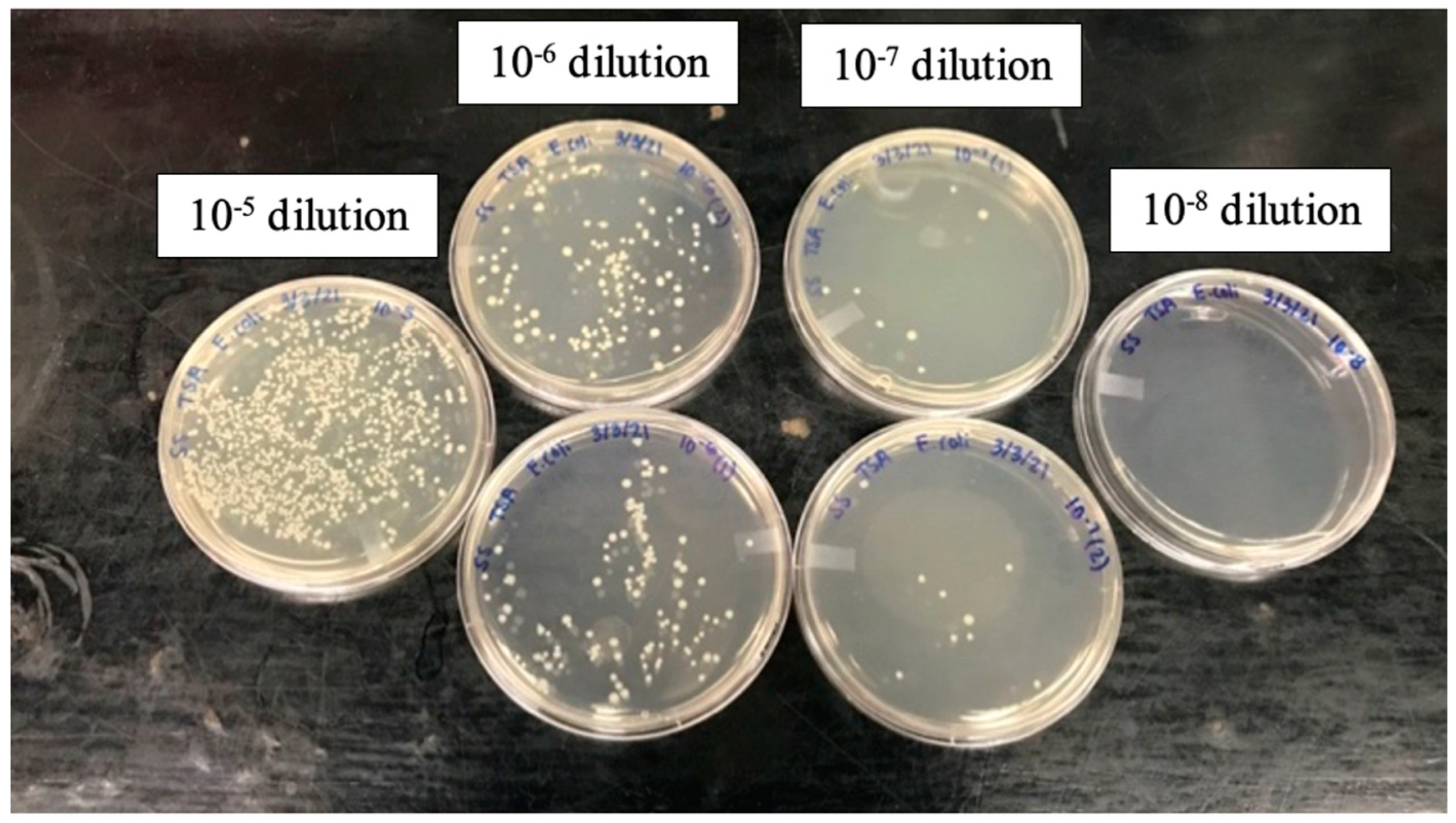
Serial dilutions were performed to determine the concentration of E. coli in colony-forming units per milliliter (CFU/mL) in the stock suspension. The stock suspension was initially diluted 10 times using 9 parts phosphate buffer solution (PBS) to 1 part of the stock suspension to obtain a 10−1 dilution. Serial dilutions were then performed to the 10−8 dilution. A volume of 0.1 mL from the 10−5, 10−6, 10−7, and 10−8 dilutions was pipetted onto individual TSA plates. These dilutions were chosen because growth of 30 to 200 colonies on the plate is required to perform the E. coli concentration calculations. Once pipetted onto the TSA plate, a plate spreader was used to create an even lawn on the agar. These plates were then incubated for 24 h at 37 °C. Plates that grew between 30 and 200 colonies were then used to calculate a concentration in CFU/mL using Equation (1). This method was performed three times on separate days with different stock suspensions of E. coli to ensure precision.
where C is the concentration of stock solution (CFU/mL), CFU is the number of colonies counted on the plate, DF is the dilution factor (10Number of times diluted (positive)), and V is the volume of solution plated.
2.2. E. coli Sensing Agents
Three different suspensions of commercial nanoparticles were investigated in this study for the detection of E. coli (Table 2). These nanoparticles were (1) branched polyethyleneimine-coated silver nanoparticles (BPEI-AgNPs) purchased from nanoComposix, (2) amine functionalized iron oxide nanoparticle suspension (amine-Fe3O4) purchased from Sigma Aldrich, and (3) cerium (Ce3/4+)-doped iron oxide-coated nanoparticles coated with BPEI (BPEI-Fe2O3), purchased from Sigma Aldrich. These nanomaterials were chosen for this investigation to examine the research hypotheses and elucidate the sensor’s operational mechanism, specifically regarding E. coli detection. The focus lies on determining whether the nanoparticle type, surface charge, or the chemical nature of the coating/surface functionalization agent primarily governs this detection process. The nanomaterials tested were all positively charged because of the amine groups on their surfaces. Furthermore, alongside the functionalized nanomaterials, two cationic chemical agents—a polyelectrolyte BPEI with a molecular weight of 1200 g/mol and a cationic surfactant (CTAB) with a molecular weight of 364.456 g/mol—were evaluated as potential sensing agents for E. coli. Both CTAB and BPEI were without a nanoparticle carrier and were procured from Fischer Scientific. BPEI is a cationic organic polymer that is positively charged due to the protonation of the amine groups that the polymer contains [20].
2.3. The Experimental Testing Program
Experiments were conducted in triplicate to systematically determine the optimal concentrations and quantities of the sensing agents required. Additionally, the sequence of addition of these agents in the testing wells was investigated to achieve the specified E. coli detection goals. The phases of the experimental testing program are outlined in Figure 2. The initial set of experiments did not involve E. coli or sensing agents; instead, it focused solely on optimizing the colorimetric reaction between β-Gal and CPRG. This reaction leads to the hydrolysis of CPRG by β-Gal, which is responsible for the color change in the solution to red. Subsequent activity assays were conducted without microbes, concentrating on identifying the type, quantity, and concentration of sensing agent capable of inhibiting β-Gal activity while maintaining the solution color unchanged (i.e., it remains yellow). The activity assays were also conducted to determine whether the surface charge or the sensing agent itself is responsible for the interactions with -Gal. Then, E. coli detection assays were performed using the optimal mixtures of sensing agents determined based on the results of the activity assays.
The mass of sensing agent required for detection of E. coli was determined based on adding enough mass to bind -Gal in the sample, which results in a yellow color solution at the lowest detectable E. coli concentration. As the concentration of E. coli increases, the sensing agent favors binding E. coli than -Gal because of the stronger Coulombic interactions between the positively charged sensing agent and the negatively charged E. coli.
The E. coli detection assay used in the current investigation was conducted according to the method by Thiramanas and Laocharoensuk [18] with modifications related to the volumes and concentrations of sensing reagents used as well as the sequence or the addition of the well plate components. Control samples were included with each well plate. Negative control samples consisted only of -Gal and CPRG, with microbes and sensing agents substituted by phosphate buffer solution. These negative control samples were used to confirm that the -Gal and CPRG were active and interacting properly. In the samples containing a sensing agent, -Gal and CPRG only served as positive controls.
2.3.1. Optimization of the Colorimetric Assay
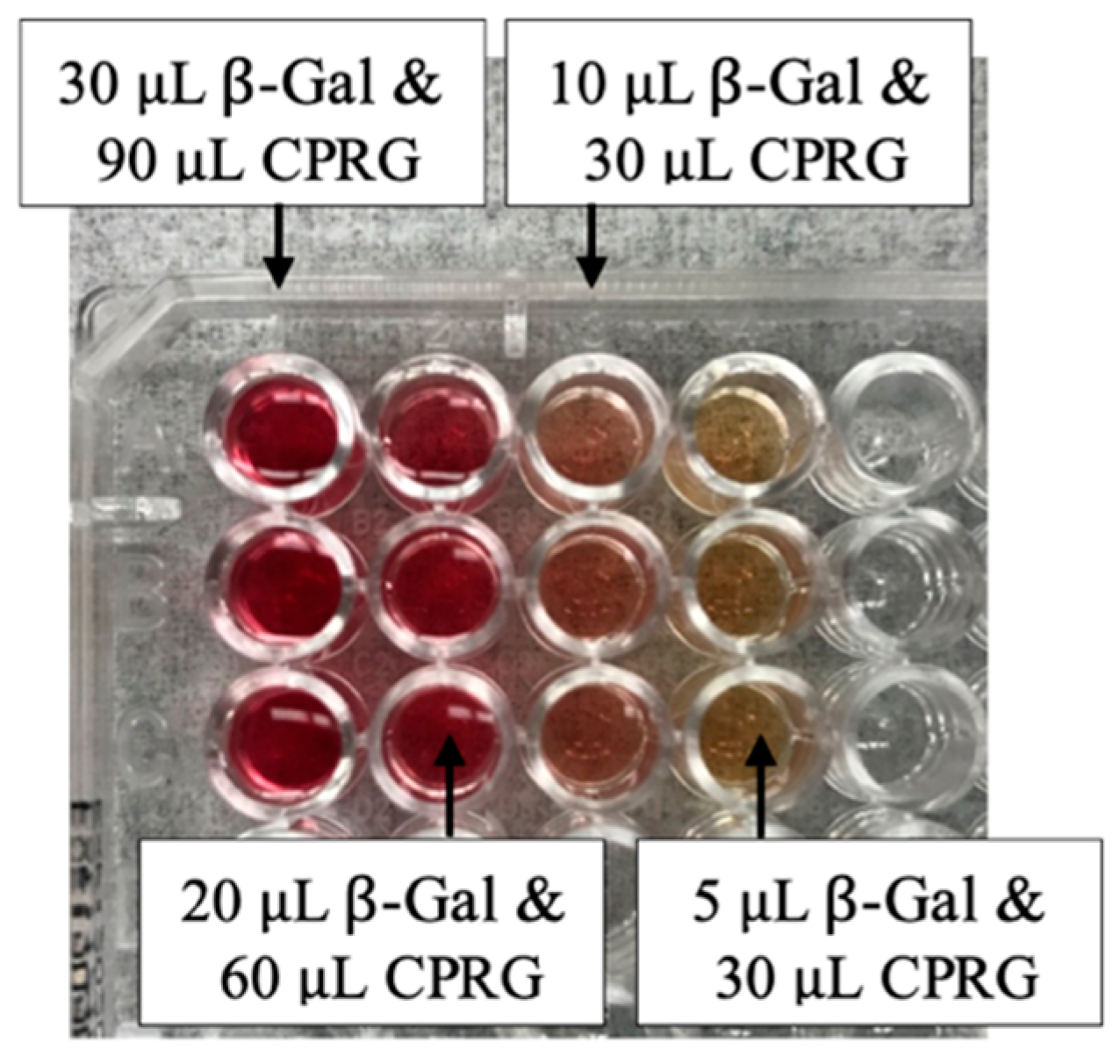
This experimental phase was conducted to determine the optimal volume and molar concentration of -Gal and CPRG to allow hydrolysis of CPRG to occur. This hydrolysis reaction results in a change in solution color to dark red. The concentrations tested were 0.5 nanomolar (nM) and 0.125 micromolar M) for -Gal and 1.5 millimolar (mM) and 0.75 mM for CPRG. These concentrations were selected based on previous research studies on colorimetric detection of E. coli [18,19]. The volumes tested for -Gal ranged from 0.5 to 180 microliter (L), while the volumes of CPRG ranged from 5 to 250 L. In these colorimetric assay optimization experiments, sensing agents and E. coli were not included. Therefore, the volume of the sensing agent solution and E. coli suspension were replaced with the same volume of phosphate buffer (PBS). The optimal volume and molar concentrations of the -Gal and CPRG were determined to be 30 L of 0.125 M -Gal and 90 L of 0.75 mM CPRG. At these optimal ratios, the color of the mixtures changed from yellow to red over a three-hour period.
2.3.2. Activity Assays
The activity assays were developed based on determining the sensing agent quantity required to bind the -Gal quantity of 30 L of 0.125 M. The experimental conditions used in these assays included the use of the optimal volumes and concentrations of -Gal (30 L of 0.125 M) and CPRG (90 L of 0.75 mM), with 100 L of PBS to represent the E. coli volume, and 30 L of the various concentrations of sensing agents listed in Table 3. The goal of this set of experiments was to determine the sensing agents that could successfully achieve the desired inhibition of the hydrolysis of CPRG by -Gal for use in the subsequent E. coli detection experiments.
2.3.3. E. coli Detection Assays
The E. coli detection assays employed the most promising sensing agents—those identified from the previously described activity assays as capable of inhibiting the hydrolysis of CPRG by β-Gal—to establish the E. coli detection limit. The optimal volumes and concentrations of -Gal (30 L of 0.125 M) and CPRG (90 L of 0.75 mM) were used in this set of experiments. The volume of E. coli suspensions in PBS buffer remained constant at 100 L in these experiments. Various concentrations of 30 L of the sensing agent were tested across a range of microbial concentrations to determine their E. coli detection limit. The experiments that were performed in these E. coli detection assays are outlined in Table 4. The well plates from each experiment were placed into a spectrophotometer (SpectraMAX Plus 384 Microplate Reader by Molecular Devices, San Jose, CA, USA, Serial Number: 126309) to measure the optical density of each well every ten minutes for a period of three hours at a wavelength of 575 nanometers (nm).
3. Results and Discussion
3.1. E. coli Stock Concentration
The stock E. coli concentration was 109 colony-forming units per milliliter (CFU/mL). This concentration was calculated based on the 10−6 dilution, in serial dilution experiments, which resulted in 30 to 200 colonies grown on a tryptic soy agar (TSA) plate (Figure 3).
3.2. Optimization of the Colorimetric Assay
Before the E. coli detection assays could be performed, the optimal solution volumes and concentrations of -Gal and CPRG had to be determined. The optimal amounts of these reagents were defined herein as the -Gal and CPRG amounts that would result in hydrolyzing CPRG. When CPRG is hydrolyzed to CPR, the solution color changes from yellow to red. To determine the optimal solution volumes and concentrations of -Gal and CPRG, varying volumes of 0.125 M -Gal and 0.75 mM CPRG were tested, as shown in Figure 4. A gradient of color change from red to yellow was obtained when the volumes of reagents were decreased. The 30 L of 0.125M -Gal and 90 L of 0.75 mM CPRG resulted in the darkest red color, indicating optimal hydrolysis conditions for colorimetric sensing. Therefore, 30 L of 0.125M -Gal and 90 L of 0.75 mM CPRG were the reagent amounts used in the subsequent activity assays and E. coli detection assays.
3.3. Activity Assay Results
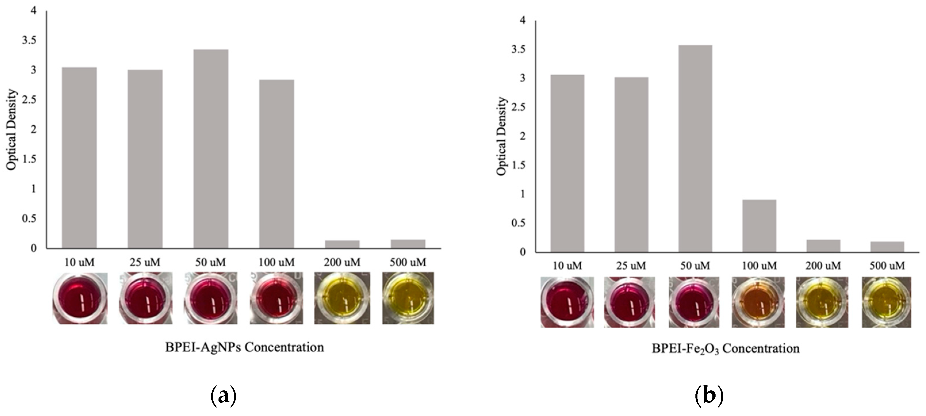
The activity assay aimed to determine the sensing agent concentration that was enough to fully inhibit the reaction between CPRG and -Gal, which would result in a yellow color solution. This experiment was conducted using the concentrations of chemical agents previously presented in Table 3. Figure 5 shows the absorbance values and color change of the activity assays after three hours. For all sensing agents, as the concentration decreased, the optical density increased, and the solution was more red. The sensing agents that showed potential for inhibiting the reaction between CPRG and -Gal were CTAB, BPEI, BPEI-AgNPs, and BPEI-Fe2O3. The lowest concentrations that resulted in inhibition of the -Gal (i.e., resulting in yellow color wells) were 5000 M of CTAB, 500 M of BPEI, and 200 M of BPEI-AgNPs. In addition, an orange color was observed at 100 M of BPEI-Fe2O3. Therefore, these materials were determined to be possible candidates for acting as sensing agents for detecting E. coli through the competitive binding approach. The optimal concentrations that caused full inhibition in this activity assay were used in the subsequent E. coli detection assays.
One of the critical outcomes of this well plate experiment is that the positively charged CTAB polymer (without a nanoparticle carrier) was able to inhibit the reaction between CPRG and -Gal. This would support the research hypothesis that the positive surface charge, rather than the sensing agent itself, is likely responsible for the mechanism of the competitive binding for the detection of E. coli. These Coulombic interactions between the positively charged sensing agents and the negatively charged bacteria or -Gal are what likely drive the competitive binding. This finding has promising applications because, theoretically, other cost-effective and more environmentally friendly sensing agents that protonate in solution and result in a positive charge could have the potential for sensing microbes. Thus, this research finding opens doors for future research investigations to optimize this sensing protocol using other positively charged polyelectrolytes.
3.4. E. coli Detection Assay Results
In this set of detection assays, the volumes and concentrations of the sensing agents used were the ones that resulted in the full inhibition of the CPRG and -Gal reaction in the previously discussed activity assays. Figure 6 shows the results of one of the trials of the E. coli detection assay. These results indicate that regardless of the concentration of E. coli tested, the color of the wells remained unchanged (no color gradient was observed), and the color was only dependent on the sensing agent tested. These results likely indicate that the microbes did not interact with the sensing agents at the testing conditions investigated. One possible explanation for this absence of interactions could be that the microbial concentrations tested were too low to be detected by the sensing agents under the given test conditions. It is hypothesized herein that the Coulombic attractions that occur between the sensing agents and microbes may only be dominant over the attraction between the sensing agents and -Gal when there are enough microbes in suspension. This is because -Gal dissolves uniformly in solution and thus, it is surrounding the nanoparticles and has higher chances of interactions. On the other hand, both E. coli and the nanoparticles are suspended in the liquid and their interactions are limited by the random collisions that occur by Brownian motion. Therefore, there is a higher likelihood that a nanoparticle will interact with a uniformly distributed enzyme than a microbe unless there are plenty (dense concentration) of microbes available in suspension. Only in the latter case will the competitive binding mechanism dominate, and the stronger Coulombic attractions will favor the binding of the sensing agent to E. coli over -Gal. To test this hypothesis, another experiment was performed at higher E. coli concentrations as described next.
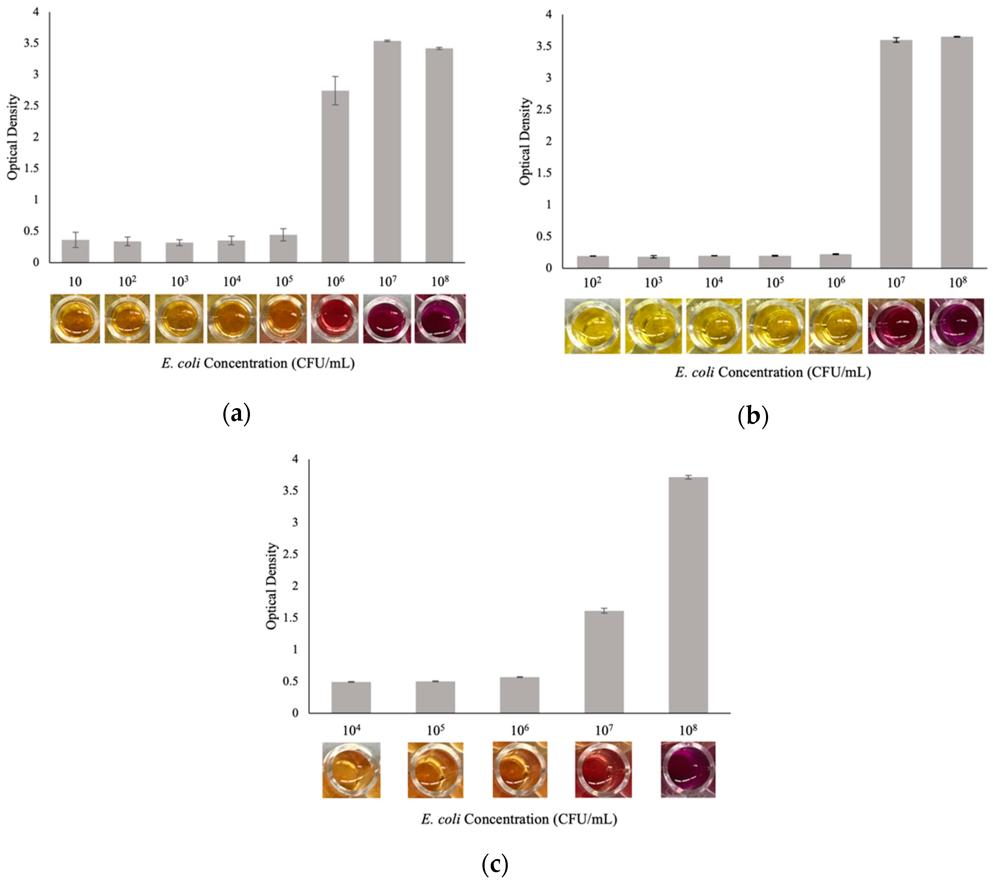
The following E. coli detection assay was then conducted at a higher range of E. coli concentrations of 10–108 CFU/mL and utilized the following sensing agents: 300 M BPEI-Fe2O3, 200 M BPEI-AgNPs, and 500 M BPEI. These concentrations of sensing agents were those that resulted in full inhibition of the CPRG and -Gal reaction in the activity assay outlined in Section 3.3. Preliminary E. coli detection assays that utilized CTAB as a sensing agent did not change color and detect E. coli, therefore, it was excluded from this detection assay. A color gradient was visually observed at 105–108 CFU/mL for BPEI-AgNPs, 107 and 108 CFU/mL for BPEI-Fe2O3, and 106–108 CFU/mL for BPEI (Figure 7). The results of this trial showed that BPEI alone, as well as BPEI-Fe2O3, and BPEI-AgNPs, have the potential for use as nanosensors for the detection of E. coli. However, more research is needed to lower the detection limit of these sensors. These results also support the hypothesis of this research that the nanoparticle type may not be the governing factor in the functionality of the nanosensors, rather it is the chemical coating/surface charge that is responsible for the mechanism of work of these nanosensors. Moreover, these results suggest that the coating itself may be sufficient for E. coli detection, as BPEI alone proved effective without a carrier nanoparticle, although further optimization is required to enhance its detection limit. These findings have not been reported in the literature before and create possibilities for a multitude of potential sensing agents that could be used without the need for a nanoparticle carrier. This would reduce the cost of the sensors and enhance their simplicity.
Research on the detection of E. coli O157:H7 in milk using automated immunomagnetic separation and an enzyme-based colorimetric assay resulted in a detection limit of 3 × 10² CFU/mL [24]. Previous studies using AuNPs covered with graphene oxide were able to detect E. coli down to 102 CFU/mL [25]. The detection limits established through the E. coli detection assays performed in the current can therefore be improved when compared to colorimetric nanosensors analyzed in the literature.
3.5. Significance
3.5.1. Material Cost Analysis
A preliminary cost analysis was performed on the sensing agents that detected E. coli in this study. The costs of these sensors were also compared to those of representative commercial E. coli detection methods. The cost analysis only considered material costs. Factors such as labor, equipment, location, supplier, taxes, and shipping fees were not accounted for in this analysis. The material cost analysis of the nanosensors was performed per sample, which is represented by one well in the well plate. One well would contain CPRG, -Gal, and the sensing agent. The volume and concentration of sensing agent used for this cost analysis were those that resulted in successful detection of E. coli (i.e., the cases where a gradient of color change was obtained with varying E. coli concentrations). The commercial microbial detection methods included only the cost of the materials (i.e., P/A bottles or IDEXX tray). Table 5 presents the results of the cost analysis. It is evident that the sensing agents tested in the study are low-cost alternatives to standard microbial detection methods. In addition, the BPEI alone was the most cost-effective sensor compared to BPEI-AgNPs and BPEI-Fe2O3.
3.5.2. Recyclability
Recyclability of this nanosensor can be improved by using positively charged nanoparticles that are magnetic, like the magnetite nanoparticles tested herein. The magnetic properties of magnetite nanoparticles allow them to be removed from solution after detection of microbes is complete, reconditioned, and then reused for further detection. Optimization of the iron oxide sensing agent is critical to improve the recyclability of the nanosensor.
4. Conclusions
This research yielded three main findings. First, the functionality of the nanosensor, which is based on the competitive binding approach, was not dependent on AuNPs. Both BPEI-AgNPs and BPEI-Fe2O3, possessing the same coating type as the AuNPs, inhibited the hydrolysis reaction in the absence of E. coli. Moreover, when the competitive binding protocol was employed, they successfully detected high concentrations of E. coli. Second, it was determined that the BPEI was not the only positively charged chemical coating that could inhibit the hydrolysis reaction. CTAB, a positively charged polymer, inhibited the hydrolysis reaction in the absence of E. coli. This might indicate that the surface charge rather than the composition of chemical coating is the governing factor for the Coulombic interactions between the positively charged sensing agents and the negatively charged -Gal and bacteria. Third, the BPEI itself (i.e., without a nanomaterial carrier) was able to detect high concentrations of microbes and inhibited the hydrolysis reaction in the absence of E. coli. These findings provide a foundation for future development of alternative competitive binding-based sensing agents (e.g., nanomaterials and positively charged polymers) that are more recyclable, environmentally friendly, and cost-effective.
One of the main limitations of the sensing agents tested in this study was the high detection limit of these sensors. Future investigation should consider optimizing the experimental testing conditions as well as the properties of the sensing agents to achieve lower detection limits. Specific suggestions for future investigation include: optimization of the chemistry (e.g., pH) of the aqueous testing media to allow for higher protonation of the positively charged sensing agents, investigating more types of positively charged polymers (e.g., EDTA), and enhancing the stability of the nanomaterials to reduce their potential aggregation in the test wells.
Author Contributions
Conceptualization, S.S., R.A.L. and A.E.B.; methodology, S.S., R.A.L., A.E.B., M.Y. and J.L.; validation, S.S., R.A.L. and A.E.B.; formal analysis, S.S., R.A.L. and A.E.B.; investigation, S.S., R.A.L., A.E.B., M.Y. and J.L.; resources, A.E.B., M.Y. and J.L.; data curation, S.S. and R.A.L.; writing—original draft preparation, S.S.; writing—review and editing, S.S., A.E.B., M.Y. and J.L.; visualization, S.S.; supervision, A.E.B., M.Y. and J.L.; project administration, A.E.B.; funding acquisition, A.E.B., M.Y. and J.L. All authors have read and agreed to the published version of the manuscript.
Funding
The main funding was provided internally by California Polytechnic State University through a Research Scholarly and Creative Activities (RSCA) Grant (20/21 RSCA).
Data Availability Statement
The raw data supporting the conclusions of this article will be made available by the authors on request.
Acknowledgments
The authors thank Trevor Harding and Joongmin Shin at California Polytechnic State University for providing access to materials characterization equipment and nanomaterials synthesis equipment, respectively.
Conflicts of Interest
The authors declare no conflicts of interest.
References
- World Health Organization (WHO). Drinking-Water. World Health Organization. Available online: https://www.who.int/news-room/fact-sheets/detail/drinking-water (accessed on 25 May 2021).
- Cabral, J.P. Water microbiology. Bacterial pathogens and water. Int. J. Environ. Res. Public Health 2010, 7, 3657–3703. [Google Scholar] [CrossRef] [PubMed]
- Ramírez-Castillo, F.Y.; Loera-Muro, A.; Jacques, M.; Garneau, P.; Avelar-González, F.J.; Harel, J.; Guerrero-Barrera, A.L. Waterborne pathogens: Detection methods and challenges. Pathogens 2015, 4, 307–334. [Google Scholar] [CrossRef] [PubMed]
- U.S. Environmental Protection Agency. Method 1103.1: Escherichia coil (E. coli) in Water by Membrane Filtration Using Membrane-Thermotolerant Escherichia coli (E. coli) Agar (mTEC); Report EPA-821-R-10-002; U.S. Environmental Protection Agency: Washington, DC, USA, 2010.
- Spatola Rossi, C.; Coulon, F.; Ma, S.; Zhang, Y.S.; Yang, Z. Microfluidics for rapid detection of live pathogens. Adv. Funct. Mater. 2023, 33, 2212081. [Google Scholar] [CrossRef]
- Tambi, A.; Brighu, U.; Gupta, A.B. Methods for detection and enumeration of coliforms in drinking water: A review. Water Supply 2023, 23, 4047–4058. [Google Scholar] [CrossRef]
- Pebdeni, A.B.; Al-Baiati, M.N.; Hosseini, M. New application of bimetallic Ag/Pt nanoplates in a colorimetric biosensor for specific detection of E. coli in water. Beilstein J. Nanotechnol. 2024, 15, 95–103. [Google Scholar] [CrossRef] [PubMed]
- Ertaş, T.; Dinç, B.; Üstünsoy, R.; Eraslan, H.; Ergenç, A.F.; Bektaş, M. Novel electrochemical biosensor for Escherichia coli using gold-coated tungsten wires and antibody functionalized short multiwalled carbon nanotubes. Instrum. Sci. Technol. 2024, 52, 109–124. [Google Scholar] [CrossRef]
- Tian, C.; Zhao, L.; Qi, G.; Zhang, S. Trace detection of E. coli O157: H7 cells by an Au nanoparticle-based SERS aptasensor. ACS Appl. Nano Mater. 2023, 6, 1386–1394. [Google Scholar] [CrossRef]
- Dimitrievska, I.; Paunovic, P.; Grozdanov, A. Recent advancements in nano sensors for air and water pollution control. Mater. Sci. Eng. 2023, 7, 113–128. [Google Scholar]
- Ahangari, A.; Mahmoodi, P.; Mohammadzadeh, A. Advanced nano biosensors for rapid detection of zoonotic bacteria. Biotechnol. Bioeng. 2023, 120, 41–56. [Google Scholar] [CrossRef] [PubMed]
- Zhu, T.; Hu, Y.; Yang, K.; Dong, N.; Yu, M.; Jiang, N. A novel SERS nanoprobe based on the use of core-shell nanoparticles with embedded reporter molecule to detect E. coli O157: H7 with high sensitivity. Microchim. Acta 2018, 185, 30. [Google Scholar] [CrossRef] [PubMed]
- Wang, X.Y.; Ji, R.Y.; Lang, W.W.; Qin, K.X.; Bai, F.Y.; Xi, H.Y.; Zheng, Y.; Xia, B.X.; Dong, L.Y.; Wang, X.H. Integrating PEGylated peptide-oriented bacteria-imprinted matrix and PdPt bimetallic-doped imidazolium zeolite framework-8 for sensitive detection of Escherichia coli with smartphone readout system. Sens. Actuators B Chem. 2024, 411, 135749. [Google Scholar] [CrossRef]
- Choi, Y.; Hwang, J.H.; Lee, S.Y. Recent trends in nanomaterials-based colorimetric detection of pathogenic bacteria and viruses. Small Methods 2018, 2, 1700351. [Google Scholar] [CrossRef] [PubMed]
- Rasheed, S.; Kanwal, T.; Ahmad, N.; Fatima, B.; Najam-ul-Haq, M.; Hussain, D. Advances and challenges in portable optical biosensors for onsite detection and point-of-care diagnostics. TrAC Trends Anal. Chem. 2024, 173, 117640. [Google Scholar] [CrossRef]
- Yang, L.; Xu, X.; Song, Y.; Huang, J.; Xu, H. Research progress of nanozymes in colorimetric biosensing: Classification, activity and application. Chem. Eng. J. 2024, 487, 150612. [Google Scholar] [CrossRef]
- John, G.S.; Johnson, A.M.; Suresh, P.A.; Unnikrishnan, N.V.; Kumar, K.A. Recent Advances in Carbon Nanotube-Based Electrochemical and Optical Biosensors. In Carbon Nanotubes for Biomedical Applications and Healthcare; Apple Academic Press: New York, NY, USA, 2024; pp. 147–166. [Google Scholar]
- Thiramanas, R.; Laocharoensuk, R. Competitive binding of polyethyleneimine-coated gold nanoparticles to enzymes and bacteria: A key mechanism for low-level colorimetric detection of gram-positive and gram-negative bacteria. Microchim. Acta 2016, 183, 389–396. [Google Scholar] [CrossRef]
- Miranda, O.R.; Li, X.; Garcia-Gonzalez, L.; Zhu, Z.J.; Yan, B.; Bunz, U.H.; Rotello, V.M. Colorimetric bacteria sensing using a supramolecular enzyme–nanoparticle biosensor. J. Am. Chem. Soc. 2011, 133, 9650–9653. [Google Scholar] [CrossRef] [PubMed]
- nanoComposix. Branched Polyethylenimine (BPEI) Surface. nanoComposix. Available online: https://nanocomposix.com/pages/branched-polyethylenimine-bpei-surface#applications (accessed on 26 April 2021).
- nanoComposix. BioPure Silver Nanospheres—BPEI. nanoComposix. Available online: https://nanocomposix.com/collections/material-silver/products/biopure-silver-nanospheres-bpei?variant=15906741551193 (accessed on 26 April 2021).
- Millipore Sigma. Iron Oxide (II, III), Magnetic Nanoparticles Solution. Sigma Aldrich. Available online: https://www.sigmaaldrich.com/catalog/product/aldrich/747300?lang=en®ion=US (accessed on 26 April 2021).
- Millipore Sigma. Cerium Doped Iron Oxide. Sigma Aldrich. Available online: https://www.sigmaaldrich.com/catalog/product/aldrich/909203?lang=en®ion=US (accessed on 26 April 2021).
- Park, J.Y.; Park, K.; Ok, G.; Chang, H.J.; Park, T.J.; Choi, S.W.; Lim, M.C. Detection of Escherichia coli O157: H7 using automated immunomagnetic separation and enzyme-based colorimetric assay. Sensors 2020, 20, 1395. [Google Scholar] [CrossRef] [PubMed]
- Gupta, R.; Kumar, A.; Kumar, S.; Pinnaka, A.K.; Singhal, N.K. Naked eye colorimetric detection of Escherichia coli using aptamer conjugated graphene oxide enclosed Gold nanoparticles. Sens. Actuators B Chem. 2021, 329, 129100. [Google Scholar] [CrossRef]
- Hach. Presence/Absence Test, pk/12. Hach. Available online: https://www.hach.com/presence-absencetest-pk-12/product?id=7640249610 (accessed on 27 May 2021).
- Analytics Shop. IDEXX Quanti-Tray/2000, 97-Well, Sterile, 100pc/PAK, Shelf Life: 18 Months from Date of Manufacture. Analytics Shop. Available online: https://www.analytics-shop.com/gb/ix982167500-gb.html (accessed on 27 May 2021).
Figure 1.
Competitive binding technique for detection of E. coli using positively charged gold nanoparticles: (a) components of the nanosensor, (b) hydrolysis of CPRG by β-Gal to form CPR, resulting in a red solution, (c) binding of PEI-AuNPs and β-Gal, resulting in a yellow solution, and (d) competitive binding of positively charged PEI-AuNPs to negatively charged β-Gal and bacteria—the nanoparticles preferentially bind to the bacteria because the bacteria is more negative than the β-Gal enzyme.
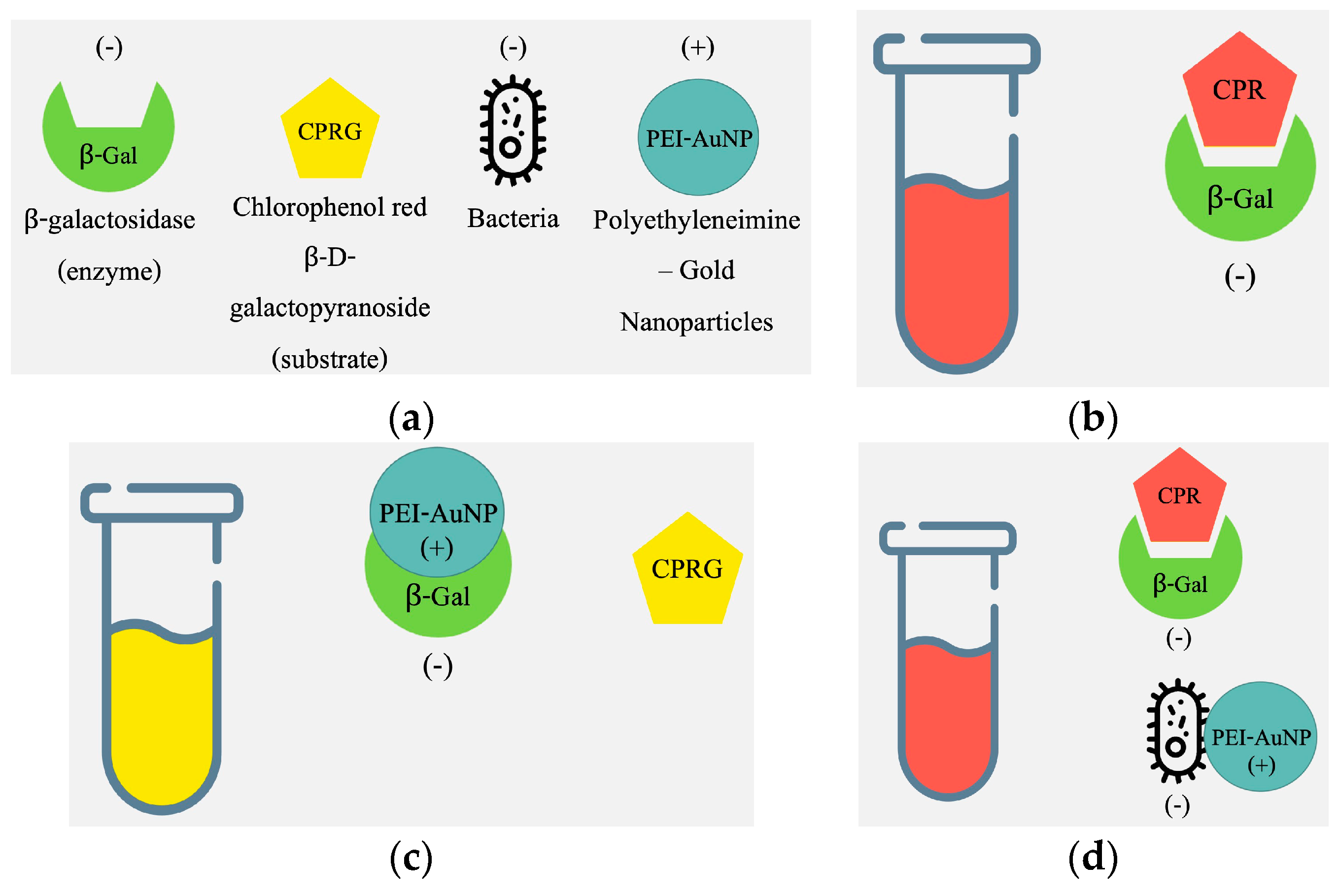
Figure 2.
Phases of the experimental testing program implemented in this study.
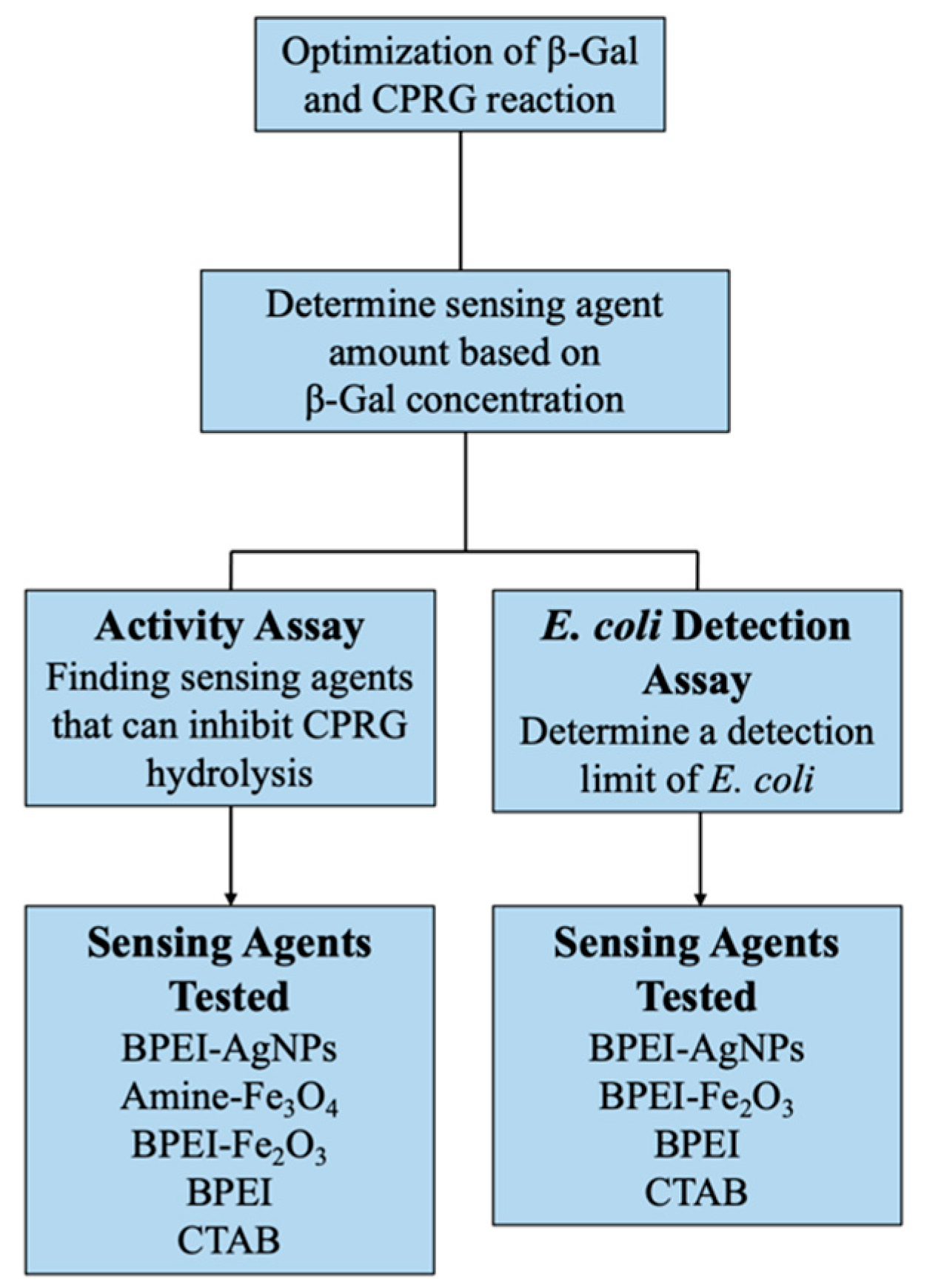
Figure 3.
TSA plates after 24 h of incubation at 37 degrees Celsius (°C). Duplicates of the 10−6 and 10−7 dilutions were performed because those were the plates closest in count to the 30 to 200 colony counts used for microbial concentration calculations.
Figure 3.
TSA plates after 24 h of incubation at 37 degrees Celsius (°C). Duplicates of the 10−6 and 10−7 dilutions were performed because those were the plates closest in count to the 30 to 200 colony counts used for microbial concentration calculations.

Figure 4.
Optimization of the amounts of CPRG and -Gal needed for the colorimetric detection assays.
Figure 4.
Optimization of the amounts of CPRG and -Gal needed for the colorimetric detection assays.

Figure 5.
Optical density results of the activity assays after three hours of incubation at room temperature for the sensing agents tested: (a) BPEI-AgNPs, (b) BPEI-Fe2O3, (c) amine-Fe3O4, (d) BPEI, and (e) CTAB.
Figure 5.
Optical density results of the activity assays after three hours of incubation at room temperature for the sensing agents tested: (a) BPEI-AgNPs, (b) BPEI-Fe2O3, (c) amine-Fe3O4, (d) BPEI, and (e) CTAB.

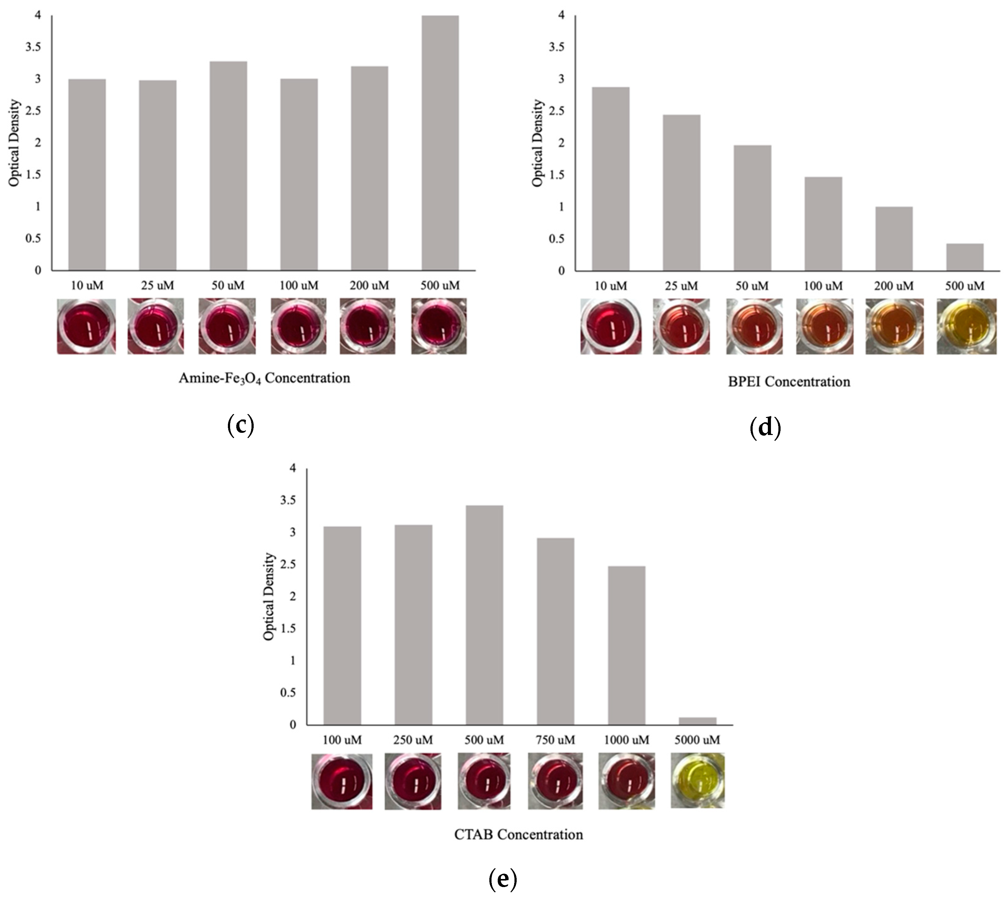
Figure 6.
Results of an E. coli detection assay after three hours in the SpectraMAX Plus. For columns 1 through 6 and column 10, the E. coli concentrations in CFU/mL ranged from 10−5 to 102. For columns 7 to 9, the E. coli concentrations were 10−1, 100, and 102 CFU/mL from top to bottom. The number of microbes in this experiment was extremely low (e.g., only one bacterium in the 101 CFU/mL) and that likely explains the lack of noticeable reactions.

Figure 7.
Optical density results of the E. coli detection assay for (a) 200 M BPEI-AgNPs, (b) 300 M BPEI-Fe2O3, and (c) 500 M BPEI after three hours of incubation at room temperature.
Figure 7.
Optical density results of the E. coli detection assay for (a) 200 M BPEI-AgNPs, (b) 300 M BPEI-Fe2O3, and (c) 500 M BPEI after three hours of incubation at room temperature.

Table 1.
Sensing agents tested in the current study for the detection of E. coli in water.
| Hypotheses Tested | Sensing Agent Investigated | Acronym |
|---|---|---|
| Nanoparticle type (i.e., Ag or Fe2O3) is not the governing factor for the sensing mechanism. Therefore, other nanoparticle types can replace gold as a nanosensor for detection of E. coli. | Silver nanoparticles coated with branched polyethyleneimine | BPEI-AgNPs |
| Cerium (Ce3/4+) doped iron oxide nanoparticles coated with branched polyethyleneimine | BPEI-Fe2O3 | |
| Amine functionalized iron oxide nanoparticles | Amine-Fe3O4 | |
| The surface charge, not the type of coating, is the governing factor for the sensing mechanism in the competitive binding approach. Therefore, other positively charged polymers (e.g., CTAB) can replace the PEI-AuNPs sensing agent. In addition, a carrier nanoparticle is not needed for the positively charged polymer to detect the microbes. | (1-Hexadecyl) trimethylammonium bromide | CTAB |
| Branched polyethyleneimine | BPEI |
Table 2.
Sensing agents tested and their characteristics.
| Nanoparticles | Concentration | Diameter | Zeta Potential | Reference |
|---|---|---|---|---|
| BPEI-AgNPs | 1 mg/mL | 40 nm | +30 to +95 mV | [21] |
| Amine-Fe3O4 | 1 mg/mL Fe | 10 nm | Not Provided | [22] |
| BPEI-Fe2O3 | 1.3 mg/mL | 7–15 nm | +29.7 mV | [23] |
Table 3.
Activity assay experiments.
| Sensing Agent | M) |
|---|---|
| BPEI-AgNPs | 10, 25, 50, 100, 200, and 500 |
| BPEI-Fe2O3 | |
| Amine-Fe3O4 | |
| BPEI | |
| CTAB | 100, 250, 500, 750, 1000, and 5000 |
Table 4.
Experiments conducted to determine E. coli detection limits.
| Sensing Agent | M) | E. coli Concentrations Tested (CFU/mL) |
|---|---|---|
| BPEI-AgNPs | 200 | 1–102 |
| 200 | 1–108 | |
| 300 | ||
| 400 | ||
| 800 | ||
| BPEI-Fe2O3 | 50 | 1–102 |
| 100 | 1–108 | |
| 200 | ||
| 200 | ||
| 400 | ||
| 600 | ||
| 800 | ||
| BPEI | 500 | 1 and 102 |
| 500 | 106–108 | |
| 800 | 1–108 | |
| CTAB | 5000 | 1 and 102 |
| 5000 | 106–108 |
Table 5.
Cost of sensing agents tested and commercial microbial detection methods.
| Cost of Individual Components of Sensing Agents | |||
|---|---|---|---|
| Individual Components | Cost per Sample ($) | Quantity Used | |
| CPRG | 0.05 | L of 0.75 mM | |
| -Gal | 0.02 | M | |
| BPEI | 0.00002 | M | |
| BPEI-AgNPs | 0.15 | M | |
| BPEI-Fe2O3 | 0.35 | M | |
| Overall Material Cost of the Sensor | |||
| Sensor (including all components) | Cost per Analysis ($) | ||
| BPEI | 0.07 | ||
| BPEI-AgNPs | 0.22 | ||
| BPEI-Fe2O3 | 0.42 | ||
| Commercial E. coli Detection Methods | |||
| Method | Cost per Test ($) | ||
| Hach P/A Test [26] | 5.04 | ||
| IDEXX Quanti-Tray 2000 [27] | 7.40 | ||
Disclaimer/Publisher’s Note: The statements, opinions and data contained in all publications are solely those of the individual author(s) and contributor(s) and not of MDPI and/or the editor(s). MDPI and/or the editor(s) disclaim responsibility for any injury to people or property resulting from any ideas, methods, instructions or products referred to in the content. |
© 2024 by the authors. Licensee MDPI, Basel, Switzerland. This article is an open access article distributed under the terms and conditions of the Creative Commons Attribution (CC BY) license (https://creativecommons.org/licenses/by/4.0/).
Share and Cite
MDPI and ACS Style
Stabler, S.; Lang, R.A.; El Badawy, A.; Yeung, M.; Lee, J. Analysis and Modification of a Colorimetric Nanosensor for Rapid Detection of Escherichia coli in Water. Crystals 2024, 14, 386. https://doi.org/10.3390/cryst14040386
AMA Style
Stabler S, Lang RA, El Badawy A, Yeung M, Lee J. Analysis and Modification of a Colorimetric Nanosensor for Rapid Detection of Escherichia coli in Water. Crystals. 2024; 14(4):386. https://doi.org/10.3390/cryst14040386
Chicago/Turabian StyleStabler, Sarah, Ruby Anne Lang, Amro El Badawy, Marie Yeung, and Jean Lee. 2024. "Analysis and Modification of a Colorimetric Nanosensor for Rapid Detection of Escherichia coli in Water" Crystals 14, no. 4: 386. https://doi.org/10.3390/cryst14040386
Note that from the first issue of 2016, this journal uses article numbers instead of page numbers. See further details here.