Article

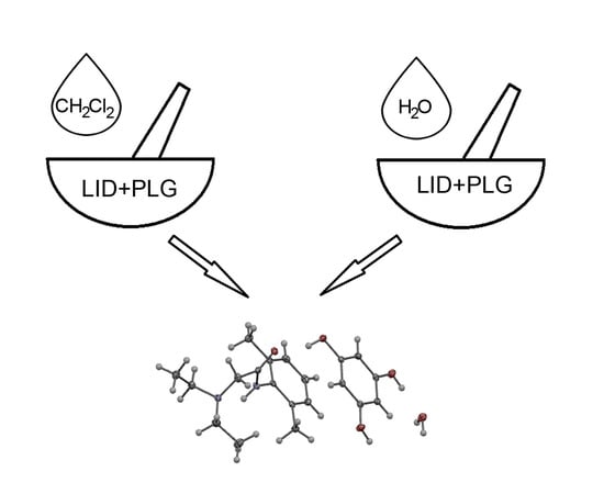
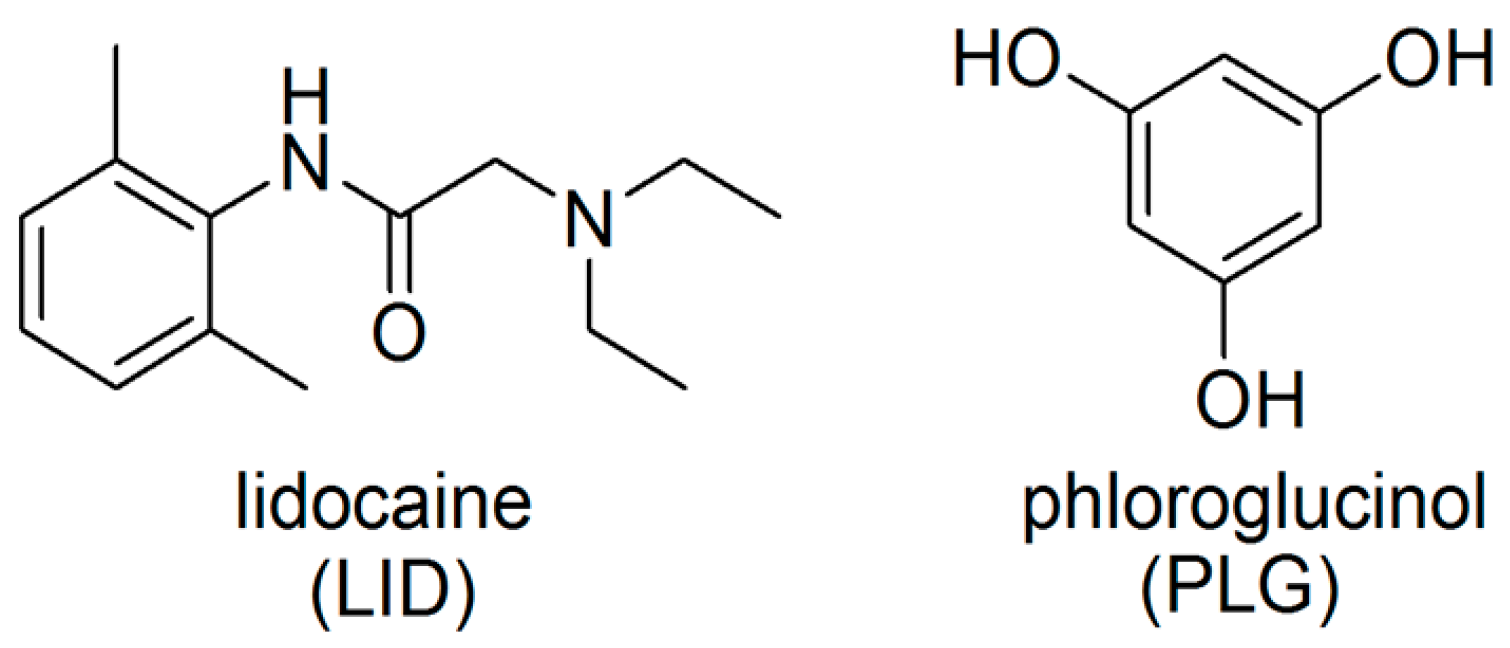
Mechanochemical Synthesis and Crystal Structure of the Lidocaine-Phloroglucinol Hydrate 1:1:1 Complex †

by
Nancy Evelyn Magaña-Vergara
1,
Porfirio De la Cruz-Cruz
2,
Ana Lilia Peraza-Campos
1,
Francisco Javier Martínez-Martínez
1 and
Juan Saulo González-González
2,*
1
CONACyT-Facultad de Ciencias Químicas, Universidad de Colima, Km 9 Carretera Colima-Coquimatlán, Coquimatlán C.P. 28400, Colima, Mexico
2
Instituto de Farmacobiología, Universidad de la Cañada, Carretera Teotitlán—San Antonio Nanahuatipán Km 1.7 s/n., Paraje Titlacuatitla, Teotitlán de Flores Magón, Oaxaca C.P. 68540, Mexico
*
Author to whom correspondence should be addressed.
This article is dedicated to Professor Narayan Hosmane on the occasion of his 70th birthday.
Crystals 2018, 8(3), 130; https://doi.org/10.3390/cryst8030130
Submission received: 8 February 2018 / Revised: 28 February 2018 / Accepted: 6 March 2018 / Published: 9 March 2018
(This article belongs to the Special Issue Polymorphism of Mechanochemically Synthesized Cocrystals)

Abstract

:
Molecular complexation is a strategy used to modify the physicochemical or biopharmaceutical properties of an active pharmaceutical ingredient. Solvent assisted grinding is a common method used to obtain solid complexes in the form of cocrystals. Lidocaine is a drug used as an anesthetic and for the treatment of chronic pain, which bears in its chemical structure an amide functional group able to form hydrogen bonds. Polyphenols are used as cocrystal coformers due to their ability to form O–H···X (X = O, N) hydrogen bond interactions. The objective of this study was to exploit the ability of phloroglucinol to form molecular complexes with lidocaine by liquid assisted grinding. The formation of the complex was confirmed by the shift of the O–H and C=O stretching bands in the IR spectra of the polycrystalline ground powders, suggesting the formation of O–H···O=C hydrogen bonds. Hydration of the complexes also was confirmed by IR spectroscopy and by powder X-ray diffraction. The molecular structure was determined by single crystal X-ray diffraction.

Graphical Abstract

1. Introduction

Drug formulation studies are performed with the aim of modifying the physicochemical and biopharmaceutical properties of an active pharmaceutical ingredient (API) to: improve its delivery its release in the target tissue, ensure the stability of the product, offer a comfortable use to patients, and make easier the production of the dosage forms [1]. Active pharmaceutical ingredients can contain solvents in the crystal structure. If the solvent is water, it is called hydrate. The ability of water to act as donor and acceptor of hydrogen bond interactions favors the incorporation of water into the crystalline lattice of APIs, reordering the intermolecular hydrogen bond pattern, obtaining hydrated complexes. Therefore, hydration studies in APIs are important because the presence of water in the crystalline lattice can affect the physicochemical and biopharmaceutical properties of the active pharmaceutical ingredient [2].
Molecular complexation is a strategy used to modify the physicochemical or biopharmaceutical properties of an API [3]. Molecular complexes, or host–guest complexes, are molecular species formed by two or more molecules that are associated by noncovalent interactions. Formation of molecular complexes involves molecular recognition between the functional groups of the molecules [4].
Lidocaine (2-(Diethylamino)-N-(2,6-dimethylphenyl)acetamide) (LID) is a drug used as an anesthetic and for the treatment of chronic pain [5]. The LID base is poorly soluble in water, and thus is formulated as its chlorhydrate salt, which is water soluble [6]. Molecular structure of LID has an amide functional group able to form hydrogen bond interactions, and an aromatic ring to form π-interactions. Molecular complexes of LID in the form of inclusion complexes [7], cocrystals [8] and eutectics [9] have been developed in order to mask the bitter taste [10] and prolong the anesthetic effect [11].
Polyphenols (di-hydroxy or tri-hydroxy benzenes) have been exploited as supramolecular bulding blocks [12,13] and as pharmaceutical cocrystals coformers [14,15,16], due to their ability to form O–H···X (X = O, N) hydrogen bond interactions.
Mechanochemistry is concerned with chemical transformations induced by mechanical means, such as compression, shear, or friction [17]. It is a low cost and green chemistry method employed in the pharmaceutical industry to obtain new solid phases of APIs such as cocrystals, salts, solvates and polymorphs. Solvent assisted grinding is a commonly used mechanochemical method to obtain solid new solid forms of APIs, and the advantages of this method are that it does not depend on the solubility of the compounds and the time reduction in the synthesis process [18,19,20].
The objective of this study was to exploit the ability of phloroglucinol (PLG) (1,3,5-benzenetriol) to form a molecular complex with lidocaine (Figure 1) by liquid assisted grinding. The complex was characterized by infrared spectroscopy, powder X-ray diffraction and X-ray single crystal diffraction.

2. Materials and Methods

2.1. Mechanochemical Synthesis and Crystallization

Lidocaine (98.0%) and phloroglucinol (99.0%) were purchased from Aldrich (St. Louis, MO, USA). Methanol (ACS grade), dichloromethane (ACS grade) and distillated water were purchased from Química Meyer, (Mexico City, Mexico). All the reagents and solvents were used as received.
Mixtures in 1:1 molar ratio of LID (0.400 g, 1.7 mmol) and PLG (0.214 g, 1.7 mmol) were ground in a mortar with a pestle for 3 min. Before starting the grinding, 0.5 mL of dichloromethane was added. After 3 min of grinding, the polycrystalline powder LID-PLG(CH2Cl2) was collected. The cycle of adding dichloromethane (0.5 mL) and grinding for 3 min was repeated three times until 12 min of grinding was complete.
The hydration study was performed grinding 1:1 mixtures of LID (0.400 g, 1.7 mmol) and PLG (0.214 g, 1.7 mmol) for 5 min. Before starting the grinding, 0.5 mL of distillated water was added. After 5 min of grinding, a polycrystalline powder LID-PLG(H2O) was obtained.
The LID-PLG(CH2Cl2) polycrystalline powder was dissolved in methanol. After the slow evaporation of the solvent at room temperature, colorless single crystals (LID-PLG(cryst)) suitable for diffraction were obtained.

2.2. IR Spectroscopy

Infrared spectra of the starting products (LID and PLG), the ground mixtures LID-PLG(CH2Cl2) and LID-PLG(H2O), and the single crystal LID-PLG(cryst) were acquired using a Bruker Tensor-27 spectrophotometer (Ettlingen, Germany) equipped with an attenuated total reflection (ATR) system accessory (16 scans, spectral range 600–4000 cm−1, resolution 4 cm−1).

2.3. X-Ray Diffraction

X-ray powder diffraction patterns of LID, PLG and LID-PLG(CH2Cl2) polycrystalline solids were obtained in a PANalytical X’Pert PRO diffractometer (Almelo, The Netherlands) with Cu Kα1 radiation (λ = 1.5405 Å, 45 kV, 40 mA) from 2.02° to 49.93° in 2θ.
The LID-PLG crystal structure was performed in a Bruker D8 QUEST (Karlsruhe, Germany) diffractometer. A summary of collection and refinement of LID-PLG(cryst) is listed in Table 1. The cell refinement and data reduction were carried out with the SAINT V8.34A (Bruker, Madison, WI, USA) [21] and SORTAV (University of Glasgow, Scotland) [22] software. The structure was solved by direct methods using SHELXL97 (University of Göttingen, Germany) [23]. H atoms on C and N were positioned geometrically and treated as riding atoms, with CH = 0.95–0.99 Å and Uiso(H) = 1.5 Ueq(C) for methyl H atoms or 1.2Ueq(C) otherwise, and N–H = 0.88 Å and Uiso(H) = 1.2Ueq(N). Mercury software (The Cambridge Crystallographic Data Centre, Cambridge, UK) [24] was used to prepare the material for publication. CCDC 1822957 contains the supplementary crystallographic data for this paper. These data can be obtained free of charge via http://www.ccdc.cam.ac.uk/conts/retrieving.html (or from the CCDC, Cambridge, UK).

3. Results and Discussion

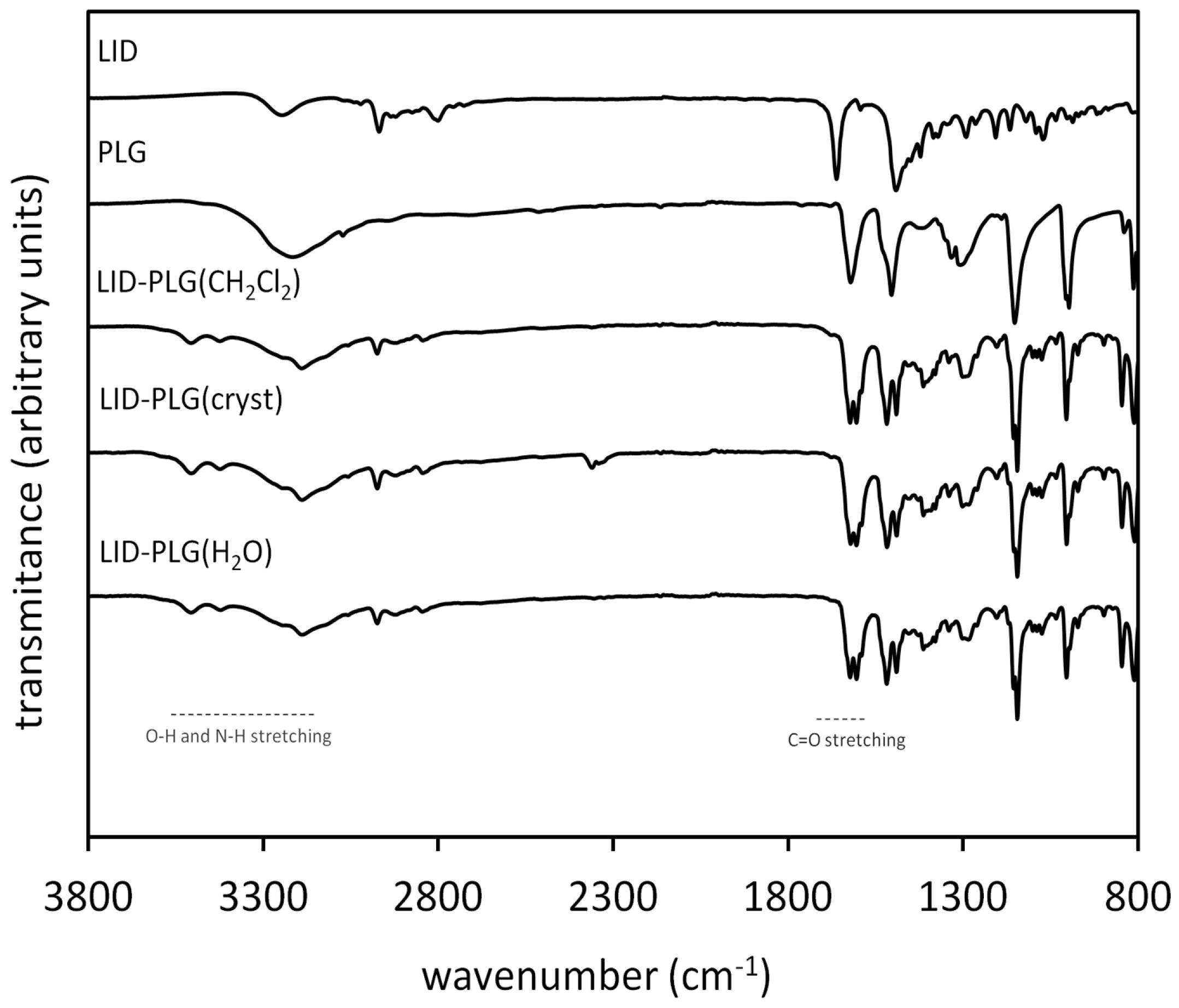
3.1. Infrared Spectroscopy

Infrared spectroscopy (IR) is a tool that allows identification of the formation of hydrogen bond interactions, by the shift of the bands of the functional groups involved in the formation of the hydrogen bonds in the infrared spectra. In the IR spectra of LID-PLG(CH2Cl2), the phenolic hydroxyl (O–H) and the amide carbonyl (C=O) bands were shifted with respect to the starting products (Figure 2), suggesting the formation of the microcrystalline complex via C=O···H–O hydrogen bond interactions. LID free base is not hygroscopic [25]; however, PLG can absorb water up to 32% of relative humidity [26]. Hydration study was performed in order to evaluate the effect of water on the formation of the complexes. In this study, LID and PLG were ground with water. Unexpectedly, the IR spectrum of LID-PLG(H2O) and the IR spectrum of the single crystal were similar to the LID-PLG(CH2Cl2), indicating that the latter incorporated water from the environment into the crystalline lattice. The O–H stretching frequency was shifted (Table 2) with ΔνO–H = −28 cm−1, +207 cm−1 and +290 cm−1; meanwhile, the C=O frequency was shifted with a ΔνC=O of −28 cm−1. These shifts are a consequence of the rearrangement of the hydrogen bond patterns with respect to the noncomplexed forms, and are in agreement with previous reports about lidocaine complexes, and dihydroxybenzenes with phenylenebis(methylene)dicarbamates and phenyldioxalamates [7,8,27,28]. The N–H stretching frequency showed a small shift (ΔνN–H = −2 cm−1, which is out of the spectral resolution), suggesting that the N–H group is not involved in the formation of the complex.

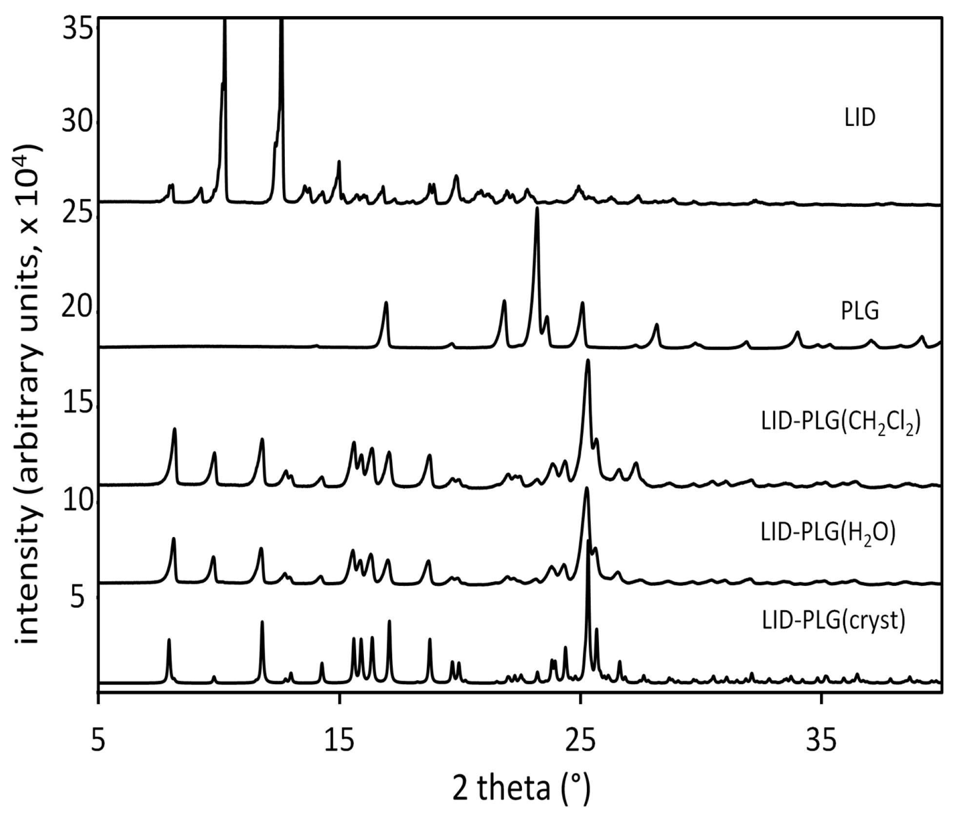
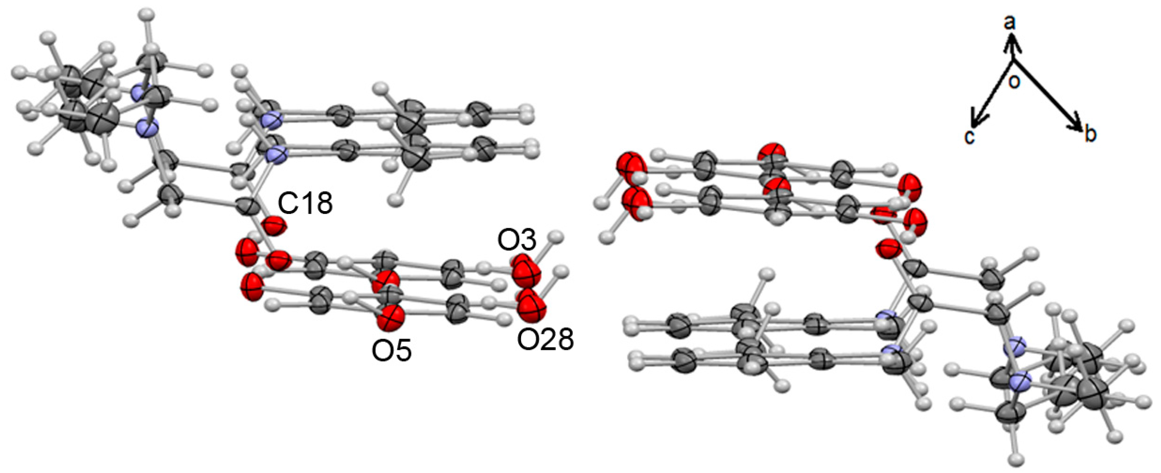
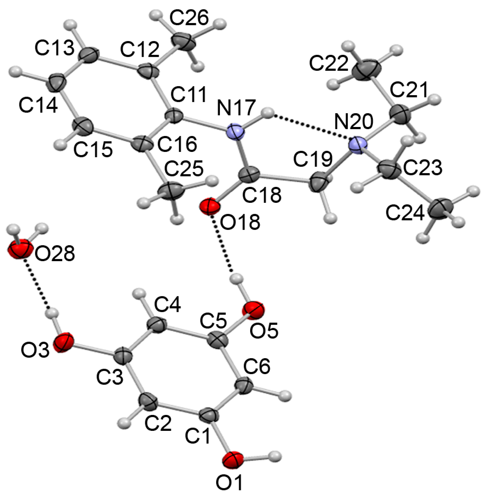
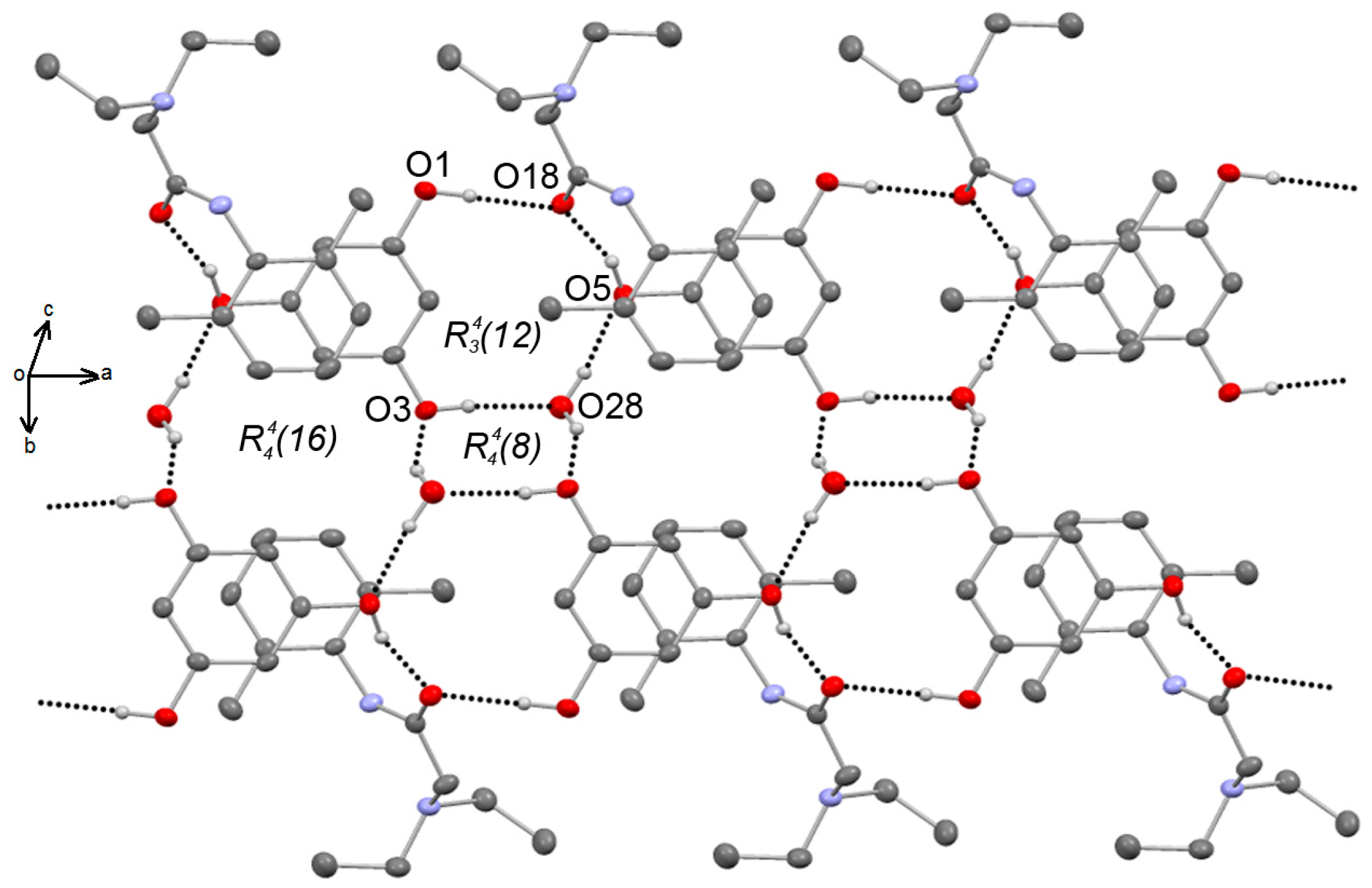
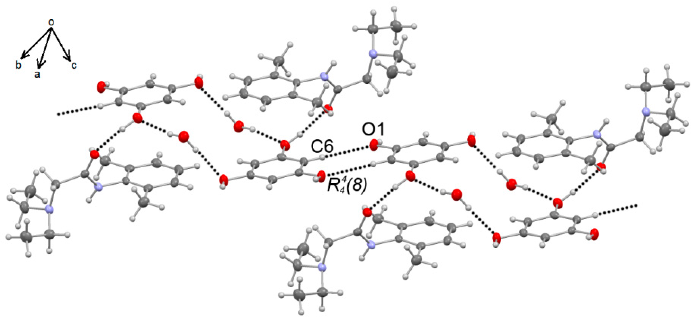
3.2. X-Ray Diffraction

The powder X-ray diffraction pattern of LID-PLG(CH2Cl2) was different with respect to starting products LID and PLG, indicating the formation of a new polycrystalline phase, belonging to the complex.
The powder X-ray diffraction pattern of LID-PLG(CH2Cl2) showed a good match with the powder pattern of LID-PLG(H2O), and with the simulated powder X-ray diffraction pattern (obtained with Mercury) of LID-PLG(cryst) (Figure 3). This indicates an adequate structural homogeneity, and the incorporation of water into the crystalline lattice of the polycrystalline powders obtained by solvent assisted grinding.
The crystal structure of LID-PLG (triclinic, P-1) confirmed the incorporation of water into the crystalline lattice (Figure 4). In the crystal structure of LID-PLG(cryst), PLG adopts the Cs conformation [25]. The amide group of LID is twisted out from the plane of the aromatic ring by −72.1(2) (torsion angle C12–C11–N17–C18), and the nitrogen atoms adopt a syn conformation with respect to the N17–C18–C19–N20 torsion angle forming the N17–H17∙∙∙N20 intramolecular S(5) hydrogen bond. From 26 crystal structures of LID reported in the Cambridge Crystallographic Data Centre Access Structures website, 22 adopt the anti conformation, and 4 adopt de syn conformation [29]. In the crystal structure of PLG dihydrate [26], each phenolic O–H is hydrogen bonded to a water molecule. Meanwhile, in LID-PLG(cryst), two phenolic O–H groups are hydrogen bonded—each one to a molecule of LID, and the remaining O–H group is hydrogen bonded to a water molecule.
In the asymmetric unit of LID-PLG(cryst), PLG is linked to LID via the O1–H1∙∙∙O18 hydrogen bond interaction, and with water by the O3–H3∙∙∙O28 hydrogen bond interaction (Figure 4) (hydrogen bond details and symmetry codes are given in Table 3), forming a heterotrimer. The first dimensional supramolecular array (Figure 5) is given by the propagation of the heterotrimer forming a supramolecular tape along the a-axis by the O5–H8∙∙∙O18i, the O28–H28A∙∙∙O5iii and the O28–H28B∙∙∙O3ii hydrogen bond interactions, depicting the R 3 4 (12) (O1–H1∙∙∙O18∙∙∙H5–O5 three centered hydrogen bond), R 4 4 (8) and R 4 4 (16) ring motifs. In this arrangement, a PLG-water-PLG corrugated supramolecular layer (similar to the PLG dihydrate [26]) is formed and the LID molecules are located above and below the layer (Figure 6). The second dimension supramolecular (2D) array is extended by the C6–H6∙∙∙O1iv soft interaction ( R 2 2 (8) motif), depicting a supramolecular tape along the (0 12 12) direction (Figure 7). In this array, a PLG-water-PLG layer with the form of a cascade was depicted.
The supramolecular architecture of free LID changed as consequence of the complexation. In the crystal structure of free LID (Refcode: LIDCAN10) [21], a supramolecular column driven by C=O∙∙∙H–N hydrogen bond interactions forming C(4) chains is observed. Meanwhile, in LID-PLG(cryst), LID forms C=O∙∙∙H–O interactions leading to the formation of a R 3 4 (12) motif.

4. Conclusions

The LID-PLG complex was obtained by the solvent assisted grinding method. Infrared spectroscopy allowed for determining the formation of the complex by the shifts of the O–H and C=O stretching bands. Hydration of the complex as a consequence of the incorporation of water from the environment also was confirmed by infrared spectroscopy and X-ray powder diffraction because the IR spectra and the powder diffraction patterns of LID-PLG(CH2Cl2), LID-PLG(H2O) and LID-PLG(cryst) were similar. The molecular structure of the LID-PLG hydrate complex was determined by X-ray single crystal diffraction, showing the incorporation of water into the crystalline lattice. The supramolecular architecture of LID-PLG(cryst) is driven by C=O∙∙∙H–O, H–O∙∙∙H–O and C=O∙∙∙H–C interactions depicting R 3 4 (12), R 4 4 (8), R 4 4 (16) and R 2 2 (8) hydrogen bond motifs.

Acknowledgments

The authors express their thanks to the Universidad de la Cañada project UNCA PFI 13/14 and Red Temática de Química Supramolecular (grant 271884) for the support of this work.

Author Contributions

Nancy Evelyn Magaña-Vergara performed the X-ray single diffraction study and revised the manuscript; Porfirio de la Cruz-Cruz synthesized the complex and performed the IR spectra spectroscopy study; Ana Lilia Peraza-Campos and Francisco Javier Martínez-Martínez performed the powder X-ray diffraction study and revised the manuscript; Juan Saulo González-González conceived the project, designed the experiments and wrote the manuscript.

Conflicts of Interest

The authors declare no conflict of interest.

References

  1. Allen, L.V.; Popovich, N.G.; Ansel, H.C. Ansel’s Pharmaceutical Dosage Forms and Drug Delivery Systems, 9th ed.; Lippincott & Wilkins: Baltimore, MD, USA, 2011; pp. 90–92. [Google Scholar]
  2. Healy, A.M.; Worku, Z.A.; Kumar, D.; Madi, A.M. Pharmaceutical solvates, hydrates and amorphous forms: A special emphasis on cocrystals. Adv. Drug Deliv. Rev. 2017, 117, 25–46. [Google Scholar] [CrossRef] [PubMed]
  3. Umeda, Y.; Fukami, T.; Furuishi, T.; Suzuki, T.; Makimura, M.; Tomono, K. Molecular Complex Consisting of Two Typical External Medicines: Intermolecular Interaction between Indomethacin and Lidocaine. Chem. Pharm. Bull. 2007, 55, 832–836. [Google Scholar] [CrossRef] [PubMed]
  4. Steed, J.W.; Atwood, J.L. Supramolecular Chemistry, 2nd ed.; John Wiley & Sons Ltd: Wiltshire, UK, 2009; pp. 2–9. [Google Scholar]
  5. Ferreira, S.M.; Campos, K.D. The analgesic effect of intravenous lidocaine in the treatment of chronic pain: A literature review. Rev. Bras. Reumatol. 2014, 54, 386–392. [Google Scholar]
  6. Badawi, H.M.; Förner, W.; Ali, S.A. The molecular structure and vibrational, 1H and 13C NMR spectra of lidocaine hydrochloride monohydrate. Spectrochim. Acta Part A Mol. Biomol. Spectrosc. 2016, 152, 92–100. [Google Scholar] [CrossRef] [PubMed]
  7. Rajendiran, N.; Mohandoss, T.; Saravanan, J. Guest: Host interactions of lidocaine and prilocaine with natural cyclodextrins: Spectral and molecular modeling studies. Spectrochim. Acta Part A Mol. Biomol. Spectrosc. 2014, 132, 387–396. [Google Scholar] [CrossRef] [PubMed]
  8. Umeda, Y.; Nagase, H.; Makimura, M.; Tomono, K.; Shiro, M.; Ueda, H. Crystal Structure of a 2:1 Complex of Indomethacin and Lidocaine. Anal. Sci. 2007, 23, x15–x16. [Google Scholar] [CrossRef]
  9. Corvis, Y.; Négrier, P.; Lazerges, M.; Massip, S.; Léger, J.M.; Espeau, P. Lidocaine/L-Menthol Binary System: Cocrystallization versus Solid-State Immiscibility. J. Phys. Chem. B 2010, 114, 5420–5426. [Google Scholar] [CrossRef] [PubMed]
  10. Lebedyeva, I.O.; Oliferenko, A.A.; Oliferenko, P.V.; Hromas, R.A.; Neubert, J.K.; Caudle, R.M.; Wickersham, J.; Castleman, W.L.; Altschuler, G.I.; Ostrov, D.A.; et al. Ionic conjugates of lidocaine and sweeteners as better tasting local anesthetics for dentistry. J. Mater. Chem. B 2015, 3, 8492–8498. [Google Scholar] [CrossRef]
  11. Berton, P.; Di Bona, K.R.; Yancey, D.; Rizvi, S.A.A.; Gray, M.; Gurau, G.; Shamshina, J.L.; Rasco, J.F.; Rogers, R.D. Transdermal Bioavailability in Rats of Lidocaine in the Forms of Ionic Liquids, Salts, and Deep Eutectic. ACS Med. Chem. Lett. 2017, 8, 498–503. [Google Scholar] [CrossRef] [PubMed]
  12. Khan, M.; Enkelmann, V.; Brunklaus, G. O-H ··· N Heterosynthon: A Robust Supramolecular Unit for Crystal Engineering. Cryst. Growth Des. 2009, 9, 2354–2362. [Google Scholar] [CrossRef]
  13. Mukherjee, A.; Grobelny, P.; Thakur, T.S.; Desiraju, G.R. Polymorphs, Pseudopolymorphs, and Co-Crystals of Orcinol: Exploring the Structural Landscape with High Throughput Crystallography. Cryst. Growth Des. 2011, 11, 2637–2653. [Google Scholar] [CrossRef]
  14. Sanphui, P.; Goud, N.R.; Khandavilli, U.B.R.; Nangia, A. Fast Dissolving Curcumin Cocrystals. Cryst. Growth Des. 2011, 11, 4135–4145. [Google Scholar] [CrossRef]
  15. Karki, S.; Friscic, T.; Fabian, L.; Jones, W. New solid forms of artemisinin obtained through cocrystallisation. CrystEngComm 2010, 12, 4038–4041. [Google Scholar] [CrossRef]
  16. Swapna, B.; Maddileti, D.; Nangia, A. Cocrystals of the Tuberculosis Drug Isoniazid: Polymorphism, Isostructurality, and Stability. Cryst. Growth Des. 2014, 14, 5991–6005. [Google Scholar] [CrossRef]
  17. Tacaks, L. The historical development of mechanochemistry. Chem. Soc. Rev. 2013, 42, 7649–7659. [Google Scholar] [CrossRef] [PubMed]
  18. Delori, A.; Friscic, T.; Jones, W. The role of mechanochemistry and supramolecular design in the development of pharmaceutical materials. CrystEngComm 2012, 14, 2350–2362. [Google Scholar] [CrossRef]
  19. Tan, D.; Loots, L.; Friscic, T. Towards medicinal mechanochemistry: Evolution of milling from pharmaceutical solid form screening to the synthesis of active pharmaceutical ingredients (APIs). Chem. Commun. 2016, 52, 7760–7781. [Google Scholar] [CrossRef] [PubMed]
  20. Do, J.L.; Friscic, T. Mechanochemistry: A Force of Synthesis. ACS Cent. Sci. 2017, 3, 13–19. [Google Scholar] [CrossRef] [PubMed]
  21. Hanson, A.W.; Banner, D.W. 2-Diethylamino-2′,6′-acetoxylidide (lidocaine). Acta Crystallogr. B Struct. Crystallogr. Cryst. Chem. 1974, 30, 2486–2488. [Google Scholar] [CrossRef]
  22. Bruker, A.X.S. APEX2, SAINT and SADABS; Bruker AXS Inc.: Madison, WI, USA, 2008. [Google Scholar]
  23. Blessing, R.H. An empirical correction for absorption anisotropy. Acta Crystallogr. Sect. A Found. Crystallogr. 1995, 51, 33–38. [Google Scholar] [CrossRef]
  24. Sheldrick, G.M. A short history of SHELX. Acta Crystallogr. Sect. A Found. Crystallogr. 2008, 64, 112–122. [Google Scholar] [CrossRef] [PubMed]
  25. Florey, K. Analytical Profiles of Drug Substances; Academic Press: Orlando, FL, USA, 1986; Volume 15, pp. 761–779. [Google Scholar]
  26. Braun, D.E.; Tocher, D.A.; Price, S.L.; Griesser, U.J. The Complexity of Hydration of Phloroglucinol: A Comprehensive Structural and Thermodynamic Characterization. J. Phys. Chem. B 2012, 116, 3961–3972. [Google Scholar] [CrossRef] [PubMed]
  27. Saucedo-Balderas, M.M.; Delgado-Alfaro, R.A.; Martínez-Martínez, F.J.; Ortegón-Reyna, D.; Bernabé-Pineda, M.; Zúñiga-Lemus, O.; González-González, J.S. Synthesis, Molecular Structure of Diethyl Phenylenebis(Methylene)Dicarbamates and FTIR Spectroscopy Molecular Recognition Study with Benzenediols. J. Braz. Chem. Soc. 2015, 26, 396–402. [Google Scholar] [CrossRef]
  28. González-González, J.S.; Martínez-Martínez, F.J.; García-Báez, E.V.; Cruz, A.; Morín-Sánchez, L.M.; Rojas-Lima, S.; Padilla-Martínez, I.I. Molecular Complexes of Diethyl N,N′-1,3-Phenyldioxalamate and Resorcinols: Conformational Switching through Intramolecular Three-Centered Hydrogen-Bonding. Cryst. Growth Des. 2014, 14, 628–642. [Google Scholar] [CrossRef]
  29. CCDC Access Structures. Available online: https://www.ccdc.cam.ac.uk/structures/?ccdc-check=1afe22a817ae3814a38887d4ad47aa67 (accessed on January 20th).
Figure 1. Compounds used in the complexation study.
Crystals 08 00130 g001
Figure 2. IR spectra of LID, PLG, LID-PLG(CH2Cl2), LID-PLG(cryst) and LID-PLG(H2O).
Crystals 08 00130 g002
Figure 3. Powder X-ray diffraction patterns of LID, PLG, LID-PLG(CH2Cl2), LID-PLG(H2O) and LID-PLG(cryst).
Crystals 08 00130 g003
Figure 4. Crystal structure of the LID-PLG monohydrate complex (LID-PLG(cryst)), showing the atom numbering scheme and the association of LID and PLG molecules with water by the O5–H5∙∙∙O18 and O3–H3∙∙∙O28 hydrogen bonds (dashed lines). Displacement ellipsoids are drawn at 50% of the probability level.
Crystals 08 00130 g004
Figure 5. 1D supramolecular array of the LID-PLG monohydrate involving the O1–H1∙∙∙O18, the O28–H28A∙∙∙O5 and the O28–H28B∙∙∙O3 hydrogen bond interactions, running along the a-axis. Dashed lines represent hydrogen bonds. Some atoms not involved in the hydrogen bonds have been omitted for clarity.
Crystals 08 00130 g005
Figure 6. Supramolecular PLG-water-PLG corrugated layer running along the a-axis, viewed from the bc plane.
Crystals 08 00130 g006
Figure 7. 2D supramolecular array of the LID-PLG monohydrate showing the C6–H6···O1 interactions running along the (0 12 12) direction. Dashed lines represent hydrogen bonds.
Crystals 08 00130 g007
Table 1. Crystallographic data and refinement for LID-PLG(cryst).
LID-PLG(Cryst)
CCDC1,822,957
Molecular formulaC20H30N2O5
Mr378.46
Crystal systemTriclinic
Space groupP-1
a, b, c (Å)8.0942 (4), 11.0731 (7), 11.9535 (8)
α, β, γ (°)74.538 (2), 71.071 (2), 83.527 (2)
V3)976.35 (10)
Z2
Radiation typeMo Kα
μ (mm−1)0.09
T (K)163
Crystal size (mm)0.3 × 0.2 × 0.1
Tmin, Tmax0.619, 0.745
No. of measured, independent and observed [I > 2σ(I)] reflections7404, 3725, 2578
Rint0.030
(sin θ/λ)max (Å−1)0.611
R[F2 > 2σ(F2)], wR(F2), S0.04, 0.101, 0.97
No. of reflections3725
No. of parameters272
H-atom treatmentH atoms treated by a mixture of independent and constrained refinement
Table 2. O–H, C=O and N–H stretching frequencies (cm−1) of the starting products and the complex.
CompoundFrequency (cm−1)
vOHΔ(vOH)vC=OΔ(vC=O)vNHΔ(NH)
LID--1662-3246-
PLG3217-----
LID-PLG(CH2Cl2)3190, 3423, 3507−27, 206, 2901623−393244−2 *
LID-PLG(H2O)3189, 3424, 3507−28, 207, 2901621−4132460
LID-PLG(cryst)3189, 3424, 3507−28, 207, 2901621−413244−2 *
* Out of the spectral resolution.
Table 3. Hydrogen bond geometry (Å, °) for LID-PLG(cryst).
D–H∙∙∙AD–HH∙∙∙AD∙∙∙AD–H∙∙∙A
N17–H17∙∙∙200.87(2)2.15(2)2.6517(2)116(2)
O1–H1∙∙∙O180.89(2)1.92(2)2.7974(2)171(2)
O3–H3∙∙∙O280.86(3)1.84(3)2.6829(2)171(2)
O5–H5∙∙∙∙O18i0.90(2)1.89(2)2.7578(2)163(2)
O28–H28B∙∙∙O3ii0.90(2)2.05(2)2.8888(2)155(2)
O28–H28A∙∙∙O5iii0.88(2)1.97(2)2.8288(2)163(2)
C6–H6∙∙∙O1iv0.952.583.5144167.0
Symmetry codes: (i) 1 + x, y, z; (ii) 1 − x, −y, 1 − z; (iii) −1 + x, y, z; (iv) 2 − x, 1 − y, −z.

