Selenium-Doped Hydroxyapatite Nanocrystals–Synthesis, Physicochemical Properties and Biological Significance

Chair and Department of Inorganic and Analytical Chemistry, Faculty of Pharmacy with Laboratory Medicine Division, Medical University of Warsaw, ul. Banacha 1, 02-097 Warsaw, Poland
*
Author to whom correspondence should be addressed.
Crystals 2018, 8(5), 188; https://doi.org/10.3390/cryst8050188
Submission received: 30 March 2018
/
Revised: 23 April 2018
/
Accepted: 24 April 2018
/
Published: 26 April 2018
(This article belongs to the Special Issue Crystal Structures of Compounds Containing Ions Selenite)
Abstract
:Hydroxyapatites (HAs), as materials with a similar structure to bone minerals, play a key role in biomaterials engineering. They have been applied as bone substitute materials and as coatings for metallic implants, which facilitates their osseointegration. One of the beneficial characteristics of HA, when used to create biocompatible materials with improved physicochemical or biological properties, is its capacity for ionic substitution. The aim of the study was to present the current state of knowledge about HAs containing selenate ions IV or VI. The enrichment of HAs with selenium aims to create a material with advantageous effects on bone tissue metabolism, as well as having anticancer and antibacterial activity. The work is devoted to both methods of obtaining Se-HA and an evaluation of its chemical structure and physicochemical properties. In addition, the biological activity of such materials in vitro and in vivo is discussed.
1. Introduction
Calcium hydroxyapatite (HA), with the general formula Ca10(PO4)6(OH)2, belongs to the group of crystalline calcium phosphates [1]. Until recently, it was considered to be the main inorganic component of bone tissue and mineralized dental tissues. However, it turned out that biological apatite is characterized by a more complex composition; nevertheless, synthetic HA has been used as a bone replacement in orthopaedics, implantology, regenerative medicine, and dental surgery [2].
Hydroxyapatite is characterized by its high biocompatibility with bone tissue and its complete non-toxicity. Importantly, it has osteoconductive properties, stimulating bone tissue to grow. Therefore, HA is one of the basic materials used to cover metallic periosteal implants [2,3]. It is also used to create bone substitute materials and implants; however, its fragility limits its application only to areas with low mechanical stresses. In turn, one of the beneficial features of HA-based materials is their porosity, which depends, inter alia, on the method of synthesis and preparation. This feature is used in the development of bone drug delivery systems [4].
This work focuses on synthetic HA modified with selenium ions IV and VI. Our aim was to present the current state of knowledge on selenium’s effect on the structure of HA and on its biological properties.
1.1. Hydroxyapatite–Structure and Function
Pure, stoichiometric HA crystallizes in a monoclinic system, space group P21/b [5]. However, it is worth mentioning that HA like this is rare and very difficult to obtain by synthesis.
The most common type of HA is the type that crystallizes in the hexagonal system, space group P6/3m [5]. The crystallographic structure is well known and has been described in many articles [6,7]. In short, hydroxyl ions are located in the corners of the rhomboidal cell base (Figure 1). Six calcium ions are associated with hydroxyl groups from the unit cell corners. These calcium atoms (denoted in the literature as Ca(2)) form equilateral triangles situated perpendicularly to the “c” axis, and moved away from each other by 60°. The remaining four calcium atoms are arranged in two separate columns along the “c” axis. They are surrounded by six oxygen atoms derived from phosphate ions. Phosphorus atoms surrounded by four oxygen atoms occupy most of the space between the calcium ions, creating almost regular tetrahedrons. The parameters of the crystal lattice are as follows: a = b = 9.432, c = 6.881 Å, γ = 120° [5,6].
The characteristic feature of HA is its susceptibility to ion substitutions, both in place of anions and cations, in the crystal lattice [7]. Certainly, ion exchange affects the size of the unit cell, the lattice parameters and, thus, the size of the crystallites. It also affects the physicochemical properties of HA, such as its solubility, thermal stability, surface area. Additionally, it is very important that “foreign ions” introduced into the HA structure can provide new biological properties. For example, silicate ions (SiO44−) stimulate the proliferation of osteoblasts, thus improving the bioactivity of HA [8]. Silver ions have a strong bactericidal effect on most Gram(−) and Gram(+) strains, which can be used in the production of biomaterials with additional antibacterial properties [9].
1.2. The Role of Selenium in Human Organisms
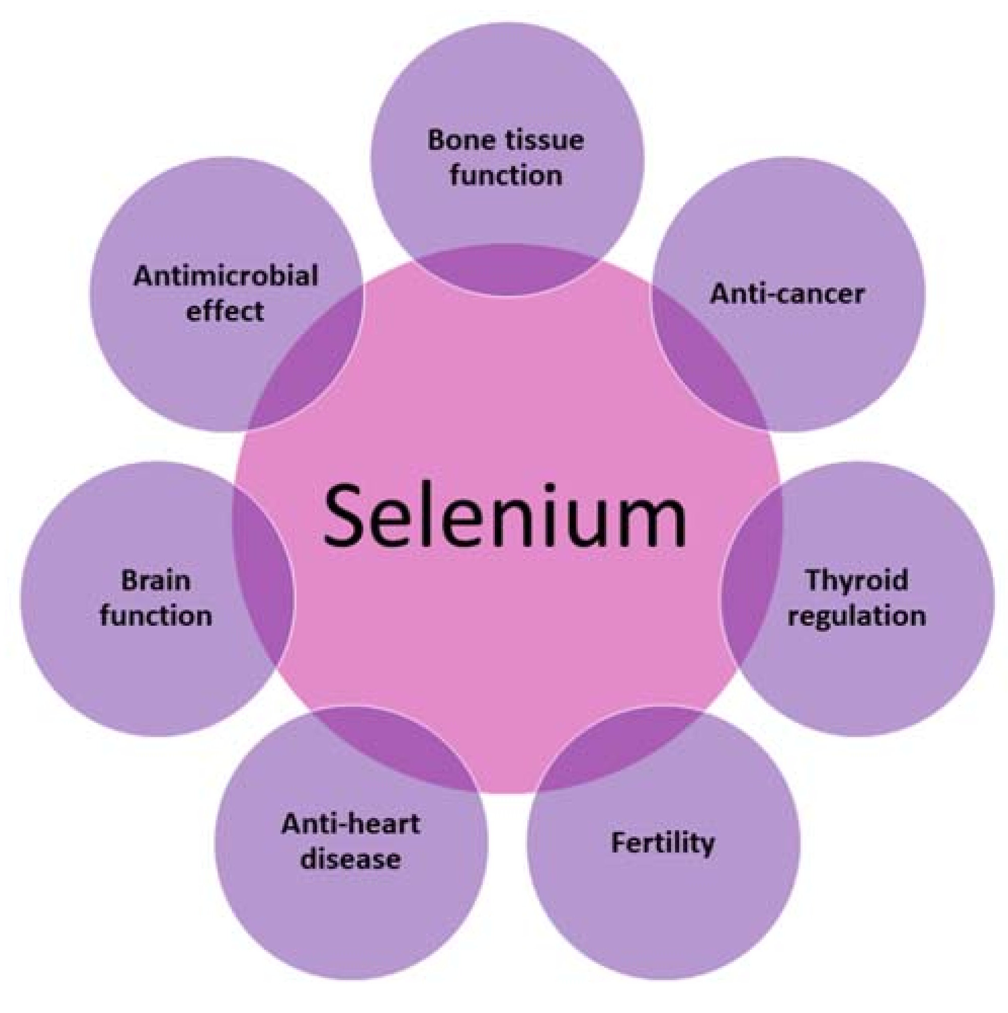
Selenium is one of the essential microelements determining the proper functioning of the human body. The characteristic feature of selenium is its narrow therapeutic window—there is a very small difference between a concentration having a beneficial effect on the human body and one at which toxic activity is exhibited [10]. Selenium exhibits antioxidant properties and protects the body against free radicals and carcinogens. It contributes towards many biochemical processes as an important component of the 25 known selenoenzymes, e.g., glutathione peroxidase or iodothyronine deiodinase [11,12,13]. To date, there have been reports on the positive effects of selenium on cardiovascular diseases, cancers, the thyroid, the brain, reproduction, bone tissue, and viral infections, as well as on the immune system (Figure 2).
Selenium also has immunostimulatory properties. It has been demonstrated that it increases the proliferation of activated T lymphocytes, increases the cytotoxicity of cancer cells, and also activates NK cells. The possible mechanism of the immunostimulatory action of selenium involves an increase in the expression of interleukin 2 receptors, which induces the proliferation of T lymphocytes [14,15]. It was proven in studies on mice that those on a diet rich in selenium showed an increased expression of interleukin 2, which was accompanied by a strengthened signal from T lymphocytes [16].
It was proven that selenium deficiency affects the development of epileptic seizures, and Parkinson’s disease, as well as leading to coordination problems and a decline in cognitive skills [17,18]. Selenium also has a significant effect on male fertility [19,20]. It participates in the biosynthesis of testosterone, as well as in the formation and development of spermatozoa [19].
The thyroid is the organ containing the highest concentration of selenium. As mentioned, selenium is a component of selenoenzymes—for example, deiodinase, which is involved in the production of active thyroid hormones—and triiodothyronine from an inactive precursor, thyroxine. Moreover, selenium in the form of glutathione peroxidase protects the thyroid cells against hydrogen peroxide, which is formed as a result of the synthesis of thyroid hormones. An excess of this compound could cause the destruction and fibrosis of the gland [13,21,22].
Studies have indicated that low blood levels of selenium are also associated with myocardial infarction incidents and an increased risk of death due to cardiovascular diseases. Selenoproteins prevent the oxidative modification of lipids, inhibit platelet aggregation, and inhibit inflammation [21,23]. Therefore, selenium, through glutathione peroxidase, helps to protect the endothelial cells of blood vessels from the deposition of oxidized low-density lipoproteins arising from the oxidation of phospholipids and cholesterol esters. Therefore, it prevents atherosclerosis and its consequences [24,25,26].
The development of cancers is mediated by, inter alia, oxidative stress, as well as impaired bodily protective functions [13,27]. In recent years, scientists have been conducting intensive research on the effect of selenium on the reduction of colon, lung, liver, thyroid, and prostate cancer. Czeczot et al. [27] found a decrease in the activity of glutathione peroxidase in hepatic liver tissue when compared to healthy tissue. This weaker level of enzyme activity may result in enhanced lipid peroxidation and an increase in the number of final peroxidation products, such as malonic aldehyde (MDA). Increased MDA levels were observed in tumour tissue. The studies prove that there may be many mechanisms by which selenium inhibits carcinogenesis and improves the effectiveness of cytostatic drugs, as well as reasons why the relationship between selenium dose and tumour growth is not linear [24,28].
Selenium is also essential for the proper functioning of bone tissue. It has been proven that selenium deficiencies may delay growth and affect the metabolism of bone tissue [29]. The mechanism of these processes is associated with the function of selenoproteins, of which at least nine are expressed in human foetal osteoblasts. Their expression seems to protect bones from oxidative stress, which is important in the regulation of inflammation and the differentiation of bone cells. An excessive level of intracellular reactive oxygen species (ROS) may contribute to the development of osteoporosis by inhibiting the differentiation of osteoblasts in bone marrow stromal cells [30,31]. It was found that the concentration of selenium in plasma is inversely proportional to the rate of bone tissue turnover and positively correlated with the incidence of low bone mineral density in healthy postmenopausal women [32]. Selenium may play an important role in cells, especially at doses higher than those required for the maximal expression of selenoproteins. It can induce cell-cycle inhibition, apoptosis, immune function or the prevention of bone resorption through the inactivation of osteoclasts. These processes can provide potential protection against rheumatoid arthritis, osteoarthritis, or osteoporosis [31].
2. Synthesis of Hydroxyapatites Doped with Selenite and Selenate Ions
HA doped with selenium ions can be obtained in two ways: through synthesis, during which ions are incorporated into the HA structure [33,34,35,36,37,38,39,40,41,42,43,44,45,46,47], or by ion exchange, where soaking in a selenium salt solution results in the exchange of ions in the HA structure [47].
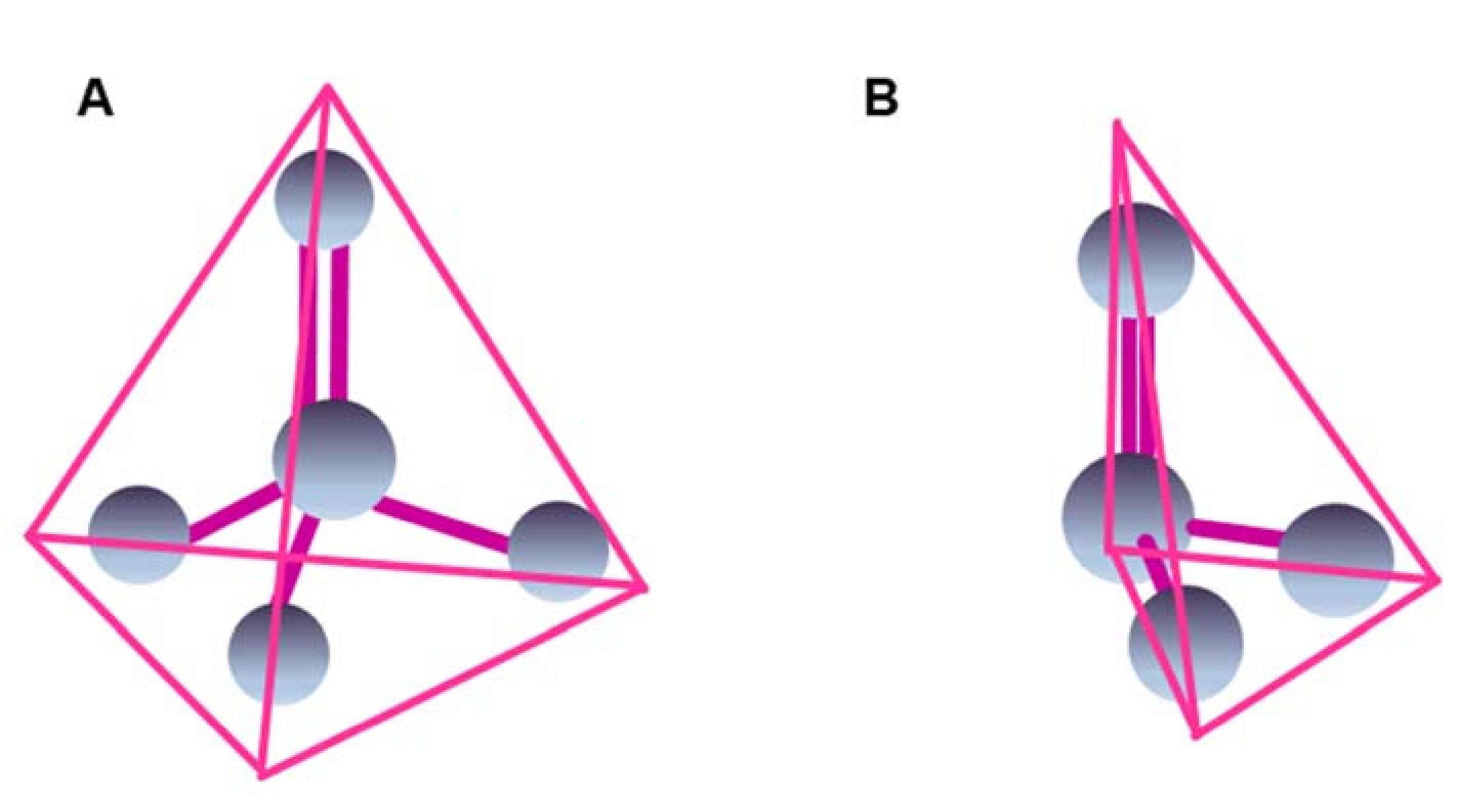
Theoretically, there are two positions in the unit cell of the HA crystal in which it is possible to incorporate selenite (SeO32−) or selenate (SeO42−) ions: into the structural channels of OH groups along the c-axis or in the orthophosphate site [29]. SeO42− ions have a structure geometrically similar to the PO43− ion, although they are much larger and measure 249 pm in diameter compared to the diameter of 238 pm of the PO43− ion [42,43,48]. SeO32− ions have a similar diameter to that of PO43− ions, measuring 239 pm; however, they differ in their geometric arrangement and have a flat trigonal pyramid structure [43,48,49] (see Figure 3). Looking at the size of SeO32− and SeO42− ions, their incorporation into the structure of HA is possible, but, as shown in the model diagram by Kolmas et al., only in place of phosphate ions [40]. Moreover, the authors noticed that, in the case of PO43− ion substitution with SeO32− or SeO42− ions, we substitute a triple charge ion with a double charge ion which, in turn, leads to the simultaneous removal of Ca2+ and OH− ions according to Equation (1) [43]:
where x is the content of selenium oxyanions, and n may be 3 or 4 depending on whether a selenite or selenate ion has been substituted.
Ca10(PO4)6(OH)2 + xSeOn2− → Ca10−x(PO4)6−x (SeOn)x(OH)2−x + xPO43− + xCa2+ + xOH−
Studies on the adsorption of selenium ions to the surface of HA crystals have examined both the adsorption of selenite and selenate ions [50,51,52]. These studies have confirmed that these ions are incorporated in the place of phosphate groups, but it was surprising that SeO32− or SeO42− ions do not stay on the surface, but diffuse into the interior to a depth of a few nanometres [51]. Moreover, the sorption capacity of the selenite ions was significantly higher than that of selenate ions [52].
During Se-HA synthesis, selenium-doped HA was most frequently obtained (both in the form of SeO32− and SeO42−) using the wet method with a co-precipitation reaction.
The obtained powders contained various amounts of selenium [33,34,35,36,37,38,39,40,41,42,43,44,45,46,47], most often up to 10% by weight [33,36,37,40,41,43,44,46]. It should be noted that the reaction efficiency was often lower than 100% [33,36,40,41,43,44,46]. Ma et al. obtained a SeO3-HA series starting from a small concentration of 3% of Se-substituted phosphate ions up to the almost full replacement of P ions with Se ions using a molar ratio of Se:P at 100:1 during the synthesis [45].
It is worth noting that the SeO3-HA obtained in the studies [34,38,39] was used to prepare biocomposites containing silk fibres [34] or lysozyme [39]. It was possible to incorporate between 6 and 30% of selenium ions relative to phosphorus ions into such materials.
Zhang et al. [42] slightly modified the wet method. The precipitated crystals were treated hydrothermally for 36 h at 160 °C. By so doing, selenium ions were incorporated in a molar ratio of Se:P ranging from 0.001 to 0.421 [42]. The solvothermal method of heating for 10 h at 120 °C was also applied by Sun et al. [35]. The reaction was carried out in a Teflon-lined reactor with the addition of PEG 20000 and oleic acid in substrate proportions that allowed a series of samples to be obtained that contained between 0% and 55% of phosphate ions replaced with selenite ions [35].
In the studies [33,37,40], a dispersion agent was used in the wet method in the form of either sodium polyacrylate [37,40] or PEG [33]. In turn, the post-precipitation method, in which the surface phosphate groups are exchanged with selenium ions, was used by Uskokovic et al. [47]. They immersed previously precipitated HA crystals in a sodium selenite solution (pH ~10) while maintaining the solid phase content within the range of 10–40% w/w for a period of 48 h. In this way, a maximum of 0.36% by weight of selenium was incorporated into the hydroxyapatite structure [47].
In the case of surface HA coating, the PLD (pulsed laser deposition) application of powders, consisting of a mixture of HA and selenium powder, is used [53,54]. In such coatings made on titanium or silicon, the plan was to incorporate up to 2.7% of selenium [53,54]. The apatite material was applied using the PLD method under reduced pressure in a steam atmosphere, heating the carrier up to 460 °C using a laser with a wavelength of 193 nm [53,54].
HA coatings can also be obtained by soaking carried out in a simulated body fluid (SBF) doped with selenate ions [55].
3. Physicochemical Examination of Hydroxyapatites Doped with Selenate and Selenite Ions
In order to confirm the HA structure during the incorporation of selenium ions, the samples were analysed using powder diffractometry (PXRD). Based on the resulting diffractograms, the size of the obtained crystallites, the crystallinity, as well as the size of the unit cell of the obtained crystals, were also calculated. The morphology of the obtained crystals can be described using images taken via transmission and scanning electron microscopy (TEM and SEM). In order to confirm the incorporation of SeO32− and SeO42− ions into the HA structure, spectra were performed by using medium-infrared spectroscopy, Raman spectroscopy, and nuclear magnetic resonance imaging. Elemental analysis of the samples was performed using energy-dispersive X-ray spectroscopy (EDS), X-ray fluorescence spectroscopy (XRF) and X-ray photoelectron spectroscopy (XPS), as well as the titration method.
3.1. Powder Diffractometry (PXRD)
In each of the discussed studies, it was confirmed, based on a comparison of the obtained results with a reference sample containing no selenium ions and/or with a model HA diffractogram originating from ICDD and JCPDS databases, that monophasic material was obtained with a preserved P63/m crystallographic system characteristic of HA.
In the study [43], diffractograms obtained for samples doped with selenate and selenite ions were compared with a diffractogram of standard HA and it was shown that the former have weakly separated wide reflections and that some of them are too wide to be distinguishable. Based on the Scherrer equation, the crystal dimensions were estimated using the reflexes (002) and (130/310) along the c and a axis, respectively. It was found that, after the introduction of selenite and selenate ions, the size of the crystals was similar to the size of crystals of biological apatites in bone tissue [43]. However, a decrease in the size of crystallites in comparison to pure HA was observed in the study [36], with dimensions of below 10 nm. The crystallinity of the obtained HA was highest for pure HA and lowest for SeO3-HA [41].
The powder diffraction method was also used in [34,37,38,39,44,46] to confirm the obtaining of monophasic HA material. The widening of reflections was observed as selenite concentration increased, which indicates a decrease in the size of crystallites, as well as a decrease in crystallinity [34,38]. The sizes of the SeO3-HA crystallites, calculated using the same method as in [46], are approximately 20 nm and are not much smaller than in pure, unsubstituted HA. A decrease in crystallite size along the c axis with an increase in Se concentration was also observed in the HA-lysozyme composites [39].
Ma et al. [45] attempted the almost total exchange of phosphate ions for selenite. Based on a comparison with the reference diffractogram, the behaviour of the hydroxyapatite monophase was determined to be highest at an Se:P ratio of below 10 [45]. At higher concentrations, reflections characteristic of calcium selenite were observed [45]. In the samples in which the HA structure was preserved, the lines were significantly widened.
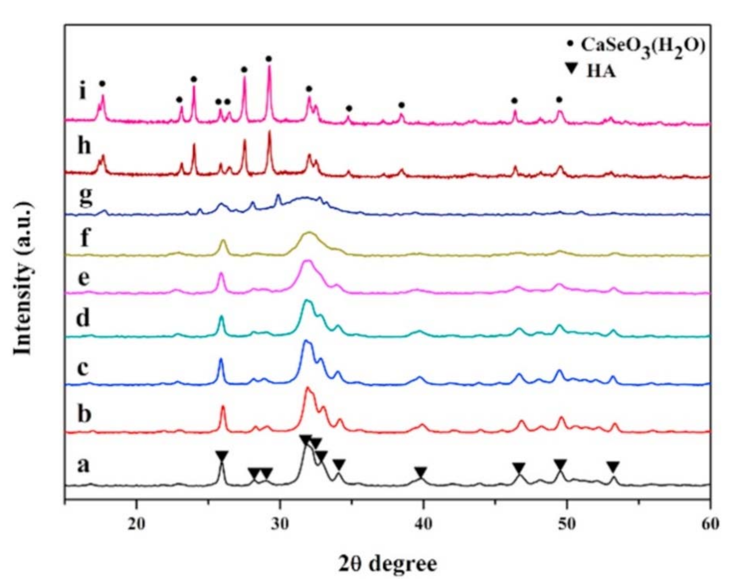
Similar results were achieved by Wei et al. [33]. They observed the reflections from the HA phase on the diffractogram in samples with a molar Se:P ratio of a maximum of 0.3 (Figure 4). In samples with a Se:P value > 1, reflections from calcium selenite were observed. The paper also describes samples that were heated at a temperature range from 700 to 1200 °C, which resulted in the sharpening of reflections, proving that the crystallinity of samples increased. In some samples heated at temperatures between 1100 and 1200 °C, the occurrence of the β-TCP phase was noticed [33].
Zhang et al. also confirmed the production of SeO3-HA in all samples using the PXRD method. The calculated crystallite size decreased from 45.3 to 17.4 nm [42]. As in other studies, it was noticed that the reflections became wider with increasing selenium concentrations. The results obtained with the PXRD method were additionally confirmed by the SAED electron diffraction method [42].
Samples of SeO3-HA obtained by the solvothermal method, with 15–40% of the phosphate ions being exchanged for selenite, have reflections typical of HA [35]. In the sample with 55% P exchanged for Se, additional reflections were noted from the calcium selenite [35].
Uskokovic et al. [47] confirmed the achievement of a monophasic hydroxyapatite material for samples with concentrations of SeO32− ions ranging from 0.1% to 1.9%. However, the sample with SeO32− concentration of 3% additionally contained reflections from the calcium pyrophosphate phase (β-Ca2P2O7), which became visible only after calcination at 800 °C. The widening of reflections with increased SeO32− ion content was also noticed [47]. The authors suggest that the decrease in crystallinity with an increase in the content of selenium ions is caused by an uneven dissolution rate in various environments. In an acidic environment, much more Ca2+ ions than PO43− ones are released, while in an alkaline environment, this mechanism is reversed. However, during the formation of HA, the first stage is the creation of a phosphate-based lattice with incorporated calcium ions, and this process is promoted by a higher concentration of phosphate ions. In turn, the replacement of some of phosphate ions with selenium ions during synthesis reduces the concentration of PO43− ions, which causes the disturbance of the structure growth and as a result, the macro structure is disturbed (long-range order). In addition, this may hinder recrystallization, as a result of which the amorphous particles change into larger crystalline structures. The result of this is that the transformation and preservation of the amorphous structure is inhibited. The authors see another possible cause of structural disruption, i.e., only a few double-charged SeO32− ions are incorporated into the crystal structure.
Based on the above-described diffractograms, the crystallite size for samples obtained by both co-precipitated and post-precipitated methods [47]. In both types of samples, the incorporation of selenite ions causes a decrease in the size of crystallites, but this is not correlated with the content of these ions in the sample [47].
Coatings obtained by soaking in SBF were analysed using the PXRD method and they were then compared with the reference HA diffractogram [55]. The reflections for the obtained HA and SeO4-HA coatings are typical of amorphous HA; they have wide lines with a position characteristic of the HA phase. Since the width of the reflections on the diffractogram of the SeO4-HA coating does not differ from the reflections of the HA coating, the authors claim that both materials have a similar crystallinity.
3.2. Examinations Using Electron Microscopy Methods (SEM, TEM)
Electron microscopy is a frequently used method when analysing materials with a crystalline structure, since it allows for an evaluation of the morphology and size of the crystals. The materials obtained in [36,43] had plate-shaped nanometre crystals; their dimensions did not exceed 10 nm in width and 30 nm in length [43]. The crystals were characterized by a strong tendency to form dense agglomerates. In turn, Wang et al. obtained crystallites similar in shape to needles during the synthesis of SeO3-HA, through the linear aggregation of single crystallites [38]. The dimensions of such nanoparticles were determined, based on TEM images, to be 20–30 nm in width and 150–200 nm in length [38,44,46]. Examinations of the size of nanoparticles using the dynamic light scattering (DLS) method indicated that there was a minimal increase in particle size in samples containing selenium ions, that it caused by a high tendency to agglomerate [38,56].
In the study [37], the synthesis method was modified by adding a dispersant, which enabled the formation of needle-shaped crystals with dimensions of below 100 nm, as confirmed by DLS and TEM measurements.
TEM and SEM images of a composite based on SeO3-HA and silk fibres showed that in the samples without selenite ions, the crystals agglomerated as polycrystalline bamboo-shaped needle bundles [34], while the sample containing 6% selenium also contained needle-shaped crystals; however, they were more clustered and proteins derived from silk fibres covered the surface of the polycrystal like a thin film. For samples with higher concentrations of selenium, a related ribbon structure with the appearance of a large grid was observed [34]. Structural disorders were also noticed in the form of separated large crystals. Simultaneous SEM images confirmed that silk fibres covered the surface of polycrystalline HA, forming a porous grid structure [34].
In contrast, HA-lysozyme composites [39] obtained by Wang et al. had narrow, sharp needles which tended to agglomerate; the dimensions, measured based on TEM images, were 10–25 nm in width and 120–150 nm in length.
Photographs taken using the field emission scanning electron microscopy (FE-SEM) method show the very irregular surfaces of composites that can be formed during the drying process, when the water molecules evaporate from the loosely adhesive interface. Images taken with this method also indicate the agglomeration of nanocomplexes.
Needle crystals were obtained in the study [40]. It was also observed that an increase in selenium content increased the size of the crystals and intensified their agglomeration [40].
The shape of the 150–200 nm long needles was recorded in TEM images by Wei et al. [33]. The authors compared samples with the Se:P molar ratios of 0.3 and 0.5, and noticed that an amorphous fraction appeared on the surface of crystals in a sample with a higher selenium content, which was confirmed by results obtained using the PXRD method.
Zhang et al. obtained rod-like crystals using the hydrothermal method, and based on SEM and TEM images, their length and width were measured to be 50 and 12 nm, respectively [42].
However, the use of the solvothermal method resulted in rod-like nanoparticles with dimensions of 8 to 150 nm [35]. Low concentrations of selenium (up to 15%) exchanged with P caused the rods to narrow and the ends to tighten, while an increase in the content of incorporated selenium to 40% caused a change in shape, where the rods transformed into needles of up to 100 nm in length. The authors of the study [35] also verified that the reaction temperature had an influence on the morphology of the nanoparticles. After reducing the reaction temperature to 100 °C, the obtained nanoparticles were shaped like rods. On the other hand, gradually raising the temperature to 200 °C resulted in the particles maintaining their length but gradually becoming narrower.
Uskokovic et al. compared images of pure HA and SeO3-HA recorded using the TEM method, and noted that pure HA is composed of two types of crystals with particles of approximately 5 nm and elongated crystals with dimensions of 10–15 nm × 50–100 nm [47]. The coexistence of the two forms is indicative of aggregative crystal growth and is the result of the tendency to grow mainly along the c axis of the crystal. In contrast, SeO3-HA obtained using the wet method had homogeneous grains measuring 20–50 nm in diameter. This is probably due to the fact that SeO32− ions block the aggregation of small particles into more crystalline needles, which causes the resulting SeO3-HA to be less crystalline than pure HA.
The HA and SeO3-HA coatings obtained by the PLD method on Ti and Si materials were analysed by SEM [54]. The researchers described the surface morphology as being typical of HA, consisting of spherically shaped aggregates; moreover, the similarity of the HA and SeO3-HA coatings indicated a similar mechanism of formation by globular groupings.
Coatings deposited using SBF doped with selenium ions were analysed based on field emission FE-SEM images [55]. The researchers noticed the first calcium phosphate compounds after four days. It turned out that, after 14 days of soaking, the whole surface was covered with semicircular compact SeO4-HA with a homogeneous morphology. When comparing the coatings, the authors noticed that their thickness increased with longer soaking times, and that the coating based on Se-doped SBF was more homogeneous [55].
3.3. Examinations Using Mid-Infrared Spectroscopy (FT-IR)
HA samples doped with selenium were analysed using mid-infrared spectroscopy in the range of 4000–400 cm−1 [36,38,43,45,46]. Based on the reference spectrum of hydroxyapatite, the occurrence of HA-specific bands was confirmed (see Table 1). Additionally, Kolmas et al. [36,43] recorded two bands at 767 cm−1 and 840 cm−1 on mid-infrared spectra for HA doped with SeO32− ions, the intensity of which increased with the content of selenite ions (Figure 5). These bands were assigned to symmetrical and asymmetrical vibrations of the Se–O bond of selenite ions in HA crystals. In turn, a band at approximately 505 cm−1 was attributed to bending vibrations [43]. For the HA doped with selenate ions, an additional band at 910 cm−1 was recorded that was not observed on the pure HA spectrum, which was attributed to the Se-O stretching vibrations of the SeO42− group. Based on the location of these bands, the authors confirmed that selenite and selenate ions had become incorporated into the structure of HA crystals.
Wang et al. [46] recorded signals assigned to the phosphate and hydroxyl groups in the spectra, confirming that they were receiving SeO3-HA. The authors distinguished 1460 and 1420 cm−1 signals, indicating the presence of carbonates in the samples. Moreover, the signal at 784 cm−1 confirmed the incorporation of the SeO32− ions, while the 874 cm−1 signal was assigned to selenite and carbonate ions. All bands derived from selenite and carbonate ions increased as the Se:P concentration increased. In [38], the same bands were observed, but attention was paid to the significant reduction of the bands from the hydroxyl groups at 3570 and 630 cm−1 when compared with the pure HA spectrum.
Ma et al. [45] noted that, as the selenium ion content increased, there was a reduction in the strong bands attributed to the symmetrical and antisymmetric vibrations of the P–O bond in the 1200–900 cm−1 range, as well as in the bands of asymmetrical bending vibrations of the O–P–O bonds (bands at 604 and 567 cm−1). They also noticed that, despite the incorporation of selenite ions, bands derived from phosphate groups were unaffected. However, the position of both the stretching and bending bands was obviously unaffected by the selenite substitutions. Similarly, when the Se:P ratio increased, the content of B-type carbonates decreased (bands at 1456 and 1413 cm−1), while the intensity of the band at 1564 cm−1, derived from A-type carbonates in the SeO3-HA samples, was higher than the intensity of the bands derived from B-type carbonates. The most intense bands from selenite appeared in the 900–800 cm−1 range, and originated from antisymmetric stretching vibrations, while the 766 cm−1 band originating from the bending vibrations of O–Se–O bonds increased as the Se concentration also increased.
On the recorded FT-IR spectra, Zhang et al. observed bands that were typical of HA, which originated from phosphates, carbonates, water and hydroxyl groups. They additionally recorded bands at 869 cm−1 and 775 cm−1, which they assigned to the antisymmetric stretching vibrations of SeO4 tetrahedron and bending vibrations (ν3) O–Se–O [42].
In the studies [33,35,40], it was observed that the intensity of the band originating from the structural vibrations of OH groups at 3570 cm−1 decreased as the Se concentration increased. The authors [35] assigned the bands 767 and 712 cm−1 to the symmetric stretching vibrations (ѵ3) of the selenite group. Moreover, when the concentration of selenite ions increased, the 856 and 823 cm−1 signals corresponding to the ѵ1 and ѵ3 vibrations of the SeO32−group became stronger.
The spectra also showed bands derived from C–H, –CH2−, and COO− groups from oleic acid, which indicates that the samples were covered in this acid [35].
Liu et al. [40] observed a band originating from the vibrations of selenite groups at 766 cm−1. This signal was observed for the first time with a sample containing a planned content of 3% Se, and it increased as the selenium content increased. There was also an increase in the intensity of the bands from CO32− ions in the 1418–1566 cm−1 range, along with an increase in selenium content, which was probably caused by the facilitation of CO32− group incorporation as the crystallinity of the samples decreased.
The examination of SeO3-HA coatings obtained by the PDL method confirmed the occurrence of HA-specific water bands, as well as phosphate and carbonate groups. It was also observed that, as the coatings increased in terms of Se content, not only did the bands from carbonates decrease, but the main band from phosphate groups (antisymmetric stretching) also decreased and its half-width increased. There was also a simultaneous loss of definition in the bending bands from the PO43− group, as well as shift of the main phosphate band from 1040 cm−1 for the pure HA coating to about 1063 cm−1 for the coating containing the highest concentration of selenium. This band shift was caused by a change in the length of bonds, which was a consequence of the incorporation of Se ions [54].
3.4. Examinations Using the Raman Spectroscopy Method
Studies on samples of hydroxyapatites doped with selenium ions, conducted using Raman spectroscopy, have been described by several authors.
When comparing coatings with pure HA and SeO3-HA, obtained using the PLD method, in material doped with Se ions, signals that were characteristic of phosphates were confirmed: the they were strongest at 960 cm−1 from bending vibrations (ѵ1), at about 1070 cm−1 from stretching vibrations (ѵ3), at 590 cm−1 from bending vibrations (ѵ2), at 430 cm−1 from bending vibrations (ѵ2) and symmetric stretching bands (ѵ1) and at 1064 cm−1 derived from carbonate groups. In addition, a band of approximately 830 cm−1 originating from the symmetric stretching vibrations of the SeO32− groups was registered for the SeO3-HA coatings [54].
Kolmas et al. recorded three bands at 911, 873, and 843 cm−1 for the sample doped with SeO42− ions, which was assigned to selenate ions [43].
In contrast, Yilmaz et al. [55] examined the SeO4-HA coating using Raman spectroscopy. In addition to the bands characteristic of phosphates at 1072, 961, 590, and 429 cm−1, as well as bands originating from the substrate at 629, 270, 209, and 136 cm−1 in a sample soaked for 14 days in a 1.5 × SBF doped with Se ions, a weak signal from selenates was also observed at 764 cm−1 [55].
3.5. Examinations Using Nuclear Magnetic Resonance Spectroscopy (NMR)
Kolmas et al. [41] subjected the obtained samples of HA doped with SeO32− and SeO42− selenium ions to ssNMR examination. Spectra were recorded for the nuclei 31P, 1H, and 77Se.
The 31P spectra were recorded using two techniques: one-pulse (Bloch-decay, BD) and 1H→31P cross-polarization (CP). One signal characteristic of hydroxyapatite samples at about 3 ppm was recorded on the spectra. It was noted that the signal in the 31P BD NMR spectra was narrowest for the pure HA sample and widest for the SeO3-HA sample; moreover, because the line width was correlated with the crystallinity of the sample, this was indicative of its reduction in the selenium-doped samples.
The signal in the 31P CP NMR spectra was deconvoluted into two components: narrow, derived from phosphate groups inside the crystal located near the protons of structural hydroxyl groups, and broad, derived from phosphate groups located near to the water-rich environment (especially from phosphors from the hydrated surface layer) [41,57]. The wide component was relatively the most intense in the spectrum of the sample containing SeO32−, indicating a more extensive hydrated surface layer and, thus, a more developed surface. In all of the recorded 1H MAS NMR spectra, two signals can be distinguished at about 0 and 5.4 ppm. The signal at about 0 ppm comes from structural hydroxyl groups. The OH groups form columns in the channels, but they are so far apart that it is impossible to create hydrogen bonds between them. In spectra registered for samples doped with selenium, the chemical shift of this signal moved toward positive values, which were highest in the sample containing selenites. The widening of the signal in the Se-HA samples was also observed to be more significant than the signal from pure HA. The authors suggest that, due to the incorporation of selenium ions into the hydroxyapatite structure, the column structure of the OH group became disturbed, resulting in the formation of weak hydrogen bonds. The content of structural hydroxyl groups was calculated, and it was observed that there was a significant loss of HA crystals with incorporated selenite ions (49% OH groups relative to stoichiometric HA) and selenate (63%) when compared to pure HA (77%). It was also proven that this reduction in the content of structural hydroxyl groups was related to the decreased size of the crystals [43,57] due to the absorption of water from the crystals’ surface into the columns of the hydroxyl groups. Thus, the authors postulate that the reason for the loss of structural hydroxyl groups is two-fold: not only are OH groups removed during substitution, but also water is present in the OH columns [41].
The signal at about 5.4 ppm is attributed to water adsorbed onto the surface of the crystals. The spectra for the 77Se nucleus were recorded using the 1H→77Se CP technique. The obtained spectra for the HA containing selenates showed two intense signals at 1045 ppm, and a much weaker one at 1027 ppm. Furthermore, for the sample containing selenites, there was an analogously strong signal at 1310 ppm and a weaker one at 1325 ppm. It was proven that the strong signals on both spectra originated from selenium ions embedded in the crystalline lattice of the crystal, while the lower intensity signals were derived from selenates and selenites in the hydrated surface layer.
4. Biological Examinations of Hydroxyapatite Materials Doped with Selenium
Biological examinations of HA materials containing selenium have focused primarily on the determination of their antibacterial, anticancer, and cytotoxic effects, as well as their influence on the development of bone tissue. In addition to in vitro studies, some research groups have also conducted experiments using an in vivo animal model (mice or rats). Below, we will try to illuminate the results and conclusions from the studies.
4.1. Antibacterial Activity
Studies to determine the antibacterial activity of SeO3-HA and SeO4−HA were carried out on the following bacterial strains: Staphylococcus aureus, Pseudomonas aeruginosa, Staphylococcus epidermidis, Escherichia coli, and Salmonella enteritidis. This is because they are the bacteria that contribute most often to infections during orthopaedic surgery. S. aureus is of particular note, as it is a very common pathogen that causes infections during bone implantation due to the ease of biofilm formation and the presence of numerous antibiotic-resistant strains.
Measurements of antibacterial activity have typically been performed through determining the optical density of the bacterial suspension, counting the CFUs attached to the material, determining the area of inhibition using the disk diffusion method, or evaluating biofilm formation. The measurements were taken after the bacteria had been incubating for an appropriate length of time on the growth medium in the presence of the examined materials.
In the case of S. aureus, all of the studies [47,53,54,58,59] observed that SeO3-HA materials significantly reduced bacterial viability, inhibiting both their growth and biofilm formation, when compared to the control and samples with pure HA. According to the study by Rodríguez-Valencia [54], the introduction of 0.6 wt % of selenium ions into the HA structure was already sufficient to inhibit the formation of an S. aureus biofilm (as well as P. aeruginosa) (Figure 6).
Additionally, according to Uskokovic [47], despite the antibacterial effect of the SeO3-HA material on S. aureus, these properties were lost over time and the bacterial population returned to a level similar to that observed in the negative control group. This may indicate that those S. aureus bacteria that survived their initial exposure to SeO3-HA did not later show susceptibility to SeO32− ions, which allowed the population to rebuild.
The results of studies carried out on E. coli bacteria are interesting. These bacteria are characterized by their ability to reduce SeO32− ions to Se0, which they can then use to synthesize the amino acids selenocysteine and selenomethionine. Due to this fact, the level of SeO32− ion concentration will determine whether E. coli growth is stimulated or inhibited. According to the study carried out by Kolmas [58], a selenium content of 3.6 wt % in the HA structure was insufficient to inhibit the growth of E. coli. In turn, according to Murugan [59], there was a decrease in the functioning and lifespan of bacteria at all applied concentrations of SeO3-HA, which was further confirmed by the study of Uskokovic [47], in which E. coli bacteria were still sensitive to the examined biomaterial 72 h after the start of incubation at the highest level of selenium content in the analysed SeO3-HA material (1.92 wt % and 3 wt %).
It is worth emphasizing that studies performed on both E. coli and S. aureus [47,59] indicate that the SeO3-HA material has a stronger effect on these strains than on the first of the mentioned strains. This may be due to the presence of a thinner layer of peptidoglycan in the cell wall in Gram-negative bacteria, which makes it easier for the active substance to penetrate inside the cell.
According to Rodríguez-Valencia [53], as the selenium content in SeO3-HA increased, there was a gradual increase in its antibacterial activity against S. epidermidis and a gradual inhibition of biofilm formation until it completely disappeared 72 h after the start of incubation. This was confirmed by the study of Yilmaz [55], in which a significant decrease in optical density was observed after 72 h of incubation in a sample with an SeO4−HA-coated titanium implant, as compared with a sample with an uncoated implant, one with a pure HA coated implant and the control sample.
Antimicrobial activity was also tested for the strains P. aeruginosa and S. enteritidis. The results of the studies confirmed the effectiveness of SeO3-HA in inhibiting the growth of these microorganisms by [47,54].
In conclusion, the studies carried out confirmed that HA material containing selenium has an antibacterial effect on individual bacterial strains. The antibacterial effect of this biomaterial is based on inducing the formation of reactive oxygen species in a bacterial cell. According to Kramer and Seko [60,61], oxidation and reduction reactions which occur during selenium metabolism lead to the formation of hydrogen peroxide and superoxide (i.e., reactive oxygen species), which, in turn, contribute to oxidative stress and damage to the bacterial cell wall. It is worth noting that the antibacterial effect of SeO3-HA is stronger for Gram-negative bacteria than it is for Gram-positive ones [47,59], while the adsorption of antibiotics on the surface of SeO3-HA (e.g., vancomycin) increases the obtained antibacterial properties [47].
4.2. Anticancer, Cytotoxic, and Osteoinductive Effects
The main assumption during the synthesis of hydroxyapatite materials doped with selenium is that biomaterial will be obtained that will effectively inhibit the development of bone cancers, while showing no cytotoxic activity on healthy tissue. Another important feature of this material is its ability to induce osteogenesis in order to stimulate bone tissue reconstruction at the site of a bone defect.
In vitro studies were performed on healthy mouse cells (mouse MC3T3-E1 preosteoblasts and mouse fibroblasts collected from the lungs), healthy human cells (human foetal hFOB osteoblasts, bone marrow stromal cells (BMSC) and human fibroblasts), mouse cancer cells (murine ATCC osteosarcoma cells and K7M2), and human cancer cells (human MG−63 osteosarcoma cells, Saos-2, and MNNG/HOS). In vivo studies were performed on Wistar rats and BALB/c mice.
4.2.1. In Vitro Studies
In vitro tests on healthy cells primarily allowed for an evaluation of the biocompatibility of the discussed material, as well as its osteogenic properties. A study on biocomposites constructed from SeO3-HA and silk fibrin (SF/Se-HA), carried out by Wang [34] on BMSC cells, clearly indicated that the presence of selenium in the biomaterial increased the intensity of cell population growth on days one to seven from the start of the study compared to the control group and SF/HA sample, in which a relatively slow increase in cell population growth was observed. In addition, after seven days of incubation, the observed cell density was highest for the biomaterial samples with the highest selenium content. An increase in selenium content resulted in an increase in the rate of osteoblast proliferation, which was additionally confirmed by Vekariya’s study [62]. In turn, the study on the effect of SeO3-HA materials on MC3T3-E1 cells, carried out by Jianpeng [35], allowed for an assessment of cytotoxicity with respect to healthy cells. They analysed the effect of two SeO3-HA materials with a ratio of Se:(P + Se) of 0.05 and 0.1, respectively (using different concentrations of the examined materials). In this case, not only was no cytotoxic activity observed in the sample containing the material with the highest selenium concentration, but it was also found to have a greater beneficial effect on the viability of MC3T3-E1 cells than in the case of pure HA samples. Rodríguez-Valencia’s studies [53,54] confirmed the lack of cytotoxic activity of SeO3-HA, where cell viability in the samples containing SeO3-HA was at a similar or even a higher level than in the pure HA samples. The morphology of the MC3T3-E1 cells also did not change with incubation time [35,54]; numerous filopodia and lamellipodia could be observed in the cell structure, which enabled them to spread on the surface of the biomaterial [54]. This confirmed the biocompatibility of SeO3-HA material with mouse preosteoblasts cells. The activity of osteoblasts was also analysed in studies carried out by Rodríguez-Valencia [53] and Uskokovic [47]. It was shown that osteoblastic activity also increased with incubation time and with increased selenium content in HA materials. Differences in the results were due to the selenium concentrations in the HA material, where osteoblastic activity was highest at higher selenium concentrations. For the first study, this was observed for sample containing the highest concentration (2.7 wt %) of selenium in the HA, while in the second study, this activity was highest at a concentration of 1.23 wt % and decreased when the selenium content increased any higher than this value. At the highest selenium concentration used (3 wt %), osteoblastic activity was significantly inhibited when compared with the pure HA sample, abnormalities in the cells’ structure were visible and some of them died within the first 24 h of incubation. This was due to the effect of selenium on ROS generation and, thus, on the level of oxidative stress in osteoblasts. A suitable amount of selenium enabled the maintenance of oxidative stress at a level where it was able to exert a beneficial effect on osteoblastic activity; however, too much of this element had the effect of increasing oxidative stress to a level at which apoptotic pathways were activated [63,64,65,66].
In vitro tests carried out on cancer cells facilitated the determination of the anticancer efficacy of the examined biomaterials. In virtually every case, these tests were carried out in parallel with studies on healthy cells, which led to a fuller picture of the biomaterial activity. Studies conducted by Wang [34,46] and Uskokovic [47] showed that SeO3-HA material had no effect on the viability and proliferation of healthy cells, while it inhibited the growth and reduced the viability of cancer cells (compared to the control group or HA sample). The effect of SeO3-HA on osteosarcoma cells was stronger at higher selenium levels. The studies of Kolmas [36] and Yilmaz [55] confirmed the abovementioned effect of SeO3-HA (and SeO4-HA) on cancer cells; however, their results also indicated inhibition of growth and the reduction of viability of healthy cells. According to Kolmas [36], this was due to the so-called “burst release” of selenium ions from the biomaterial surface during the first 10 h following the beginning of the study, leading to a high concentration of selenium ions and, thus, causing the effective reduction of viability of both cancer cells and healthy osteoblasts.
Wang [46] confirmed that the anticancer effect of SeO3-HA is due to the induction of apoptosis in cancer cells. In order to illuminate the mechanism of its anticancer activity, both Wang [46] and Wang [37] (convergence of names) performed a number of studies. They both measured the fluorescence intensity of the ROS marker to determine whether SeO3-HA causes an increase in the amount of reactive oxygen species generated. The results of both studies confirmed the above dependence. In other words, they found that Se-HA enhanced the formation of intracellular ROS, which in turn induced apoptosis and activated caspases. Caspase-3 is a key factor in the apoptosis process and is activated by caspase-8 and caspase-9, which initiate two different apoptotic pathways: the caspase-8 extrinsic and the caspase-9 intrinsic apoptotic pathways. In his study, Wang [37] confirmed the presence of activated caspase-3 and caspase-8 and caspase-9 in samples with a high selenium content (not only in the sample with Se-HA, but also in the one with Na2SeO3), which clearly indicated that that caspase-dependent apoptosis was associated with the presence of high selenium content (see Figure 7).
Finally, it is worth mentioning the study performed by Kolmas [43], which tested the toxicity of HA material doped with SeO32− or SeO42− ions in Vibrio fisheri bacteria and Spirostomum ambiguum protozoa. The results indicated that SeO32− ions were toxic to a certain degree for both microorganisms, while this effect was not visible in the case of SeO42− ions, which may indicate that selenate ions (VI) are less toxic than selenite ions (IV).
4.2.2. In Vivo Studies
In in vivo studies on Wistar rats, Wang [44] applied the examined biomaterials in previously prepared skull bone defects. These materials were a biocomposite made of hydroxyapatite and chitosan (HA/Ch) and the biocomposite SeO3-HA/Ch. The control group was made up of rats with a cranial bone defect, and no biomaterials were applied in this group. No complications were observed with the graft, and all of the rats survived until the day of euthanasia (which took place four, eight, or 12 weeks after implantation). The histological examination showed that there was inflammation within the graft or necrosis of the tissue, which was indicative of the biocompatibility of the material with the tissue (this was due, inter alia, to the high porosity of the material). The scaffolding created by the implant began to partially degrade eight weeks after implantation. It was replaced by newly-formed bone tissue, which, however, was less abundant than in the control group. Twelve weeks after implantation, an increasing number of bone connections between the host bone and the implant could be observed in groups with SeO3-HA/Ch and HA/Ch biomaterials. In addition, in the case of SeO3-HA/Ch, larger quantities of stromal cells, ossein, and fibrous cartilage cells were visible within the defect. Furthermore, it should be noted that characteristic polymorphonuclear cells were present, which supported the formation of new bones in the sample with SeO3-HA/Ch [67]. In addition, for the SeO3-HA/Ch group, the formation of new blood vessels was visible (neovascularization). The determination of calcein in all of the implantable materials tested was performed 12 weeks after implantation. The introduction of selenium into the structure resulted in the recruitment of progenitor cells and osteoclasts, which had a positive effect on scaffold degradation and bone remodelling. However, it is difficult to obtain a complete bone-width between the material and the host bone by osteogenesis and neovascularization. According to [68,69], a significant amount of time is needed to bind HA to the host bone, so more research is needed to confirm this. Basing their results on the number of bone beams, Wang [37] also stated that bone quality was higher in the SeO3-HA/Ch group, since this parameter was highest in this sample.
In turn, Wang [37] carried out an in vivo study on osteosarcoma cells in BALB/c mice. In addition to applying SeO3-HA materials with different levels of selenium content, control groups were prepared, which were HA solutions with various amounts of Na2SeO3 added. For the sample of SeO3-HA material with the highest selenium content, the author found that cancer growth was inhibited and the volume and weight of tumours were reduced. The control group sample with an identical selenium concentration gave a similar result. Although all of the mice survived until the end of the experiment without any visible changes in body weight, the analysis of various biochemical parameters (AST, BUN, CREA, and LDH) indicated differences between the various groups in terms of systemic toxicity. It should be taken into account that in vivo toxicity may have decreased under the influence of selenium ions by inducing apoptosis in cancer cells, impairing the metabolism of these cells and protecting healthy tissues. On the other hand, too high a dose of selenium may cause side effects in terms of organ function in the examined mice, thus leading to increased toxicity. The toxicity level was lowest in the SeO3-HA group with the highest selenium content, which was indicated by significantly reduced biochemical parameters. This may be explained by the gradual release of selenium from the SeO3-HA material, which was additionally regulated by the acidic pH of the cancer tissue environment. Therefore, the concentration of selenium in the tumour was high, with a small quantity of selenium ions leaking out of the cancerous tissue. For the control group, where Na2SeO3 was mixed with HA, the whole substance immediately decomposed and spread beyond the cancer tissue into healthy tissues, which caused an increase in toxicity. Studies on healthy BALB/c mice confirmed the lack of toxicity of SeO3-HA materials in comparison with the HA/Na2SeO3 control sample, which showed a significant increase in systemic toxicity. This means that the use of SeO3-HA material enables the avoidance of selenium toxicity because it is gradually released from the material. Wang [37] also confirmed that the mechanism of the activity of SeO3-HA materials in the in vivo model was analogous to that presented in in vitro studies.
Finally, it is worth mentioning that Wang [38] also showed the beneficial anticancer effects of SeO3-HA material on hepatocellular carcinoma cells in a study on BALB/c mice, which increased the survival of the mice and improved their biochemical parameters.
5. Conclusions and Future Perspectives
Hydroxyapatite that contains selenium ions is a material with high application potential. The studies presented in this review have demonstrated that it is possible to introduce both IV and VI selenium ions into the interior of the crystals, preserving the hexagonal structure of the HA. The obtained crystals are nanocrystalline, with a strongly-developed specific surface and a tendency to form large agglomerations. IV and VI selenite ions can also be located in the hydrated surface layer and strongly adsorb onto the surface of HA crystals. Se-HA was successfully used to form composites with silk fibres, sodium alginate and lysozyme. The studies demonstrated that it has the ability to cover metallic implants. Biological tests confirmed the antibacterial activity of SeO3-HA materials; however, it had a stronger effect on Gram-negative strains than Gram-positive ones. In vitro studies indicated the beneficial effect of apatite materials containing selenium on osteoblastic activity. However, it is worth noting that high concentrations of selenium caused a significant increase in the toxicity levels of normal cells. In vitro cancer cell research, supported by in vivo tests, clearly confirmed the inhibition of growth and the reduction of vitality.
These promising results obtained for hydroxyapatite materials containing selenium mean that the subject is still relevant. It is possible to find some attempts in the literature to synthesize co-substituted hydroxyapatites, e.g., those containing selenium IV ions along with Mn2+ manganese, Zn2+ zinc or Fe3+ iron ions [59,70,71]. There have also been attempts to use Se-HA to create systems for the delivery of medicinal substances to bone tissue, e.g., antiretroviral drugs (from the group of bisphosphonates) or antibiotics [36].
The future steps of the studies on Se-HA materials should be focused on appropriate selenium concentration causing low toxicity towards normal cells and sufficient toxicity towards cancer cells. Moreover, the physicochemical studies on selenium-doped materials should be continued. For example, thermal stability of Se-HA is not yet analysed. Next, bioceramics based on Se-HA materials should be developed.
The future research should also focus on multifunctional composite materials containing selenium-doped HA which could be used as drug delivery systems.
Acknowledgments
This work was supported by the research programme (UMO-2016/22/E/ST5/00564) of the National Science Center, Poland. Figure 7 was prepared using Vesta (K. Momma and F. Izumi, “VESTA 3 for three-dimensional visualization of crystal, volumetric and morphology data,” J. Appl. Crystallogr., 44, 1272-1276 (2011).
Conflicts of Interest
The authors declare no conflict of interest.
References
- Habraken, W.; Habibovic, P.; Epple, M.; Bohner, M. Calcium phosphates in biomedical applications: Materials for the future? Mater. Today 2016, 19, 69–87. [Google Scholar] [CrossRef]
- Dorozhkin, S.V.; Epple, M. Biological and medical significance of calcium phosphates. Angew. Chem. Int. Ed. Engl. 2002, 41, 3130–3146. [Google Scholar] [CrossRef]
- Miyazaki, T.; Kawashita, M. Electrochemical deposition of hydroxyapatite and its biomedical applications. In Hydroxyapatite Coatings for Biomedical Applications; Zhang, S., Ed.; CRC Press: Boca Raton, FL, USA, 2013; pp. 31–54. [Google Scholar]
- Palazzo, B.; Sidoti, M.C.; Roveri, N.; Tampieri, A.; Sandri, M.; Bertolazzi, L.; Galbusera, F.; Dubini, G.; Vena, P.; Contro, R. Controlled drug delivery from porous hydroxyapatite grafts: An experimental and theoretical approach. Mater. Sci. Eng. C 2005, 25, 207–213. [Google Scholar] [CrossRef]
- Sakae, T.; Nakada, H.; John, P.L. Historical review of biological apatite crystallography. J. Hard Tissue Biol. 2015, 24, 111–122. [Google Scholar] [CrossRef]
- Kay, M.I.; Young, R.A.; Posner, A.S. Crystal structure of hydroxyapatite. Nature 1964, 204, 1050–1052. [Google Scholar] [CrossRef] [PubMed]
- Šupová, M. Substituted hydroxyapatites for biomedical applications: A review. Ceram. Int. 2015, 41, 9203–9231. [Google Scholar] [CrossRef]
- Szurkowska, K.; Kolmas, J. Hydroxyapatites enriched in silicon—Bioceramic materials for biomedical and pharmaceutical applications. Prog. Nat. Sci. Mater. Int. 2017, 27, 401–409. [Google Scholar] [CrossRef]
- Kolmas, J.; Piotrowska, U.; Kuras, M.; Kurek, E. Effect of carbonate substitution on physicochemical and biological properties of silver containing hydroxyapatites. Mater. Sci. Eng. C 2017, 74, 124–130. [Google Scholar] [CrossRef] [PubMed]
- Kieliszek, M.; Błażejak, S. Selenium: Significance and outlook for supplementation. Nutrition 2013, 29, 713–718. [Google Scholar] [CrossRef] [PubMed]
- Fairweather-Tait, S.J.; Collings, R.; Hurst, R. Selenium bioavailability: Current knowledge and future research requirements. Am. J. Clin. Nutr. 2010, 91, 1484S–1491S. [Google Scholar] [CrossRef] [PubMed]
- Lenz, M.; Lens, P.N.L. The essential toxin: The changing perception of selenium in environmental sciences. Sci. Total Environ. 2009, 407, 3620–3633. [Google Scholar] [CrossRef] [PubMed]
- Navarro-Alarcon, M.; Cabrera-Vique, C. Selenium in food and the human body: A review. Sci. Total Environ. 2008, 400, 115–141. [Google Scholar] [CrossRef] [PubMed]
- Wood, S.M.; Beckham, C.; Yosioka, A.; Darban, H.; Watson, R.R. β-carotene and selenium supplementation enhances immune response in aged humans. Integr. Med. 2000, 2, 85–92. [Google Scholar] [CrossRef]
- Broome, C.S.; McArdle, F.; Kyle, J.A.M.; Andrews, F.; Lowe, N.M.; Hart, C.A.; Arthur, J.R.; Jackson, M.J. An increase in selenium intake improves immune function and poliovirus handling in adults with marginal selenium status. Am. J. Clin. Nutr. 2004, 80, 154–162. [Google Scholar] [CrossRef] [PubMed]
- Hoffmann, F.W.; Hashimoto, A.C.; Shafer, L.A.; Dow, S.; Berry, M.J.; Hoffmann, P.R. Dietary selenium modulates activation and differentiation of CD4(+) T cells in mice through a mechanism involving cellular free thiols. J. Nutr. 2010, 140, 1155–1161. [Google Scholar] [CrossRef] [PubMed]
- Ashrafi, M.R.; Shabanian, R.; Abbaskhanian, A.; Nasirian, A.; Ghofrani, M.; Mohammadi, M.; Zamani, G.R.; Kayhanidoost, Z.; Ebrahimi, S.; Pourpak, Z. Selenium and intractable epilepsy: Is there any correlation? Pediatr. Neurol. 2007, 36, 25–29. [Google Scholar] [CrossRef] [PubMed]
- Burk, R.F.; Hill, K.E. Selenoprotein p—Expression, functions, and roles in mammals. Biochim. Biophys. Acta (BBA)—Gen. Subj. 2009, 1790, 1441–1447. [Google Scholar] [CrossRef] [PubMed]
- Hawkes, W.C.; Turek, P.J. Effects of dietary selenium on sperm motility in healthy men. J. Androl. 2001, 22, 764–772. [Google Scholar] [PubMed]
- Ursini, F.; Heim, S.; Kiess, M.; Maiorino, M.; Roveri, A.; Wissing, J.; Flohé, L. Dual function of the selenoprotein PHGPx during sperm maturation. Science 1999, 285, 1393–1396. [Google Scholar] [CrossRef] [PubMed]
- Rayman, M.P. Selenium and human health. Lancet 2012, 379, 1256–1268. [Google Scholar] [CrossRef]
- Contempré, B.; de Escobar, G.M.; Denef, J.-F.; Dumont, J.E.; Many, M.-C. Thiocyanate induces cell necrosis and fibrosis in selenium- and iodine-deficient rat thyroids: A potential experimental model for myxedematous endemic cretinism in Central Africa. Endocrinology 2004, 145, 994–1002. [Google Scholar] [CrossRef] [PubMed] [Green Version]
- Helmersson, J.; Ärnlöv, J.; Vessby, B.; Larsson, A.; Alfthan, G.; Basu, S. Serum selenium predicts levels of F2-isoprostanes and prostaglandin F2α in a 27 year follow-up study of Swedish men. Free Radic. Res. 2005, 39, 763–770. [Google Scholar] [CrossRef] [PubMed]
- Holben, D.H.; Smith, A.M. The diverse role of selenium within selenoproteins. J. Am. Diet. Assoc. 1999, 99, 836–843. [Google Scholar] [CrossRef]
- Ju, W.; Li, X.; Li, Z.; Wu, G.R.; Fu, X.F.; Yang, X.M.; Zhang, X.Q.; Gao, X.B. The effect of selenium supplementation on coronary heart disease: A systematic review and meta-analysis of randomized controlled trials. J. Trace Elem. Med. Biol. 2017, 44, 8–16. [Google Scholar] [CrossRef] [PubMed]
- Thomas, J.P.; Geiger, P.G.; Girotti, A. Lethal damage to endothelial cells by oxidized low density lipoprotein: Role of selenoperoxidases in cytoprotection against lipid hydroperoxide- and iron-mediated reactions. J. Lipid Res. 1993, 34, 479–490. [Google Scholar] [PubMed]
- Czeczot, H.; Scibior, D.; Skrzycki, M.; Podsiad, M. Glutathione and GSH-dependent enzymes in patients with liver cirrhosis and hepatocellular carcinoma. Acta Biochim. Pol. 2006, 53, 237–242. [Google Scholar] [PubMed]
- Schomburg, L. Dietary selenium and human health. Nutrients 2017, 9, 22. [Google Scholar] [CrossRef] [PubMed]
- Moreno-Reyes, R.; Egrise, D.; Nève, J.; Pasteels, J.L.; Schoutens, A. Selenium deficiency-induced growth retardation is associated with an impaired bone metabolism and osteopenia. J. Bone Miner. Res. 2001, 16, 1556–1563. [Google Scholar] [CrossRef] [PubMed]
- Xu, Z.S.; Wang, X.Y.; Xiao, D.M.; Hu, L.F.; Lu, M.; Wu, Z.Y.; Bian, J.S. Hydrogen sulfide protects MC3T3-E1 osteoblastic cells against H2O2-induced oxidative damage—Implications for the treatment of osteoporosis. Free Radic. Biol. Med. 2011, 50, 1314–1323. [Google Scholar] [CrossRef] [PubMed]
- Zeng, H.; Cao, J.J.; Combs, G.F. Selenium in bone health: Roles in antioxidant protection and cell proliferation. Nutrients 2013, 5, 97–110. [Google Scholar] [CrossRef] [PubMed]
- Hoeg, A.; Gogakos, A.; Murphy, E.; Mueller, S.; Köhrle, J.; Reid, D.M.; Glüer, C.C.; Felsenberg, D.; Roux, C.; Eastell, R.; et al. Bone turnover and bone mineral density are independently related to selenium status in healthy euthyroid postmenopausal women. J. Clin. Endocrinol. Metab. 2012, 97, 4061–4070. [Google Scholar] [CrossRef] [PubMed]
- Wei, L.; Pang, D.; He, L.; Deng, C. Crystal structure analysis of selenium-doped hydroxyapatite samples and their thermal stability. Ceram. Int. 2017, 43, 16141–16148. [Google Scholar] [CrossRef]
- Wang, Y.; Hao, H.; Zhang, S. Biomimetic coprecipitation of silk fibrin and calcium phosphate: Influence of selenite ions. Biol. Trace Elem. Res. 2017, 178, 338–347. [Google Scholar] [CrossRef] [PubMed]
- Sun, J.; Zheng, X.; Li, H.; Fan, D.; Song, Z.; Ma, H.; Hua, X.; Hui, J. Monodisperse selenium-substituted hydroxyapatite: Controllable synthesis and biocompatibility. Mater. Sci. Eng. C 2017, 73, 596–602. [Google Scholar] [CrossRef] [PubMed]
- Kolmas, J.; Pajor, K.; Pajchel, L.; Przekora, A.; Ginalska, G.; Oledzka, E.; Sobczak, M. Fabrication and physicochemical characterization of porous composite microgranules with selenium oxyanions and risedronate sodium for potential applications in bone tumors. Int. J. Nanomed. 2017, 12, 5633–5642. [Google Scholar] [CrossRef] [PubMed]
- Wang, Y.; Wang, J.; Hao, H.; Cai, M.; Wang, S.; Ma, J.; Li, Y.; Mao, C.; Zhang, S. In vitro and in vivo mechanism of bone tumor inhibition by selenium-doped bone mineral nanoparticles. ACS Nano 2016, 10, 9927–9937. [Google Scholar] [CrossRef] [PubMed]
- Yanhua, W.; Hao, H.; Li, Y.; Zhang, S. Selenium-substituted hydroxyapatite nanoparticles and their in vivo antitumor effect on hepatocellular carcinoma. Colloids Surf. B Biointerfaces 2016, 140, 297–306. [Google Scholar] [CrossRef] [PubMed]
- Wang, Y.; Hao, H.; Zhang, S. Lysozyme loading and release from Se doped hydroxyapatite nanoparticles. Mater. Sci. Eng. C 2016, 61, 545–552. [Google Scholar] [CrossRef] [PubMed]
- Liu, Y.; Ma, J.; Zhang, S. Synthesis and thermal stability of selenium-doped hydroxyapatite with different substitutions. Front. Mater. Sci. 2015, 9, 392–396. [Google Scholar] [CrossRef]
- Kolmas, J.; Kuras, M.; Oledzka, E.; Sobczak, M. A solid-state NMR study of selenium substitution into nanocrystalline hydroxyapatite. Int. J. Mol. Sci. 2015, 16, 11452–11464. [Google Scholar] [CrossRef] [PubMed]
- Zhang, W.; Chai, Y.; Cao, N.; Wang, Y. Synthesis and characterization of selenium substituted hydroxyapatite via a hydrothermal procedure. Mater. Lett. 2014, 134, 123–125. [Google Scholar] [CrossRef]
- Kolmas, J.; Oledzka, E.; Sobczak, M.; Nałęcz-Jawecki, G. Nanocrystalline hydroxyapatite doped with selenium oxyanions: A new material for potential biomedical applications. Mater. Sci. Eng. C 2014, 39, 134–142. [Google Scholar] [CrossRef] [PubMed]
- Wang, Y.; Lv, P.; Ma, Z.; Zhang, J. Enhanced healing of rat calvarial critical size defect with selenium-doped lamellar biocomposites. Biol. Trace Elem. Res. 2013, 155, 72–81. [Google Scholar] [CrossRef] [PubMed]
- Ma, J.; Wang, Y.; Zhou, L.; Zhang, S. Preparation and characterization of selenite substituted hydroxyapatite. Mater. Sci. Eng. C 2013, 33, 440–445. [Google Scholar] [CrossRef] [PubMed]
- Wang, Y.; Ma, J.; Zhou, L.; Chen, J.; Liu, Y.; Qiu, Z. Dual functional selenium-substituted hydroxyapatite. Interface Focus 2012, 2, 378–386. [Google Scholar] [CrossRef] [PubMed]
- Uskokovic, V.; Iyer, M.A.; Wu, V.M. One ion to rule them all: The combined antibacterial, osteoinductive and anticancer properties of selenite-incorporated hydroxyapatite. J. Mater. Chem. B 2017, 5, 1430–1445. [Google Scholar] [CrossRef] [PubMed]
- Renard, F.; Montes-Hernandez, G.; Ruiz-Agudo, E.; Putnis, C.V. Selenium incorporation into calcite and its effect on crystal growth: An atomic force microscopy study. Chem. Geol. 2013, 340, 151–161. [Google Scholar] [CrossRef]
- Aurelio, G.; Fernández-Martínez, A.; Cuello, G.J.; Román-Ross, G.; Alliot, I.; Charlet, L. Structural study of selenium(IV) substitutions in calcite. Chem. Geol. 2010, 270, 249–256. [Google Scholar] [CrossRef]
- Duc, M.; Lefevre, G.; Fedoroff, M.; Jeanjean, J.; Rouchaud, J.C.; Monteil-Rivera, F.; Dumonceau, J.; Milonjic, S. Sorption of selenium anionic species on apatites and iron oxides from aqueous solutions. J. Environ. Radioact. 2003, 70, 61–72. [Google Scholar] [CrossRef]
- Monteil-Rivera, F.; Masset, S.; Dumonceau, J.; Fedoroff, M.; Jeanjean, J. Sorption of selenite ions on hydroxyapatite. J. Mater. Sci. Lett. 1999, 18, 1143–1145. [Google Scholar] [CrossRef]
- Monteil-Rivera, F.; Fedoroff, M.; Jeanjean, J.; Minel, L.; Barthes, M.-G.; Dumonceau, J. Sorption of selenite (SeO32−) on hydroxyapatite: An exchange process. J. Colloid Interface Sci. 2000, 221, 291–300. [Google Scholar] [CrossRef] [PubMed]
- Rodríguez-Valencia, C.; Freixeiro, P.; Serra, J.; Ferreirós, C.M.; González, P.; López-Álvarez, M. In vitro evaluation of the antibacterial and osteogenic activity promoted by selenium-doped calcium phosphate coatings. Biomed. Mater. 2017, 12, 015028. [Google Scholar] [CrossRef] [PubMed]
- Rodriguez-Valencia, C.; Lopez-Alvarez, M.; Cochon-Cores, B.; Pereiro, I.; Serra, J.; Gonzalez, P. Novel selenium-doped hydroxyapatite coatings for biomedical applications. J. Biomed. Mater. Res. A 2013, 101, 853–861. [Google Scholar] [CrossRef] [PubMed]
- Yilmaz, B.; Evis, Z.; Tezcaner, A.; Banerjee, S. Surface characterization and biocompatibility of selenium-doped hydroxyapatite coating on titanium alloy. Int. J. Appl. Ceram. Technol. 2016, 13, 1059–1068. [Google Scholar] [CrossRef]
- Aksakal, B.; Say, Y.; Buyukpinar, Ç.; Bakirdere, S. Biodegradation of hydroxyapatite coated Rex-734 alloy with silver and selenium/chitosan substitutions: In vitro analysis. Ceram. Int. 2017, 43, 12609–12615. [Google Scholar] [CrossRef]
- Pajchel, L.; Kolodziejski, W. Solid-state MAS NMR, TEM, and TGA studies of structural hydroxyl groups and water in nanocrystalline apatites prepared by dry milling. J. Nanopart. Res. 2013, 15, 1868. [Google Scholar] [CrossRef] [PubMed]
- Kolmas, J.; Groszyk, E.; Piotrowska, U. Nanocrystalline hydroxyapatite enriched in selenite and manganese ions: Physicochemical and antibacterial properties. Nanoscale Res. Lett. 2015, 10, 278. [Google Scholar] [CrossRef] [PubMed]
- Murugan, N.; Kavitha, L.; Shinyjoy, E.; Rajeswari, D.; Vimala, K.; Kannan, S.; Gopi, D. Smart rose flower like bioceramic/metal oxide dual layer coating with enhanced anti-bacterial, anti-cancer, anti-corrosive and biocompatible properties for improved orthopedic applications. RSC Adv. 2015, 5, 85831–85844. [Google Scholar] [CrossRef]
- Kramer, G.F.; Ames, B.N. Mechanims of mutagenicity and toxicity of sodium selenite (Na2SeO3) in Salmonella typhimurium. Mutat. Res. Fundam. Mol. Mech. Mutagen. 1988, 201, 169–180. [Google Scholar] [CrossRef]
- Seko, Y.; Imura, N. Active oxygen generation as a possible mechanism of selenium toxicity. Biomed. Environ. Sci. 1997, 10, 333–339. [Google Scholar] [PubMed]
- Vekariya, K.K.; Kaur, J.; Tikoo, K. Alleviating anastrozole induced bone toxicity by selenium nanoparticles in SD rats. Toxicol. Appl. Pharmacol. 2013, 268, 212–220. [Google Scholar] [CrossRef] [PubMed]
- Taskin, E.; Dursun, N. The protection of selenium on adriamycin-induced mitochondrial damage in rat. Biol. Trace Elem. Res. 2012, 147, 165–171. [Google Scholar] [CrossRef] [PubMed]
- Qi, H.P.; Wei, S.Q.; Gao, X.C.; Yu, N.N.; Hu, W.Z.; Bi, S.; Cui, H. Ursodeoxycholic acid prevents selenite-induced oxidative stress and alleviates cataract formation: In vitro and in vivo studies. Mol. Vis. 2012, 18, 151–160. [Google Scholar] [PubMed]
- Rooban, B.N.; Sasikala, V.; Gayathri Devi, V.; Sahasranamam, V.; Abraham, A. Prevention of selenite induced oxidative stress and cataractogenesis by luteolin isolated from Vitex negundo. Chem. Biol. Interact. 2012, 196, 30–38. [Google Scholar] [CrossRef] [PubMed]
- Rajamannan, N.M. Oxidative-mechanical stress signals stem cell niche mediated LRP5 osteogenesis in eNOS(−/−) null mice. J. Cell. Biochem. 2012, 113, 1623–1634. [Google Scholar] [CrossRef] [PubMed]
- Zhang, W.; Wang, X.; Wang, S.; Zhao, J.; Xu, L.; Zhu, C.; Zeng, D.; Chen, J.; Zhang, Z.; Kaplan, D.L.; et al. The use of injectable sonication-induced silk hydrogel for VEGF165 and BMP-2 delivery for elevation of the maxillary sinus floor. Biomaterials 2011, 32, 9415–9424. [Google Scholar] [CrossRef] [PubMed]
- Andreas, S.S.; Parag, K.J.; Wasim, S.K. Clinical applications of mesenchymal stem cells in the treatment of fracture non-union and bone defects. Curr. Stem Cell Res. Ther. 2012, 7, 127–133. [Google Scholar]
- Bauer, T.W.; Togawa, D. Bone graft substitutes: Towards a more perfect union. Orthopedics 2003, 26, 925–926. [Google Scholar] [CrossRef] [PubMed]
- Zhang, W.; Xu, X.; Chai, Y.; Wang, Y. Synthesis and characterization of Zn2+ and SeO32− co-substituted nano-hydroxyapatite. Adv. Powder Technol. 2016, 27, 1857–1861. [Google Scholar] [CrossRef]
- Alshemary, A.Z.; Engin Pazarceviren, A.; Tezcaner, A.; Evis, Z. Fe3+/SeO42− dual doped nano hydroxyapatite: A novel material for biomedical applications. J. Biomed. Mater. Res. B Appl. Biomater. 2018, 106, 340–352. [Google Scholar] [CrossRef] [PubMed]
Figure 1.
Crystallographic structure of HA.
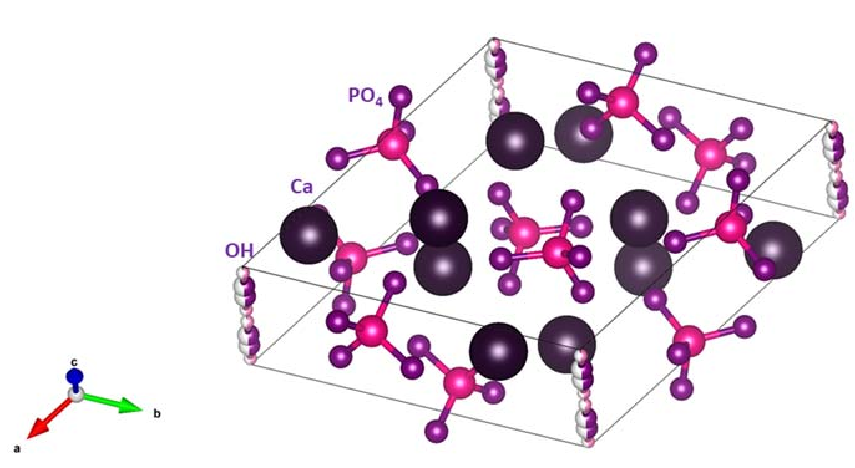
Figure 2.
The health benefits of selenium.

Figure 3.
Tetrahedral (A) and trigonal pyramid (B) arrangements of phosphate and selenite (A) and selenite (B) ions.
Figure 3.
Tetrahedral (A) and trigonal pyramid (B) arrangements of phosphate and selenite (A) and selenite (B) ions.

Figure 4.
XRD patterns of the pure HA and Se-HA samples with different Se/P ratios: (a) HA; (b) Se/P = 0.05; (c) Se/P = 0.08; (d) Se/P = 0.1; (e) Se/P = 0.3; (f) Se/P = 0.5; (g) Se/P = 1; (h) Se/P = 3; and (i) Se/P = 5. Reprinted from [33] with permission from Elsevier.
Figure 4.
XRD patterns of the pure HA and Se-HA samples with different Se/P ratios: (a) HA; (b) Se/P = 0.05; (c) Se/P = 0.08; (d) Se/P = 0.1; (e) Se/P = 0.3; (f) Se/P = 0.5; (g) Se/P = 1; (h) Se/P = 3; and (i) Se/P = 5. Reprinted from [33] with permission from Elsevier.

Figure 5.
The FT-IR transmission spectra of the studied selenite-doped HA (HA-xSeO3) and HA undoped. Reprinted from [43] with permission from Elsevier.
Figure 5.
The FT-IR transmission spectra of the studied selenite-doped HA (HA-xSeO3) and HA undoped. Reprinted from [43] with permission from Elsevier.

Figure 6.
SEM micrographs of the aspect of the biofilm after inoculating P. aeruginosa (a and b) and S. aureus (c and d) on the RHA2.5Se (b and d) coatings compared to HA (a and c). White arrows indicate non-colonized areas and black ones indicate the shape of an exemplar of that bacterium. Reprinted from [54] with permission from John Wiley and Sons.

Figure 7.
Preparation and working principle of antitumor nanoparticles. Se-HANs were fabricated using selenite to replace phosphate of HANs. Intratumoral injection of Se-HANs was performed on the xenograft osteosarcoma model. Se-HANs were internalized into tumor cells by nonspecific endocytosis and rapidly degraded in the acidic lysosome to release selenium. Cell apoptosis was activated by a primary, selenium-induced, caspase-dependent apoptosis pathway synergistically orchestrated with the ROS generation. Reprinted from [37] with permission. Copyright (2016) American Chemical Society.


| Bands (Wavenumber cm−1) | Assignment |
|---|---|
| 3700–2500 | ν3 and ν1 stretching modes of hydrogen-bonded H2O molecules |
| 3570 | stretching modes of structural hydroxyl groups |
| 1630–1640 | Bending modes of hydrogen-bonded H2O molecules |
| 1200–900 | ν3 and ν1 of PO43− |
| 605–500 | ν4 PO43− |
| 475–470 | ν2 PO43− |
© 2018 by the authors. Licensee MDPI, Basel, Switzerland. This article is an open access article distributed under the terms and conditions of the Creative Commons Attribution (CC BY) license (http://creativecommons.org/licenses/by/4.0/).
Share and Cite
MDPI and ACS Style
Pajor, K.; Pajchel, L.; Kolodziejska, B.; Kolmas, J. Selenium-Doped Hydroxyapatite Nanocrystals–Synthesis, Physicochemical Properties and Biological Significance. Crystals 2018, 8, 188. https://doi.org/10.3390/cryst8050188
AMA Style
Pajor K, Pajchel L, Kolodziejska B, Kolmas J. Selenium-Doped Hydroxyapatite Nanocrystals–Synthesis, Physicochemical Properties and Biological Significance. Crystals. 2018; 8(5):188. https://doi.org/10.3390/cryst8050188
Chicago/Turabian StylePajor, Kamil, Lukasz Pajchel, Barbara Kolodziejska, and Joanna Kolmas. 2018. "Selenium-Doped Hydroxyapatite Nanocrystals–Synthesis, Physicochemical Properties and Biological Significance" Crystals 8, no. 5: 188. https://doi.org/10.3390/cryst8050188
Note that from the first issue of 2016, this journal uses article numbers instead of page numbers. See further details here.