Synthesis and Crystal Structures of Cadmium(II) Cyanide with Branched-Butoxyethanol


1
Department of Chemistry, Faculty of Science, Toho University, 2-2-1 Miyama, Funabashi, Chiba 274-8510, Japan
2
Research Centre for Materials with Integrated Properties, Toho University, 2-2-1 Miyama, Funabashi, Chiba 274-8510, Japan
*
Author to whom correspondence should be addressed.
Crystals 2018, 8(5), 221; https://doi.org/10.3390/cryst8050221
Submission received: 27 December 2017
/
Revised: 12 March 2018
/
Accepted: 15 May 2018
/
Published: 17 May 2018
(This article belongs to the Special Issue Crystal Structure Analysis of Supramolecular and Porous Solids)
Abstract
:Two novel 3D cadmium(II) cyanide coordination polymers with branched-butoxyethanol compounds (iBucel = iso-butoxyethanol, tBucel = tert-butoxyethanol), [{Cd(CN)2(iBucel)2}{Cd(CN)2(H2O)(iBucel)}2{Cd(CN)2}6∙2(iBucel)]n I and [{Cd(CN)2(H2O)1.06(tBucel)0.94}{Cd(CN)2(tBucel)}2{Cd(CN)2}2∙1.06(tBucel)]n II, were synthesized and characterized by structural determination. Complex I contains two distinct Cd(II) coordination geometries: octahedral and tetrahedral. In contrast, complex II contains three distinct Cd(II) coordination geometries: octahedral, square-pyramidal, and tetrahedral. In the two complexes, branched-butoxyethanol molecules behave as both a ligand and a guest in the Cd(CN)2 cavities. The framework in I contains octahedral and tetrahedral Cd(II) in a 3:6 ratio. In I, the coordination environments of octahedral Cd(II) are cis-O-Cd-O. The framework in II contains octahedral, square-pyramidal, and tetrahedral Cd(II) in a 1:2:2 ratio. In II, the coordination environment of octahedral Cd(II) is disordered trans-O-Cd-O and the axial oxygen ligand is either a water or tBucel molecule. In II, the square-pyramidal Cd(II) geometry is formed by one tBucel ligand and four cyanide ligands. The Cd(CN)2 frameworks of the two complexes exhibit different structures.
1. Introduction
Because of their unique chemical properties, cadmium cyanide systems included with various guest species have been found to be one of the most important and mineralomimetic materials since the discovery of the clathrate complexes [1,2,3,4,5,6,7,8,9,10,11,12,13,14,15,16,17,18,19,20,21,22]. The discovery of the clathrate Cd(CN)2·CCl4 [4] is very important in the history of the simple, yet subtle, cadmium(II) cyanide clathrates and their related systems. Cadmium(II) cyanide is a 3D porous coordination polymer clathrating various guest molecules (for example, CCl4 [4,5,6], C6H6 [9] CH2ClCHCl2 [10], and Bu2O [10]) by van der Waals force in its cavity space. Interestingly, the Cd(CN)2 host structures change according to the guest [4,5,6,7,8,9,10,11,12,13,14,15,16,17,18,19,20], and the hosts form a mineralomimetic framework. The coordination geometry of Cd(II) in Cd(CN)2 is normally a tetrahedral four-coordination geometry (denoted as CdT). However depending on the influence of other ligands (for example, H2O [8,13,14,15,16,17,18,19]), the geometry might also be trigonal-bipyramidal five-coordination geometry (CdTB), or octahedral six-coordination geometry (CdOC). For Cd(CN)2 clathrates containing a lipophilic guest, the coordination geometries of Cd(II) were CdT and the Cd(CN)2 frameworks were cristobalite-like or tridymite-like structures [4,5,6,7,8,9,10,11,21]. In contrast, the Cd(CN)2 clathrates with the water molecule(s) coordinating to Cd(II) ion contain alcohol or short dialkyl-ether (alkyl group with a carbon number less than 3) as a guest, and the host frameworks function as zeolite-mimetic structures [8,13,14,15,16,17,18,19]. Incidentally, for Cd(CN)2, there was no guest molecule found to facilitate a doubly interpenetrated diamond-like framework. Furthermore, the coordination geometry of Cd(II) was CdT [3,4,5,6,7]. Thus, we surmised that the hydrophilic groups of guest molecules influence the Cd(II) coordination environment. We are interested in the effect of the coexistence of two kinds of hydrophilic groups on the Cd(II) coordination environment. Alkoxyethanol (Rcel) has both hydroxyl and etheric groups as shown in Scheme 1. We recently reported three crystal structures of cadmium cyanide coordination polymers with an alkoxyethanol of normal-alkyl chain structure; that is, [Cd(CN)2(Etcel)]n, [{Cd(CN)2(Bucel)}3{Cd(CN)2}]n, and [{Cd(CN)2(H2O)2}{Cd(CN)2}3·2(Hexcel)]n (Etcel = 2-ethoxyethanol, Bucel = 2-butoxyethanol, Hexcel = 2-hexyloxyethanol) [22]. In [Cd(CN)2(Etcel)]n and [{Cd(CN)2(Bucel)}3{Cd(CN)2}]n, hydroxyl oxygen atoms of Rcel ligands coordinate to the Cd(II) ions, and the Cd(II) ions exhibit CdTB. In contrast, in [{Cd(CN)2(H2O)2}{Cd(CN)2}3·2(Hexcel)]n, Hexcel molecules do not coordinate to the Cd(II) ions, and two water molecules are located at the cis-positions of CdOC. The three Cd(CN)2 framework structures varied according to differences in the normal-alkyl chain of the Rcel molecules. We report herein, the synthesis and crystal structures of two novel cadmium(II) cyanide complexes with branched-butoxyethanol compounds of formulae [{Cd(CN)2(iBucel)2}{Cd(CN)2(H2O)(iBucel)}2{Cd(CN)2}6∙2(iBucel)]n I and [{Cd(CN)2(H2O)1.06(tBucel)0.94}{Cd(CN)2(tBucel)}2{Cd(CN)2}2∙1.06(tBucel)]n II (iBucel = iso-butoxyethanol, tBucel = tert-butoxyethanol (Scheme 1)).
2. Materials and Methods
2.1. Synthesis
2.1.1. Synthesis of [{Cd(CN)2(iBucel)2}{Cd(CN)2(H2O)(iBucel)}2{Cd(CN)2}6∙2(iBucel)]n, I
An aqueous solution (45 mL) containing CdCl2·2.5H2O (2 mmol), and K2[Cd(CN)4] (4 mmol), was stirred for 30 min at room temperature. After the solution was filtered through a membrane filter, colorless crystals of I were obtained from the filtrate with 3 mL of iBucel kept in a sealed container at room temperature for two weeks. Elemental analysis found, I: C; 28.44 H; 3.84, and N; 11.21%. Calculated for C54H88N18O14Cd9: C; 29.15, H; 3.99, and N; 11.33%. IR(nujol mull, cm−1): νOH = 3355(br), νCN = 2180(s), νCOC = 1112(s).
2.1.2. Synthesis of [{Cd(CN)2(H2O)1.06(tBucel)0.94}{Cd(CN)2(tBucel)}2{Cd(CN)2}2∙(1.06tBucel)]n, II
An aqueous solution (45 mL) containing CdCl2·2.5H2O (2 mmol), and K2[Cd(CN)4] (4 mmol), was stirred for 30 min at room temperature. After the solution was filtered through a membrane filter, colorless crystals of II were obtained from the filtrate with 3 mL of tBucel kept in a sealed container at room temperature for a week. Elemental analysis II: C; 30.71 H; 4.11, and N; 10.70%. Calculated for C34H58.12N10O9.06Cd5: C; 31.08, H; 4.46, and N; 10.66%. IR(nujol mull, cm−1): νOH = 3319(br), νCN = 2179(s), νCOC = 1089(s).
2.2. Single Crystal X-ray Diffraction
The structural characterization for I and II was determined by the single crystal X-ray diffraction using a Bruker ApexII Smart CCD area-detector diffractometer (Bruker, Madison, WI, USA) with monochromated Mo-Kα radiation (λ = 0.71073 Å) under a temperature controlled N2 gas flow. The diffraction data were treated using APEX2 [23] and SAINT ver. 7.23A [24]. The absorption data were collected using SADABS ver. 2014/5 [25]. The structures were solved using the direct method, expanded using Fourier techniques, and refined using a full-matrix least-square refinement. H atoms of water molecules and of the hydroxyl group of alkoxyethanol behaving as a ligand were located in difference syntheses and refined isotropically. The remaining H atoms were placed at calculated positions and all H atoms were allowed to ride on a parent atom.
The overall structural solution was found using SHELXTL ver. 2014/7 [26]. Torsion angles and hydrogen bonds of all complexes were determined using SHELX [26] and PLATON ver. 30118 [27]. CCDC 1813424–1813425 contain the supplementary crystallographic data for this paper. These data can be obtained free of charge via http://www.ccdc.cam.ac.uk/conts/retrieving.html (or from the CCDC, located at 12 Union Road, Cambridge CB2 1EZ, UK; Fax: +44-1223-336033; [email protected]).
3. Results
Crystal Structure
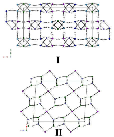
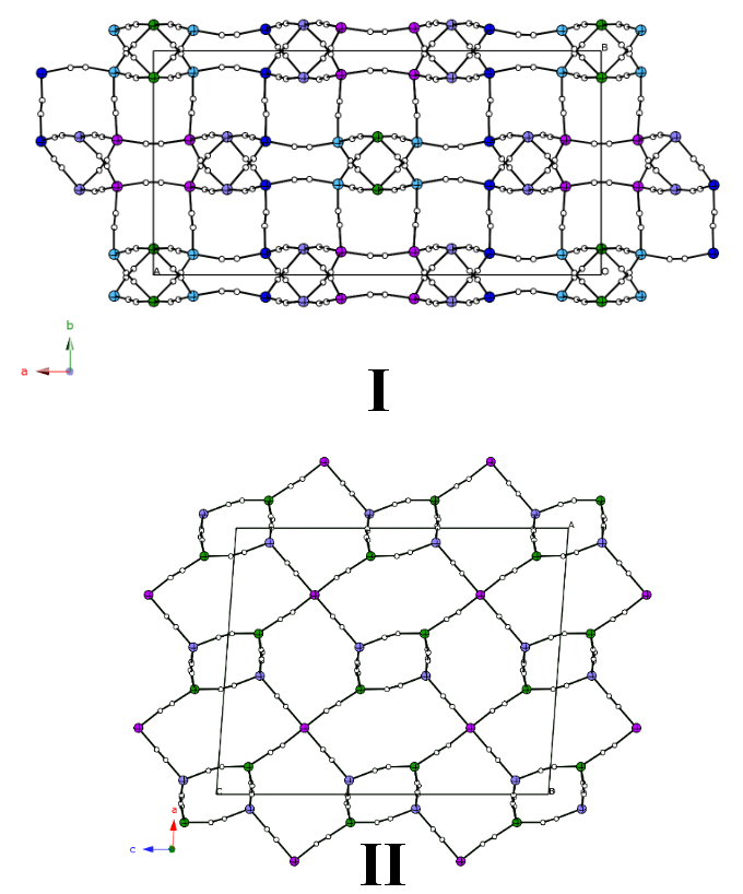
Single crystals of complexes I and II were prepared by a method similar to that found in the literature [4,22]. Crystal data for I and II are listed in Table 1. Complex I crystallizes in the space group C2/c. The space group of complex II was transformed from C2/c to I2/a of the alternate setting of C2/c so that its monoclinic angle would be less than 120 degrees [28,29]. In both complexes, all cyanide bridges between the two Cd(II) ions, Cd(CN)2, form a three-dimensional framework with large cavities (Figure 1, Figure S1, Figure 2 and Figure S2). We observed from the IR spectra that the peaks of C≡N stretching (νCN) were at approximately 2180 cm−1. The νCN values showed a blue shift compared with that observed for terminal C≡N of K2[Cd(CN)2] (2145 cm−1). This observation supports the view that C≡N bridges two Cd(II) ions. The exact arrangements of cyanides (Cd–NC–Cd or Cd–CN–Cd) cannot be determined by single X-ray diffraction because these arrangements are disordered. Nishikiori et al. [13,30,31] showed that the arrangements of cyanides in a Cd(CN)2 host are disordered, by 113Cd–CP/MAS NMR.
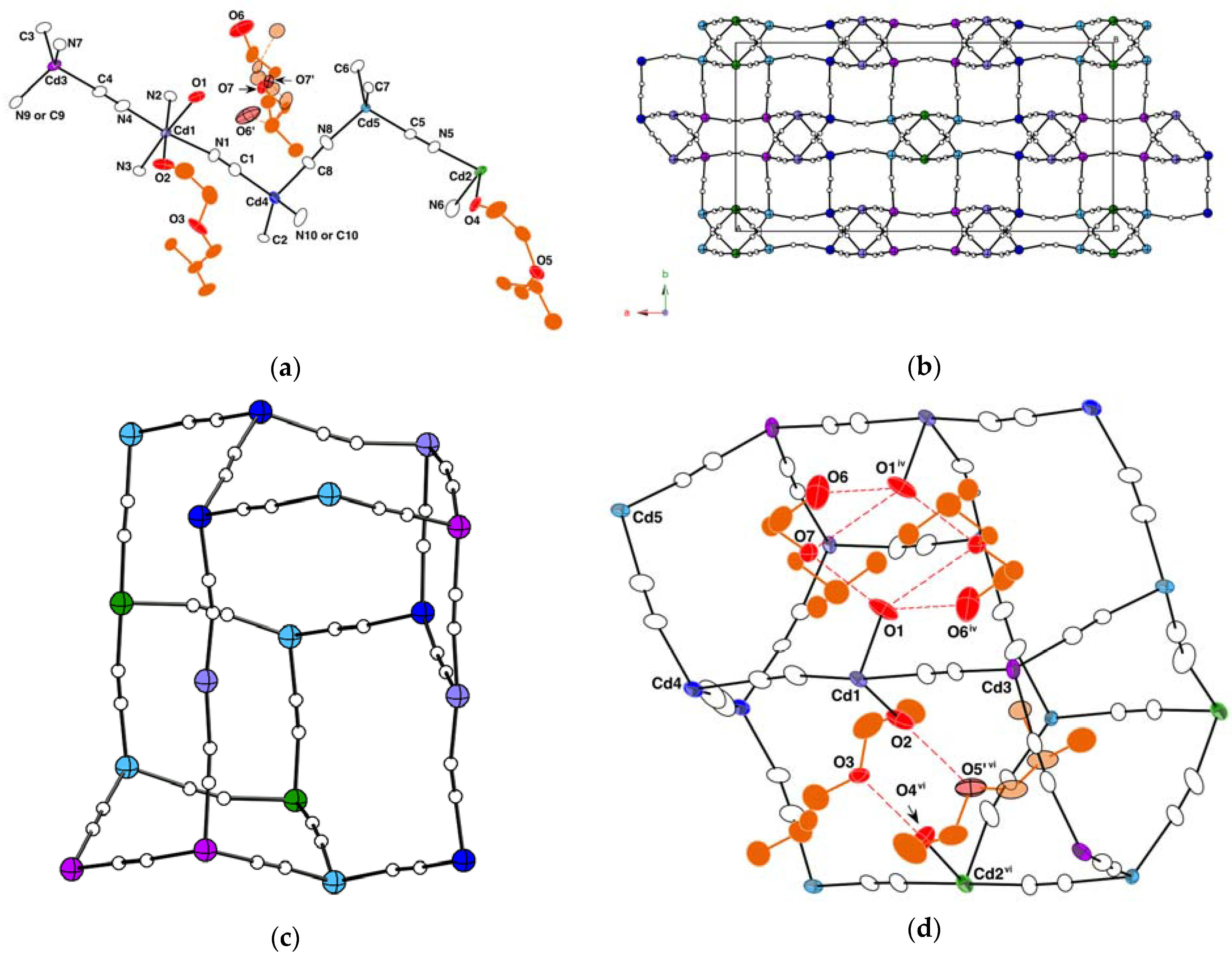
The crystal structure of complex I is shown in Figure 1 and Figure S1, and selected parameters are listed in Table 2. Complex I has five crystallographically independent Cd(II) sites denoted as Cd1, Cd2, Cd3, Cd4, and Cd5 (Figure 1a). Cd1 and Cd2 have octahedral six-coordination geometries, and the remaining sites have tetrahedral four-coordination geometries. In the unit cell, Cd2 is located on a special position, and the other Cd(II) sites are located on general positions. Therefore, the complex contains CdOC and CdT in a 3:6 ratio. The iBucel molecules exist as both a ligand and a guest in the Cd(CN)2 cavities (Figure 1a and Figure S1c). Because all iBucel molecules are strongly disordered (Figure 1a), the R factor is relatively high (Table 1). Around Cd1, two oxygen atoms are located at the cis-positions (Figure 1a) and four cyanides are located at the remaining positions. At the cis-position, one oxygen (O2) atom is located in the hydroxyl group of the iBucel ligand and another oxygen (O1) is located in the water molecule. Around Cd2, two oxygen (O3) atoms of iBucel ligands are located at the cis-positions (Figure 1a) and four cyanides are located at the remaining positions. Atoms labeled N9/C9 or N10/C10 are hybrids because of the disordered arrangement of cyanide (Figure 1) and because of the effects of the midpoints of the C≡N bonds on the symmetry of the lattice [2,3,4,5,6,7,8,9,10,11,12,13,22,30,31]. CdT are coordinated by only four cyanides. The TG and DTG curves (Figure S3) were observed in four steps of weight loss ranging from approximately 45 °C to 100 °C (first weight loss), from 100 °C to 160 °C (second and third weight losses), and from 160 °C to 230 °C (fourth weight loss). The first weight loss seems to have caused the desorption of iBucel guests from complex I. Because the second and third weight losses suggest that the elimination of the iBucel ligands occur near each other, we conclude that these two DTG peaks overlap. The fourth weight loss seems to involve the elimination of water ligands. The values of bond lengths around Cd(II) ions decrease in the order CdOC-O ≈ CdOC-(CN) ≈ CdT-(CN). The cis-O-Cd-O angles are similar to those in complex [{Cd(CN)2(H2O)2}{Cd(CN)2}3·2(Hexcel)]n [22] and other complexes with cis-O-Cd-O [14,15,16]. The Cd(CN)2 framework forms a NOQ net and these cavities’ shapes are a distorted [42.52.62.82] tile [32,33,34] (Figure 1c). The iBucel guest connects the water ligand through weak hydrogen bonds between etheric oxygen and water oxygen, and between hydroxyl oxygen and water oxygen. The iBucel ligands are connected with the neighboring iBucel ligands by hydrogen bonds between one ligand’s hydroxyl oxygen atoms (O2 and O4) and the other’s etheric oxygen atoms (O5′ and O3) (Figure 1d and Table 2), and these iBucel ligands form (O-CH2-CH2-O)2 eight-membered rings as in the case of complexes [Cd(CN)2(Etcel)]n and [{Cd(CN)2(Bucel)}3{Cd(CN)2}]n [22].
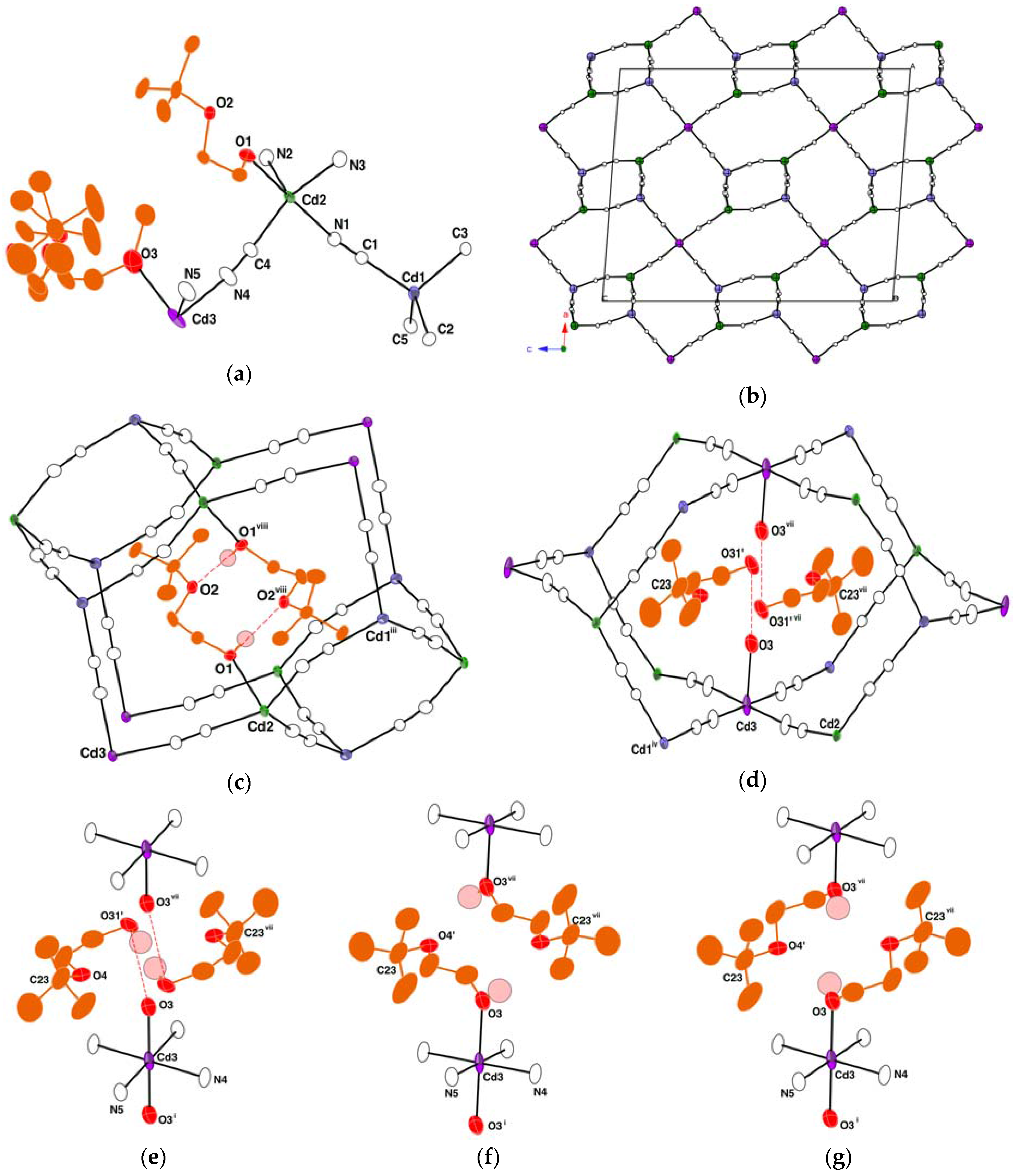
For complex II, the crystal structure is shown in Figure 2 and Figure S2, and selected parameters are listed in Table 3. Complex II has three crystallographically independent Cd(II) sites denoted as Cd1, Cd2, and Cd3 (Figure 2a). Cd1 has a four-coordination geometry, Cd2 has a five-coordination geometry, and Cd3 has a six-coordination geometry. In the unit cell, Cd1 and Cd2 are located on general positions, and Cd3 is located on a special position. Therefore, the complex contains Cd1, Cd2, and Cd3 in a 2:2:1 ratio. Cd1 is CdT and is coordinated by only four cyanides. Around the Cd2, one hydroxyl oxygen atom O1 of tBucel ligand is located at one of the positions and four cyanides are located at the remaining positions. The five-coordination geometry is estimated by a simple distortion parameter τ proposed by Addison et al. [22,35,36]. The τ value is simply defined by τ = (θ1 − θ2)/60, where θ1 is the largest basal angle and θ2 is a second largest; a perfect square pyramid is characterized by τ = 0, while τ = 1 represents a perfect trigonal bipyramid [22,35,36]. For the Cd2, τ is 0.46 from the selected bond angles, as shown in Table 3. Therefore, Cd2 has a distorted square pyramid five-coordination geometry (CdSP). The N2 atom of cyanide is located at the apex of the square pyramid. The N1 atom of cyanide is located at the trans-position of O1. The Cd2-N1 bond length is longer than the other Cd2-(CN) bonds (Table 3). The Cd2-O1 bond length is slightly longer than the CdTB-O bond lengths of complexes [Cd(CN)2(Etcel)]n and [{Cd(CN)2(Bucel)}3{Cd(CN)2}]n [22]. The tBucel ligand on Cd2 is connected with the neighboring tBucel on Cd2 by hydrogen bonds between one ligand’s hydroxyl oxygen atom O1 and the other’s etheric oxygen atom O2 (Figure 2c and Table 3), as in the case of complexes [Cd(CN)2(Etcel)]n, [{Cd(CN)2(Bucel)}3{Cd(CN)2}]n [22], and I. Around the Cd3, two oxygen (O3) atoms are located at the axial positions and four cyanides are located at the equatorial positions. The equatorial plane of Cd3 is almost parallel to the xz plane of the cell. The axial positions are disordered. The O3 atom is either the oxygen of the water ligand (Figure 2d) or the hydroxyl oxygen of the tBucel ligand (Figure 2e,f). Furthermore, the tBucel molecule over the axial position of Cd3 is either a guest or ligand. Specifically, if O3 is water, then the tBucel molecule on the axial position is a guest; otherwise, the tBucel is a ligand. The conformation of the tBucel ligand on Cd3 comprises two types (Figure 2e,f). However, C23 denotes that the center carbon atom of the tert-butyl group is ordered regardless of the above-mentioned disorder. The Cd(CN)2 cavity around O3 is similar to Cd(CN)2∙2/3H2O∙t-BuOH (t-BuOH = tert-butanol) [18,19] (Figure 2d). We suggest that one of the factors affecting the topologies of Cd(CN)2 cavities, as in Figure 2d, are variations in the tert-butyl groups. The occupancies of disordered parts are 0.531(3) (Figure 2e), 0.322(3) (Figure 2f), and 0.146(3) (Figure 2g). The TG and DTG curves (Figure S4) were observed in three steps of weight loss ranging from approximately 80 °C to 145 °C, from 143 °C to 175 °C, and from 175 °C to 230 °C. The first weight loss seems to be due to the release of 1.06 tBucel guest molecules and 0.94 tBucel ligands coordinated to Cd3. The peak shoulder on the DTG curve in the first weight loss seems to be due to the release of the 1.06 tBucel guests. The second weight loss suggests that the tBucel ligands coordinated to Cd2 were removed from II. The third weight loss seems to involve the elimination of the 1.06 H2O ligand. The Cd3-O3 bond length is in the range of CdOC-O bonds of complexes with trans-O-CdOC-O [14,16,17,18,19].
4. Conclusions
We synthesized and crystallographically characterized two novel 3D cadmium(II) cyanide coordination polymers with branched-butoxyethanol; [{Cd(CN)2(iBucel)2}{Cd(CN)2(H2O)(iBucel)}2{Cd(CN)2}6∙2(iBucel)]n I and [{Cd(CN)2(H2O)1.06(tBucel)0.94}{Cd(CN)2(tBucel)}2{Cd(CN)2}2∙1.06(tBucel)]n II. Complex I contains two distinct Cd(II) coordination geometries, namely CdOC and CdT, in a 3:6 ratio. In contrast, complex II contains three distinct Cd(II) coordination geometries: CdOC, CdSP, and CdT, in a 1:2:2 ratio. In complexes I and II, branched-butoxyethanol molecules behave as both a ligand and guest in the Cd(CN)2 cavities. Rcel ligands coordinated to Cd(II) ions tend to form (O-CH2-CH2-O)2 eight-membered rings constructed by hydrogen bonds between the two ethyleneglycol fragments (Figure 1d and Figure 2c) [22]. The (O-CH2-CH2-O)2 eight-membered rings may assist in the coordination of alkoxyethanol molecules to Cd(II) ions and in the stabilization of that coordination. The Cd(CN)2 frameworks of complexes I and II exhibit different structures. Furthermore, these Cd(CN)2 framework structures of the two complexes are different from the framework structure of complex [{Cd(CN)2(Bucel)}3{Cd(CN)2}]n containing a Bucel ligand and having a butyl group. This suggests that the Cd(CN)2 framework and coordination environment of Cd(II) are strongly affected by not only the number of carbons of the alkyl group of the Rcel molecule but also by the shape of the alkyl group of the Rcel molecule. Because of the differences in their alkyl groups, the Cd(CN)2 frameworks of cadmium(II) cyanide coordination polymers with alkoxyethanol [22] change more dramatically than the frameworks of complexes with either dialkyl-ether [16,17] or with alkyl-alcohol [8,13,14,18,19]. The effect of the alkyl group of alkoxyethanol compounds was found to be stronger than the effect of either alkyl-alcohol or dialkyl-ether compounds. These new cadmium cyanide host-guest compounds also demonstrate potential advantages of the clathrate chemistry for molecular recognition, a theme with a long history of research on inclusion compounds [1].
Supplementary Materials
The following materials are available online at https://www.mdpi.com/2073-4352/8/5/221/s1, Figure S1: Cd(CN)2 network structure of I; (a) along the b axis, (b) along the (102) direction and (c) iBucel guest molecule in Cd(CN)2 framework of I. Figure S2: Cd(CN)2 network structure of II; (a) along the c axis and (b) along the (101) direction. Figure S3: TG and DTG plots of I. Figure S4: TG and DTG plots of II. comp_I.cif and comp_II.cif: Crystal analysis data for complexes I and II, respectively.
Author Contributions
Takafumi Kitazawa conceived and designed the experiments; Takeshi Kawasaki performed the experiments and analyzed data; Takeshi Kawasaki and Takafumi Kitazawa wrote the paper.
Acknowledgments
The work was supported by JSPS KAKENHI Grant Number JP15K05485.
Conflicts of Interest
The authors declare no conflict of interest.
References
- Dyadin, Y.A.; Terekhova, I.S.; Rodionova, T.V.; Soldatov, D.V. Half century history of clathrate chemistry. J. Struct. Chem. 1999, 40, 645–653. [Google Scholar] [CrossRef]
- Zhdanov, H. The crystalline structure of Zn(CN)2. C. R. Acad. Sci. USSR 1941, 31, 352–354. [Google Scholar]
- Shugam, E.; Zhdanov, H. The crystal structure of cyanides. II. Structure of cadmium cyanide. Acta Physicochim. USSR 1945, 20, 247–252. [Google Scholar]
- Kitazawa, T.; Nishikiori, S.; Kuroda, R.; Iwamoto, T. Novel Clathrate Compound of Cadmium Cyanide Host with an Adamantane-like Caity. Cadmium Cyanide-Carbon Tetrachloride(1/1). Chem. Lett. 1988, 17, 1729–1732. [Google Scholar] [CrossRef]
- Iwamoto, T.; Nishikiori, S.; Kitazawa, T.; Yuge, H. Mineralomimetic chemistry as a modern aspect of co-ordination chemistry. J. Chem. Soc. Dalton Trans. 1997, 4127–4136. [Google Scholar] [CrossRef]
- Phillips, A.E.; Goodwin, A.L.; Halder, G.J.; Southon, P.D.; Kepert, C.J. Nanoporosity and Exceptional Negative Thermal Expansion in Single-Network Cadmium Cyanide. Angew. Chem. Int. Ed. 2008, 47, 1396–1399. [Google Scholar] [CrossRef] [PubMed] [Green Version]
- Hoskins, B.F.; Robson, R. Design and Construction of New Class of Scaffolding-like Materials Comprising Infinite Polymeric Frameworks of 3D-Linked Molecular Rods. A Reappraisal of the Zn(CN)2 and Cd(CN)2 Structures and the Synthesis and Structure of the Diamond-Related Frameworks [N(CH3)4][CuIZnII(CN)4] and CuI[4,4′,4′′,4′′′-tetracyanotetraphenylmethane]BF4∙xC6H6NO2. J. Am. Chem. Soc. 1990, 112, 1546–1554. [Google Scholar]
- Abrahams, B.F.; Hardie, M.J.; Hoskins, B.F.; Robson, R.; Williams, G.A. Topological Rearrangement within a Single Crystal from a Honeycomb [Cd(CN)2]n 3D Nets to a Diamond Net. J. Am. Chem. Soc. 1992, 114, 10641–10643. [Google Scholar] [CrossRef]
- Kitazawa, T.; Nishimura, A. Mineralomimetic cadmium cyanide benzene clathrate. J. Struct. Chem. 1999, 40, 721–725. [Google Scholar] [CrossRef]
- Kitazawa, T. A new mineralomimetic Cd(CN)2 host framework which is intermediate between H- and l-cristobalite-like frameworks. Chem. Commun. 1999, 10, 891–892. [Google Scholar] [CrossRef]
- Kitazawa, T. A new type of mineralomimetic cadmium cyanide host framework containing methyl acetate. J. Mater. Chem. 1998, 8, 671–674. [Google Scholar] [CrossRef]
- Kitazawa, T.; Nishikiori, S.; Kuroda, R.; Iwamoto, T. Two novel metal-complex host structures consisting of cyanocadmate coordination polyhedra. Clay-like and zeolite-like structures. Chem. Lett. 1988, 17, 459–462. [Google Scholar] [CrossRef]
- Nishikiori, S.; Ratcliffe, C.I.; Ripmeester, J.A. Crystal Structure of Cd5(CN)10(H2O)4·4C6H11OH Studied by X-ray Diffraction and Solid-State 113Cd NMR. A New Type of Cristobalite-like Framework Host with a Site Interacting with Cyclohexanol by Hydrogen Bonding. J. Am. Chem. Soc. 1992, 114, 8590–8595. [Google Scholar] [CrossRef]
- Kim, J.; Whang, D.; Lee, J.I.; Kim, K. Guest-dependent [Cd(CN)2]n Host Structures of Cadmium Cyanide-Alcohol Clathrates: Two New [Cd(CN)2]n Frameworks formed with PrnOH and PriOH Guests. J. Chem. Soc. Chem. Commun. 1993, 18, 1400–1402. [Google Scholar] [CrossRef]
- Kim, J.; Whang, D.; Koh, Y.-S.; Kim, K. Two New [Cd(CN)2]n Frameworks with Linear Channels of Large, Elongated Hexagonal Cross-section: Structures of Cadmium Cyanide-Guest (Guest = dmf and Me2SO) Clathrates. J. Chem. Soc. Chem. Commun. 1994, 5, 637–638. [Google Scholar] [CrossRef]
- Kitazawa, T.; Kukuyama, T.; Takahashi, M.; Takeda, M. Cadmium Cyanide-Ether Clathrates: Crystal Structures of Cd8(CN)16(H2O)6·6G (G = Et2O or Pri2O) and Cd3(CN)6(H2O)2·2Prn2O. J. Chem. Soc. Dalton Trans. 1994, 20, 2933–2937. [Google Scholar] [CrossRef]
- Yuge, H.; Chong-Hyeak, K.; Iwamoto, T.; Kitazawa, T. Hofmann-H2O-type and Hofmann-H2O-Td-type host structures accommodating 1,4-dioxane: Crystal structures of trans-bis(morpholine-N)cadmium(II) tetaracyanonikelate(II), trans-diaquacadmium(II) tetracyanonickelate(II)-(1,4-dioxane)(1/2) and trans-diaquacadmium(II) tetracyanocadmate(II) (1,4-dioxane)(1/2). Inorg. Chim. Acta 1997, 257, 217–224. [Google Scholar]
- Abrahams, B.F.; Hoskins, B.F.; Liu, J.; Robson, R. The Archetype for a New Class of Simple Extended 3D Honeycomb Frameworks. The Synthesis and X-ray Crystal Structure of Cd(CN)5/3(OH)1/3∙1/3(C6H12N4), Cd(CN)2∙1/3(C6H12N4), and Cd(CN)2∙2/3H2O∙tBuOH (C6H12N4 = Hexamethylenetetramine) Revealing Tow Topologycallyt Equivalent but Geometrically Different Frameworks. J. Am. Chem. Soc. 1991, 113, 3045–3051. [Google Scholar]
- Abrahams, B.F.; Hoskins, B.F.; Lam, Y.-H.; Robson, R.; Separovic, F.; Woodberry, P. A Reexamination of the Structure of “Honeycomb Cadmium Cyanide”. J. Solid State Chem. 2001, 156, 51–56. [Google Scholar] [CrossRef]
- Ruiz, E.; Alvarez, S. Theoretical Study of Host∙Guest Interactions in Clathrates with a Cd(CN)2 Host. Inorg. Chem. 1995, 34, 5845–5851. [Google Scholar] [CrossRef]
- Nishikiori, S. Reversible reconstructive transition of the [CuZn(CN)4]− framework host induced by guest exchange. CrystEngComm 2014, 16, 10173–10176. [Google Scholar] [CrossRef]
- Kawasaki, T.; Kitazawa, T. Three-Dimensional Cadmium(II) Cyanide Coordination Polymers with Ethoxy-, Butoxy- and Hexyloxy-ethanol. Crystals 2016, 6. [Google Scholar] [CrossRef]
- Bruker. APEX2, Version 2.0-2; Bruker AXS Inc.: Madison, WI, USA, 2006. [Google Scholar]
- Bruker. SAINT, Version 7.23A; Bruker AXS Inc.: Madison, WI, USA, 2007. [Google Scholar]
- Sheldrick, G.M. SADABS: Program for Empirical Absorption Correction; University of Göttingen: Göttingen, Germany, 1996. [Google Scholar]
- Sheldrick, G.M. Crystal structure refinement with SHELXL. Acta Crystallogr. Sect. C 2015, 71, 3–8. [Google Scholar] [CrossRef] [PubMed]
- Spek, A.L. Structure validation in chemical crystallography. Acta Crystallogr. Sect. D 2009, 65, 148–155. [Google Scholar] [CrossRef] [PubMed]
- Mighell, A.D. Monoclinic I- and C-centered cells. Acta Crystallogr. Sect. B 2003, 59, 300–302. [Google Scholar] [CrossRef]
- Feast, G.C.; Haestier, J.; Page, L.W.; Robertson, J.; Thompson, A.L.; Watkin, D.J. An unusual methylene aziridine refined in P21/c and the nonstandard setting P21/n. Acta Crystallogr. Sect. C 2009, 65, o635–o638. [Google Scholar] [CrossRef] [PubMed]
- Nishikiori, S.; Ratcliffe, C.I.; Ripmeester, J.A. 113Cd NMR studies of Hofmann-type clathrates and related compounds: Evidence form two room temperature orientational glasses. Can. J. Chem. 1990, 68, 2270–2273. [Google Scholar] [CrossRef]
- Nishikiori, S.; Ratcliffe, C.I.; Ripmeester, J.A. Framework ordering in solid cadmium cyanides from cadmium-113 NMR spectroscopy. J. Chem. Soc. Chem. Commun. 1991, 10, 735–736. [Google Scholar] [CrossRef]
- Blatov, V.A.; Delgado-Friedrichs, O.; O’Keeffe, M.; Proserpio, D.M. Three-periodic nets and tilings: Natural tilings for nets. Acta Crystallogr. Sect. A 2007, 63, 418–425. [Google Scholar] [CrossRef] [PubMed]
- O’Keefee, M.; Peskov, M.A.; Ramsden, S.J.; Yaghi, O.M. The Reticular Chemistry Structure Resource (RCSR) Database of, and Symbols for, Crystal Nets. Acc. Chem. Res. 2008, 41, 1782–1789. [Google Scholar] [CrossRef] [PubMed]
- Anurova, N.A.; Blatov, V.A.; Ilyushin, G.D.; Proserpio, D.M. Natural Tilings for Zeolite-Type Frameworks. J. Phys. Chem. C 2010, 114, 10160–10170. [Google Scholar] [CrossRef]
- Addison, A.W.; Rao, T.N.; Reedijk, J.; van Rijin, J.; Verschoor, G.C. Synthesis, structure, and spectroscopic properties of copper(II) compounds containing nitrogen–sulphur donor ligands; the crystal and molecular structure of aqua[1,7-bis(N-methylbenzimidazol-2′-yl)-2,6-dithiaheptane]copper(II) perchlorate. J. Chem. Soc. Dalton Trans. 1984, 7, 1349–1356. [Google Scholar] [CrossRef]
- Siewe, A.D.; Kim, J.-Y.; Kim, S.; Park, I.-H.; Lee, S.S. Regioisomer-Dependent Endo- and Exocyclic Coordination of Bis-Dithiamacrocycles. Inorg. Chem. 2014, 53, 393–398. [Google Scholar] [CrossRef] [PubMed]
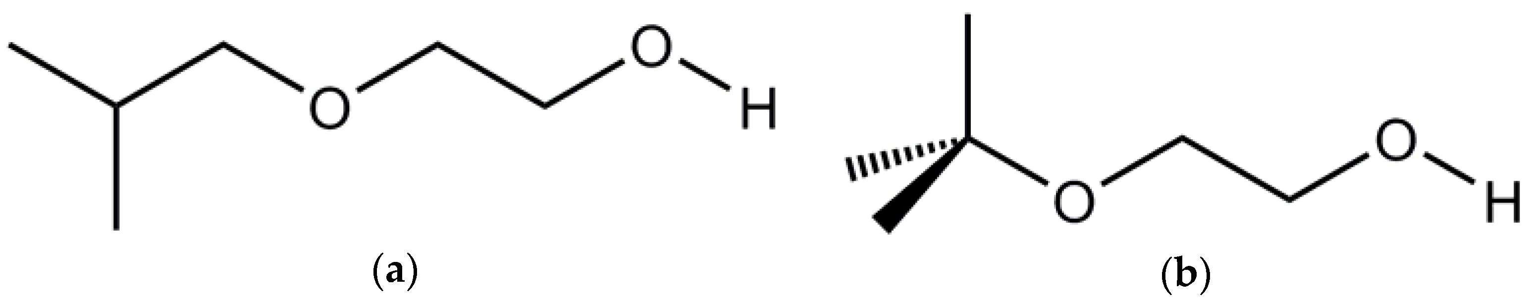
Scheme 1.
The structural formulae of alkoxyethanol in this work: (a) iso-butoxyethanol (iBucel); and (b) tert-butoxyethanol (tBucel).
Scheme 1.
The structural formulae of alkoxyethanol in this work: (a) iso-butoxyethanol (iBucel); and (b) tert-butoxyethanol (tBucel).

Figure 1.
The crystal structure of I. H atoms and disordered parts are omitted for clarity: (a) Asymmetric unit of I. Displacement ellipsoids are drawn at the 50% probability level. Because the arrangements of cyanides (Cd-NC-Cd or Cd-CN-Cd) are disordered, the cyanide atoms are labeled arbitrarily; (b) The Cd(CN)2 network structure of I along the c axis; (c) [42.52.62.82] tile; (d) Hydrogen bonds between iBucel and water molecules in [42.52.62.82] cavities of cadmium cyanide network. Displacement ellipsoids are drawn at the 50% probability level. (Symmetry codes: iv = −x + 1/2, −y + 1/2, −z + 2; vi = −x + 1/2, −y + 1/2, −z + 1).

Figure 2.
The crystal structure of II. Because arrangements of cyanides (Cd-NC-Cd or Cd-CN-Cd) are disordered, the cyanide atoms are labeled arbitrarily: (a) Asymmetric unit containing disordered parts. Displacement ellipsoids are drawn at the 50% probability level. H atoms are omitted for clarity; (b) The Cd(CN)2 network view along the b axis; (c) Hydrogen bonds between neighboring tBucel ligands on Cd2 in the cavities of cadmium cyanide network of II. Displacement ellipsoids are drawn at the 50% probability level. H atoms, except for OH hydrogens, are omitted for clarity; (d) Cd(CN)2 cavity around O3. Displacement ellipsoids are drawn at the 50% probability level. H atoms are omitted for clarity; (e–g). The disordered parts of the axial positions on the Cd3 and hydrogen bonds between the neighboring ligands. (e) Shows where water molecules coordinate to the Cd3. (f,g) show how the tBucel ligands coordinate to the Cd3. All hydrogen bonds might not be found due to the complexity of the observed disorder. (Symmetry codes: i = −x + 1/2, −y + 3/2, −z + 3/2; iii = −x, −y + 2, −z + 2; iv = x + 1/2, −y + 2, z; vii = −x + 1/2, −y + 1/2, −z + 3/2; viii = −x + 1/2, y, −z + 2).


Table 1.
The crystal data.
| Complex | I | II |
|---|---|---|
| Empirical formula | C54H88Cd9N18O14 | C34H58.12Cd5N10O9.06 |
| Formula weight | 2225.02 | 1314.03 |
| Temperature (K) | 90 | 90 |
| Crystal system | Monoclinic | Monoclinic |
| Space group | C2/c | I2/a |
| a (Å) | 33.8994(11) | 21.8261(7) |
| b (Å) | 16.6003(5) | 8.8155(3) |
| c (Å) | 15.8715(5) | 27.1515(12) |
| β (ᵒ) | 101.562(1) | 94.2899(4) |
| V (A3) | 8750.3(5) | 5209.5(3) |
| Z | 4 | 4 |
| dcalc (g cm−3) | 1.689 | 1.675 |
| μ (mm−1) | 2.199 | 2.059 |
| F(000) | 4328 | 2578 |
| Reflections collected | 33716 | 19,666 |
| Rint | 0.0197 | 0.0127 |
| Data/restraints/parameters | 13296/205/478 | 7912/7/319 |
| GOF | 1.072 | 1.054 |
| R1, wR2 [I > 2σ(I)] | 0.0967, 0.2291 | 0.0256, 0.0636 |
| Δρmax, Δρmin (e Å−3) | 4.314, −7.405 | 1.347, −1.357 |
Table 2.
The selected bond lengths and angles in I.
| CdOC-OW/Å | |||||
| Cd1-O1 | 2.372(7) | ||||
| CdOC-OOH/Å | |||||
| Cd1-O2 | 2.343(8) | Cd2-O4 | 2.323(8) | ||
| CdOC-(CN)/Å | |||||
| Cd1-N1 | 2.282(9) | Cd1-N2 | 2.310(8) | Cd1-N3 | 2.261(7) |
| Cd1-N4 | 2.282(9) | Cd2-N5 | 2.286(8) | Cd2-N6 | 2.309(10) |
| CdT-(CN)/Å | |||||
| Cd3-C3 | 2.189(10) | Cd3-C4 | 2.214(10) | Cd3-N7 | 2.214(9) |
| Cd3-N9 | 2.224(9) | Cd4-C1 | 2.191(11) | Cd4-C2 | 2.161(9) |
| Cd4-C8 | 2.218(10) | Cd4-N10 | 2.223(14) | Cd5-C6 | 2.176(9) |
| Cd5-C5 | 2.190(9) | Cd5-C7 | 2.228(8) | Cd5-N8 | 2.205(9) |
| O-CdOC-O/ᵒ | |||||
| O1-Cd1-O2 | 86.2(3) | O4-Cd2-O4 i | 85.5(5) | ||
| trans((CN)-CdOC-O)/ᵒ | |||||
| N3-Cd1-O1 | 174.4(3) | N2-Cd1-O2 | 172.5(3) | N6-Cd2-O4 i | 172.3(4) |
| cis((CN)-CdOC-OOH)/ᵒ | |||||
| N3-Cd1-O2 | 89.1(3) | N1-Cd1-O2 | 89.1(4) | N4-Cd1-O2 | 88.7(4) |
| N6-Cd2-O4 | 87.1(4) | N5-Cd2-O4 | 87.6(3) | ||
| cis((CN)-CdOC-OW)/ᵒ | |||||
| N2-Cd1-O1 | 86.7(3) | N1-Cd1-O1 | 85.0(3) | N4-Cd1-O1 | 86.1(3) |
| trans((CN)-CdOC-(CN))/ᵒ | |||||
| N4-Cd1-N1 | 171.0(4) | N5 i-Cd2-N5 | 178.3(4) | ||
| cis((CN)-CdOC-(CN))/ᵒ | |||||
| N3-Cd1-N1 | 91.8(3) | N3-Cd1-N4 | 97.0(3) | N1-Cd1-N2 | 92.5(4) |
| N4-Cd1-N2 | 88.5(3) | N5-Cd2-N6 | 90.8(3) | ||
| N3-Cd1-N2 | 98.1(3) | N6 i-Cd2-N6 | 100.3(7) | ||
| (CN)-CdT-(CN)/ᵒ | |||||
| C3-Cd3-C4 | 115.1(4) | C3-Cd3-N7 | 109.4(3) | C4-Cd3-N7 | 105.2(3) |
| C3-Cd3-N9 | 113.0(3) | C4-Cd3-N9 | 107.0(3) | N7-Cd3-N9 | 106.6(3) |
| C2-Cd4-C1 | 118.5(3) | C2-Cd4-C8 | 115.2(4) | C1-Cd4-C8 | 106.3(4) |
| C2-Cd4-N10 | 105.3(4) | C1-Cd4-N10 | 106.4(5) | C8-Cd4-N10 | 103.8(5) |
| C6-Cd5-C5 | 117.1(4) | C6-Cd5-N8 | 115.9(4) | C5-Cd5-N8 | 107.6(3) |
| C6-Cd5-C7 | 104.8(3) | C5-Cd5-C7 | 106.0(3) | N8-Cd5-C7 | 104.1(3) |
| Hydrogen bonds/Å | |||||
| O1···O6 iv | 2.84(3) | O1···O7′ iv | 2.922(14) | O1···O7 iv | 3.323(18) |
| O1···O7′x iv | 2.922(14) | O2···O5′ vi | 2.865(18) | O4···O3 vi | 2.743(11) |
| O6···O1 iv | 2.84(3) | O6′···O1 | 3.19(3) | C11···O6 iv | 3.48(3) |
Symmetry codes: i = −x, y, −z + 1/2; iv = −x + 1/2, −y + 1/2, −z + 2; vi = −x + 1/2, −y + 1/2, −z + 1.
Table 3.
The selected bond lengths and angles in II.
| CdT-(CN)/Å | |||||
| Cd1-C1 | 2.196(2) | Cd1-C2 | 2.219(2) | Cd1-C3 | 2.234(2) |
| Cd1-C5 | 2.192(2) | ||||
| CdSP-O/Å | |||||
| Cd2-O1 | 2.5875(17) | ||||
| CdSP-(CN)/Å | |||||
| Cd2-N1 | 2.3408(19) | Cd2-N3 | 2.215(2) | Cd2-C4 | 2.188(2) |
| Cd2-N2 | 2.215(2) | ||||
| CdOC-O/Å | |||||
| Cd3-O3 | 2.343(3) | ||||
| CdOC-(CN)/Å | |||||
| Cd3-N4 | 2.290(2) | Cd3-N5 | 2.309(2) | ||
| (CN)-CdT-(CN)/ᵒ | |||||
| C1-Cd1-C2 | 107.10(8) | C1-Cd1-C3 | 109.86(7) | C2-Cd1-C3 | 101.73(8) |
| C5-Cd1-C1 | 116.97(8) | C5-Cd1-C2 | 111.16(8) | C5-Cd1-C3 | 108.93(8) |
| (CN)-CdSP-O/ᵒ | |||||
| N1-Cd2-O1 | 167.42(6) | N3-Cd2-O1 | 85.81(6) | C4-Cd2-O1 | 81.76(7) |
| N2-Cd2-O1 | 92.99(6) | ||||
| (CN)-CdSP-(CN)/ᵒ | |||||
| C4-Cd2-N3 | 139.92(8) | C4-Cd2-N1 | 96.07(8) | N3-Cd2-N1 | 88.04(7) |
| C4-Cd2-N2 | 113.43(8) | N3-Cd2-N2 | 105.12(7) | N2-Cd2-N1 | 99.21(7) |
| cis(CN)-CdOC-O/ᵒ | |||||
| N4-Cd3-O3 | 90.07(9) | N5-Cd3-O3 | 89.45(9) | ||
| N4-Cd3-O3 i | 89.93(9) | N5-Cd3-O3 i | 90.55(9) | ||
| cis(CN)-CdOC-O/ᵒ | |||||
| N4-Cd3-N5 | 87.66(8) | N4-Cd3-N5 i | 92.34(8) | ||
| hydrogen bonds | |||||
| O1...O2 viii | 2.808(2) | O31′...O3 | 3.104(8) | ||
| C12...O31 vii | 3.178(8) | C12...O31′ vii | 3.514(8) | C21...O3 | 3.373(7) |
| C21B...O4′ | 3.21(3) | ||||
i = ‒x + 1/2, ‒y + 3/2, ‒z + 3/2; vii = ‒x + 1/2, ‒y + 1/2, ‒z + 3/2; viii = ‒x + 1/2, y, ‒z + 2. Symmetry codes: i = ‒x + 3/2, ‒y + 1/2, ‒z; vii = ‒x + 3/2, ‒y + 3/2, ‒z; viii = ‒x + 2, y, ‒z + 1/2.
© 2018 by the authors. Licensee MDPI, Basel, Switzerland. This article is an open access article distributed under the terms and conditions of the Creative Commons Attribution (CC BY) license (http://creativecommons.org/licenses/by/4.0/).
Share and Cite
MDPI and ACS Style
Kawasaki, T.; Kitazawa, T. Synthesis and Crystal Structures of Cadmium(II) Cyanide with Branched-Butoxyethanol. Crystals 2018, 8, 221. https://doi.org/10.3390/cryst8050221
AMA Style
Kawasaki T, Kitazawa T. Synthesis and Crystal Structures of Cadmium(II) Cyanide with Branched-Butoxyethanol. Crystals. 2018; 8(5):221. https://doi.org/10.3390/cryst8050221
Chicago/Turabian StyleKawasaki, Takeshi, and Takafumi Kitazawa. 2018. "Synthesis and Crystal Structures of Cadmium(II) Cyanide with Branched-Butoxyethanol" Crystals 8, no. 5: 221. https://doi.org/10.3390/cryst8050221
Note that from the first issue of 2016, this journal uses article numbers instead of page numbers. See further details here.