

MuSK Myasthenia Gravis—Potential Pathomechanisms and Treatment Directed against Specific Targets

1
Department of Neurology, Wroclaw Medical University, Borowska 213, 50-556 Wroclaw, Poland
2
Department of Molecular and Cellular Biology, Wroclaw Medical University, Borowska 211A, 50-556 Wroclaw, Poland
*
Author to whom correspondence should be addressed.
Cells 2024, 13(6), 556; https://doi.org/10.3390/cells13060556
Submission received: 13 February 2024
/
Revised: 17 March 2024
/
Accepted: 19 March 2024
/
Published: 21 March 2024
(This article belongs to the Special Issue Spotlight on Neuromuscular Diseases: Pathomechanisms and New Therapeutic Targets)
Abstract
:Myasthenia gravis (MG) is an autoimmune disease in which autoantibodies target structures within the neuromuscular junction, affecting neuromuscular transmission. Muscle-specific tyrosine kinase receptor-associated MG (MuSK-MG) is a rare, often more severe, subtype of the disease with different pathogenesis and specific clinical features. It is characterized by a more severe clinical course, more frequent complications, and often inadequate response to treatment. Here, we review the current state of knowledge about potential pathomechanisms of the MuSK-MG and their therapeutic implications as well as ongoing research in this field, with reference to key points of immune-mediated processes involved in the background of myasthenia gravis.
1. Introduction
Myasthenia gravis (MG) is an autoimmune disease of the postsynaptic part of the neuromuscular junction (NMJ). Immunologically, MG is a heterogeneous group caused by different, pathogenic antibodies against important synapse proteins. These antibodies include Ig1 or Ig3 class antibodies against acetylcholine receptor (AChR), Ig4 class antibodies against muscle-specific kinase receptor (MuSK), and antibodies against lipoprotein receptor-related protein 4 (LRP4). Patients with MG have a similar clinical presentation, but the immunopathology is unusually heterogeneous [1,2,3,4].
Approximately 5–8% of myasthenia gravis patients are positive for antibodies against muscle-specific tyrosine kinase receptors [5,6]. Its prevalence varies between countries and ethnic groups. Higher rates of MuSK-MG patients are observed in southern Europe, with a pronounced prevalence in females, who account for more than 70% of patients. The disease has an earlier age of onset, with a peak incidence in the latter part of the third decade of life, and rarely occurs after the age of 70 [5]. In contrast to AChR-MG, no significant thymus alterations, such as thymic hyperplasia, have been reported in MuSK-MG patients [7]. Furthermore, it is postulated that thymectomy does not improve clinical outcomes in these patients [8].
MuSK-MG is a rare, often more severe subtype of the disease with different pathogenesis and specific clinical features. MuSK-MG usually has an acute onset, involving predominantly facial and bulbar muscles. Symptoms usually develop progressively, over the course of several weeks. Initial respiratory crises are common. The disease can lead to generalized muscle weakness to the stage of muscle atrophy. The muscle groups mainly involved are facial muscles and the tongue. Severe skeletal muscle involvement can also be confirmed [4,5,6,7]. The atypical onset of the disease, such as ocular involvement, lack of variable symptoms, failure of acetylcholinesterase inhibitors, and negative electrophysiological studies, impede the diagnosis of MuSK-MG [5,9,10].
Here, we highlight the immunological mechanisms of the MuSK subtype of MG, the commonality with other autoimmune nervous system disorders, and note the most recent approaches to treatment.
2. Methods
The authors conducted a literature search focused on the topic of the pathomechanisms and treatment of MuSK myasthenia gravis. The key search terms applied in PubMed via MEDLINE were “myasthenia gravis” or “MG” or “MuSK MG” and “pathomechanisms” and “immunology” and “treatment” (Figure 1). The online search covered the publication period from database inception, i.e., 2010, until 31 December 2023. Reviews and research studies, classified according to their relevance, were initially included, with the subsequent exclusion of conference abstracts and papers written in languages other than English. In addition, reference lists from the eligible publications were searched for their relevance to the topic.
3. MUSK: From Gene to Functions
Signal transmission is involved between the motoneuron and the muscle fiber muscle, a specialized structure called the neuromuscular synapse or neuromuscular junction. The motoneuron, together with the muscle fiber (or a group of fibers of the same type) innervated by it, forms a motor unit.
In an NMJ, one can distinguish three essential elements:
- The presynaptic part, including the motoneuron endings;
- The synaptic gap into which synaptic vesicles are secreted from the motoneuron axon and from which the neurotransmitter, acetylcholine, is released;
Formation of NMJs involves a complex signaling process, both spatially and temporally, between motoneurons and muscle myotubes, the end result of which is the clustering of acetylcholine receptors (AChRs) on the postsynaptic side of the junction and a differentiated nerve terminal on the presynaptic side. The key proteins in NMJ formation include a neuronally derived heparan-sulfate proteoglycan, agrin, and three muscle proteins: downstream of kinase-7 (Dok7), low-density lipoprotein receptor-related protein-4 (LRP4), and rapsyn [13,14]. LRP4 serves as a cis-acting (in muscle) transmembrane ligand for MuSK; agrin acts as an allosteric regulator of LRP4’s interaction with MuSK; Dok7 functions as a cytoplasmic activator of MuSK, whereas rapsyn binds directly to AChR to facilitate its clustering [15,16]. Muscle-specific kinase was identified as a postsynaptic integral membrane protein playing a crucial role in the development of the neuromuscular junction synapse (Figure 2). The absence of NMJs is lethal. The inability to form or maintain normal NMJs results in neuromuscular transmission pathologies such as myasthenia gravis and congenital myasthenic syndromes (CMS).
MuSK was described for the first time as a novel Trk-related receptor tyrosine kinase (RTK) enriched in the electric organ of Torpedo californica, a species of electric ray in the family Torpedinidae [15,16,17,18]. Human gene coding MuSK is located on chromosome 9q31.3 and consists of 11 constitutive and five alternative exons. Six transcript variants of MuSK have been identified due to alternative splicing [19]. Initially, MuSK expression was considered tissue-specific and limited to skeletal muscle cells. However, more detailed investigations have shown the highest level of its transcripts in the small intestine and similar to skeletal muscle expression in the testis, bladder, and lung. The expression in brain tissue is extremely low. However, it is detectable in some brain regions, especially the epithalamus. The detection of MuSK transcripts in mouse and human vascular leptomeningeal cells (VLMCs) additionally indicated alternative functions of MuSK in both neuronal and non-neuronal cells [20]. Valenzuela et al. noticed the high transcript expression of two main MuSK isoforms during the early embryonic development of rat myotome, which then persisted in time of skeletal muscle formation. However, its mRNA has dramatically decreased after birth [21]. Despite many similarities between rodent and human MUSK genes, there are also differences. Nasrin et al. detected three alternative splicing isoforms of MuSK transcripts in human skeletal muscle.
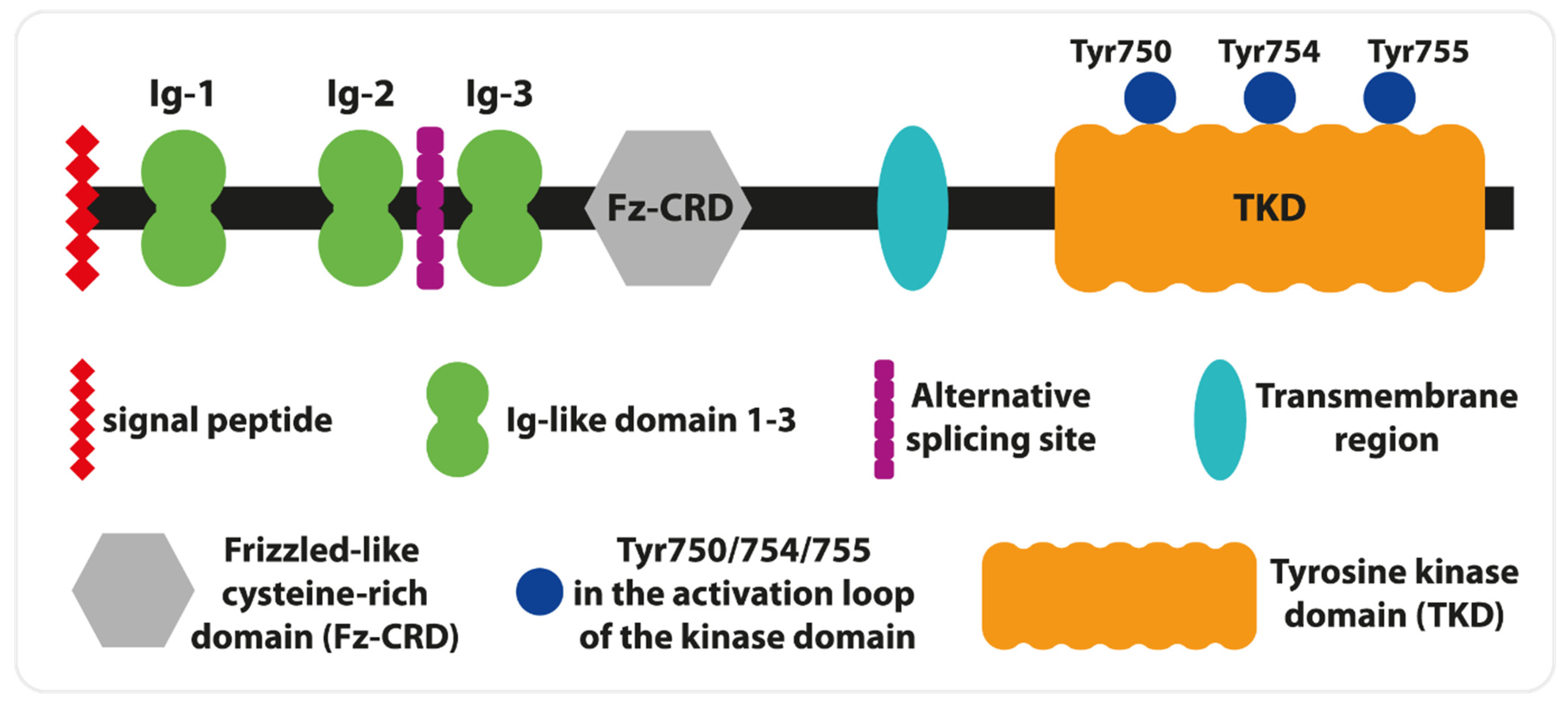
Furthermore, they have observed unique for the human gene, alternative exon 10 defective skipping. The exclusion of this exon from mRNA is more frequent in undifferentiated and poorly differentiated human myoblast and myogenic cells than in skeletal muscle [19]. Interestingly, two MuSK isoforms (one identical to the skeletal muscle variant) are expressed in the brain. It was shown that hippocampal MuSK isoforms play a crucial role in cholinergic response and help memory formation [20]. These data obtained on animal models confirm observations of MG patients with memory deficits [22]. Human MuSK is a transmembrane glycoprotein of type I and consists of several extracellular domains, a single transmembrane helix, and a cytoplasmic tail with a tyrosine kinase domain (TKD, Figure 2). The N-terminal fragment of MuSK includes a signal peptide, followed by three immunoglobulin-like domains (Ig), and a frizzle-like cysteine-rich domain (Fz-CRD). The first two Ig are crucial for lipoprotein receptor-related protein 4 (LRP4) interactions as well as homodimerization. The central point of LPR4— the binding site on Ig1—is determined by Ile96, and its mutation decreases the interaction of MuSK with LRP4 [20]. These interactions can be enhanced by agrin. Thus, agrin plays a role as an allosteric and paracrine regulator, and its presence is not necessary for MuSK activation [23]. Furthermore, LRP4 binding to MuSK impacts the hydrophobic surface situated opposite to Ile96 and aids direct interactions between Leu83 and Met48, which leads to Ig1- dimerization and autophosphorylation reactions [24]. The role of Fz-CRD is believed to be essential for MuSK activation by Wnt signaling proteins in the lack of agrin [18].
The transmembrane domain is linked with TKD by a cytoplasmic juxtamembrane segment. The autophosphorylation of Tyr553 within this segment creates the docking site for the cytoplasmic protein Dok7. Similar to LRP4, Dok7 binding increases the strength of MuSK dimerization, which is crucial for further activation of its kinase domain [25,26]. For full activation, two (Tyr754, Tyr 755) of three (Tyr, 750, Tyr754, Tyr 755) tyrosines within the activation loop of TKD need to be phosphorylated in the established order (Figure 3). In contrast, the lack of phosphate groups on these tyrosines autoinhibits the activity of MuSK [27]. Simultaneously with autophosphorylation of the activation loop, Dok7 is phosphorylated at Tyr369 and Tyr406, which permits the binding of the adapter molecule Crk and NMJ formation [28].
MuSK: From Gene to Disease
Currently, MuSK-MG is only diagnosed by detecting the autoantibodies against MuSK in patients’ serum or plasma. AChR-MG, as well as LPR4-MG, correlates with increased levels of IgG1 and IgG3 autoantibodies [29]. In contrast, antibodies against MuSK belong mainly to the subclass IgG4. These IgG4s can block direct interaction between MuSK and complex collagen Q-AChR and inhibit agrin-induced phosphorylation of MuSK, leading to attenuation of AChR clustering [30,31]. IgG4 antibodies do not affect MuSK phosphorylation in cases of a lack of agrin stimuli. The same final effect of clustering of AChR inhibition can be achieved by the less common IgG1-3 anti-MuSK antibodies. In contrast, IgG1-3 can act independently on agrin stimuli and leads to increased MuSK phosphorylation. Thus, both inhibition and overphosphorylation of MuSK can attenuate AChR clustering and lead to disorders in NMJs [31,32]. The pathogenicity and acute course of the disease result directly from the quantitative composition of individual IgG fractions, as well as the reduction of their galactosylation levels [33,34].
It has been shown that HLA class II DR14, DR16, and DQ5 alleles are dominant in MuSK-MG and can be predisposed to the production of autoimmunogenic IgG4 antibodies. On the other hand, HLA-DR13 can be beneficial and protects against pathological IgG4 [5,6,35,36,37].
The role of cellular immune response in MuSK-MG is still unknown. The contribution of Th1 and Th17 cells in this disease subclass has been postulated. The unclear mechanism of the regulation of the activity of T cell subsets causes additional constraints in the development of effective therapies [38]. Myasthenia gravis is associated with different circulating miRNA profiles [39]. However, their role, especially in the pathogenesis and development of MuSK-MG, is poorly studied. Elevated miRNAs in the serum of patients with MuSK-MG include miR-151a-3p, let-7a-5p, let-7f-5p, and miR-423-5p. Furthermore, downregulation of miR-210-3p and miR-324-3p has been found in the plasma of MuSK-MG relative to healthy controls. Despite altered microRNA expression in MuSK-MG patients’ PBMC, its targets and the signaling pathways that may play key roles in the development of the disease remain unclear. For comparison, the state of knowledge about AChR-MG is much more advanced, and increased expression of miR150-5p, as well as miR21-5p, is associated with the differentiation and cellular response of T cells. Moreover, miR30e-5p has been found to be a promising predictive biomarker for the disease. It is noteworthy, however, that studies remain very limited and require proper grouping of patients. Circulating miRNAs are not used in routine clinical practice for the diagnosis of MG [40,41,42].
Although MuSK-MG is classified as an autoimmunological disease, some mutations of the MuSK gene may lead to similar symptoms. According to the National Center for Biotechnology Information data (NCBI, www.ncbi.nlm.nih.gov), 52,922 different mutant variants of the MUSK gene were detected. The mutations that abolish the protein’s activity and functions are usually lethal. Crucial are the amino acids responsible for interactions with LRP4 (Ile96, Leu83, and Met48), autophosphorylation of the tyrosine kinase domain (Tyr 553), and Dok-7 interaction (Val790, Met605, Ala727). Other amino acids can result in diminished expression of MuSK (c220insC). Similarly, dysfunctional mutations in all NMJ proteins can lead to congenital myasthenic syndrome [43,44,45].
4. Specificity of Neurophysiological Diagnostic Tests
Extended neurophysiological assessment in patients with myasthenia gravis includes repetitive nerve stimulation (RNS), quantitative EMG (QEMG), single fiber electromyography (SFEMG), and electromyography (EMG) with nerve conduction study.
The SFEMG test was developed in the 1970s by Ekstedt and Stålberg [46,47]. Initiating research into quantifying muscle fatigue, Ekstedt and Stålberg developed a multi-electrode electrode for recording action potentials from single muscle fibers (SFAPs), which was inspired by the electrode used by Buchthal et al. [48]. The criterion for SFAPs was a rapidly increasing positive–negative peak of constant shape with successive discharges. Differences in the timing of SFAPs resulted in a ‘jitter phenomenon’, which was attributed to differences in the time at which muscle action potentials are initiated at the motor endplate. This multi-electrode electrode was also used in the analysis of the propagation velocity of individual muscle fibers. SFEMG allows quantitative measurement of, among other things, the variability of neuromuscular transmission (jitter) during successive discharges of an individual muscle fiber action potential. The test is performed by activating the muscle with a voluntary contraction, recording potentials from the muscles of the face (orbicularis oculi, frontalis) or upper extremity (extensor digitorum brevis). The average jitter is automatically calculated from 20 pairs of potentials recorded from several electrode positions. An increase in jitter with any intermittent impulse blocking indicates a significant disturbance in neuromuscular transmission. Measurement of jitter has proven to be the most sensitive method in detecting this type of pathology. This method is highly sensitive but not completely specific for myasthenia gravis. Abnormal SFEMG findings, i.e., increased jitter and blocking, also occur in Lambert–Eaton myasthenic syndrome, as well as in some other neuromuscular diseases such as amyotrophic lateral sclerosis, other neurogenic lesions, and some myopathies (including progressive external ophthalmoplegia, muscular dystrophies, and myositis) [47,48,49]. In myopathy, reinnervation, fiber splitting, and denervation in the late stage of fibrosis may be responsible for the increase in fiber density (FD) parameters. However, jitter is increased in most patients with mitochondrial diseases that primarily affect the extraocular muscles. In progressive extraocular ophthalmoplegia (PEO), as in MG, abnormal jitter can be obtained, making it impossible to diagnose these disorders with SFEMG alone [49,50,51,52,53,54,55].
The results of RNS testing in myasthenia gravis patients with anti-MuSK antibodies are similar to those from SFEMG testing, showing a higher rate of positive results (sensitivity 86%) for facial muscle testing compared to MG cases with AchR antibodies (sensitivity 82%). This reflects the greater propensity for facial muscle involvement in these MuSK antibody-positive cases and highlights the importance of including facial muscles in RNS protocols when assessing these patients [56]. However, Padua et al. conducted a study in a group of patients with seronegative myasthenia gravis (SNMG) which distinguished patients with (USK(+)) and patients without (MUSK(−)) anti-MuSK antibodies. The authors revealed that the RNS test was abnormal in significantly more MUSK(−) than MUSK(+) patients (p < 0.00001), while MuSK- patients had a more severe neurophysiological pattern with SFEMG [57].
Comparisons between RNS and jitter analysis, between MuSK-MG and AchR-MG patients, have shown that RNS is less sensitive (52%) in MG patients with antibodies to muscle-specific kinase compared with MG patients with antibodies to the acetylcholine receptor (93%) (p < 0.01) [58,59]. Nemoto et al. found positive jitter in 93% of patients with AchR antibodies, but only in 50% of patients with MuSK antibodies, and the range of jitter was greater in AchR-antibody patients versus AchR-negative patients (MCD: 76 μs in patients with AchR antibodies, 36 μs in patients with MuSK antibodies) [60]. In contrast, Nikolic et al. found no significant difference in pathological jitter detection between the two subtypes of MG patients (90% in patients with MuSK antibodies vs. 93% in patients with AchR antibodies, p > 0.05) [61]. However, the extent of jitter may be partly due to the severity of the dysfunction in different muscles. Kuwabara et al. found abnormal jitter in the extensor digitorum communis (EDC) muscle in only one of three MuSK-positive patients, but all three had increased jitter in the frontalis muscle [62]. In contrast, all AchR-positive patients (n = 11) showed equally abnormal jitter in both muscles. Similar results were reported in a different study by Farrugia et al., where the greater of patients with MuSK antibodies (n = 13) had normal jitter in the EDC notwithstanding abnormal jitter in the orbicularis oculi muscle [63]. Since patients with MuSK antibodies are thought to have predominant muscle weakness in the bulbar, facial, and neck compared to patients with MG with AchR antibodies, SFEMG should be undertaken in the most apparent muscles when MG with MuSK antibodies is suspected in order to increase sensitivity [58]. In contrast, a similar degree of SFEMG abnormalities was present in proximal muscles between MuSK(+) and AchR(+) patients [64].
5. Non-Neurological Manifestations of MuSK-MG
Myasthenia gravis also leads to reduced psychological and social well-being. The literature presents papers on quality of life (QoL) in the MuSK-MG patient population compared to AchR MG patients [65,66,67,68]. To assess health-related QoL, the SF-36 questionnaire and scales are most commonly used: the Hamilton scale for depression and anxiety, the Multidimensional Perceived Social Support Scale, and the Illness Acceptance Scale. In the study by Stankovic et al. [65], QoL scores in the physical domain were indistinguishable in MuSK-MG and AchR-MG patients, while the mental domain and total SF-36 scores were even better in MuSK groups. Social support was better in the MuSK group. The SF-36 total score correlated with anxiety (rho = 0.49, p < 0.01), depression (rho = 0.54, p < 0.01), and MSPSS (rho = −0.35, p < 0.05), and depression was an independent predictor of worse QoL. The authors conclude that, in addition to therapy for weakness, psychiatric treatment and various forms of psychosocial conditioning should form part of regular treatment protocols in MG [65].
6. Molecular Commonalities between MuSK-MG and Other Autoimmune Diseases of the Nervous System
The IgG4 autoantibody subclass is implicated in a broad spectrum of more than 12 multisystem or fibroinflammatory autoimmune diseases, referred to as IgG4-related diseases (IgG4-RD) [69,70]. IgG4 neurological disorders (IgG4-ND) are now developing into an immunopathologically distinct spectrum of diseases, as recently indicated, due to their association with pathogenic IgG4 antibodies targeting neuron-specific antigens. The main IgG4 antibody-mediated neurological disorders (IgG4-ND) include MuSK myasthenia, autoimmune nodopathies with antibodies against nodal-paranodal cell-adhesion molecules (neurofascin-155 (NF155), contactin-associated protein 1 (Caspr1), and neurofascin isoforms (NF140/186), Morvan syndrome, or neuromyotonia, anti-LGI1- and CASPR2-associated limbic encephalitis, and several cases of the anti-IgLON5 and anti-DPPX-spectrum CNS diseases. However, because IgG4 antibody titers appear to be decreased in remission and increased in exacerbation, they may serve as potential biomarkers of treatment response, further supporting a pathogenic role for self-reacting B cells. Patients with autoimmune nodopathy usually have characteristic symptoms that emphasize the subacute onset of severe neuropathy, tremor, and sensory ataxia [69,70,71,72].
Most significantly, they respond poorly to IVIg and plasmapheresis, but excellently to rituximab, which induces long-term remissions. Although patients with anti-LGI-1 and CASPR2 antibodies are characterized by clinical heterogeneity, they also demonstrate considerable overlap in clinical symptomatology; anti-LGI1 antibodies are most commonly associated with epilepsy and limbic encephalitis, while anti-CASPR2 antibodies are associated with neuromyotonia, Morvan syndrome, and neuropathic pain. Anti-IgLON5 antibodies define a complex syndrome of chronic progressive brainstem symptomatology, gait instability, distinct non-rapid eye movement (REM) and REM parasomnias, sleep-disordered breathing, obstructive sleep apnea, cognitive decline, and movement disorders as recently identified, most commonly craniofacial dyskinesias, chorea, dystonia, and abnormal eye movements [3,71,72,73,74,75].
7. Treatment
The majority of patients with MuSK-MG have little or no therapeutic response to treatment with anticholinesterase drugs and experience an increase in cholinergic symptoms even at low doses [4]. Modoni et al. demonstrated that cholinergic hyperactivity to standard doses of acetylcholine esterase inhibitors (AchE-Is) is a relatively common symptom in patients with MuSK-MG, independent of AchE-I treatment, and may be an inherent feature of the disease [75]. In addition, the response to standard doses of pyridostigmine used in AchR-MG is ineffective and poorly tolerated due to its side effects. Among symptomatic medications for MuSK-MG, 3,4-diaminopyridine (3,4-DAP), ephedrine, and albuterol have recently been considered. The use of 3,4-DAP in patients with MuSK-MG has been described as moderately to mildly effective, with no notable side effects [5,76,77,78,79,80].
Conventional immunosuppressants are not commonly able to replace steroids in the maintenance of the satisfactory long-term control of symptoms. In MuSK-MG patients with exacerbated symptoms, high-dose prednisone, combined with plasma exchange, is recommended. Intravenous immunoglobulin should also be considered in these patients. In patients with contraindications to steroids, traditional immunosuppressive drugs (azathioprine, tacrolimus, mycophenolate, methotrexate, and cyclosporine) have been used, but achieving and ensuring long-term and complete symptom control is usually more difficult than in Ach-R MG patients. However, the majority of MuSK-MG patients are refractory to treatment. In these cases, the use of rituximab has shown promising results leading to sustained symptom control [80,81,82,83].
According to expert opinion, the treatment of MuSK-MG with rituximab (RTX), a monoclonal antibody directed against the CD20 receptor, is highly effective. RTX is more successful in MuSK-MG than in other MG subgroups and can be used for treatment at an earlier stage [84,85,86]. Rituximab is a chimeric murine/human monoclonal antibody produced by genetic engineering of Chinese hamster ovary tissue culture cells. It is a glycosylated immunoglobulin containing fixed sequences of human IgG1 and variable sequences of mouse light and heavy chains. It binds selectively to the transmembrane antigen CD20, which is found on the surface of B lymphocytes (circulating naïve and memory B cells) and is absent on other cells. RTX induces the death of cells containing the CD20 antigen by mechanisms dependent on both the complement system and those associated with antibody-dependent cellular cytotoxicity, as well as by apoptosis. B lymphocyte stem cells are devoid of the CD20 antigen, and the B lymphocyte population can be reconstituted after treatment with rituximab [85,86,87]. Marino et al. studied the long-term effects of RTX in nine treatment-resistant patients with MuSK-MG, with follow-up periods of 17 months to 13 years. Their data demonstrated that the therapeutic effects of RTX can continue for several years following treatment, suggesting that by depleting autoreactive B-cell clones, RTX can markedly disrupt the immunopathogenic circuitry responsible for maintaining the disease [83]. It is recognized that B-cell activity depends on T–B lymphocyte cross-talk and cooperation. Future studies are needed to investigate the effect of RTX on such interaction, particularly in relation to specific T- and B-cell repertoires.
Other antibodies targeting B lymphocytes (CD20, CD19) with potential relevance in myasthenia gravis, but without documented efficiency, include ocrelizumab, ofatumumab, obinutuzumab, ublituximab, and ibalizumab. In addition, the potential efficacy of the proteasome inhibitor was described in a report on the favorable impact of this drug in a patient with severe myasthenia gravis with anti-MuSK antibodies [88,89,90,91,92].
Promising therapeutic targets in patients with MuSK-MG are monoclonal antibodies against molecules involved in B-cell activation and against B cells at different stages of their maturation (e.g., plasmablasts). Precision medicine using chimeric autoantibodies against the T-cell receptor (CAAR-T) can also be effective. These are designed to target antigen-specific B cells in MuSK-MG. Other drugs are monoclonal antibodies against FcRn receptors: rozanolixizumab and efgartigimod. The principle of action of FcRn, a neonatal Fc receptor, is to bind to the Fc region and rescue IgG from acidic lysosomal degradation. This promotes recycling. The mechanism of action is very similar to that of IVIG. Efgartigimod is the first FcRn inhibitor to be approved for use in AChR-MG [93,94,95,96]. Clinical trials have shown that FcRn inhibitors are more effective in MuSK-MG patients than in AchR-MG patients. IgG4-dominant MuSK-MG is a weak complement-activating subclass of immunoglobulin, and complement inhibition is not effective [97,98,99]. Results from the REGAIN study showed that eculizumab was most effective in patients with anti-AChR antibodies [100,101]. Eculizumab is a humanized monoclonal antibody directed against the C5 component of the complement system. It inhibits the final step of complement activation and the formation of membrane attack complexes (MACs). It does this by blocking the conversion of C5a to C5b. With regard to the potential efficacy of the proteasome inhibitor bortezomib, at least one case of a beneficial effect of this drug has been described in a patient with severe myasthenia gravis with anti-MuSK antibodies [102,103].
In MuSK-MG, the thymus is usually atrophic. Thymic follicular hyperplasia (TFH) occurs in rare cases. They are characterized by a more severe course and less responsive to standard immunosuppression [104]. For patients with MuSK-MG, thymectomy is not currently recommended. However, anterior mediastinal imaging is required in all patients with an established diagnosis of MG, regardless of antibody type [105].
8. Conclusions
MuSK myasthenia gravis is a more aggressive and difficult-to-treat form of neuromuscular junction disease. In recent years, new information has become available on the potential pathomechanisms of this form of MG. Advances in research into immunopathogenesis will contribute to the correct diagnosis of this autoimmune disease and the application of effective treatment. Further research is needed on the role of thymus in MuSK MG pathogenesis and its role as a potential therapeutic target too.
Author Contributions
E.D. conceptualized and wrote the manuscript; D.B. conceptualized and wrote the manuscript; M.W.-P. conceptualized and wrote the manuscript. All authors have read and agreed to the published version of the manuscript.
Funding
This research received no external funding.
Institutional Review Board Statement
Not applicable.
Informed Consent Statement
Not applicable.
Data Availability Statement
Not applicable.
Acknowledgments
Conflicts of Interest
The authors declare no conflicts of interest.
Abbreviations
| AchE-Is | acetylcholinesterase inhibitors |
| AP | action potential |
| Caspr1 | contactin-associated protein 1 |
| CMS | congenital myasthenic syndromes |
| 3,4-DAP | 3,4-diaminopyridine |
| Dok7 | downstream of kinase-7 |
| EDC | extensor digitorum communis |
| EGF | epidermal growth factor |
| IgG4-RD | IgG4-related diseases |
| LRP4 | low-density lipoprotein receptor-related protein 4 |
| MASC | myotube-associated specificity component |
| MG | myasthenia gravis |
| MuSK | muscle-specific tyrosine kinase |
| NF155 | neurofascin-155 |
| NMJ | neuromuscular junction |
| QoL | quality of life |
| RNS | Repetitive Nerve Stimulation |
| RTX | Rituximab |
| SFAPs | single muscle fibers |
References
- Fichtner, M.L.; Jiang, R.; Bourke, A.; Nowak, R.J.; O’Connor, K.C. Autoimmune Pathology in Myasthenia Gravis Disease Subtypes Is Governed by Divergent Mechanisms of Immunopathology. Front. Immunol. 2020, 11, 776. [Google Scholar] [CrossRef] [PubMed]
- Dresser, L.; Wlodarski, R.; Rezania, K.; Soliven, B. Myasthenia Gravis: Epidemiology, Pathophysiology and Clinical Manifestations. J. Clin. Med. 2021, 10, 2235. [Google Scholar] [CrossRef] [PubMed]
- Dziadkowiak, E.; Waliszewska-Prosół, M.; Wieczorek, M.; Bladowska, J.; Budrewicz, S.; Ejma, M. Myasthenia Gravis-An Analysis of Multimodal Evoked Potentials. Brain Sci. 2021, 11, 1057. [Google Scholar] [CrossRef] [PubMed]
- El-Salem, K.; Yassin, A.; Al-Hayk, K.; Yahya, S.; Al-Shorafat, D.; Dahbour, S.S. Treatment of MuSK-Associated Myasthenia Gravis. Curr. Treat. Options Neurol. 2014, 16, 283. [Google Scholar] [CrossRef] [PubMed]
- Rodolico, C.; Bonanno, C.; Toscano, A.; Vita, G. MuSK-Associated Myasthenia Gravis: Clinical Features and Management. Front. Neurol. 2020, 11, 660. [Google Scholar] [CrossRef] [PubMed]
- Evoli, A.; Alboini, P.E.; Damato, V.; Iorio, R.; Provenzano, C.; Bartoccioni, E.; Marino, M. Myasthenia gravis with antibodies to MuSK: An update. Ann. N. Y. Acad. Sci. 2018, 1412, 82–89. [Google Scholar] [CrossRef]
- Leite, M.I.; Ströbel, P.; Jones, M.; Micklem, K.; Moritz, R.; Gold, R.; Niks, E.H.; Berrih-Aknin, S.; Scaravilli, F.; Canelhas, A.; et al. Fewer thymic changes in MuSK antibody-positive than in MuSK antibody-negative MG. Ann. Neurol. 2005, 57, 444–448. [Google Scholar] [CrossRef]
- Clifford, K.M.; Hobson-Webb, L.D.; Benatar, M.; Burns, T.M.; Barnett, C.; Silvestri, N.J.; Howard, J.F., Jr.; Visser, A.; Crum, B.A.; Nowak, R.; et al. Thymectomy may not be associated with clinical improvement in MuSK myasthenia gravis. Muscle Nerve 2019, 59, 404–410. [Google Scholar] [CrossRef]
- Borges, L.S.; Richman, D.P. Muscle-Specific Kinase Myasthenia Gravis. Front. Immunol. 2020, 11, 707. [Google Scholar] [CrossRef]
- Mori, S.; Shigemoto, K. Mechanisms associated with the pathogenicity of antibodies against muscle-specific kinase in myasthenia gravis. Autoimmun. Rev. 2013, 12, 912–917. [Google Scholar] [CrossRef]
- Lepore, E.; Casola, I.; Dobrowolny, G.; Musarò, A. Neuromuscular Junction as an Entity of Nerve-Muscle Communication. Cells 2019, 8, 906. [Google Scholar] [CrossRef]
- Rodríguez Cruz, P.M.; Cossins, J.; Beeson, D.; Vincent, A. The Neuromuscular Junction in Health and Disease: Molecular Mechanisms Governing Synaptic Formation and Homeostasis. Front. Mol. Neurosci. 2020, 13, 610964. [Google Scholar] [CrossRef]
- Hubbard, S.R.; Gnanasambandan, K. Structure and activation of MuSK, a receptor tyrosine kinase central to neuromuscular junction formation. Biochim. Biophys. Acta 2013, 1834, 2166–2169. [Google Scholar] [CrossRef]
- Burden, S.J. SnapShot: Neuromuscular Junction. Cell 2011, 144, 826.e1. [Google Scholar] [CrossRef]
- Zhang, B.; Luo, S.; Wang, Q.; Suzuki, T.; Xiong, W.C.; Mei, L. LRP4 serves as a coreceptor of agrin. Neuron 2008, 60, 285–297. [Google Scholar] [CrossRef]
- Kim, N.; Stiegler, A.L.; Cameron, T.O.; Hallock, P.T.; Gomez, A.M.; Huang, J.H.; Hubbard, S.R.; Dustin, M.L.; Burden, S.J. Lrp4 is a receptor for Agrin and forms a complex with MuSK. Cell 2008, 135, 334–342. [Google Scholar] [CrossRef]
- Koneczny, I.; Herbst, R. Myasthenia Gravis: Pathogenic Effects of Autoantibodies on Neuromuscular Architecture. Cells 2019, 8, 671. [Google Scholar] [CrossRef] [PubMed]
- Herbst, R. MuSk function during health and disease. Neurosci. Lett. 2020, 716, 134676. [Google Scholar] [CrossRef] [PubMed]
- Nasrin, F.; Rahman, M.A.; Masuda, A.; Ohe, K.; Takeda, J.; Ohno, K. HnRNP C, YB-1 and hnRNP L coordinately enhance skipping of human MUSK exon 10 to generate a Wnt-insensitive MuSK isoform. Sci. Rep. 2014, 4, 6841. [Google Scholar] [CrossRef] [PubMed]
- Vergoossen, D.L.E.; Keo, A.; Mahfouz, A.; Huijbers, M.G. Timing and localization of myasthenia gravis-related gene expression. Eur. J. Neurosci. 2021, 54, 5574–5585. [Google Scholar] [CrossRef]
- Valenzuela, D.M.; Stitt, T.N.; DiStefano, P.S.; Rojas, E.; Mattsson, K.; Compton, D.L.; Nunez, L.; Park, J.S.; Stark, J.L.; Gies, D.R.; et al. Receptor tyrosine kinase specific for the skeletal muscle lineage: Expression in embryonic muscle, at the neuromuscular junction, and after injury. Neuron 1995, 15, 573–584. [Google Scholar] [CrossRef] [PubMed]
- Bohbot, V.D.; Jech, R.; Bures, J.; Nadel, L.; Růzicka, E. Spatial and nonspatial memory involvement in myasthenia gravis. J. Neurol. 1997, 244, 529–532. [Google Scholar] [CrossRef]
- Zhang, W.; Coldefy, A.S.; Hubbard, S.R.; Burden, S.J. Agrin binds to the N-terminal region of Lrp4 protein and stimulates association between Lrp4 and the first immunoglobulin-like domain in muscle-specific kinase (MuSK). J. Biol. Chem. 2011, 286, 40624–40630. [Google Scholar] [CrossRef] [PubMed]
- Stiegler, A.L.; Burden, S.J.; Hubbard, S.R. Crystal structure of the agrin-responsive immunoglobulin-like domains 1 and 2 of the receptor tyrosine kinase MuSK. J. Mol. Biol. 2006, 364, 424–433. [Google Scholar] [CrossRef] [PubMed]
- Okada, K.; Inoue, A.; Okada, M.; Murata, Y.; Kakuta, S.; Jigami, T.; Kubo, S.; Shiraishi, H.; Eguchi, K.; Motomura, M.; et al. The muscle protein Dok-7 is essential for neuromuscular synaptogenesis. Science 2006, 312, 1802–1805. [Google Scholar] [CrossRef] [PubMed]
- Bergamin, E.; Hallock, P.T.; Burden, S.J.; Hubbard, S.R. The cytoplasmic adaptor protein Dok7 activates the receptor tyrosine kinase MuSK via dimerization. Mol. Cell 2010, 39, 100–109. [Google Scholar] [CrossRef]
- Till, J.H.; Becerra, M.; Watty, A.; Lu, Y.; Ma, Y.; Neubert, T.A.; Burden, S.J.; Hubbard, S.R. Crystal structure of the MuSK tyrosine kinase: Insights into receptor autoregulation. Structure 2002, 10, 1187–1196. [Google Scholar] [CrossRef] [PubMed]
- Hallock, P.T.; Xu, C.F.; Park, T.J.; Neubert, T.A.; Curran, T.; Burden, S.J. Dok-7 regulates neuromuscular synapse formation by recruiting Crk and Crk-L. Genes Dev. 2010, 24, 2451–2461. [Google Scholar] [CrossRef]
- Rødgaard, A.; Nielsen, F.C.; Djurup, R.; Somnier, F.; Gammeltoft, S. Acetylcholine receptor antibody in myasthenia gravis: Predominance of IgG subclasses 1 and 3. Clin. Exp. Immunol. 1987, 67, 82–88. [Google Scholar]
- Otsuka, K.; Ito, M.; Ohkawara, B.; Masuda, A.; Kawakami, Y.; Sahashi, K.; Nishida, H.; Mabuchi, N.; Takano, A.; Engel, A.G.; et al. Collagen Q and anti-MuSK autoantibody competitively suppress agrin/LRP4/MuSK signaling. Sci. Rep. 2015, 5, 13928. [Google Scholar] [CrossRef]
- Cao, M.; Liu, W.W.; Maxwell, S.; Huda, S.; Webster, R.; Evoli, A.; Beeson, D.; Cossins, J.A.; Vincent, A. IgG1-3 MuSK Antibodies Inhibit AChR Cluster Formation, Restored by SHP2 Inhibitor, Despite Normal MuSK, DOK7, or AChR Subunit Phosphorylation. Neurol. Neuroimmunol. Neuroinflamm. 2023, 10, e200147. [Google Scholar] [CrossRef]
- Koneczny, I.; Cossins, J.; Waters, P.; Beeson, D.; Vincent, A. MuSK myasthenia gravis IgG4 disrupts the interaction of LRP4 with MuSK but both IgG4 and IgG1-3 can disperse preformed agrin-independent AChR clusters. PLoS ONE 2013, 8, e80695. [Google Scholar] [CrossRef]
- Vergoossen, D.L.E.; Plomp, J.J.; Gstöttner, C.; Fillié-Grijpma, Y.E.; Augustinus, R.; Verpalen, R.; Wuhrer, M.; Parren, P.W.H.I.; Dominguez-Vega, E.; van der Maarel, S.M.; et al. Functional monovalency amplifies the pathogenicity of anti-MuSK IgG4 in myasthenia gravis. Proc. Natl. Acad. Sci. USA 2021, 118, e2020635118. [Google Scholar] [CrossRef]
- Hajdukovic, L.; Palibrk, A.; Peric, S.; Basta, I.; Minic, R.; Jankovic, M.; Lavrnic, D. Galactosylation of serum immunoglobulin G in myasthenia gravis with different autoantibodies. Scand. J. Clin. Lab. Investig. 2023, 83, 348–355. [Google Scholar] [CrossRef]
- Hong, Y.; Li, H.F.; Romi, F.; Skeie, G.O.; Gilhus, N.E. HLA and MuSK-positive myasthenia gravis: A systemic review and meta-analysis. Acta Neurol. Scand. 2018, 138, 219–226. [Google Scholar] [CrossRef] [PubMed]
- Alahgholi-Hajibehzad, M.; Yilmaz, V.; Gülsen-Parman, Y.; Aysal, F.; Oflazer, P.; Deymeer, F.; Saruhan-Direskeneli, G. Association of HLA-DRB1∗14, -DRB1∗16 and -DQB1∗05 with MuSK-myasthenia gravis in patients from Turkey. Hum. Immunol. 2013, 74, 1633–1635. [Google Scholar] [CrossRef] [PubMed]
- Kanai, T.; Uzawa, A.; Kawaguchi, N.; Sakamaki, T.; Yoshiyama, Y.; Himuro, K.; Oda, F.; Kuwabara, S. HLA-DRB1*14 and DQB1*05 are associated with Japanese anti-MuSK antibody-positive myasthenia gravis patients. J. Neurol. Sci. 2016, 363, 116–118. [Google Scholar] [CrossRef]
- Yi, J.S.; Guidon, A.; Sparks, S.; Osborne, R.; Juel, V.C.; Massey, J.M.; Sanders, D.B.; Weinhold, K.J.; Guptill, J.T. Characterization of CD4 and CD8 T cell responses in MuSK myasthenia gravis. J. Autoimmun. 2014, 52, 130–138. [Google Scholar] [CrossRef]
- Saghazadeh, A.; Rezaei, N. MicroRNA expression profiles of peripheral blood and mononuclear cells in myasthenia gravis: A systematic review. Int. Immunopharmacol. 2022, 112, 109205. [Google Scholar] [CrossRef] [PubMed]
- Beretta, F.; Huang, Y.F.; Punga, A.R. Towards Personalized Medicine in Myasthenia Gravis: Role of Circulating microRNAs miR-30e-5p, miR-150-5p and miR-21-5p. Cells 2022, 11, 740. [Google Scholar] [CrossRef]
- Punga, A.R.; Andersson, M.; Alimohammadi, M.; Punga, T. Disease specific signature of circulating miR-150-5p and miR-21-5p in myasthenia gravis patients. J. Neurol. Sci. 2015, 356, 90–96. [Google Scholar] [CrossRef]
- Punga, T.; Bartoccioni, E.; Lewandowska, M.; Damato, V.; Evoli, A.; Punga, A.R. Disease specific enrichment of circulating let-7 family microRNA in MuSK+ myasthenia gravis. J. Neuroimmunol. 2016, 292, 21–26. [Google Scholar] [CrossRef] [PubMed]
- Chevessier, F.; Girard, E.; Molgó, J.; Bartling, S.; Koenig, J.; Hantaï, D.; Witzemann, V. A mouse model for congenital myasthenic syndrome due to MuSK mutations reveals defects in structure and function of neuromuscular junctions. Hum. Mol. Genet. 2008, 17, 3577–3595. [Google Scholar] [CrossRef] [PubMed]
- Chevessier, F.; Faraut, B.; Ravel-Chapuis, A.; Richard, P.; Gaudon, K.; Bauché, S.; Prioleau, C.; Herbst, R.; Goillot, E.; Ioos, C.; et al. MUSK, a new target for mutations causing congenital myasthenic syndrome. Hum. Mol. Genet. 2004, 13, 3229–3240. [Google Scholar] [CrossRef] [PubMed]
- Maselli, R.A.; Arredondo, J.; Cagney, O.; Ng, J.J.; Anderson, J.A.; Williams, C.; Gerke, B.J.; Soliven, B.; Wollmann, R.L. Mutations in MUSK causing congenital myasthenic syndrome impair MuSK-Dok-7 interaction. Hum. Mol. Genet. 2010, 19, 2370–2379. [Google Scholar] [CrossRef]
- Ekstedt, J. Human single muscle fiber action potentials. Extracellular recording during voluntary and chemical activation. With some comments on end-plate physiology and on the fiber arrangement of the motor unit. Acta Physiol. Scand. Suppl. 1964, (Suppl. 226:1+). [Google Scholar] [PubMed]
- Stålberg, E.; Ekstedt, J.; Broman, A. Neuromuscular transmission in myasthenia gravis studied with single fibre electromyography. J. Neurol. Neurosurg. Psychiatry 1974, 37, 540–547. [Google Scholar] [CrossRef]
- Buchthal, F.; Guld, C.; Rosenfalck, F. Multielectrode study of the territory of a motor unit. Acta Physiol. Scand. 1957, 39, 83–104. [Google Scholar] [CrossRef]
- Sanders, D.B.; Arimura, K.; Cui, L.; Ertaş, M.; Farrugia, M.E.; Gilchrist, J.; Kouyoumdjian, J.A.; Padua, L.; Pitt, M.; Stålberg, E. Guidelines for single fiber EMG. Clin. Neurophysiol. 2019, 130, 1417–1439. [Google Scholar] [CrossRef]
- Oh, S.J.; Ohira, M. Single-fiber EMG and clinical correlation in Lambert-Eaton myasthenic syndrome. Muscle Nerve 2013, 47, 664–667. [Google Scholar] [CrossRef]
- Emeryk-Szajewska, B. Electrophysiological investigations in diagnosis and evaluation of ALS progress. Neurol. Neurochir. Pol. 2001, 35 (Suppl. S1), 11–24. [Google Scholar] [PubMed]
- Cui, L.Y.; Liu, M.S.; Tang, X.F. Single fiber electromyography in 78 patients with amyotrophic lateral sclerosis. Chin. Med. J. 2004, 117, 1830–1833. [Google Scholar] [PubMed]
- Hatanaka, Y.; Oh, S.J. Single-fiber electromyography in sporadic inclusion body myopathy. Clin. Neurophysiol. 2007, 118, 1563–1568. [Google Scholar] [CrossRef] [PubMed]
- Jian, F.; Cui, L.Y.; Li, B.H.; Du, H. Changes of single fiber electromyography in patients with inflammatory myopathies. Chin. Med. Sci. J. 2005, 20, 1–4. [Google Scholar] [PubMed]
- Padua, L.; Stalberg, E.; LoMonaco, M.; Evoli, A.; Batocchi, A.; Tonali, P. SFEMG in ocular myasthenia gravis diagnosis. Clin. Neurophysiol. 2000, 111, 1203–1207. [Google Scholar] [CrossRef] [PubMed]
- Oh, S.J.; Hatanaka, Y.; Hemmi, S.; Young, A.M.; Scheufele, M.L.; Nations, S.P.; Lu, L.; Claussen, G.C.; Wolfe, G.I. Repetitive nerve stimulation of facial muscles in MuSK antibody-positive myasthenia gravis. Muscle Nerve. 2006, 33, 500–504. [Google Scholar] [CrossRef] [PubMed]
- Padua, L.; Tonali, P.; Aprile, I.; Caliandro, P.; Bartoccioni, E.; Evoli, A. Seronegative myasthenia gravis: Comparison of neurophysiological picture in MuSK+ and MuSK- patients. Eur. J. Neurol. 2006, 13, 273–276. [Google Scholar] [CrossRef]
- Yoganathan, K.; Stevenson, A.; Tahir, A.; Sadler, R.; Radunovic, A.; Malek, N. Bedside and laboratory diagnostic testing in myasthenia. J. Neurol. 2022, 269, 3372–3384. [Google Scholar] [CrossRef]
- Abraham, A.; Alabdali, M.; Alsulaiman, A.; Breiner, A.; Barnett, C.; Katzberg, H.D.; Lovblom, L.E.; Bril, V. Repetitive nerve stimulation cutoff values for the diagnosis of myasthenia gravis. Muscle Nerve 2017, 55, 166–170. [Google Scholar] [CrossRef]
- Nemoto, Y.; Kuwabara, S.; Misawa, S.; Kawaguchi, N.; Hattori, T.; Takamori, M.; Vincent, A. Patterns and severity of neuromuscular transmission failure in seronegative myasthenia gravis. J. Neurol. Neurosurg. Psychiatry 2005, 76, 714–718. [Google Scholar] [CrossRef]
- Nikolic, A.; Basta, I.; Stojanovic, V.R.; Stevic, Z.; Lavrnic, D. Electrophysiological profile of the patients with MuSK positive myasthenia gravis. Neurol. Res. 2014, 36, 945–949. [Google Scholar] [CrossRef]
- Kuwabara, S.; Nemoto, Y.; Misawa, S.; Takahashi, H.; Kawaguchi, N.; Hattori, T. Anti-MuSK-positive myasthenia gravis: Neuromuscular transmission failure in facial and limb muscles. Acta Neurol. Scand. 2007, 115, 126–128. [Google Scholar] [CrossRef]
- Farrugia, M.E.; Kennett, R.P.; Newsom-Davis, J.; Hilton-Jones, D.; Vincent, A. Single-fiber electromyography in limb and facial muscles in muscle-specific kinase antibody and acetylcholine receptor antibody myasthenia gravis. Muscle Nerve 2006, 33, 568–570. [Google Scholar] [CrossRef] [PubMed]
- Rostedt Punga, A.; Ahlqvist, K.; Bartoccioni, E.; Scuderi, F.; Marino, M.; Suomalainen, A.; Kalimo, H.; Stålberg, E.V. Neurophysiological and mitochondrial abnormalities in MuSK antibody seropositive myasthenia gravis compared to other immunological subtypes. Clin. Neurophysiol. 2006, 117, 1434–1443. [Google Scholar] [CrossRef] [PubMed]
- Stankovic, M.; Peric, S.; Stojiljkovic Tamas, O.; Stankovic, T.; Nikolic, A.; Lavrnic, D.; Basta, I. Quality of life in patients with MuSK positive myasthenia gravis. Acta Neurol. Belg. 2018, 118, 423–427. [Google Scholar] [CrossRef] [PubMed]
- Wu, X.; Li, R.Y.; Ye, X.B.; Wang, N. Reduced quality of life in myasthenia gravis patients: A study on 185 patients from China. Front. Neurol. 2023, 13, 1072861. [Google Scholar] [CrossRef] [PubMed]
- Lehnerer, S.; Jacobi, J.; Schilling, R.; Grittner, U.; Marbin, D.; Gerischer, L.; Stascheit, F.; Krause, M.; Hoffmann, S.; Meisel, A. Burden of disease in myasthenia gravis: Taking the patient’s perspective. J. Neurol. 2022, 269, 3050–3063. [Google Scholar] [CrossRef] [PubMed]
- Gelinas, D.; Parvin-Nejad, S.; Phillips, G.; Cole, C.; Hughes, T.; Silvestri, N.; Govindarajan, R.; Jefferson, M.; Campbell, J.; Burnett, H. The humanistic burden of myasthenia gravis: A systematic literature review. J. Neurol. Sci. 2022, 437, 120268. [Google Scholar] [CrossRef] [PubMed]
- Waliszewska-Prosół, M.; Ejma, M. Hashimoto Encephalopathy-Still More Questions than Answers. Cells 2022, 11, 2873. [Google Scholar] [CrossRef]
- Dziadkowiak, E.; Waliszewska-Prosół, M.; Nowakowska-Kotas, M.; Budrewicz, S.; Koszewicz, Z.; Koszewicz, M. Pathophysiology of the Different Clinical Phenotypes of Chronic Inflammatory Demyelinating Polyradiculoneuropathy (CIDP). Int. J. Mol. Sci. 2021, 23, 179. [Google Scholar] [CrossRef]
- Dalakas, M.C. Autoimmune Neurological Disorders with IgG4 Antibodies: A Distinct Disease Spectrum with Unique IgG4 Functions Responding to Anti-B Cell Therapies. Neurotherapeutics 2022, 19, 741–752. [Google Scholar] [CrossRef] [PubMed]
- Stathopoulos, P.; Alexopoulos, H.; Dalakas, M.C. Autoimmune antigenic targets at the node of Ranvier in demyelinating disorders. Nat. Rev. Neurol. 2015, 11, 143–156. [Google Scholar] [CrossRef] [PubMed]
- Dalakas, M.C. IgG4-Mediated Neurologic Autoimmunities: Understanding the Pathogenicity of IgG4, Ineffectiveness of IVIg, and Long-Lasting Benefits of Anti-B Cell Therapies. Neurol. Neuroimmunol. Neuroinflamm. 2021, 9, e1116. [Google Scholar] [CrossRef] [PubMed]
- Sabater, L.; Gaig, C.; Gelpi, E.; Bataller, L.; Lewerenz, J.; Torres-Vega, E.; Contreras, A.; Giometto, B.; Compta, Y.; Embid, C.; et al. A novel non-rapid-eye movement and rapid-eye-movement parasomnia with sleep breathing disorder associated with antibodies to IgLON5: A case series, characterisation of the antigen, and post-mortem study. Lancet Neurol. 2014, 13, 575–586. [Google Scholar] [CrossRef] [PubMed]
- Tian, X.; Deng, Z.; Wang, S.; Wang, Y. Basic Research and Clinical Reports Associated with Low Serum IgG4 Concentrations. Int. Arch. Allergy Immunol. 2020, 181, 149–158. [Google Scholar] [CrossRef] [PubMed]
- Modoni, A.; Mastrorosa, A.; Spagni, G.; Evoli, A. Cholinergic hyperactivity in patients with myasthenia gravis with MuSK antibodies: A neurophysiological study. Clin. Neurophysiol. 2021, 132, 1845–1849. [Google Scholar] [CrossRef] [PubMed]
- Huda, S.; Waters, P.; Woodhall, M.; Leite, M.I.; Jacobson, L.; De Rosa, A.; Maestri, M.; Ricciardi, R.; Heckmann, J.M.; Maniaol, A.; et al. IgG-specific cell-based assay detects potentially pathogenic MuSK-Abs in seronegative MG. Neurol. Neuroimmunol. Neuroinflamm. 2017, 4, e357. [Google Scholar] [CrossRef]
- Evoli, A.; Alboini, P.E.; Damato, V.; Iorio, R. 3,4-Diaminopyridine may improve myasthenia gravis with MuSK antibodies. Neurology 2016, 86, 1070–1071. [Google Scholar] [CrossRef]
- Haran, M.; Schattner, A.; Mate, A.; Starobin, D.; Haran, G.; Shtalrid, M. Can a rare form of myasthenia gravis shed additional light on disease mechanisms? Clin. Neurol. Neurosurg. 2013, 115, 562–566. [Google Scholar] [CrossRef]
- König, N.; Stetefeld, H.R.; Dohmen, C.; Mergenthaler, P.; Kohler, S.; Schönenberger, S.; Bösel, J.; Lee, D.H.; Gerner, S.T.; Huttner, H.B.; et al. MuSK-antibodies are associated with worse outcome in myasthenic crisis requiring mechanical ventilation. J. Neurol. 2021, 268, 4824–4833. [Google Scholar] [CrossRef]
- Guptill, J.T.; Sanders, D.B. Update on muscle-specific tyrosine kinase antibody positive myasthenia gravis. Curr. Opin. Neurol. 2010, 23, 530–535. [Google Scholar] [CrossRef]
- Evoli, A.; Padua, L. Diagnosis and therapy of myasthenia gravis with antibodies to muscle-specific kinase. Autoimmun. Rev. 2013, 12, 931–935. [Google Scholar] [CrossRef]
- Marino, M.; Basile, U.; Spagni, G.; Napodano, C.; Iorio, R.; Gulli, F.; Todi, L.; Provenzano, C.; Bartoccioni, E.; Evoli, A. Long-Lasting Rituximab-Induced Reduction of Specific-But Not Total-IgG4 in MuSK-Positive Myasthenia Gravis. Front. Immunol. 2020, 11, 613. [Google Scholar] [CrossRef]
- Narayanaswami, P.; Sanders, D.B.; Wolfe, G.; Benatar, M.; Cea, G.; Evoli, A.; Gilhus, N.E.; Illa, I.; Kuntz, N.L.; Massey, J.; et al. International Consensus Guidance for Management of Myasthenia Gravis: 2020 Update. Neurology 2021, 96, 114–122. [Google Scholar] [CrossRef]
- Sanders, D.B.; Wolfe, G.I.; Benatar, M.; Evoli, A.; Gilhus, N.E.; Illa, I.; Kuntz, N.; Massey, J.M.; Melms, A.; Murai, H.; et al. International consensus guidance for management of myasthenia gravis: Executive summary. Neurology 2016, 87, 419–425. [Google Scholar] [CrossRef]
- Vesperinas-Castro, A.; Cortés-Vicente, E. Rituximab treatment in myasthenia gravis. Front. Neurol. 2023, 14, 1275533. [Google Scholar] [CrossRef] [PubMed]
- Hofmann, K.; Clauder, A.K.; Manz, R.A. Targeting B Cells and Plasma Cells in Autoimmune Diseases. Front. Immunol. 2018, 9, 835. [Google Scholar] [CrossRef] [PubMed]
- Huijbers, M.G.; Plomp, J.J.; van der Maarel, S.M.; Verschuuren, J.J. IgG4-mediated autoimmune diseases: A niche of antibody-mediated disorders. Ann. N. Y. Acad. Sci. 2018, 1413, 92–103. [Google Scholar] [CrossRef] [PubMed]
- Sánchez-Tejerina, D.; Sotoca, J.; Llaurado, A.; López-Diego, V.; Juntas-Morales, R.; Salvado, M. New Targeted Agents in Myasthenia Gravis and Future Therapeutic Strategies. J. Clin. Med. 2022, 11, 6394. [Google Scholar] [CrossRef] [PubMed]
- Mantegazza, R.; Bernasconi, P.; Cavalcante, P. Myasthenia gravis: From autoantibodies to therapy. Curr. Opin. Neurol. 2018, 31, 517–525. [Google Scholar] [CrossRef] [PubMed]
- Beecher, G.; Putko, B.N.; Wagner, A.N.; Siddiqi, Z.A. Therapies Directed Against B-Cells and Downstream Effectors in Generalized Autoimmune Myasthenia Gravis: Current Status. Drugs 2019, 79, 353–364. [Google Scholar] [CrossRef]
- Lazaridis, K.; Tzartos, S.J. Autoantibody Specificities in Myasthenia Gravis; Implications for Improved Diagnostics and Therapeutics. Front. Immunol. 2020, 11, 212. [Google Scholar] [CrossRef]
- Vakrakou, A.G.; Karachaliou, E.; Chroni, E.; Zouvelou, V.; Tzanetakos, D.; Salakou, S.; Papadopoulou, M.; Tzartos, S.; Voumvourakis, K.; Kilidireas, C.; et al. Immunotherapies in MuSK-positive Myasthenia Gravis; an IgG4 antibody-mediated disease. Front. Immunol. 2023, 14, 1212757. [Google Scholar] [CrossRef]
- Howard, J.F., Jr.; Bril, V.; Vu, T.; Karam, C.; Peric, S.; Margania, T.; Murai, H.; Bilinska, M.; Shakarishvili, R.; Smilowski, M.; et al. Safety, efficacy, and tolerability of efgartigimod in patients with generalised myasthenia gravis (ADAPT): A multicentre, randomised, placebo-controlled, phase 3 trial. Lancet Neurol. 2021, 20, 526–536. [Google Scholar] [CrossRef] [PubMed]
- Stathopoulos, P.; Kumar, A.; Heiden, J.A.V.; Pascual-Goñi, E.; Nowak, R.J.; O’Connor, K.C. Mechanisms underlying B cell immune dysregulation and autoantibody production in MuSK myasthenia gravis. Ann. N. Y. Acad. Sci. 2018, 1412, 154–165. [Google Scholar] [CrossRef] [PubMed]
- Matic, A.; Alfaidi, N.; Bril, V. An evaluation of rozanolixizumab-noli for the treatment of anti-AChR and anti-MuSK antibody-positive generalized myasthenia gravis. Expert Opin. Biol. Ther. 2023, 23, 1163–1171. [Google Scholar] [CrossRef]
- Keller, C.W.; Pawlitzki, M.; Wiendl, H.; Lünemann, J.D. Fc-Receptor Targeted Therapies for the Treatment of Myasthenia gravis. Int. J. Mol. Sci. 2021, 22, 5755. [Google Scholar] [CrossRef]
- Albazli, K.; Kaminski, H.J.; Howard, J.F., Jr. Complement Inhibitor Therapy for Myasthenia Gravis. Front. Immunol. 2020, 11, 917. [Google Scholar] [CrossRef]
- Dalakas, M.C. Role of complement, anti-complement therapeutics, and other targeted immunotherapies in myasthenia gravis. Expert Rev. Clin. Immunol. 2022, 18, 691–701. [Google Scholar] [CrossRef]
- Dhillon, S. Eculizumab: A Review in Generalized Myasthenia Gravis. Drugs 2018, 78, 367–376. [Google Scholar] [CrossRef] [PubMed]
- Howard, J.F., Jr.; Utsugisawa, K.; Benatar, M.; Murai, H.; Barohn, R.J.; Illa, I.; Jacob, S.; Vissing, J.; Burns, T.M.; Kissel, J.T.; et al. Safety and efficacy of eculizumab in anti-acetylcholine receptor antibody-positive refractory generalised myasthenia gravis (REGAIN): A phase 3, randomised, double-blind, placebo-controlled, multicentre study. Lancet Neurol. 2017, 16, 976–986. [Google Scholar] [CrossRef] [PubMed]
- Schneider-Gold, C.; Reinacher-Schick, A.; Ellrichmann, G.; Gold, R. Bortezomib in severe MuSK-antibody positive myasthenia gravis: First clinical experience. Ther. Adv. Neurol. Disord. 2017, 10, 339–341. [Google Scholar] [CrossRef] [PubMed]
- DeHart-McCoyle, M.; Patel, S.; Du, X. New and emerging treatments for myasthenia gravis. BMJ Med. 2023, 2, e000241. [Google Scholar] [CrossRef] [PubMed]
- Maggi, L.; Andreetta, F.; Antozzi, C.; Confalonieri, P.; Cornelio, F.; Scaioli, V.; Mantegazza, R. Two cases of thymoma-associated myasthenia gravis without antibodies to the acetylcholine receptor. Neuromuscul. Disord. 2008, 18, 678–680. [Google Scholar] [CrossRef]
- Marx, A.; Yamada, Y.; Simon-Keller, K.; Schalke, B.; Willcox, N.; Ströbel, P.; Weis, C.A. Thymus and autoimmunity. Semin. Immunopathol. 2021, 43, 45–64. [Google Scholar] [CrossRef]
Figure 1.
Flow chart of study selection.
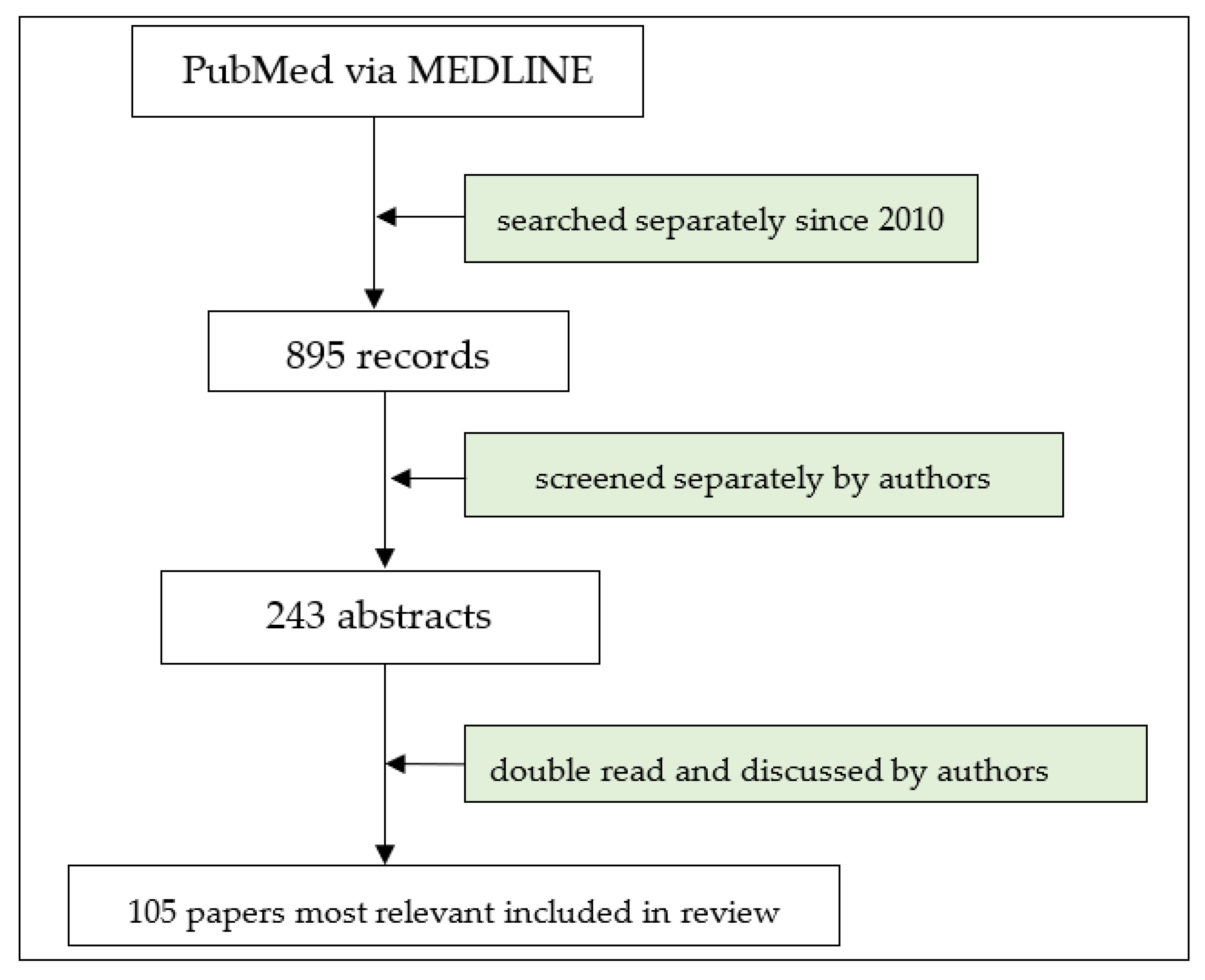
Figure 2.
Developing NMJ: The key proteins in NMJ formation include a neuronally derived heparan-sulfate proteo-glycan, agrin, and three muscle proteins: downstream of kinase-7 (Dok7), low-density lipoprotein receptor-related protein-4 (LRP4), and rapsyn [13,14]. The low-density lipoprotein receptor-related protein-4 (LRP4) serves as a cis-acting (in muscle) transmembrane ligand for MuSK; agrin acts as an allosteric regulator of LRP4 interaction with MuSK; downstream of kinase-7 (Dok7) functions as a cytoplasmic activator of MuSK, whereas rapsyn binds directly to AChR to facilitate its clustering (based on [9], own modification).

Figure 3.
MuSK Structure (modified from [16]).
Figure 3.
MuSK Structure (modified from [16]).
Disclaimer/Publisher’s Note: The statements, opinions and data contained in all publications are solely those of the individual author(s) and contributor(s) and not of MDPI and/or the editor(s). MDPI and/or the editor(s) disclaim responsibility for any injury to people or property resulting from any ideas, methods, instructions or products referred to in the content. |
© 2024 by the authors. Licensee MDPI, Basel, Switzerland. This article is an open access article distributed under the terms and conditions of the Creative Commons Attribution (CC BY) license (https://creativecommons.org/licenses/by/4.0/).
Share and Cite
MDPI and ACS Style
Dziadkowiak, E.; Baczyńska, D.; Waliszewska-Prosół, M. MuSK Myasthenia Gravis—Potential Pathomechanisms and Treatment Directed against Specific Targets. Cells 2024, 13, 556. https://doi.org/10.3390/cells13060556
AMA Style
Dziadkowiak E, Baczyńska D, Waliszewska-Prosół M. MuSK Myasthenia Gravis—Potential Pathomechanisms and Treatment Directed against Specific Targets. Cells. 2024; 13(6):556. https://doi.org/10.3390/cells13060556
Chicago/Turabian StyleDziadkowiak, Edyta, Dagmara Baczyńska, and Marta Waliszewska-Prosół. 2024. "MuSK Myasthenia Gravis—Potential Pathomechanisms and Treatment Directed against Specific Targets" Cells 13, no. 6: 556. https://doi.org/10.3390/cells13060556
Note that from the first issue of 2016, this journal uses article numbers instead of page numbers. See further details here.