Review

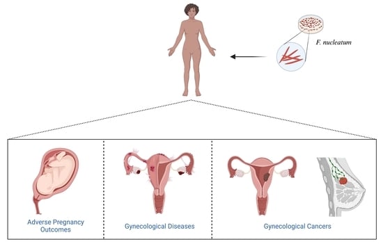
Fusobacterium nucleatum: An Overview of Evidence, Demi-Decadal Trends, and Its Role in Adverse Pregnancy Outcomes and Various Gynecological Diseases, including Cancers

by
Arunita Ghosh
1,2,
Ken Jaaback
3,4,
Angela Boulton
5,
Michelle Wong-Brown
2,4,
Steve Raymond
5,
Partha Dutta
6,7,
Nikola A. Bowden
2,4,* and
Arnab Ghosh
1,2,*
1
School of Biomedical Sciences and Pharmacy, University of Newcastle, Callaghan, NSW 2308, Australia
2
Drug Repurposing and Medicines Research Program, Hunter Medical Research Institute, New Lambton Heights, NSW 2305, Australia
3
Hunter New England Centre for Gynecological Cancer, John Hunter Hospital, Newcastle, NSW 2305, Australia
4
School of Medicine and Public Health, University of Newcastle, Callaghan, NSW 2308, Australia
5
Newcastle Private Hospital, Newcastle, NSW 2305, Australia
6
Department of Medicine, Division of Cardiology, University of Pittsburgh, Pittsburgh, PA 15261, USA
7
Department of Immunology, University of Pittsburgh, Pittsburgh, PA 15261, USA
*
Authors to whom correspondence should be addressed.
Cells 2024, 13(8), 717; https://doi.org/10.3390/cells13080717
Submission received: 7 March 2024 / Revised: 14 April 2024 / Accepted: 15 April 2024 / Published: 20 April 2024
(This article belongs to the Section Cellular Pathology)

Abstract

:
Gynecological and obstetric infectious diseases are crucial to women’s health. There is growing evidence that links the presence of Fusobacterium nucleatum (F. nucleatum), an anaerobic oral commensal and potential periodontal pathogen, to the development and progression of various human diseases, including cancers. While the role of this opportunistic oral pathogen has been extensively studied in colorectal cancer in recent years, research on its epidemiological evidence and mechanistic link to gynecological diseases (GDs) is still ongoing. Thus, the present review, which is the first of its kind, aims to undertake a comprehensive and critical reappraisal of F. nucleatum, including the genetics and mechanistic role in promoting adverse pregnancy outcomes (APOs) and various GDs, including cancers. Additionally, this review discusses new conceptual advances that link the immunomodulatory role of F. nucleatum to the development and progression of breast, ovarian, endometrial, and cervical carcinomas through the activation of various direct and indirect signaling pathways. However, further studies are needed to explore and elucidate the highly dynamic process of host–F. nucleatum interactions and discover new pathways, which will pave the way for the development of better preventive and therapeutic strategies against this pathobiont.

Graphical Abstract

1. Introduction

The human microbiome is a diverse collection of microorganisms, such as bacteria, archaea, viruses, and eukaryotes, that exist both inside and outside of the human body. These microorganisms inhabit and interact with the human body by commensalism, mutualistic, or pathogenic behavior, impacting human health either beneficially or detrimentally by contributing to sound health or disease through the enhancement or impairment of metabolic and immune functions [1,2].
Among the trillions of microorganisms present in the human body, bacteria are by far the most predominant [3,4]. Studies in recent decades have delineated the structure and the functional capacity of the bacterial microbiome in both the healthy state and a variety of disease states [2,5]. In healthy individuals, pathogenic and symbiotic bacteria coexist without causing harm to the host. However, when there is a disturbance in this balance or a change in the host’s bacterial community due to factors such as infectious illnesses, specific diets, or the prolonged use of antibiotics or other bactericidal medications, dysbiosis can occur. This can cease the normal beneficial interactions, thereby increasing the host’s susceptibility to various infections, and the nature of those depends on the anatomical sites involved [2,6]. An article published in ‘The Lancet’ in 2022 [7] reported on a systematic analysis of the Global Burden of Disease Study 2019 and found that bacterial infections are the second-leading cause of death worldwide, responsible for one in eight of all global deaths. Pathogenic bacteria play a role in different inflammatory diseases, including cancers [8,9,10]. Additionally, emerging evidence [11,12,13,14,15,16] has demonstrated a clear role of intestinal and genital bacteria in the development of various pregnancy complications, gynecological diseases (GDs), and gynecological cancers (GCs). Among these bacteria, Listeria monocytogens, Chlamydia trachomatis, Neisseria gonorrhoeae, Treponema pallidum, Trichomonas vaginalis, Escherichia coli, Shigella sps, Staphylococcus epidermidis, Porphyromonas somerae and Fusobacterium nucleatum have been most prevalent in various gynecological complications over the last few years [16,17,18,19,20,21,22,23,24].
However, one bacterium that has received considerable attention in cancer microbiota studies is Fusobacterium nucleatum (F. nucleatum). This is due to its high abundance and relationship to poor prognosis in various types of cancers (colorectal, head and neck cancer, esophageal cancer, pancreatic cancer, and prostatic cancer), including GCs (cervical carcinoma, and breast cancer) [24,25,26]. Recent worldwide studies have also reported the association of F. nucleatum with pregnancy complications (chorioamnionitis, spontaneous abortion, preterm birth, stillbirth, neonatal sepsis, preeclampsia, etc.) [27,28,29]. Further, studies have also stated the association of F. nucleatum with various GDs, including polycystic ovarian syndrome (PCOS), endometriosis, and bacterial vaginosis (BV) [30,31,32], in addition to cancers.
An obligate anaerobic Gram-negative bacillus F. nucleatum belongs to the genus Fusobacterium, so-named based on its slender shape and spindle-like tips at both ends. It often exists as a commensal in various sites of the body, especially in the human oral cavity [33]. F. nucleatum is a heterogeneous species, with five known subspecies (i.e., animalis, fusiforme, nucleatum, polymorphum, and vincentii) [34]. Originally, an oral pathobiont, F. nucleatum, is known to coaggregate with various microbial species in the oral cavity, playing a pivotal role in dental plaque formation [16,24,35,36]. However, it has often been implicated in various extra-oral diseases, including cancers, due to its transmission via the hematogenous route [16,37] and by its virulence mechanisms, including the ability to induce aberrant inflammation and tumorigenesis [16]. F. nucleatum, an adhesive bacterium, acts as an opportunistic pathogen in patients with compromised health conditions, particularly when it invades sterile locations such as the root canal [25,35].
Various studies have provided epidemiological and/or experimental evidence that a significant association between F. nucleatum and colorectal cancers (CRCs) exists, also delineating its crucial role in the pathogenicity, development, and prognosis of CRC [38,39,40,41,42]. Additionally, both molecular mechanism studies and epidemiological evidence have shown a positive correlation between pre-existing inflammatory lesions, such as periodontitis, and an increased risk of cancer [43]. Extensive research studies with epidemiological evidence and mechanistic linkage involving F. nucleatum and various GDs are underway. The role of F. nucleatum in GDs, including GCs, is emerging and discloses the versatile ways in which this bacterium contributes to the development, growth, spread, and treatment response to these diseases.
This review aims to delve into recent evidence and future directions of F. nucleatum, particularly in gynecology, including their genetics and mechanistic role in promoting adverse pregnancy outcomes (APOs), various GDs, including cancers, and the challenges of developing diagnostics and therapeutics for F. nucleatum. This review focuses on the current evidence for F. nucleatum in disease pathogenesis and/or tumor development, highlighting the similarities and differences between F. nucleatum-associated GDs. We also explore the diverse facets of this bacterium’s interaction with the host with potential gynecological implications. Thus, this review provides a comprehensive understanding of the role of F. nucleatum in APOs and GDs, including cancers (Figure 1), and a potential rationale for future research studies based on F. nucleatum as a predictive biomarker and/or a target for anti-tumor therapy.

2. Evidence Acquisition and Synthesis

An extensive literature search was conducted using the electronic databases PubMed and Google Scholar from 2019 until 2023. The keywords used were F. nucleatum in association with various APOs (chorioamnionitis, neonatal deaths, preterm births, stillbirths, and spontaneous abortion), different GDs (polycystic ovary syndrome, salpingitis, endometriosis, and BV), and cancers (breast cancer, ovarian cancer, endometrial cancer, and cervical cancer). The retrieved papers were carefully chosen based on title and abstracts. Then, the full text of the selected papers was evaluated. The references of the reviews were manually searched to ensure no relevant references were missed. Full article texts, including literature reviews and chapters were incorporated for analysis relating to the review’s objectives. However, regarding the clinical terminology, the most relevant information was incorporated from online sources, irrespective of the article length and type. Only articles written in English were considered for this review, and preprints were excluded. The figures and graphical abstract were created with BioRender.com.

3. F. nucleatum in Adverse Pregnancy Outcomes

APO is a broad term comprising health complications that affect the mother, newborn baby, or both during pregnancy, labor, delivery, and the postpartum period [44]. These health complications vary from pregnancy to pregnancyand include chorioamnionitis, preterm birth, spontaneous abortions, stillbirth, neonatal sepsis, low birth weight, preeclampsia, and gestational diabetes mellitus (GDM) (please refer to Box 1) [16,23,34,45,46,47,48,49,50]. APOs are responsible for an enormous burden of maternal and infant mortality and morbidity worldwide [51,52].
Box 1. Adverse pregnancy outcomes Cells 13 00717 i001 definition, etiologies, and symptoms.
Chorioamnionitis
Acute inflammation can occur in the chorion, amnion, or both of the extraplacental membranes or chorionic plate. This is known as the maternal inflammatory response (MIR). In some cases, there may also be acute inflammatory cell extravasation from the umbilical cord vasculature or chorionic plate vessels, known as the fetal inflammatory response (FIR). These responses can occur prior to, during, or after labor [53]. The inflammation is often caused by chronic, subacute, or acute infection, typically due to ascending polymicrobial bacterial infection after membrane rupture [54]. Symptoms may include fever, maternal and/or fetal tachycardia, uterine tenderness and inflammation, foul-smelling amniotic fluid, or an elevated white blood cell (WBC) count, which may lead to pregnancy complications [55].
 
Preterm birth
Any birth that occurs before 37 completed weeks of gestation. It is a major cause of neonatal mortality worldwide [56]. Various factors contribute to preterm birth, including stress, infection, placental abruption, placenta previa, substance use, history of abortion, inadequate prenatal care, smoking, maternal age (<18 or >40), poor nutrition, fetal anomaly, fetal growth restriction, oligohydramnios, polyhydramnios, vaginal bleeding, premature preterm rupture of membranes (PPROM), and environmental factors. Some common signs of preterm labor include regular contractions before the expected gestational age, cervical changes, pelvic pressure, menstrual-like cramps, watery vaginal discharge, and lower back pain [57].
 
Spontaneous abortions
Natural loss of pregnancy prior to twenty weeks of gestation [58], or with a fetus born weighing < 500 g [27]. It is estimated that about 50% of miscarriages are caused by fetal chromosomal abnormalities. Other contributing factors include advanced maternal age, alcohol consumption, smoking, cocaine use, and chronic diseases such as diabetes, celiac disease, and autoimmune conditions like anti-phospholipid antibody syndrome [58]. Rapid conception after delivery and infections such as cervicitis, vaginitis, HIV, syphilis, and malaria are also common risk factors. Symptoms often include abdominal and pelvic cramping, vaginal bleeding, and tachycardia. In cases of significant bleeding, patients may experience symptoms of hypovolemia, even without sepsis [58].
 
Stillbirth
The death of a fetus with a birth weight of 500 g, or if birth weight is unobtainable, gestational age of 22 weeks or a crown-to-heel length of 25 cm. According to the World Health Organization (WHO), all fetuses and infants weighing at least 500 g at birth should be included in statistics. However, for international reporting and comparisons, WHO also recognizes a higher limit (1000 g/28 weeks/35 cm) for third-trimester stillbirths [59]. This can be caused by various factors, such as intrapartum complications, hypertension, diabetes, infection, congenital and genetic abnormalities, and placental dysfunction. Certain risk factors, including advanced maternal age, teenage pregnancies, maternal nutritional status, infections, prior pregnancy losses, complicated pregnancies, and multiple pregnancies, can increase the likelihood of stillbirth. Symptoms may include a cessation of fetal movement and kicks, vaginal spotting or bleeding, and back pain for the mother [60].
 
Neonatal sepsis
An infection in the bloodstream in newborn infants less than 28 days old. This remains a leading cause of morbidity and mortality among neonates. It is divided into two groups, based on the time of presentation after birth: early-onset sepsis (EOS) and late-onset sepsis (LOS). EOS refers to sepsis in neonates at or before 72 h of life, while LOS occurs at or after 72 h of life. EOS is typically caused by the transmission of pathogens from the female genitourinary system to the newborn or fetus, while LOS is usually transmitted from the surrounding environment after delivery, such as through contact with healthcare workers or caregivers. Risk factors for this condition include advanced maternal age, chorioamnionitis, a weakened immune system, and the use of invasive devices. Symptoms and signs may include irritability, lethargy, and poor feeding, as well as respiratory distress, fever, hypothermia, hypotension, and shock. In some cases, hyperglycemia, hypoglycemia, acidosis, or hyperbilirubinemia may be the only indicators of the condition when other factors are absent [61].
 
Preeclampsia
A multisystem condition arising during pregnancy including hypertension and proteinuria. According to Karrar and Hong (2023) [62], the initial parameters for identifying preeclampsia are a systolic blood pressure of 140 mm Hg or higher, or a diastolic blood pressure of 90 mm Hg or higher on two separate occasions at least 4 h apart. Alternatively, a shorter interval timing of a systolic blood pressure of 160 mm Hg or higher or a diastolic blood pressure of 110 mm Hg or higher, can also indicate preeclampsia. These criteria must be met after 20 weeks of gestation. There are various potential causes of preeclampsia, including uteroplacental ischemia, maternal infection and inflammation, maternal intestinal dysbiosis, maternal obesity, sleep disorders, hydatidiform mole, fetal diseases, autoimmune disorders, placental aging, breakdown of maternal-fetal immune tolerance, and endocrine disorders [63]. Common signs and symptoms of preeclampsia include new onset headache, right upper quadrant or epigastric pain with associated nausea or vomiting, shortness of breath, and increased swelling [62].
 
Gestational diabetes
Any degree of glucose intolerance that occurs or is first recognized during pregnancy and typically resolves after the baby is born. It is recommended to screen for gestational diabetes between 24 and 28 weeks of pregnancy using a 50 g, 1 h oral glucose tolerance test. If the results are abnormal, with a value of 130 mg/dL (7.22 mmol/L) or higher, a confirmatory test is necessary using a 100 g, 3 h oral glucose tolerance test. The following values are considered abnormal: a value over 180 mg/dL in the first hour, over 155 mg/dL in the second hour, and over 140 mg/dL in the third hour. A diagnosis of gestational diabetes is established if two or more of these values are abnormal [64]. However, according to the Australian Diabetes in Pregnancy Society (ADIPS), a single abnormal result of either fasting > or = to 5.1 mmol/L or 1 h > or = to 10.0 mmol/L or 2 h > or = to 8.5 mmol/L is considered diagnostic for gestational diabetes. The etiology is thought to be related to dysfunction of the pancreatic beta cells and/or delayed response of the beta cells to changes in blood sugar levels, as well as increased insulin resistance due to hormones released by the placenta. Contributing factors may include a body mass index (BMI) over 25, decreased physical activity, a first-degree relative with diabetes mellitus, a history of gestational diabetes or a previous newborn with macrosomia, and metabolic comorbidities such as hypertension, low HDL, triglycerides over 250, polycystic ovarian syndrome, an abnormal oral glucose tolerance test, acanthosis nigricans, or a medical history of cardiovascular diseases. Clinical signs may include disproportionate weight gain, obesity, an elevated BMI, and fatigue during pregnancy [64].
F. nucleatum, although often found in healthy human placentas [65], has by far been the most prevalent bacterium implicated in various APOs in the last 5 years [16,29,66,67]. Chorioamnionitis and preterm birth, the common complications of pregnancy causingsignificant maternal, perinatal, and long-term adverse outcomes, have often been associated with a high abundance of F. nucleatum in the placenta, amniotic fluid and fetal membranes [15,16,25,28,48,50,65,68,69,70,71,72,73,74]. A recent case report from Italy by Bonasoni et al. [75] has shown that F. nucleatum was present in the placental membrane of a 26-year-old woman who presented at the hospital with painful uterine contractions at 23 weeks gestation. She delivered a preterm infant with premature rupture of membranes, but unfortunately, the baby girl died soon after birth. The placental histopathology revealed severe necrotizing chorioamnionitis and funisitis, with Fusiform bacteria consistent with F. nucleatum. The neonatal autopsy showed organ congestion and mild pericardial, pleural, and abdominal serous effusions, but no congenital anomalies were found. Histological analysis showed an abundance of neutrophils in the lungs and scattered granulocytes in the gastric lumen and large bowel. The fetal inflammatory response was evident with acute funisitis, chorionic vasculitis, and acute deciduitis. Microbiologically, F. nucleatum was only isolated from the infant’s lung. It is worth noting that despite -severe inflammation, the mother did not experience any symptoms (usually, F. nucleatum chorioamnionitis presents with a raised temperature). However, the mother presented with mild and diffuse gingivitis and had undergone a dental procedure one day prior to delivery, indicating a probable association between periodontal disease and hematological dissemination from the oral cavity, leading to chorioamnionitis and its consequent effects on the fetus. A similar, but symptomatic case of a 31-year-old female at 21 weeks gestation, was reported from Canada [76]. This was the first reported case to reveal the practicability of performing 16S rRNA sequencing on formalin-fixed, paraffin-embedded placental tissues (FFPE) in microbiological identification of F. nucleatum and the first to establish the use of this molecular technique in the neonatal death investigation [76]. A study from Germany by Heusler et al. [65] proposed that low concentrations of F. nucleatum might promote the invasion of trophoblast cells and induce the secretion of mediators for pregnancy establishment. However, in contrast, unrestrained infections caused by F. nucleatum in early pregnancy might impact placental development. In a longitudinal study on pregnant Japanese women [77] real-time PCR with TaqMan probes and ELISA revealed that F. nucleatum levels in placenta samples were significantly higher in the threatened preterm labor (TPL) group compared to the healthy group before delivery. Additionally, the presence of F. nucleatum in placental tissues was found to be significantly higher in the TPL-Healthy delivery (HD) group compared to the healthy-HD group. The authors also affirmed the significant association of F. nucleatum in placental tissues with TPL, indicating it may be a potential risk factor. A review article from Qatar by Saadaoui et al. [49] threw the spotlight on the role of F. nucleatum in preterm birth, specifically in cases of clinical chorioamnionitis. The article also noted a temporal relationship between orogenital interaction with a male partner with periodontitis and the onset of clinical infection. Another review article from Italy [72] reported that meconium from preterm infants born to mothers with chorioamnionitis contained high levels of pathogenic bacteria, including F. nucleatum. Further, Payne et al. [66] from Australia demonstrated a significant enhancement of the microbial risk algorithm by the inclusion of F. nucleatum in the novel vaginal bacterial DNA test that was efficacious at the prediction of spontaneous preterm birth in a cohort of mostly white, low- to medium-risk Australian women during midgestational period. A study from China [78] -corroborated a significant positive correlation between the Apgar score (appearance, pulse, grimace, activity, and respiration) and the presence of F. nucleatum in the vagina. The Apgar score is a rapid method for assessing neonates immediately after birth or in response to resuscitation. A report by Walsh et al. [79] from the United States of America (USA) specified the role of F. nucleatum in triggering a more robust inflammatory response, leading to a higher incidence of preterm birth in African American women with a prevalence of F. nucleatum in their vaginal microbiome, compared to L. crispatus, which is prevalent in the vaginal microbiome of women of European ancestry. A case-control study from Brazil [67] conducted on 120 postpartum women, comprising 40 cases (gestational age < 37 weeks; preterm delivery) and 80 controls (gestational age ≥ 37 weeks; full-term delivery), revealed significantly higher proportions of F. nucleatum in the subgingival biofilm of cases compared to controls. Additionally, a 2022 cross-sectional case-control study in the USA [74] used umbilical cord blood specimens to evaluate the presence, abundance, and composition of bacteria through endpoint PCR of full-length 16S rRNA and the V4 amplicon sequence variants (ASVs). The study revealed F. nucleatum subsp. animalis as the most prevalent F. nucleatum strain detected in the umbilical cord blood microbiome in early preterm live birth cases.
The last five years have seen an increase in research on the link between F. nucleatum and spontaneous abortion/miscarriage, a pregnancy complication that may have physical and psychological effects on women [15,27]. In a matched case-control study conducted in Thailand [27], 85 women who experienced spontaneous abortion (<20 weeks of gestation) were compared to 85 controls of similar age, gestational age. The study found no significant differences in the levels of F. nucleatum in subgingival plaque between case and control groups. However, when subgroup analysis was conducted to compare levels of F. nucleatum between individuals with and without periodontitis in both case and control groups, with periodontitis had higher levels of F. nucleatum in both the spontaneous abortion and normal pregnancy groups.
F. nucleatum has been widely reported in various sources such as amniotic fluid, cord blood, fetal membranes, placental and fetal tissues of patients with stillbirth, fetal death, neonatal sepsis, and low birth weight [16,71,73,80]. It has also been found in vaginal samples of pregnant women at 28 weeks of pregnancy, and has been associated with early-onset neonatal sepsis [50]. In mice, F. nucleatum has been shown to induce stillbirths when given intravenously [25,47]. This is primarily caused by placental inflammation mediated by Toll-like receptor 4 (TLR4). In mice lacking TLR4 or treated with a TLR4 antagonist, F. nucleatum was found to colonize the placenta without eliciting an inflammatory response, resulting in a reduced fetal death rate [47]. In humans, F. nucleatum was isolated as a pure culture from the lung and stomach of a stillborn infant. An identical clone was also observed in the mother’s subgingival plaque, while no fusobacteria were detected in her vaginal and rectal flora [47].
Recent reports have also linked F. nucleatum to hypertension and preeclampsia [28,48,81,82,83,84,85]. Preeclampsia affects 5% to 7% of all pregnant women and is responsible for over 70,000 maternal deaths and 500,000 fetal deaths worldwide every year. It is the leading cause of maternal death, severe maternal morbidity, maternal intensive care admissions, caesarean section, and prematurity in the USA [86]. While higher levels of F. nucleatum have been associated with uncontrolled type 2 diabetes mellitus (T2DM), recent reports have not directly linked this bacterium to GDM. However, one recent review article has linked the genus Fusobacterium to GDM [48]. The above studies point towards the need for accurate species and strain-level identification of Fusobacterium in oral as well as vaginal microbiomes for predicting the risk of various APOs, which, in turn, might help to delineate appropriate therapeutic strategies. Moreover, clinical studies to reconnoiter the microbial profiles and the composition of multiple biofilms during the different trimesters of pregnancy should also be proposed.
F. nucleatum, detected in the neonatal microbiome, was found to be absent in the mother’s urogenital tract but present in their oral cavity. This suggests that the bacteria may have been transferred through the bloodstream due to its ability to adhere to vascular epithelium [87]. A review article from China [16] also highlighted that injecting saliva or subgingival plaque samples into mice can lead to placental infection with oral commensal species, including F. nucleatum, suggestingthat oral bacteria have the ability to travel to the feto–placental unit. Additionally, there is evidence of F. nucleatum detectionin amniotic fluid, placenta, and neonatal aspirates, matching those found in maternal oral samples [33,47]. These studies emphasize the importance of understanding the need to protect pregnant women from periodontal diseases. This further sheds light on the need to detect the environmental uniqueness of the placenta that permits it to harbor these potentially pathogenic oral commensals. These reports further corroborated the incidence of oral–uterine translocation via the bloostream [29,47,88].
Interestingly, recent reports have linked the abundance of F. nucleatum to various external factors like smoking, oral hygiene and diet [71,89,90,91,92]. Studies have shown that smokers have a higher abundance of F. nucleatum in their subgingival plaque samples compared to non-smokers [71,89]. Additionally, adherence to a Western diet, characterized by high fat and processed carbohydrate intake and low fiber consumption, has been linked to increased levels of F. nucleatum [90]. The consumption of inflammatory foods, such as refined grains, red and processed meats, and carbonated beverages, has also been associated with F. nucleatum infections [93]. In a review article from France, Martinon et al. [94] reported the significant bactericidal effects of a gel containing 1% curcumin (a bioactive substance of turmeric) on F. nucleatum in patients with periodontal diseases. A Polish study [37] stated that the use of zinc ions, commonly found in commercial anti-malodor mouthwashes, can inhibit F. nucleatum. A Columbian systematic review and meta-analysis conducted by Merchant et al. [95] indicated that treating maternal periodontitis patients infected with F. nucleatum using chlorhexidine mouthwash, scaling and root planing (SRP) reduces the risk of preterm birth and low birth weight babies. These reports highlight the importance of considering multiple external factors that impact a pregnant woman’s oral microbiota and potentially lead to APOs. This further emphasizes the need for a thorough understanding of the origin of the offending bacteria and their potential routes of invasion to the placenta and amniotic cavity, as well as the importance of maintaining good oral hygiene.
Several virulence factors and enzymes possessed by F. nucleatum are known to aid the bacterium in colonizing the fetal and placental membranes. However, Fusobacterium adhesion A (FadA), a unique adhesin of F. nucleatum, is best characterized and known to play a critical role in bacterial dissemination and colonization in the placenta. This is followed by spread to the fetal membranes, leading to acute inflammation of the placental and fetal membranes, causing chorioamnionitis and ultimately preterm birth [16,47,71] (Figure 2A). The active form of Fusobacterium adhesion A is an amyloid-like complex, termed FadAc, which comprises the intact pre-Fusobacterium adhesin A (129 amino acids) and the cleaved Fusobacterium adhesin A without the signal peptide. Under stress and disease conditions, F. nucleatum secretes amyloid-like Fusobacterium adhesin A, acting as a molecular switch to change the bacterium from a commensal to a pathogen [34,96]. The fadA-deletion mutant is significantly impaired in mediating placental colonization. The binding of FadA to vascular endothelial cadherin causes the loosening of the tight junction, allowing F. nucleatum and other oral bacteria to penetrate the endothelium. This may explain why F. nucleatum is often found in intrauterine infections, not only as a sole infectious agent but also concomitantly with other oral species [47].
Apart from FadA adhesin, Fusobacterium apoptosis-inducing protein 2 (Fap2) and the two Coaggregation regulator Response regulator and Sensor kinase– Arginine (R)- inhibitable adhesin D (CarRS- RadD) systems have been reported to be involved in F. nucleatum colonization of placenta [34]. Fap2, involved in interspecies coaggregation and cell adhesion, is a huge type V autotransporter with more than 3000 amino acids [34]. A recent study [29] involving human samples and a mouse pregnancy model has demonstrated that the fusobacterial outer membrane protein, Fap2, and host placenta displaying D-galactose-β (1–3)-N-acetyl-D-galactosamine (Gal-GalNAc) are involved in fusobacterial placenta localization and enrichment. Fascinatingly, Fap2-dependent fusobacterial attachment has been perceived not only in the placenta but also in the blood vessels leading to the placenta, signifying that enhanced Gal-GalNAc display is coordinated both in fetal and maternal tissues (Figure 2B). Interestingly, Fap2, mediating placental colonization by fusobacteria, is also found to activate the human (but not mouse) TIGIT killing-suppressing receptor expressed on T cells and natural killer (NK) cells (Figure 2B). Thus, galactose-sensitive adhesin, Fap2 of F. nucleatum contributes to its virulence for successful colonization in the placenta by evasion of host immune surveillance. However, a two-component signal transduction system, CarRS, consisting of the response regulator CarR and the sensor kinase CarS, regulates the expression of the outer membrane transporter adhesin RadD. Disruption or Interruption of CarR is reported to increase the fetal survival rate in mice, while the disruption of either CarS or RadD decreases their survival, unveiling a hypervirulence phenotype. It is still uncertain if the involvement of CarRS in placental infection is facilitated directly by RadDor through the regulation of other virulence factors, such as Fusobacterium adhesin A and/or Fap2 [34,68]. However, a recent study from USA [97] has reported the essential role of the multigene locus encoding a single, fused methionine sulfoxide reductase (MsrAB) and thioredoxin (Trx)- and cytochrome c (CcdA)-like proteins in attachment and colonization of F. nucleatum to the placental tissues, thereby expediting the initiation of infection and its spread to the amniotic fluid. This study explicitly adduces that the MsrAB system governed by the two-component system ModRS epitomizes a major oxidative stress defense pathway that keeps the bacteria away from oxidative damage in immune cells, thereby conferring virulence by enabling adhesion and invasion of target tissues.
Additionally, a study conducted by Garcia-So and colleagues from the USA [98] demonstrated that F. nucleatum triggers placental inflammation through maternal, TLR4-mediated signaling. The study revealed a spatiotemporal pattern of placental inflammatory response, with NF-κB activation (Figure 2C) first observed in maternal endothelial cells, followed by decidual cells surrounding the endothelium, and constant induction of inflammatory cytokines and chemokines. Moreover, the study highlighted the beneficial role of purified omega-3 fatty acids, specifically eicosapentaenoic acid (EPA) and docosahexaenoic acid (DHA), in suppressing inflammatory responses in endothelial cells mediated by both TLR2 and TLR4, thereby protecting against placental inflammation. Furthermore, the study showed that omega-3 fatty acids inhibit F. nucleatum proliferation in the placenta and increase fetal and neonatal survival. Therefore, Garcia-So et al. [98] not only elucidated the mechanism by which F. nucleatum causes intrauterine infection and triggers placental inflammation, leading to various APOs such as preterm birth, stillbirth, and neonatal sepsis but also provided a prophylactic measure to protect against such infections.
Furthermore, Park and colleagues [28] stated that receptor-interacting protein kinase 2 (Ripk2) might aid in the F. nucleatum-induced production of IL-6 by initiating NF-κB signaling in murine macrophages and human decidual stromal cells (hDSCs). Ripk2 was also reported to upregulate inducible nitric oxide synthase (iNOS) gene expression and NO production in macrophages. This promoted the production of CXCL8 and CCL2, which was reduced by Ripk2 inhibitors, SB203580 and PP2. These findings advocate the fact that APOs result from F. nucleatum infection due to the induction of aberrant production of cytokines and chemokines through nucleotide-binding oligomerization domain (NOD) 1/2-Ripk2-mediated signaling. This study also revealed that Ripk2 deficiency led to the reduced production of tumor necrosis factor α (TNF-α) in the absence of TLR4, thereby betokening the redundancy of Ripk2 in some immune responses in macrophages against F. nucleatum. This study [28] displayed for the first time the contribution of Ripk2 to cytokine or chemokine production in response to F. nucleatum in murine macrophages and hDSCs. It would be highly advantageous to regulate Ripk2 signaling to prevent APOs, especially in chorioamnionitis caused by bacterial infections and use it as a molecular drug target. However, further studies using animal models are required to expound the immunomodulatory role of Ripk2 in F. nucleatum-induced APOs and the beneficial role of Ripk2 inhibitors in the prevention of APOs. Moreover, the use of cutting-edge technologies is also warranted to fully unravel the complex regulatory networks of molecular and cellular events underlying the role of F. nucleatum in APOs. Table 1 summarizes the main findings from research articles and case reports linking the association between F. nucleatum and its potential adverse effects during pregnancy.

4. F. nucleatum in Gynecological Diseases

In the last 5 years, there have been several reports from various parts of the world indicating the involvement of F. nucleatum in various GDs like polycystic ovary syndrome (PCOS), endometriosis, and pelvic inflammatory disease (PID) like salpingitis and (BV) (please refer to Box 2) [15,30,31,32,75,99,100,101,102,103].
Box 2. Gynecological diseases Cells 13 00717 i001 definition, histopathologies, etiologies, and symptoms.
Polycystic ovary syndrome
A complex endocrine and metabolic disorder classically characterized by anovulation, infertility, obesity, insulin resistance, and polycystic ovaries. It is also known as hyperandrogenic anovulation (HA) or Stein–Leventhal syndrome. It defines a typical condition where at least one ovary has an ovarian volume greater than 10 mL and at least one ovary has an estimated 12 small cysts, with diameters ranging from 2 to 9 mm [104]. However, due to advances in ultrasound technology, the initial Rotterdam criteria have been changed to >20 follicles in either ovary [105]. The most common contributing factors include obesity and insulin resistance. Fetal androgen exposure may also contribute to this condition [106]. According to the recently updated (2023) guidelines of the WHO, possible symptoms include heavy, long, intermittent, unpredictable, or absent periods, infertility, acne or oily skin, excessive hair on the face or body, male-pattern baldness or hair thinning, and weight gain, especially around the belly.
 
Pelvic inflammatory disease
Classically defined as an infection that originates in the cervical–vaginal region and spreads to the upper genital tract, resulting in a combination of symptoms such as acute salpingitis, perihepatitis, endometritis, oophoritis, pelvic peritonitis, and/or tubo-ovarian abscess [107]. The majority of PID cases are caused by sexually transmitted infections.
  • Salpingitis is particularly defined as an infection and inflammation in the oviducts (fallopian tubes). These tubes are responsible for transporting oocytes and sperm, as well as facilitating fertilization and early embryonic development. The inflammation can be acute or chronic and can range in severity from mild to severe. It is typically caused by an infection that spreads from the lower tract to the upper genital tract [108]. It is also referred to as salpingitis isthmica nodosa (SIN) and is believed to be a part of the chronic pelvic inflammatory disease (PID) spectrum in some patients. Histopathologically, SIN is characterized by nodular thickening of the muscularis layer of the fallopian tube and the formation of inclusion cysts or diverticula due to overgrown epithelium. It is strongly associated with both infertility and ectopic pregnancies [109]. The current known causes of salpingitis include infection, cellular invasion, and congenital malformations [110]. Clinically, it is often manifested by edema, congestion of the fallopian tubes, and inflammation of the peritoneal structures [109].
  • Perihepatitis, or Fitz–Hugh–Curtis syndrome, is a rare and chronic complication of PID that primarily affects premenopausal women. It is characterized by inflammation of the liver capsule and adhesion of the peritoneum, resulting in right upper quadrant pain [111]. The condition can be caused by various factors, including spontaneous ascending infection where microbes from the cervix or vagina travel to the endometrium, through the fallopian tubes, and into the peritoneal cavity; lymphatic spread, such as infection of the parametrium from an intrauterine device; and hematogenous spread, such as with tuberculosis. Common symptoms include acute pain and/or chronic tenderness in the right upper abdomen [112].
  • Endometritis is an infectious inflammation of the endometrium, which is the innermost uterine layer. When the inflammation spreads into the muscular layer, the process is termed endomyometritis, and when it extends through to the parametrium, it is called endoparametritis. Histopathologically, acute endometritis is usually characterized by microabscesses and neutrophil invasion of the superficial endometrial epithelium, glandular lumens, and endometrial cavity. However, chronic endometritis is characterized by the infiltration of endometrial stromal plasmacytes (ESPCs), micropolyposis, edematous changes in the proliferative phase, and dissociated maturation between the stroma and epithelium. Additionally, B cells can accumulate in the endometrial stroma and glands. This condition is caused by the migration of normal bacterial flora from the cervix and vagina into the uterine cavity but can also be caused by bacteria from outside the genital tract. Symptoms of endometritis typically include irregular bleeding, pelvic discomfort, and leukorrhea [113].
  • Oophoritis is a condition in which the ovaries become inflamed due to certain infections, potentially leading to impaired ovarian function. This inflammation can result in atrophic and fibrotic ovaries and, in rare cases, the replacement of ovarian stroma by foamy macrophages and histiocytes [114,115]. Various factors can contribute to the development of oophoritis, including sexual transmission, infection during pregnancy, and peripubertal infection, depending on the specific virus or pathogenic agent. Common clinical symptoms of oophoritis include anorexia, fever, suprapubic pain, menorrhagia, vaginal bleeding, adnexal tenderness, and/or a pelvic mass [114].
  • Peritonitis is a medical condition characterized by inflammation of the peritoneum, which is the membrane that lines the abdominal cavity [116]. This inflammation can be caused by various factors, such as underlying health conditions or the presence of infectious agents. In some cases, it may also present as granulomas with central caseous necrosis, although this is rare. Some of the known causes of peritonitis include gastroduodenal perforations, intestinal volvulus, ruptured abscesses, traumatic bowel perforation, perforated peptic ulcers, tubo-ovarian abscesses, and amoebic colonic perforations [117]. The classic symptoms of peritonitis include severe abdominal pain, tenderness and rigidity, fever, chills, and altered mental status [118].
  • Tubo-ovarian abscess (TOA) is a complex infectious mass that forms in the adnexa as a result of PID. It is often caused by bacteria from the lower genital tract that travel up to the fallopian tube, ovary, and potentially other nearby pelvic organs. Common risk factors include being of reproductive age, having an intrauterine device (IUD) inserted, having multiple sexual partners, and having a previous episode of PID. TOAs are typically polymicrobial and often contain a high proportion of anaerobic bacteria. Symptoms may include an adnexal mass, fever, elevated white blood cell count (WBC), lower abdominal or pelvic pain, and/or vaginal discharge [119].
Endometriosis
Defined as a chronic inflammatory hormone-dependent condition associated with pelvic pain and infertility. It occurs when uterine tissue grows outside of the uterus, leading to inflammation [120,121]. This can result in the formation of scar tissue, known as adhesions or fibrosis, in the pelvis and other parts of the body. The WHO has identified several types of endometriosis lesions, including superficial endometriosis on the pelvic peritoneum, cystic ovarian endometriosis (endometrioma) in the ovaries, and deep endometriosis in the recto-vaginal septum, bladder, and bowel. In rare cases, endometriosis may also be found outside of the pelvis. According to the WHO, the causes of endometriosis include retrograde menstruation, cellular metaplasia, and the spread of stem cells through the body via blood and lymphatic vessels. Symptoms, as described by the WHO, include severe pain during periods, sexual intercourse, bowel movements, and/or urination, as well as chronic pelvic pain, abdominal bloating, nausea, fatigue, and sometimes depression, anxiety, and infertility.
 
Bacterial vaginosis
Defined as a condition in which the vaginal microbiome presents an overgrowth of obligate and facultative anaerobes that disrupts the vaginal microbiome balance. It is a common and recurring infection among women of reproductive age and has been linked to adverse health outcomes and a decreased quality of life [122]. This condition is associated with significant negative healthcare outcomes, including an increased susceptibility to sexually transmitted infections, urogenital infections, pelvic inflammatory disease, and an increased risk of abnormal pregnancy [123]. Histopathologically, it is characterized by the presence of clue cells, which are epithelial cells of the cervix that are embedded with bacteria. It is typically caused by a decrease in the number of normal hydrogen peroxide-producing Lactobacilli, leading to an overgrowth of anaerobic bacteria [124]. Common clinical symptoms include a foul fishy odor and itching in the perineal region [125].
F. nucleatum has been frequently linked directly or indirectly to PCOS in several studies [15,31,99]. A systematic literature review from Macedonia [15] also found a high incidence of F. nucleatum in the saliva of PCOS patients. Similar facts have also been reported in a recently published chapter [126]. A nationwide cohort study conducted in Taiwan [92] did not directly link F. nucleatum to PCOS, however, it suggested the plausible association between chronic periodontitis in patients, often caused by periodontal pathogens such as F. nucleatum, and the development of the systemic antibody responses that might influence the pathogenesis of PCOS. Additionally, a case-control study from India [31] reported significantly elevated levels of F. nucleatum in PCOS patients with periodontitis and/or gingivitis. The authors of this study [31] concluded that hormonal imbalances in women with PCOS may impact their salivary levels of potential periodontal pathogens and systemic antibody responses, making them more susceptible to periodontal diseases such as gingival inflammation. These findings from different parts of the world support the idea that fluctuating sex hormones, combined with high levels of male hormones (androgens) in women with PCOS, may have a quantitative impact on the oral microflora, potentially leading to oral dysbiosis and contribute to gingival inflammation and periodontal health issues.
In the last five years, several studies have been published that have linked F. nucleatum to various PIDs [127,128,129,130,131]. One specific type of PID, salpingitis, has been found to be associated with F. nucleatum in sexually inactive patients [100]. A review article from India [132] also stated that F. nucleatum can cause different degrees of endometritis. In a case report from Canada, a 69-year-old woman with a 2-month history of postmenopausal bleeding and pelvic cramping was found to have F. nucleatum in her endometrial culture [101]. F. nucleatum has also been linked to endometritis in cows [133,134]. Reports from various parts of the world have stated the association of F. nucleatum with endometritis in either humans or animals, or both [135,136,137]. A 2019 case report [138] from the United Kingdom (UK), of a Pakistani woman with a 1-month history of lethargy, weight loss of 3 kg, and ascites, was reported to develop spontaneous bacterial peritonitis after 4 weeks of the initial ascitic sampling, with a positive culture of F. nucleatum. After a liver biopsy, she was diagnosed with “small hepatic vein” Budd–Chiari syndrome (BCS), which was found to be complicated by F. nucleatum-induced peritonitis. This case report further highlighted the possibility the patient might have developed peritonitis due to F. nucleatum-induced liver complications, owing to bowel content leakage traveling through the portal vein. Although no reports in the last 5 years have directly linked F. nucleatum with perihepatitis, oophoritis or TOA, other species of fusobacterium were reported to have associations with TOA [139].
A recent translational [30] study in Japan has demonstrated the pathogenic role of F. nucleatum in the development of endometriosis. The study found that 64% of women with endometriosis had F. nucleatum infiltration in the endometrium, compared to less than 10% of women without the disease. This is a benchmark publication that revealed that F. nucleatum infection of endometrial cells activates transforming growth factor–β (TGF-β) signaling, leading to the transition from quiescent fibroblasts to transgelin (TAGLN)-positive myofibroblasts, thereby conferring the bacterium with the ability to proliferate, adhere, and migrate in vitro. Additionally, Muraoka and colleagues [30] showed that F. nucleatum inoculation in a syngeneic mouse model of endometriosis caused a noticeable rise in TAGLN-positive myofibroblasts and increased the number and weight of endometriotic lesions (Figure 3A). This study also highlighted the potential of targeting F. nucleatum in the endometrium with antibiotic treatment as a therapeutic option for patients with endometriosis. This milestone work, which established a direct link between bacterial infection and endometriosis was featured in the news of ‘The Lancet Microbe’ [140] and the ‘Daily Briefing of Nature’ [141].
Several reports from various regions of the world have linked F. nucleatum to BV in the last five years [102,103,142]. Agarwal et al. [103] displayed that F. nucleatum foraging and growth on mammalian sialoglycans is facilitated by sialidase activity, which is a diagnostic feature of BV. This sialidase activity serves as a source of nutrients that would otherwise be inaccessible due to the lack of endogenous F. nucleatum sialidase. Additionally, their experiments on a mouse model revealed that F. nucleatum may also contribute to the community by enhancing sialidase activity, a biochemical feature of human dysbiosis (Figure 3B). This study also indicated that mutually beneficial relationships between vaginal bacteria can actually support the colonization of pathogens and may aid in maintaining features of dysbiosis. Thus, this study on BV is crucial, as its findings are discordant with the simplistic dogma that the absence of “healthy” lactobacilli is the sole mechanism that produces an accommodative environment for pathogens during vaginal dysbiosis. Furthermore, this study shed light on why women with BV are at a higher risk of vaginal colonization by pathogens such as F. nucleatum. Table 2 provides a summary of the key findings from research articles and case reports on the association between F. nucleatum and various GDs.

5. F. nucleatum in Gynecological Cancers

Recently, worldwide reports have stated the incidence of a high abundance of F. nucleatum in various types of GCs [breast cancer (BC), ovarian cancer (OC), endometrial cancer (EC), and cervical cancer (CC) (please refer to Box 3)]. Moreover, F. nucleatum-associated cancers are found to have poor prognostic value. A few of these reports also delineated the potential pathogenic role of this bacterium in the formation and progression of these cancers [14,24,142,143,144,145,146,147,148,149].
Box 3. Gynecological cancers Cells 13 00717 i001 definition, types, histopathologies, etiologies, symptoms, and staging.
Breast cancer
Defined as the uncontrolled growth of epithelial cells originating in the ducts or breast lobules. Depending on its relationship to the basement membrane, it can be classified as either invasive or non-invasive. Non-invasive neoplasms are typically divided into two main types: lobular carcinoma in situ (LCIS) and ductal carcinoma in situ (DCIS). LCIS is identified by its adherence to the structure of a normal lobule, with enlarged and filled acini. On the other hand, DCIS is more varied in appearance and can be further categorized into four types: papillary, cribriform, solid, and comedo. Invasive ductal cancer typically presents as a cohesive mass and can appear as discrete abnormalities on mammograms. It is often palpable as a lump in the breast, typically smaller than lobular cancers. In contrast, invasive lobular cancer tends to spread through the breast in a single-file pattern, making it difficult to detect on mammography or physical examination until it has advanced. Tubular and mucinous tumors are typically low-grade (grade I) lesions, while medullary cancer is characterized by abnormal invasive cells with high-grade nuclear features, numerous mitoses, and a lack of an in situ component. BC develops due to DNA damage and genetic mutations, often as a result of exposure to estrogen. Inherited DNA defects or pro-cancerous genes, such as BRCA1 and BRCA2, can also increase the risk of developing BC. Therefore, a family history of OC or BC can increase an individual’s risk of developing BC. Other contributing factors may include advanced age, histologic abnormalities, early menarche, late childbirth, nulliparity, and late menopause [150]. According to WHO recommendations, the symptoms of BC can include a breast lump or thickening, often without pain, changes in the size, shape, or appearance of the breast, dimpling, redness, pitting, or other changes in the skin, changes in nipple appearance or the skin surrounding the nipple (areola), and abnormal or bloody fluid from the nipple. BC is classified using the TNM classification system, which groups patients into four stage groupings based on the size of the primary tumor (T), the status of regional lymph nodes (N), and the presence of distant metastasis (M) [150].
 
Ovarian cancer
Defined as a malignant neoplasm originating from the ovaries or fallopian tubes. It can be classified into different types, including epithelial, germ cell, stromal, and other types, such as mesothelial-mesenchymal, mixed cell, and secondary tumors [151]. The most common histological types of epithelial OC are serous, endometrioid, clear cell, and mucinous tumors. Less common subtypes include Brenner and seromucinous. Low-grade serous carcinoma (LGSOC) is a subtype of serous OC that is characterized by minimal nuclear atypia, rare mitosis, and fewer molecular abnormalities. On the other hand, high-grade serous carcinoma (HGSOC) is characterized by significant nuclear atypia, high mitotic activity (>12 per 10 high-power fields), and more molecular abnormalities as observed through cytogenetic analysis. LGSOCs are typically diagnosed at a younger age and have a better prognosis compared to HGSOCs, which tend to present at an older age. LGSOCs also have a higher frequency of KRAS and BRAF mutations, while HGSOCs have a higher frequency of p53 and BRCA 1 and 2 gene mutations and an absence of KRAS/BRAF mutations [152]. HGSOCs have a more aggressive clinical course and are genetically less stable [151]. Ovarian endometrioid carcinomas (OECs) are derived from endometriosis and are characterized by cystic areas that appear as soft masses with bloody fluid, as well as less common solid areas with extensive hemorrhage and necrosis. They also have microsatellite instability. OECs are typically diagnosed at an early stage, leading to a better prognosis [152]. Mucinous ovarian cancers (MOCs) are a unique subtype of OC with an unclear etiology, including whether they originate from the ovary or are the result of metastatic disease from other organs [153]. KRAS mutations are common in these tumors. Histopathological analysis may reveal the presence of glands with architectural and cytological features of adenocarcinoma, but they may lack stromal invasion [152]. Ovarian clear cell carcinoma (OCCC) is a rare subtype of epithelial OC with distinct molecular characteristics, specific biological and clinical behavior, poor prognosis, and high resistance to chemotherapy [154]. Histopathologically, OCCC shows cystic growth patterns, cellular clearing, and a characteristic hobnail growth pattern. Immunohistochemically, stage I and II tumors show overexpression of BAX, while metastatic lesions show higher expression of the anti-apoptotic protein BCL-2 compared to primary tumors. The most significant risk factor for OC is a positive family history of breast or ovarian cancer, and a personal history of BC also increases the risk. OC mainly affects postmenopausal women of advanced age. The symptoms of OC typically become apparent in the late stages (stage III or stage IV). This disease is often clinically manifested by a combination of symptoms, including abdominal fullness, bloating, nausea, abdominal distention, early satiety, fatigue, changes in bowel movements, urinary symptoms, back pain, dyspareunia, and weight loss. The staging of OC is determined by the 8th edition of the American Joint Committee of Cancer (AJCC) and the International Federation of Gynecology and Obstetrics (FIGO) staging system, which uses the TNM classification. This system has four stages, with each stage indicating increasing severity and decreasing chances of survival [152].
 
Endometrial cancer
Defined as a malignancy of the inner epithelial lining of the uterus [155]. EC is broadly classified into two types: Type I (association with unopposed estrogen stimulation, comprising low-grade cells that are more common and have a favorable prognosis) and Type II (not estrogen-driven, comprising high-grade cells that are less common and have an unfavorable prognosis). Type I ECs are mainly composed of grade I or grade II endometrioid adenocarcinomas, while Type II ECs comprise grade III endometrioid adenocarcinomas, serous, clear cell, undifferentiated, and carcinosarcomas [155]. Type I, low-grade endometrioid carcinomas (LGECs) are usually confined to the uterus during diagnosis and have a comparatively favorable prognosis compared to high-grade serous endometrial carcinomas (HGSECs) and other type II endometrial carcinomas, which have significantly poorer prognoses and are often disseminated at the time of diagnosis. The majority of endometrial LGECs show microsatellite instability (MSI) and carry PTEN mutations, while HGSECs commonly exhibit nuclear pleomorphism and nearly ubiquitous TP53 mutations. ECs with high-grade solid, endometrioid, and/or transitional cell-like (SET) morphologies resemble pure HGSC. Many HGSC-SET carcinomas also display evidence of TP53 mutations [156]. Endometrial clear cell carcinoma (ECCC) is a rare and aggressive type II endometrial carcinoma that is more common in older women and patients with advanced-stage disease [157]. It is typically characterized by a combination of papillary, tubulocystic, and/or solid architectural patterns, with cuboidal or polygonal cells containing nuclei with varying degrees of pleomorphism (although overt pleomorphism is usually absent). Hobnail tumor cells and cytoplasmic clearing are often present but are not necessary for diagnosis [158]. Endometrial undifferentiated carcinomas (EUCs) are composed of sheets of monotonous, typically dyscohesive cells that may have a rhabdoid appearance. They often have a limited expression of cytokeratins and are generally negative for epithelial membrane antigen, PAX8, and hormone receptors. They also lack membranous E-cadherin and typically demonstrate loss of expression of DNA mismatch repair proteins and SWI-SNF chromatin remodeling proteins [158]. Endometrial carcinosarcoma is a rare and immensely aggressive disease characterized by a biphasic growth of malignant epithelial (carcinomatous) and mesenchymal (sarcomatous) components [159]. Carcinosarcomas are classified into homologous and heterologous types, depending on whether the mesenchymal component displays differentiation that is intrinsic (endometrial stromal sarcoma or leiomyosarcoma) or extrinsic (chondrosarcoma, rhabdomyosarcoma, etc.) to the uterus [158]. Possible etiologic factors for EC may include exposure to endogenous or exogenous estrogen unopposed by progesterone or progestins, insulin resistance, and hyperandrogenemia [156]. Abnormal vaginal and postmenopausal bleeding are the most commonly reported symptoms, often accompanied by abdominal complaints, vaginal discharge, uterine prolapse, and urinary incontinence [160]. The extent of EC is surgically pathologically “staged” according to the International Federation of Gynecology and Obstetrics (FIGO) criteria published in 2009 [156]. It has four stages, with increasing severity.
 
Cervical cancer
Defined as a malignant tumor of the cervix that can be divided into two histological types, adenocarcinoma (AC) and squamous cell carcinoma (SCC). Human papillomavirus (HPV) is the primary cause of CCs, accounting for more than 75 percent of cases, with high-risk HPV 16 and 18 being the most common types [161]. The majority of squamous cell carcinomas (SCCs) are also HPV-positive. Histopathologically, invasive SCCs of the cervix present as a network of anastomosing bands or single cells with intervening inflammatory or desmoplastic stroma. The cervical stroma in the tumor is typically infiltrated by plasma cells and lymphocytes, with rare instances of an eosinophilic response or foreign body type giant cell reaction [162]. According to the World Health Organization (WHO), HPV-independent SCCs are defined as squamous tumors with stromal or exophytic invasion and negative P16 immunohistochemistry (IHC). These types of SCCs have a higher rate of node metastases and a poorer prognosis [161]. HPV-associated adenocarcinomas can be identified by the presence of conspicuous apical mitoses and karyorrhexis at low power magnification. These adenocarcinomas are classified into two histological types: usual and mucinous. The usual type is the most common and includes glands with smooth luminal surfaces and pseudostratified columnar epithelial cells with enlarged stretched-out, and hyperchromatic nuclei. The mucinous type is further subtyped into mucinous, not otherwise specified (NOS) adenocarcinoma, intestinal adenocarcinoma, signet-ring cell adenocarcinoma, and invasive stratified mucin-producing carcinoma [163]. HPV-independent adenocarcinomas are negative or patchy for P16. These types of adenocarcinomas are typically diagnosed at a later stage, with extrauterine spread and a higher prevalence of destructive invasion [162]. Contributing factors for HPV-associated CC include early age at first intercourse, multiple sexual partners, smoking, herpes simplex virus (HSV), human immunodeficiency virus (HIV), co-infection with other genital infections, and oral contraceptive use [161]. According to the WHO guidelines (2022), symptoms of early-stage CC include irregular bleeding or spotting between periods in women of reproductive age, postmenopausal bleeding or spotting, bleeding after sexual intercourse, and increased vaginal discharge, sometimes with a foul odor. As the cancer progresses, more severe symptoms may appear, such as persistent back, leg, or pelvic pain, weight loss, fatigue, loss of appetite, foul-smelling discharge, and vaginal discomfort, and swelling of one or both lower extremities. The degree of CC is pathologically divided into four stages using the FIGO staging system.
In recent years, a plethora of articles have been published, which have given multifarious views regarding the direct and indirect role of F. nucleatum in BC development, progression and its prognostic outcomes [16,24,33,42,80,134,143,148,157,164,165,166,167,168,169,170,171,172,173,174,175,176]. A few of the current reports [14,24,26,167,177,178,179,180,181] have stated that BC tissues are found to be enriched with F. nucleatum and the bacterial load progressively increases with the tumor size and the advanced stages of cancer. These reports have suggested that F. nucleatum may contribute to poorer clinical outcomes and shorter survival. In a study conducted by Bernhard et al. [143] in Brazil, 44 women with BCs were evaluated for the microbial abundances of over 40 bacterial species within the subgingival plaque samples (n = 144). F. nucleatum was found to have one of the highest mean counts of infectious pathogens in the samples. This study indicated a strong association between oral F. nucleatum-induced chronic inflammation and BC, suggesting it as one of the contributing factors for female BCs. A cohort study by Nejman et al. [182] in Israel reported that F. nucleatum, earlier found to be enriched in colorectal tumors, is also predominant in the breast tumor samples. This study highlighted that the organs such as the breast, which was previously considered sterile, has the potential to harbor microbes. Further, a recent review and meta-analysis based on women between the ages of 18 and 96 years from the Netherlands, has directly pointed out the significant relationship between oral F. nucleatum species and their critical role in female-specific BC pathogenesis, thereby accentuating their biomarker potentiality [171]. Furthermore, an earlier review article from the USA [183] used a random forest classifier and identified F. nucleatum as one of the 14 potential microbial markers for postmenopausal women with BC. Contradictorily, a review article by Chadha et al. [164] from India specified that urine and fecal microbiome analyses of postmenopausal BC patients exhibited reduced counts of F. nucleatum. Another study from India [184] did not find an enrichment of F. nucleatum in BC transcriptome samples. Nonetheless, it is worth noting that F. nucleatum strain ATCC 23726 has been shown to specifically colonize mouse BC tissues and promote tumor growth and metastasis [14,185]. In a groundbreaking publication by Parhi and colleagues [14], it was documented that oral F. nucleatum species can translocate and colonize the lactiferous ducts in breast tissues through a hematogenous route. This colonization relies on neoplastic tissues expressing D-galactose–β (1–3)-N-acetyl-D-galactosamine (Gal-GalNAc), which are extensively displayed on BC cells. Their scholarly work using experimentations executed on murine models exhibited that the high Gal-GalNAc level in BC tissues, acting as an oncoantigen, plays a critical role by serving as a ligand to Fap2 (a surface lectin from F. nucleatum) in the specificity of tumor colonization by F. nucleatum. This, in turn, promotes mammary tumor growth and its metastatic progression (Figure 4A). They further advocated that this effect may be mediated by the prevention of the accumulation of tumor-infiltrating T cells in the tumor microenvironment (TME) and/or increased expression of matrix metalloproteinase-9 (MMP-9). Additionally, their work also revealed that antibiotic therapy using metronidazole can counteract the metastatic progression of mammary tumors, thereby suppressing BC aggravation. This work further highlighted the fact that targeting F. nucleatum, specifically Fap 2, might be advantageous in the treatment of BCs. In this study, the researchers utilized a method to visualize bacteria in tissue slices from an in vivo model of BC. This method has been recently published [186], and provides direct visualization of fusobacterial colonization in BC tissue using multiphoton microscopy. The establishment and validation of this protocol by Parhi and colleagues has gained significant histopathological importance in recent times, as it enables direct visualization without causing any damage to the tissues, allowing for the identification of all structures. Furthermore, a review by Van der Merwe and colleagues [187] from South Africa that elucidated the possible mechanisms by which F. nucleatum might promote BC progression, also speculated that this particular bacterium may promote BC progression by activating the TLR4/MyD88 pathway through its immunomodulatory effects, as indicated in the literature for colorectal cancer. However, this hypothesis proposed by Van der Merwe and colleagues requires further investigation and validation. If the onco-immunological implications of F. nucleatum are confirmed through experimental research, it could provide valuable insights into the microbial and immune-therapeutic aspects of this disease, potentially leading to advanced treatment options. This review further professed thatnovel methods of impeding the binding of F. nucleatum to tumors such as Gal/GalNAc antagonists or Fap2 antibodies should be considered. Withal, a recent study from China [179] using in vivo experimentation techniques, has validated that the F. nucleatum-derived small extracellular vesicles (F. nucleatum-EVs) can significantly augment the cell viability, proliferation, migration, and invasion of BC cells, thus inducing a promotive function on mammary tumor growth and metastasis. They also found that knocking down TLR4 in BC cells efficaciously counteracted the effects of F. nucleatum-derived EVs. This indicated the TLR4 activation-dependent contributive role of F. nucleatum-derived EVs in BC tumor growth and metastasis. Furthermore, exosomes, which are small extracellular vesicles (EVs), play a crucial role in mediating cellular communication by delivering various bioactive molecules, such as oncogenes, oncomiRs, proteins, and even pharmacological compounds. These molecules can be transferred to target cells, altering their transcriptome and influencing tumor-related signaling pathways. While numerous studies have investigated the involvement of exosomes in BC biology, including therapeutic resistance and the surrounding microenvironment [188], the role of exosomes derived from F. nucleatum in BC progression remains largely unexplored. However, a study by Guo et al. [189] from China demonstrated that exosomal miR-1246/92b-3p/27a-3p derived from F. nucleatum-infected CRC cells promotes metastasis in uninfected cells, contributing to CRC progression. Additionally, a recent study from China [190] has identified that F. nucleatum-infected gastric cancer (GC) cells produce exosomes that increase the expression of the long non-coding RNA (lncRNA) HOXA transcript at the distal tip (HOTTIP), promoting GC invasion through the miR-885-3p/EphB2/PI3K/AKT axis. Therefore, further research is needed to fully understand the role of F. nucleatum-derived EVs in BC and their associated pro-inflammatory and inflammatory responses, which could potentially lead to the development of novel therapeutic agents. A 2023 review article [178] examined the molecular consequences of the F. nucleatum within the TME, likely indicating the probable actionable pathways modulated by this anaerobic bacterium that may have significance in BC patients. Little and her colleagues have put forward a question to the new age oncologists and cancer researchers concerning whether F. nucleatum is capable of modulating the local TME, promoting an inflammatory state and further interacting with and influencing infiltrating immune cells in the case of BC as suggestive of CRC in the literature. They also recommended the use of advanced in vitro models such as organoids to replicate the hypoxic environment of the tumors and study the impact on the survival and growth of the anaerobic bacterium F. nucleatum. A three-dimensional (3D) tumor spheroid model was successfully used to examine the effects of co-culturing viable F. nucleatum with human epithelial colon cancer cells, including gene expression, metabolomics, and morphology [191]. Little et al. [178] also highlighted the need for the development of cost-effective assays to detect and quantify F. nucleatum in BC patients. Their review work emphasized that it may be of great interest to immunobiologists and cancer researchers to unravel the immunogenetics concerning the potential interaction between F. nucleatum and immune checkpoint inhibitors (ICI) in the breast. Another review article [192] also highlighted that formate-producing F. nucleatum stimulates the aryl hydrocarbon receptor (AhR) signaling pathway, thereby promoting cell migration and eliciting cancer stem cell (CSC) traits, high metastatic activity and active Wnt signaling. This pro-malignant effect of AhR ligands was seen in the BC cell line MCF-7. Thus, these works provided valuable insights into the immunomodulatory role of the F. nucleatum in BC development, progression and its effect on treatment effectiveness in patients.
In recent years, several reports have been published linking F. nucleatum to OCs [144,146,147,193]. One such case series, presented by Almohaya et al. [144] from Saudi Arabia, highlighted the incidence of F. nucleatum-associated bacteremia in a 72-year-old female patient with metastatic OC. The patient had a high white blood cell count (28.4 × 10⁹) and c-reactive protein level (124 mg/L), anddespite receiving 14 days of antibiotic treatment (meropenem and moxifloxacin), she passed away within 30 days. Another study in 2022 from Korea [146] reported the incidence of F. nucleatum-associated bacteremia in an OC patient. However, the bacterial isolates were found to be susceptible to all the 10 antimicrobials tested that belonged to the groups: penicillin, cephalosporins, carbapenems, beta-lactamase inhibitors, macrolides, fluoroquinolones, chloramphenicol, and metronidazole. Additionally, a study from China [193] summarized the clinical characteristics of patients with F. nucleatum infection. The study reported the bacterium’s association with a female patient aged below 70 years suffering from OC who died within 2 days. A recent study from the USA [147] has utilized the differential abundant analysis to demonstrate the enrichment of F. nucleatum, in patients with other OC histologies, in comparison to the serous OC patients within the whole OC cohort. These studies not only depicted a possible relationship between F. nucleatum infection and OC, but also provided clinically appropriate data for the implementation of empirical therapies against F. nucleatum. CXC motif chemokine ligand 16 (CXCL16), a chemotactic cytokine belonging to the α-chemokine subfamily, and its receptor CXC motif chemokine receptor 6 (CXCR6) have been shown to play significant roles in the progression of various cancers (invasion and migration), including OCs [189,194]. Further, in a study by Guo et al. from China [189], F. nucleatum was found to stimulate the production of CXCL16 in CRC patients. The observations from these studies suggested that F. nucleatum can stimulate the production of CXCL16, which can further activate CXCR6 and potentially aid in the implantation of OC cells and the formation of peritoneal metastasis. However, this plausible chemokine-related F. nucleatum-induced initiation and formation of OCs need experimental validation in future studies which, in turn, is expected to open avenues to target OCs by controlling immune cell trafficking. Moreover, CSCs have an imperative role in ovarian tumor initiation, invasion, metastasis, local recurrence following curative resection and therapeutic resistance. The process of epithelial–mesenchymal transition (EMT) is also considered a vital step in OC proliferation and CSC metastases [195,196]. EMT is characterized by the repression of E-cadherin (an important component of adherens junctions), occludins, claudins, Epcam, α6β4 integrin, and different cytokeratins (important for stabilization of desmosomes) and up-regulation of vimentin, fibronectin, neural cadherin (N-cadherin), β1 and β3 integrins, and matrix MMPs [197]. The regulatory crosstalk between CSC and EMT is known to increase cancer cell mesenchymal characteristics on the CSCs and help to promote OC cells to gain stemness [196]. Studies have also reported F. nucleatum can induce CSC characteristics by activating IL-6/STAT3 and eliciting EMT-resembling activation [196,198]. Another report from China [199] showed that F. nucleatum significantly upregulated the expression of lncRNA Keratin7-antisense (KRT7-AS) and Keratin7 (KRT7) in CRC cells. However, it is currently unclear whether KRT7-AS is directly involved in the initiation and development of OC, but earlier, it was reported that KRT7 regulated EMT in OC via the TGF-β/Smad2/3 pathway [200]. It is also known to regulate cell-matrix adhesion through integrin-β1-focal adhesion kinase signaling, signifying numerous potential links between KRT7-AS and OC [200,201]. In OC, silencing of the TLR4 gene expression is known to cause a reduction in the expression of MMP2 and MMP9 and decrease levels of mesenchymal markers in lipopolysaccharide (LPS)-treated OC cells [202]. TLR4/MyD88 signaling is also found to be associated with chemoresistance to paclitaxel in OC (Figure 4B) [203]. On top of that, F. nucleatum has been reported to be targeted by TLR4-mediated innate immune signaling and alters the chemotherapeutic response in CRC patients [202,204]. While these studies could not directly identify the pathways involved in F. nucleatum-induced OCs, they shed light on the probable ones, thereby accentuating the need for further exploration. Additionally, a study from the USA [142] highlighted an earlier observation that lanthionine (highly accumulated during F. nucleatum infection) biosynthesis, which generates hydrogen sulfide (H2S), is associated with advanced-stage OC in a mouse model. These studies strongly implied the probable mechanistic association between F. nucleatum and OCs and opened the way for the discovery of new therapeutics by targeting biological pathways.
Although there have not been many recent studies directly linking F. nucleatum to ECs, two recent reports published in 2022 [205] and 2023 [149] from Florida and Poland, respectively, have strongly professed a high abundance of this bacterium in vaginal and/or cervical samples collected from women suffering from EC. The exact mechanism by which F. nucleatum contributes to the development and progression of ECs, either directly or indirectly, is still unknown. However, this review proposes a few potential causes that should be further investigated in future studies. In patients with CRC, the presence of F. nucleatum is known to activate the Wnt/β-catenin signaling pathway [205]. This pathway is crucial for normal cellular proliferation during the menstrual cycle, but when it is dysregulated in the endometrium, it can lead to endometrial hyperplasia and potentially EC. Thus, the observations mentioned above indicate the potential carcinogenic role of F. nucleatum in the development of ECs through oncogenic activation or induction of aberrant Wnt/β-catenin signaling (Figure 4C). Additionally, type 1 ECs are often known to be associated with some unique molecular alterations, also termed microsatellite instability (MSI), which is the result of the defects in DNA mismatch repair proteins. This is due to the accumulation of mutation loads in cancer-related genes and the generation of neoantigens, which stimulate the anti-tumor immune response of the host [206,207,208,209]. Moreover, F. nucleatum infection has also been linked to MSI in CRC and head and neck cancer patients due to impaired DNA mismatch repair (MMR) pathways [210,211,212]. Thus, mechanistically, the capacity of F. nucleatum to create genotoxic oxidant species by causing epigenetic changes and inducing inflammation-associated microsatellite instability via impairment of MMR to promote DNA damage and cell proliferation, may possibly underlie the pathobiont’s associations with ECs (Figure 4D). Further exploration of these target pathways concerning F. nucleatum and EC may provide valuable therapeutic insights into ECs. Furthermore, studies have shown that abnormal expression of lncRNAs is involved in the development of ECs by affecting pathways related to the cell cycle, DNA replication, and mismatch repair [213,214]. Additionally, the process of EMT in ECs has been linked to the expression of transcription factors such as Snail, Slug, Twist2, Zeb1, and Zeb2 [215,216]. While no direct link has been established between F. nucleatum and the expression of lncRNAs in promoting EC, a recent study from China [217] has demonstrated that F. nucleatum can promote CRC metastasis by upregulating the expression of lncRNA endogenous retroviral-associated adenocarcinoma RNA (EVADR). This lncRNA acts as a scaffold for Y-box binding protein 1 (YBX1) which, in turn, enhances the translation of EMT-related factors such as Snail, Slug, and Zeb1. Therefore, Lu and colleagues have highlighted the crucial role of F. nucleatum in regulating lncRNAs and EMT-related factors. This not only provides new insights into the molecular mechanisms underlying CRC metastasis but also suggests that exploring the dysregulated expression of lncRNAs induced by F. nucleatum in EC cells may lead to new therapeutic targets for treating patients with EC.
The association of F. nucleatum with CC has been implicated in a few studies published in the last 5 years from various parts of the world [16,142,145,172]. This pathobiont is mostly found in patients suffering from HPV-related CC, cervical intraepithelial neoplasia, and invasive cervical carcinoma [142]. In a 2020 study conducted in China on 112 patients with squamous carcinoma of the cervix, high levels of F. nucleatum were found, particularly in cases of recurrent lesions [145]. Apart from this, Huang and colleagues made several novel observations. They found that patients with high burdens of intratumorally infiltrated F. nucleatum displayed poor rates of both overall survival and progression-free survival (PFS). Thus, this anaerobic bacterium can serve as a potential CC diagnostic and prognostic biomarker. Interestingly, the levels of F. nucleatum were positively correlated with tumor differentiation. These observations by Huang et al. [145] might help to improve or change therapeutic strategies and provide a better prognostic outcome for afflicted patients. Furthermore, the study showed that CC cells from patients with high levels of intratumorally infiltrated F. nucleatum exhibited characteristics of CSCs. Their scholarly work also implied that induction of CSC characteristics by F. nucleatum might be through the activation of certain specific transcription factors, such as NANOG, Octamer-binding transcription factor 4 (OCT4), SRY-Box Transcription Factor 2 (SOX2) and associated signaling pathway, such as WNT/β-catenin and insulin-like growth factor 1 (IGF-1) receptor pathway. Hence, increased F. nucleatum burden might further activate many other sequences of metastasis, such as C-X-C Motif Chemokine Receptor 4 (CXCR4), epithelial cellular adhesion molecule (Ep-CAM), Slug, Snail1 and Zinc Finger E-Box Binding Homeobox (Zeb1/2). Therefore, this study by Huang et al. [145] not only delineated a cogent role of F. nucleatum in the onset of CC and its consequent development and progression but also betokened a plausible role of the F. nucleatum in the dynamics underlying CCs. Thus, Huang and his colleagues provided appropriate rationale and merit to further investigate their findings in a larger cohort and explore novel mechanisms concerning F. nucleatum-associated CCs to aid inthe development of better treatment options for CC patients. Table 3 outlines the specific findings from research articles and case reports on the link between F. nucleatum and various GCs.

6. Conclusions, Future Challenges, and Perspectives

F. nucleatum, an opportunistic pathogen, and also one of the most variegated bacterial species, has recently been found to play a significant role in various infectious and systemic diseases, including tumorigenesis, particularly in colorectal and breast carcinomas. This has accrued considerable attention over the past decade; however, so far, the growing pieces of evidence remain full of correlative observations and associations that immensely outnumber the field’s mechanistic studies. To the best of our knowledge, this literature review is the first one to identify a knowledge gap relating to the mechanistic role of F. nucleatum in various aspects of gynecology, particularly OCs and ECs. Addressing this gap in future studies could provide valuable insights into the basic biology of F. nucleatum-associated GC initiation and progression and shed light on its potential pathogenic, mechanistic and contributory roles in these diseases. Thus, this review highlights the need for further mechanism-driven research, which may garner the attention of clinicians and researchers. Further, this is the first review of its kind that successfully provides a well-defined overview of the present developments of F. nucleatum concerning its epidemiological evidence and mechanistic linkage in almost all notable GDs, including cancers. Moreover, here, we not only summarize the detrimental effects of F. nucleatum on women’s health but also discuss the available treatment options and possible therapeutic strategies to combat F. nucleatum infections causing or contributing to various APOs and GDs, including cancers. Numerous recent findings discussed in this review have shed light on the interplay between F. nucleatum; APOs, F. nucleatum; GDs and F. nucleatum; and GCs, some of which may break the ground for promising novel therapies, especially for GCs.
Nevertheless, there is still much to be discovered in order for this field to progress. Several epidemiological studies reviewed here only showed the correlation between F. nucleatum and various APOs and GDs, inclusive of cancers. However, the explicit causation and mechanisms of pathogenesis of F. nucleatum in relation to diseases impacting women’s health, as well as its sensitivity to immunomodulation and immune escape, have yet to be uncovered. A few conclusions obtained in various studies were contentious. Therefore, in order to identify targeted therapies for F. nucleatum-associated APOs and other GDs, including cancers, researchers must delve deeper into the basic biology of F. nucleatum, not only in the field of gynecology or gynecological oncology, but also in its natural habitat and other disease-associated conditions and locations. It is also important for scholars to identify the pathways or mechanisms through which F. nucleatum interacts with its host. Immune mechanisms involved in this phenomenon deserve further exploration. Preclinical models, especially organoids and humanized gnotobiotic mouse models that mimic human tissue-specific microenvironments have been recently used to study human cancer genetics and the human microbiome. However, their use to study the effects of F. nucleatum in various GDs including GCs, is yet to be executed.
Currently, advancements in detection methods for microbial entities and microbe-derived small molecules have greatly improved our ability to accurately identify and quantify F. nucleatum strains and levels in the mouth and gut. This is particularly crucial for pregnant women and immunocompromised individuals. By understanding the bacterial load and the molecular mechanisms involved in the transformation of this oral commensal into a pathobiont, which can impact women’s health, researchers and clinicians can not only develop effective diagnostic and therapeutic strategies but can also educate women on preventive measures. The potential for using F. nucleatum as a disease-predictive biomarker in various GDs, including carcinomas, is promising. However, there is still much to be discovered and explored regarding the interactions between F. nucleatum and the host, which ultimately determine a female patient’s response to a specific treatment regimen and, potentially, their long-term impact on women’s health.

Author Contributions

Conceptualization, A.G. (Arunita Ghosh), A.G. (Arnab Ghosh) and N.A.B.; original draft preparation, A.G. (Arunita Ghosh); writing—review, A.G. (Arunita Ghosh); preparing-figures, A.G. (Arunita Ghosh) and A.G. (Arnab Ghosh); editing, A.G. (Arnab Ghosh), N.A.B., K.J., A.B., M.W.-B., S.R. and P.D.; supervision, A.G. (Arnab Ghosh) and N.A.B. All authors have read and agreed to the published version of the manuscript.

Funding

N.A.B. is funded by the Hunter Medical Research Institute Vanessa McGuigan HMRI Fellowship in Ovarian Cancer (HMRI778).

Acknowledgments

The authors would like to express their sincere gratitude to Hunter Medical Research Institute and University of Newcastle, Australia for their continuous support.

Conflicts of Interest

The authors declare that they have no conflicts of interest.

References

  1. Berg, G.; Rybakova, D.; Fischer, D.; Cernava, T.; Verges, M.C.; Charles, T.; Chen, X.; Cocolin, L.; Eversole, K.; Corral, G.H.; et al. Microbiome definition re-visited: Old concepts and new challenges. Microbiome 2020, 8, 103. [Google Scholar] [CrossRef]
  2. Ogunrinola, G.A.; Oyewale, J.O.; Oshamika, O.O.; Olasehinde, G.I. The Human Microbiome and Its Impacts on Health. Int. J. Microbiol. 2020, 2020, 8045646. [Google Scholar] [CrossRef] [PubMed]
  3. Singh, R.; Zogg, H.; Wei, L.; Bartlett, A.; Ghoshal, U.C.; Rajender, S.; Ro, S. Gut Microbial Dysbiosis in the Pathogenesis of Gastrointestinal Dysmotility and Metabolic Disorders. J. Neurogastroenterol. Motil. 2021, 27, 19–34. [Google Scholar] [CrossRef] [PubMed]
  4. Rappuoli, R.; Young, P.; Ron, E.; Pecetta, S.; Pizza, M. Save the microbes to save the planet. A call to action of the International Union of the Microbiological Societies (IUMS). One Health Outlook 2023, 5, 5. [Google Scholar] [CrossRef] [PubMed]
  5. Hou, K.; Wu, Z.X.; Chen, X.Y.; Wang, J.Q.; Zhang, D.; Xiao, C.; Zhu, D.; Koya, J.B.; Wei, L.; Li, J.; et al. Microbiota in health and diseases. Signal Transduct. Target. Ther. 2022, 7, 135. [Google Scholar] [CrossRef] [PubMed]
  6. Aggarwal, N.; Kitano, S.; Puah, G.R.Y.; Kittelmann, S.; Hwang, I.Y.; Chang, M.W. Microbiome and Human Health: Current Understanding, Engineering, and Enabling Technologies. Chem. Rev. 2023, 123, 31–72. [Google Scholar] [CrossRef] [PubMed]
  7. Collaborators, G.B.D.A.R. Global mortality associated with 33 bacterial pathogens in 2019: A systematic analysis for the Global Burden of Disease Study 2019. Lancet 2022, 400, 2221–2248. [Google Scholar] [CrossRef]
  8. Kouzu, K.; Tsujimoto, H.; Kishi, Y.; Ueno, H.; Shinomiya, N. Role of Microbial Infection-Induced Inflammation in the Development of Gastrointestinal Cancers. Medicines 2021, 8, 45. [Google Scholar] [CrossRef]
  9. Akbar, N.; Khan, N.A.; Muhammad, J.S.; Siddiqui, R. The role of gut microbiome in cancer genesis and cancer prevention. Health Sciences Review 2022, 2, 100010. [Google Scholar] [CrossRef]
  10. Xu, S.; Xiong, Y.; Fu, B.; Guo, D.; Sha, Z.; Lin, X.; Wu, H. Bacteria and macrophages in the tumor microenvironment. Front. Microbiol. 2023, 14, 1115556. [Google Scholar] [CrossRef]
  11. Sipos, A.; Ujlaki, G.; Miko, E.; Maka, E.; Szabo, J.; Uray, K.; Krasznai, Z.; Bai, P. The role of the microbiome in ovarian cancer: Mechanistic insights into oncobiosis and to bacterial metabolite signaling. Mol. Med. 2021, 27, 33. [Google Scholar] [CrossRef] [PubMed]
  12. Han, M.; Wang, N.; Han, W.; Ban, M.; Sun, T.; Xu, J. Gut Microbes in Gynecologic Cancers: Causes or Biomarkers and Therapeutic Potential. Front. Oncol. 2022, 12, 902695. [Google Scholar] [CrossRef] [PubMed]
  13. Ventolini, G.; Vieira-Baptista, P.; De Seta, F.; Verstraelen, H.; Lonnee-Hoffmann, R.; Lev-Sagie, A. The Vaginal Microbiome: IV. The Role of Vaginal Microbiome in Reproduction and in Gynecologic Cancers. J. Low. Genit. Tract. Dis. 2022, 26, 93–98. [Google Scholar] [CrossRef] [PubMed]
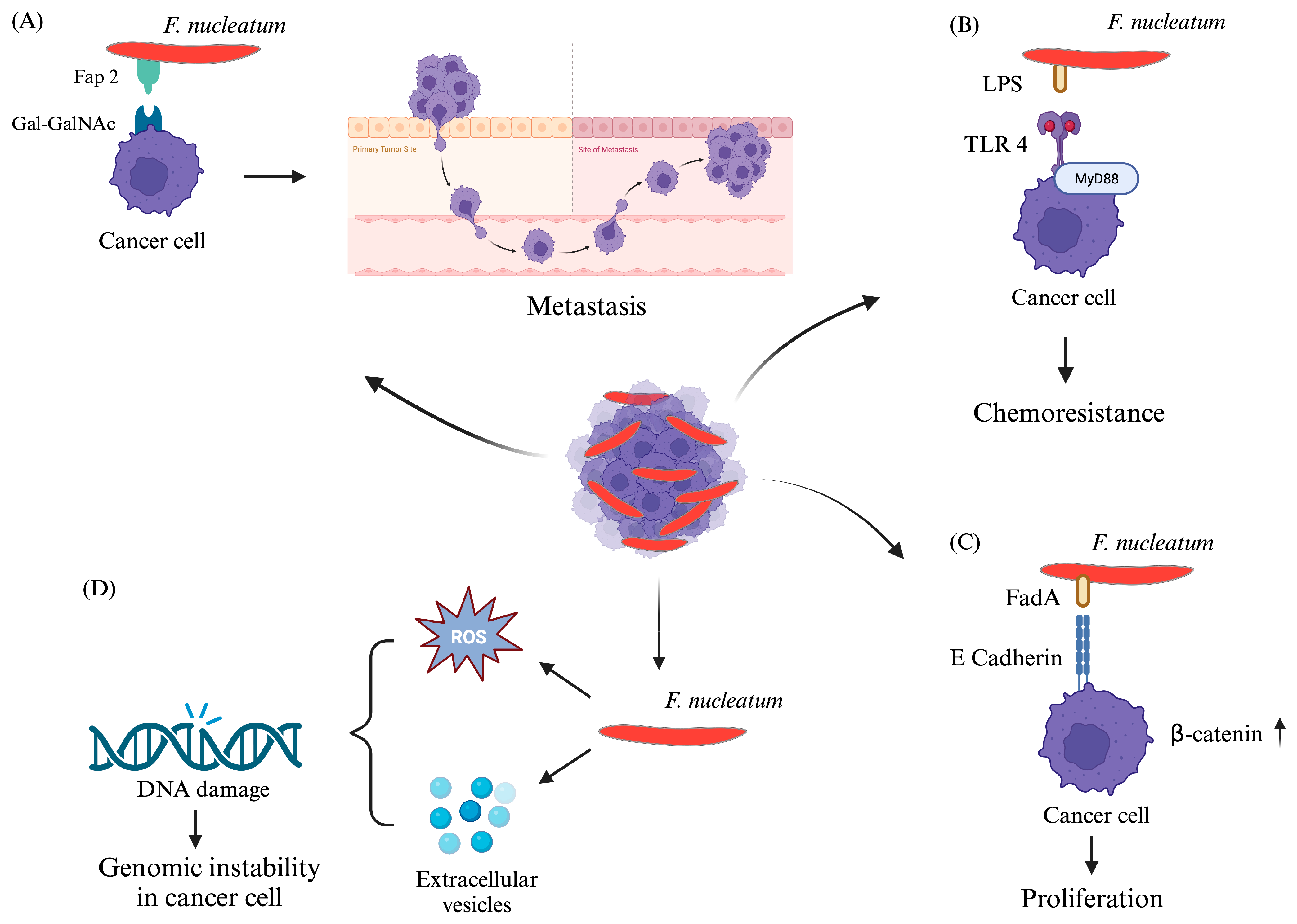
  14. Parhi, L.; Alon-Maimon, T.; Sol, A.; Nejman, D.; Shhadeh, A.; Fainsod-Levi, T.; Yajuk, O.; Isaacson, B.; Abed, J.; Maalouf, N.; et al. Breast cancer colonization by Fusobacterium nucleatum accelerates tumor growth and metastatic progression. Nat. Commun. 2020, 11, 3259. [Google Scholar] [CrossRef] [PubMed]
  15. Tefiku, U.; Popovska, M.; Cana, A.; Zendeli-Bedxeti, L.; Recica, B.; Spasovska-Gjorgovska, A.; Spasovski, S. Determination of the Role of Fusobacterium nucleatum in the Pathogenesis in and Out the Mouth. Pril 2020, 41, 87–99. [Google Scholar] [CrossRef]
  16. Fan, Z.; Tang, P.; Li, C.; Yang, Q.; Xu, Y.; Su, C.; Li, L. Fusobacterium nucleatum and its associated systemic diseases: Epidemiologic studies and possible mechanisms. J. Oral. Microbiol. 2023, 15, 2145729. [Google Scholar] [CrossRef] [PubMed]
  17. Trabert, B.; Waterboer, T.; Idahl, A.; Brenner, N.; Brinton, L.A.; Butt, J.; Coburn, S.B.; Hartge, P.; Hufnagel, K.; Inturrisi, F.; et al. Antibodies Against Chlamydia trachomatis and Ovarian Cancer Risk in Two Independent Populations. J. Natl. Cancer Inst. 2019, 111, 129–136. [Google Scholar] [CrossRef] [PubMed]
  18. Liu, Y.; Jeraldo, P.; Jang, J.S.; Eckloff, B.; Jen, J.; Walther-Antonio, M. Bacterial Single Cell Whole Transcriptome Amplification in Microfluidic Platform Shows Putative Gene Expression Heterogeneity. Anal. Chem. 2019, 91, 8036–8044. [Google Scholar] [CrossRef] [PubMed]
  19. Parida, S.; Sharma, D. Microbial Alterations and Risk Factors of Breast Cancer: Connections and Mechanistic Insights. Cells 2020, 9, 1091. [Google Scholar] [CrossRef]
  20. Wang, L.; Zhou, J.; Gober, H.J.; Leung, W.T.; Huang, Z.; Pan, X.; Li, C.; Zhang, N.; Wang, L. Alterations in the intestinal microbiome associated with PCOS affect the clinical phenotype. Biomed. Pharmacother. 2021, 133, 110958. [Google Scholar] [CrossRef]
  21. Boutriq, S.; Gonzalez-Gonzalez, A.; Plaza-Andrades, I.; Laborda-Illanes, A.; Sanchez-Alcoholado, L.; Peralta-Linero, J.; Dominguez-Recio, M.E.; Bermejo-Perez, M.J.; Lavado-Valenzuela, R.; Alba, E.; et al. Gut and Endometrial Microbiome Dysbiosis: A New Emergent Risk Factor for Endometrial Cancer. J. Pers. Med. 2021, 11, 659. [Google Scholar] [CrossRef] [PubMed]
  22. Giampaolino, P.; Foreste, V.; Di Filippo, C.; Gallo, A.; Mercorio, A.; Serafino, P.; Improda, F.P.; Verrazzo, P.; Zara, G.; Buonfantino, C.; et al. Microbiome and PCOS: State-of-Art and Future Aspects. Int. J. Mol. Sci. 2021, 22, 2048. [Google Scholar] [CrossRef]
  23. Kumar, M.; Saadaoui, M.; Al Khodor, S. Infections and Pregnancy: Effects on Maternal and Child Health. Front. Cell Infect. Microbiol. 2022, 12, 873253. [Google Scholar] [CrossRef] [PubMed]
  24. Alon-Maimon, T.; Mandelboim, O.; Bachrach, G. Fusobacterium nucleatum and cancer. Periodontology 2000 2022, 89, 166–180. [Google Scholar] [CrossRef]
  25. Brennan, C.A.; Garrett, W.S. Fusobacterium nucleatum—Symbiont, opportunist and oncobacterium. Nat. Rev. Microbiol. 2019, 17, 156–166. [Google Scholar] [CrossRef]
  26. Zhao, T.; Wang, X.; Fu, L.; Yang, K. Fusobacterium nucleatum: A new player in regulation of cancer development and therapeutic response. Cancer Drug Resist. 2022, 5, 436–450. [Google Scholar] [CrossRef] [PubMed]
  27. Chanomethaporn, A.; Chayasadom, A.; Wara-Aswapati, N.; Kongwattanakul, K.; Suwannarong, W.; Tangwanichgapong, K.; Sumanonta, G.; Matangkasombut, O.; Dasanayake, A.P.; Pitiphat, W. Association between periodontitis and spontaneous abortion: A case-control study. J. Periodontol. 2019, 90, 381–390. [Google Scholar] [CrossRef] [PubMed]
  28. Park, J.Y.; Lee, T.S.; Noh, E.J.; Jang, A.R.; Ahn, J.H.; Kim, D.Y.; Jung, D.H.; Song, E.J.; Lee, Y.J.; Lee, Y.J.; et al. Receptor-interacting protein kinase 2 contributes to host innate immune responses against Fusobacterium nucleatum in macrophages and decidual stromal cells. Am. J. Reprod. Immunol. 2021, 86, e13403. [Google Scholar] [CrossRef]
  29. Parhi, L.; Abed, J.; Shhadeh, A.; Alon-Maimon, T.; Udi, S.; Ben-Arye, S.L.; Tam, J.; Parnas, O.; Padler-Karavani, V.; Goldman-Wohl, D.; et al. Placental colonization by Fusobacterium nucleatum is mediated by binding of the Fap2 lectin to placentally displayed Gal-GalNAc. Cell Reports 2022, 38, 110537. [Google Scholar] [CrossRef]
  30. Muraoka, A.; Suzuki, M.; Hamaguchi, T.; Watanabe, S.; Iijima, K.; Murofushi, Y.; Shinjo, K.; Osuka, S.; Hariyama, Y.; Ito, M.; et al. Fusobacterium infection facilitates the development of endometriosis through the phenotypic transition of endometrial fibroblasts. Sci. Transl. Med. 2023, 15, eadd1531. [Google Scholar] [CrossRef]
  31. Achu Joseph, R.; Ajitkumar, S.; Kanakasabapathy Balaji, S.; Santhanakrishnan, M. Evaluation of Microbial Profile in Patients with Polycystic Ovary Syndrome and Periodontal Disease: A Case-Control Study. Int. J. Fertil. Steril. 2023, 17, 248–253. [Google Scholar] [CrossRef] [PubMed]
  32. Rokos, T.; Holubekova, V.; Kolkova, Z.; Hornakova, A.; Pribulova, T.; Kozubik, E.; Biringer, K.; Kudela, E. Is the Physiological Composition of the Vaginal Microbiome Altered in High-Risk HPV Infection of the Uterine Cervix? Viruses 2022, 14, 2130. [Google Scholar] [CrossRef] [PubMed]
  33. Chen, Y.; Huang, Z.; Tang, Z.; Huang, Y.; Huang, M.; Liu, H.; Ziebolz, D.; Schmalz, G.; Jia, B.; Zhao, J. More Than Just a Periodontal Pathogen -the Research Progress on Fusobacterium nucleatum. Front. Cell Infect. Microbiol. 2022, 12, 815318. [Google Scholar] [CrossRef] [PubMed]
  34. Xu, B.; Han, Y.W. Oral bacteria, oral health, and adverse pregnancy outcomes. Periodontology 2000 2022, 89, 181–189. [Google Scholar] [CrossRef] [PubMed]
  35. Chen, Y.; Shi, T.; Li, Y.; Huang, L.; Yin, D. Fusobacterium nucleatum: The Opportunistic Pathogen of Periodontal and Peri-Implant Diseases. Front. Microbiol. 2022, 13, 860149. [Google Scholar] [CrossRef] [PubMed]
  36. Britton, T.A.; Wu, C.; Chen, Y.W.; Franklin, D.; Chen, Y.; Camacho, M.I.; Luong, T.T.; Das, A.; Ton-That, H. The respiratory enzyme complex Rnf is vital for metabolic adaptation and virulence in Fusobacterium nucleatum. mBio 2024, 15, e0175123. [Google Scholar] [CrossRef] [PubMed]
  37. Stokowa-Sołtys, K.; Wojtkowiak, K.; Jagiełło, K. Fusobacterium nucleatum—Friend or foe? J. Inorg. Biochem. 2021, 224, 111586. [Google Scholar] [CrossRef] [PubMed]
  38. Umana, A.; Sanders, B.E.; Yoo, C.C.; Casasanta, M.A.; Udayasuryan, B.; Verbridge, S.S.; Slade, D.J. Utilizing Whole Fusobacterium Genomes to Identify, Correct, and Characterize Potential Virulence Protein Families. J. Bacteriol. 2019, 201, e00273-19. [Google Scholar] [CrossRef] [PubMed]
  39. Sun, C.H.; Li, B.B.; Wang, B.; Zhao, J.; Zhang, X.Y.; Li, T.T.; Li, W.B.; Tang, D.; Qiu, M.J.; Wang, X.C.; et al. The role of Fusobacterium nucleatum in colorectal cancer: From carcinogenesis to clinical management. Chronic Dis. Transl. Med. 2019, 5, 178–187. [Google Scholar] [CrossRef]
  40. Cochrane, K.; Robinson, A.V.; Holt, R.A.; Allen-Vercoe, E. A survey of Fusobacterium nucleatum genes modulated by host cell infection. Microb. Genom. 2020, 6, e000300. [Google Scholar] [CrossRef]
  41. Boehm, E.T.; Thon, C.; Kupcinskas, J.; Steponaitiene, R.; Skieceviciene, J.; Canbay, A.; Malfertheiner, P.; Link, A. Fusobacterium nucleatum is associated with worse prognosis in Lauren’s diffuse type gastric cancer patients. Sci. Rep. 2020, 10, 16240. [Google Scholar] [CrossRef]
  42. McIlvanna, E.; Linden, G.J.; Craig, S.G.; Lundy, F.T.; James, J.A. Fusobacterium nucleatum and oral cancer: A critical review. BMC Cancer 2021, 21, 1212. [Google Scholar] [CrossRef]
  43. Nwizu, N.; Wactawski-Wende, J.; Genco, R.J. Periodontal disease and cancer: Epidemiologic studies and possible mechanisms. Periodontology 2000 2020, 83, 213–233. [Google Scholar] [CrossRef]
  44. Tadese, M.; Dagne, K.; Wubetu, A.D.; Abeway, S.; Bekele, A.; Misganaw Kebede, W.; Baye Mulu, G. Assessment of the adverse pregnancy outcomes and its associated factors among deliveries at Debre Berhan Comprehensive Specialized Hospital, Northeast Ethiopia. PLoS ONE 2022, 17, e0271287. [Google Scholar] [CrossRef]
  45. Selma-Royo, M.; Tarrazo, M.; Garcia-Mantrana, I.; Gomez-Gallego, C.; Salminen, S.; Collado, M.C. Shaping Microbiota During the First 1000 Days of Life. Adv. Exp. Med. Biol. 2019, 1125, 3–24. [Google Scholar] [CrossRef]
  46. Schoenmakers, S.; Steegers-Theunissen, R.; Faas, M. The matter of the reproductive microbiome. Obstet. Med. 2019, 12, 107–115. [Google Scholar] [CrossRef]
  47. Figuero, E.; Han, Y.W.; Furuichi, Y. Periodontal diseases and adverse pregnancy outcomes: Mechanisms. Periodontology 2000 2020, 83, 175–188. [Google Scholar] [CrossRef]
  48. Siena, M.; Laterza, L.; Matteo, M.V.; Mignini, I.; Schepis, T.; Rizzatti, G.; Ianiro, G.; Rinninella, E.; Cintoni, M.; Gasbarrini, A. Gut and Reproductive Tract Microbiota Adaptation during Pregnancy: New Insights for Pregnancy-Related Complications and Therapy. Microorganisms 2021, 9, 473. [Google Scholar] [CrossRef]
  49. Saadaoui, M.; Singh, P.; Al Khodor, S. Oral microbiome and pregnancy: A bidirectional relationship. J. Reprod. Immunol. 2021, 145, 103293. [Google Scholar] [CrossRef]
  50. Moreno, I.; Capalbo, A.; Mas, A.; Garrido-Gomez, T.; Roson, B.; Poli, M.; Dimitriadis, E.; Santamaria, X.; Vilella, F.; Simon, C. The human periconceptional maternal-embryonic space in health and disease. Physiol. Rev. 2023, 103, 1965–2038. [Google Scholar] [CrossRef]
  51. Blanco, E.; Marin, M.; Nunez, L.; Retamal, E.; Ossa, X.; Woolley, K.E.; Oludotun, T.; Bartington, S.E.; Delgado-Saborit, J.M.; Harrison, R.M.; et al. Adverse pregnancy and perinatal outcomes in Latin America and the Caribbean: Systematic review and meta-analysis. Rev. Panam. Salud Publica 2022, 46, e21. [Google Scholar] [CrossRef] [PubMed]
  52. Muluneh, A.G.; Asratie, M.H.; Gebremariam, T.; Adu, A.; Enyew, M.M.; Cherkos, E.A.; Melkamu, S.; Berta, M.; Mamo, W.; Kassahun, D.; et al. Lifetime adverse pregnancy outcomes and associated factors among antenatal care booked women in Central Gondar zone and Gondar city administration, Northwest Ethiopia. Front. Public. Health 2022, 10, 966055. [Google Scholar] [CrossRef] [PubMed]
  53. Goldstein, J.A.; Gallagher, K.; Beck, C.; Kumar, R.; Gernand, A.D. Maternal-Fetal Inflammation in the Placenta and the Developmental Origins of Health and Disease. Front. Immunol. 2020, 11, 531543. [Google Scholar] [CrossRef]
  54. Lukanovic, D.; Batkoska, M.; Kavsek, G.; Druskovic, M. Clinical chorioamnionitis: Where do we stand now? Front. Med. 2023, 10, 1191254. [Google Scholar] [CrossRef]
  55. Jain, V.G.; Willis, K.A.; Jobe, A.; Ambalavanan, N. Chorioamnionitis and neonatal outcomes. Pediatr. Res. 2022, 91, 289–296. [Google Scholar] [CrossRef]
  56. Granese, R.; Gitto, E.; D’Angelo, G.; Falsaperla, R.; Corsello, G.; Amadore, D.; Calagna, G.; Fazzolari, I.; Grasso, R.; Triolo, O. Preterm birth: Seven-year retrospective study in a single centre population. Ital. J. Pediatr. 2019, 45, 45. [Google Scholar] [CrossRef]
  57. Suman, V.; Luther, E.E. Preterm Labor. In StatPearls; StatPearls Publishing LLC.: Treasure Island, FL, USA, 2023. [Google Scholar]
  58. Alves, C.; Jenkins, S.M.; Rapp, A. Early Pregnancy Loss (Spontaneous Abortion). In StatPearls; StatPearls Publishing LLC.: Treasure Island, FL, USA, 2022. [Google Scholar]
  59. Kasa, G.A.; Woldemariam, A.Y.; Adella, A.; Alemu, B. The factors associated with stillbirths among sub-saharan African deliveries: A systematic review and meta-analysis. BMC Pregnancy Childbirth 2023, 23, 835. [Google Scholar] [CrossRef]
  60. Maslovich, M.M.; Burke, L.M. Intrauterine Fetal Demise. In StatPearls; StatPearls Publishing LLC.: Treasure Island, FL, USA, 2022. [Google Scholar]
  61. Singh, M.; Alsaleem, M.; Gray, C.P. Neonatal Sepsis. In StatPearls; StatPearls Publishing LLC.: Treasure Island, FL, USA, 2022. [Google Scholar]
  62. Karrar, S.A.; Hong, P.L. Preeclampsia. In StatPearls; StatPearls Publishing LLC.: Treasure Island, FL, USA, 2023. [Google Scholar]
  63. Jung, E.; Romero, R.; Yeo, L.; Gomez-Lopez, N.; Chaemsaithong, P.; Jaovisidha, A.; Gotsch, F.; Erez, O. The etiology of preeclampsia. Am. J. Obstet. Gynecol. 2022, 226, S844–S866. [Google Scholar] [CrossRef]
  64. Quintanilla Rodriguez, B.S.; Mahdy, H. Gestational Diabetes. In StatPearls; StatPearls Publishing LLC.: Treasure Island, FL, USA, 2023. [Google Scholar]
  65. Heusler, M.; Einenkel, R.; Ehrhardt, J.; Muzzio, D.O.; Zygmunt, M. Low Abundance Fusobacterium nucleatum Supports Early Pregnancy Development—An In Vitro Study. Front. Immunol. 2021, 12, 698045. [Google Scholar] [CrossRef]
  66. Payne, M.S.; Newnham, J.P.; Doherty, D.A.; Furfaro, L.L.; Pendal, N.L.; Loh, D.E.; Keelan, J.A. A specific bacterial DNA signature in the vagina of Australian women in midpregnancy predicts high risk of spontaneous preterm birth (the Predict1000 study). Am. J. Obstet. Gynecol. 2021, 224, 206-e1. [Google Scholar] [CrossRef]
  67. Lima, K.M.; Alves, C.M.; Vidal, F.C.; Gomes-Filho, I.S.; Costa, J.C.; Coletta, R.D.; Rodrigues, V.P.; Lopes, F.F. Fusobacterium nucleatum and Prevotella in women with periodontitis and preterm birth. Med. Oral. Patol. Oral. Cir. Bucal 2023, 28, e450–e456. [Google Scholar] [CrossRef]
  68. Wu, C.; Chen, Y.W.; Scheible, M.; Chang, C.; Wittchen, M.; Lee, J.H.; Luong, T.T.; Tiner, B.L.; Tauch, A.; Das, A.; et al. Genetic and molecular determinants of polymicrobial interactions in Fusobacterium nucleatum. Proc. Natl. Acad. Sci. USA 2021, 118, e2006482118. [Google Scholar] [CrossRef]
  69. Zhao, F.; Hu, X.; Ying, C. Advances in Research on the Relationship between Vaginal Microbiota and Adverse Pregnancy Outcomes and Gynecological Diseases. Microorganisms 2023, 11, 991. [Google Scholar] [CrossRef] [PubMed]
  70. Al-Nasiry, S.; Ambrosino, E.; Schlaepfer, M.; Morré, S.A.; Wieten, L.; Voncken, J.W.; Spinelli, M.; Mueller, M.; Kramer, B.W. The Interplay Between Reproductive Tract Microbiota and Immunological System in Human Reproduction. Front. Immunol. 2020, 11, 378. [Google Scholar] [CrossRef]
  71. Dessì, A.; Bosco, A.; Pintus, R.; Orrù, G.; Fanos, V. Fusobacterium nucleatum and alteration of the oral microbiome: From pregnancy to SARS-COV-2 infection. Eur. Rev. Med. Pharmacol. Sci. 2021, 25, 4579–4596. [Google Scholar] [CrossRef] [PubMed]
  72. Coscia, A.; Bardanzellu, F.; Caboni, E.; Fanos, V.; Peroni, D.G. When a Neonate Is Born, So Is a Microbiota. Life 2021, 11, 148. [Google Scholar] [CrossRef] [PubMed]
  73. Chattopadhyay, I.; Lu, W.; Manikam, R.; Malarvili, M.B.; Ambati, R.R.; Gundamaraju, R. Can metagenomics unravel the impact of oral bacteriome in human diseases? Biotechnol. Genet. Eng. Rev. 2023, 39, 85–117. [Google Scholar] [CrossRef]
  74. Vander Haar, E.L.; Wu, G.; Gyamfi-Bannerman, C.; Thomas, C.; Wapner, R.J.; Reddy, U.M.; Zhao, L.; Silver, R.M.; Goldenberg, R.L.; Han, Y.W. Microbial Analysis of Umbilical Cord Blood Reveals Novel Pathogens Associated with Stillbirth and Early Preterm Birth. mBio 2022, 13, e0203622. [Google Scholar] [CrossRef]
  75. Bonasoni, M.P.; Comitini, G.; Pati, M.; Bardaro, M.; Russello, G.; Carretto, E.; Dalla Dea, G.; Palicelli, A.; Bernardelli, G.; Chesi, E.; et al. Fulminant Sepsis and Perinatal Death at 23 Weeks Due to Fusobacterium nucleatum. Fetal Pediatr. Pathol. 2023, 42, 456–463. [Google Scholar] [CrossRef]
  76. Chan, E.; Brundler, M.A.; Zhang, K. Identification of Fusobacterium nucleatum in formalin-fixed, paraffin-embedded placental tissues by 16S rRNA sequencing in a case of extremely preterm birth secondary to amniotic fluid infection. Pathology 2019, 51, 320–322. [Google Scholar] [CrossRef]
  77. Ye, C.; Katagiri, S.; Miyasaka, N.; Kobayashi, H.; Khemwong, T.; Nagasawa, T.; Izumi, Y. The periodontopathic bacteria in placenta, saliva and subgingival plaque of threatened preterm labor and preterm low birth weight cases: A longitudinal study in Japanese pregnant women. Clin. Oral. Investig. 2020, 24, 4261–4270. [Google Scholar] [CrossRef] [PubMed]
  78. Sun, M.; Geng, H.; Bai, J.; Feng, J.; Xu, N.; Liu, Y.; Liu, X.; Liu, G. Characterization of cervical canal and vaginal bacteria in pregnant women with cervical incompetence. Front. Microbiol. 2022, 13, 986326. [Google Scholar] [CrossRef] [PubMed]
  79. Walsh, S.W.; Nugent, W.H.; Alam, S.M.K.; Washington, S.L.; Teves, M.; Jefferson, K.K.; Strauss, J.F., 3rd. Protease Amplification of the Inflammatory Response Induced by Commensal Bacteria: Implications for Racial Disparity in Term and Preterm Birth. Reprod. Sci. 2020, 27, 246–259. [Google Scholar] [CrossRef] [PubMed]
  80. Surlin, P.; Nicolae, F.M.; Surlin, V.M.; Patrascu, S.; Ungureanu, B.S.; Didilescu, A.C.; Gheonea, D.I. Could Periodontal Disease through Periopathogen Fusobacterium nucleatum be an Aggravating Factor for Gastric Cancer? J. Clin. Med. 2020, 9, 3885. [Google Scholar] [CrossRef] [PubMed]
  81. Beckers, K.F.; Sones, J.L. Maternal microbiome and the hypertensive disorder of pregnancy, preeclampsia. Am. J. Physiol. Heart Circ. Physiol. 2020, 318, H1–H10. [Google Scholar] [CrossRef] [PubMed]
  82. Amir, M.; Brown, J.A.; Rager, S.L.; Sanidad, K.Z.; Ananthanarayanan, A.; Zeng, M.Y. Maternal Microbiome and Infections in Pregnancy. Microorganisms 2020, 8, 1996. [Google Scholar] [CrossRef] [PubMed]
  83. Bodurska, T.; Konova, E.; Pachkova, S.; Yordanov, A. Endometrial Microbiome and Women’s Reproductive Health—Review of the Problem Endometrial Microbiome and Reproductive Health. J. Pure Appl. Microbiol. 2021, 15, 1727–1734. [Google Scholar] [CrossRef]
  84. Bhattacharya, K.; Dutta, S.; Sengupta, P.; Bagchi, S. Reproductive tract microbiome and therapeutics of infertility. Middle East Fertil. Soc. J. 2023, 28, 11. [Google Scholar] [CrossRef]
  85. Ye, C.; Kapila, Y. Oral microbiome shifts during pregnancy and adverse pregnancy outcomes: Hormonal and Immunologic changes at play. Periodontology 2000 2021, 87, 276–281. [Google Scholar] [CrossRef]
  86. Rana, S.; Lemoine, E.; Granger, J.P.; Karumanchi, S.A. Preeclampsia: Pathophysiology, Challenges, and Perspectives. Circ. Res. 2019, 124, 1094–1112. [Google Scholar] [CrossRef]
  87. Farhat, S.; Hemmatabadi, M.; Ejtahed, H.S.; Shirzad, N.; Larijani, B. Microbiome alterations in women with gestational diabetes mellitus and their offspring: A systematic review. Front. Endocrinol. 2022, 13, 1060488. [Google Scholar] [CrossRef] [PubMed]
  88. Pereira, M.L.; Levy, M.; Nissapatorn, V.; de Oliveira, G.L.V. Editorial: Women in microbiome in health and disease 2021. Front Cell Infect Microbiol 2022, 12, 1054190. [Google Scholar] [CrossRef] [PubMed]
  89. Lee, Y.H.; Chung, S.W.; Auh, Q.S.; Hong, S.J.; Lee, Y.A.; Jung, J.; Lee, G.J.; Park, H.J.; Shin, S.I.; Hong, J.Y. Progress in Oral Microbiome Related to Oral and Systemic Diseases: An Update. Diagnostics 2021, 11, 1283. [Google Scholar] [CrossRef] [PubMed]
  90. Ibragimova, S.; Ramachandran, R.; Ali, F.R.; Lipovich, L.; Ho, S.B. Dietary Patterns and Associated Microbiome Changes that Promote Oncogenesis. Front. Cell Dev. Biol. 2021, 9, 725821. [Google Scholar] [CrossRef] [PubMed]
  91. Akimbekov, N.S.; Digel, I.; Yerezhepov, A.Y.; Shardarbek, R.S.; Wu, X.; Zha, J. Nutritional factors influencing microbiota-mediated colonization resistance of the oral cavity: A literature review. Front. Nutr. 2022, 9, 1029324. [Google Scholar] [CrossRef] [PubMed]
  92. Pignatelli, P.; Nuccio, F.; Piattelli, A.; Curia, M.C. The Role of Fusobacterium nucleatum in Oral and Colorectal Carcinogenesis. Microorganisms 2023, 11, 2358. [Google Scholar] [CrossRef] [PubMed]
  93. Appunni, S.; Rubens, M.; Ramamoorthy, V.; Tonse, R.; Saxena, A.; McGranaghan, P.; Kaiser, A.; Kotecha, R. Emerging Evidence on the Effects of Dietary Factors on the Gut Microbiome in Colorectal Cancer. Front. Nutr. 2021, 8, 718389. [Google Scholar] [CrossRef] [PubMed]
  94. Martinon, P.; Fraticelli, L.; Giboreau, A.; Dussart, C.; Bourgeois, D.; Carrouel, F. Nutrition as a Key Modifiable Factor for Periodontitis and Main Chronic Diseases. J. Clin. Med. 2021, 10, 197. [Google Scholar] [CrossRef]
  95. Merchant, A.T.; Gupta, R.D.; Akonde, M.; Reynolds, M.; Smith-Warner, S.; Liu, J.; Tarannum, F.; Beck, J.; Mattison, D. Association of Chlorhexidine Use and Scaling and Root Planing with Birth Outcomes in Pregnant Individuals with Periodontitis: A Systematic Review and Meta-analysis. JAMA Netw. Open 2022, 5, e2247632. [Google Scholar] [CrossRef]
  96. Meng, Q.; Gao, Q.; Mehrazarin, S.; Tangwanichgapong, K.; Wang, Y.; Huang, Y.; Pan, Y.; Robinson, S.; Liu, Z.; Zangiabadi, A.; et al. Fusobacterium nucleatum secretes amyloid-like FadA to enhance pathogenicity. EMBO Rep. 2021, 22, e52891. [Google Scholar] [CrossRef]
  97. Scheible, M.; Nguyen, C.T.; Luong, T.T.; Lee, J.H.; Chen, Y.W.; Chang, C.; Wittchen, M.; Camacho, M.I.; Tiner, B.L.; Wu, C.; et al. The Fused Methionine Sulfoxide Reductase MsrAB Promotes Oxidative Stress Defense and Bacterial Virulence in Fusobacterium nucleatum. mBio 2022, 13, e0302221. [Google Scholar] [CrossRef] [PubMed]
  98. Garcia-So, J.; Zhang, X.; Yang, X.; Rubinstein, M.R.; Mao, Y.; Kitajewski, J.; Liu, K.; Han, Y.W. Omega-3 fatty acids suppress Fusobacterium nucleatum-induced placental inflammation originating from maternal endothelial cells. JCI Insight 2019, 4, e125436. [Google Scholar] [CrossRef] [PubMed]
  99. Tong, C.; Wang, Y.H.; Yu, H.C.; Chang, Y.C. Increased Risk of Polycystic Ovary Syndrome in Taiwanese Women with Chronic Periodontitis: A Nationwide Population-Based Retrospective Cohort Study. J. Womens Health 2019, 28, 1436–1441. [Google Scholar] [CrossRef] [PubMed]
  100. Rubino, C.; Barbati, F.; Regoli, M.; Bencini, E.; Mattei, A.; Fierro, F.; Brizzi, I.; Indolfi, G. Recurrent Bilateral Salpingitis in a Sexually Inactive Adolescent: Don’t Forget about the Appendix. J. Pediatr. Adolesc. Gynecol. 2021, 34, 217–219. [Google Scholar] [CrossRef] [PubMed]
  101. Mercer, V.J.; Naseemuddin, A.; Webb, A. Monckeberg’s arteriosclerosis: A case report of chronic endometritis presenting as postmenopausal bleeding. Menopause 2021, 29, 247–249. [Google Scholar] [CrossRef] [PubMed]
  102. Liu, J.X.; Yin, M.M.; Gao, Y.L.; Shang, J.; Zheng, C.H. MSF-LRR: Multi-Similarity Information Fusion through Low-Rank Representation to Predict Disease-Associated Microbes. IEEE/ACM Trans. Comput. Biol. Bioinform. 2023, 20, 534–543. [Google Scholar] [CrossRef] [PubMed]
  103. Agarwal, K.; Robinson, L.S.; Aggarwal, S.; Foster, L.R.; Hernandez-Leyva, A.; Lin, H.; Tortelli, B.A.; O’Brien, V.P.; Miller, L.; Kau, A.L.; et al. Glycan cross-feeding supports mutualism between Fusobacterium and the vaginal microbiota. PLoS Biol. 2020, 18, e3000788. [Google Scholar] [CrossRef] [PubMed]
  104. Singh, S.; Pal, N.; Shubham, S.; Sarma, D.K.; Verma, V.; Marotta, F.; Kumar, M. Polycystic Ovary Syndrome: Etiology, Current Management, and Future Therapeutics. J. Clin. Med. 2023, 12, 1454. [Google Scholar] [CrossRef] [PubMed]
  105. Teede, H.J.; Tay, C.T.; Laven, J.; Dokras, A.; Moran, L.J.; Piltonen, T.T.; Costello, M.F.; Boivin, J.; Redman, L.M.; Boyle, J.A.; et al. Recommendations from the 2023 International Evidence-based Guideline for the Assessment and Management of Polycystic Ovary Syndrome. Fertil Steril 2023, 120, 767–793. [Google Scholar] [CrossRef]
  106. Rasquin, L.I.; Anastasopoulou, C.; Mayrin, J.V. Polycystic Ovarian Disease. In StatPearls; StatPearls Publishing LLC.: Treasure Island, FL, USA, 2022. [Google Scholar]
  107. Greydanus, D.E.; Bacopoulou, F. Acute pelvic inflammatory disease: A narrative review. Pediatr. Med. 2019, 2, 36. [Google Scholar] [CrossRef]
  108. Owhor, L.E.; Reese, S.; Kolle, S. Salpingitis Impairs Bovine Tubal Function and Sperm-Oviduct Interaction. Sci. Rep. 2019, 9, 10893. [Google Scholar] [CrossRef] [PubMed]
  109. Revzin, M.V.; Moshiri, M.; Katz, D.S.; Pellerito, J.S.; Mankowski Gettle, L.; Menias, C.O. Imaging Evaluation of Fallopian Tubes and Related Disease: A Primer for Radiologists. Radiographics 2020, 40, 1473–1501. [Google Scholar] [CrossRef] [PubMed]
  110. Barkwill, D.; Tobler, K.J. Salpingitis Isthmica Nodosa. In StatPearls; StatPearls Publishing LLC.: Treasure Island, FL, USA, 2023. [Google Scholar]
  111. Kaga, M.; Ito, S.; Ueda, T. Two Cases of Perihepatitis with the Liver Capsule Irritation Sign: A New Physical Examination Technique. Cureus 2023, 15, e34327. [Google Scholar] [CrossRef] [PubMed]
  112. Basit, H.; Pop, A.; Malik, A.; Sharma, S. Fitz-Hugh-Curtis Syndrome. In StatPearls; StatPearls Publishing LLC.: Treasure Island, FL, USA, 2023. [Google Scholar]
  113. Taylor, M.; Jenkins, S.M.; Pillarisetty, L.S. Endometritis. In StatPearls; StatPearls Publishing LLC.: Treasure Island, FL, USA, 2023. [Google Scholar]
  114. Giunta, I.; Zayat, N.; Muneyyirci-Delale, O. Histologic features, pathogenesis, and long-term effects of viral oophoritis. F&S Rev. 2021, 2, 342–352. [Google Scholar] [CrossRef]
  115. Dawande, P.; Wankhade, R.; Pande, M. Xanthogranulomatous Oophoritis: A Rare Case Report. Cureus 2023, 15, e43724. [Google Scholar] [CrossRef] [PubMed]
  116. Chen, Q.; Zhu, T.; Liu, J.K.; Ding, J.; Chen, L. Conservative management of multi-trauma induced peritonitis: Experience, outcomes, and indications. World J. Clin. Cases 2023, 11, 5897–5902. [Google Scholar] [CrossRef] [PubMed]
  117. Kumar, D.; Garg, I.; Sarwar, A.H.; Kumar, L.; Kumar, V.; Ramrakhia, S.; Naz, S.; Jamil, A.; Iqbal, Z.Q.; Kumar, B. Causes of Acute Peritonitis and Its Complication. Cureus 2021, 13, e15301. [Google Scholar] [CrossRef] [PubMed]
  118. Dhaliwal, A.; Razick, D.I.; Le, N.; Akhtar, M.; Jakobsen, J. Culture-Negative Fibrinous Peritonitis in a Postpartum Female. Cureus 2023, 15, e43339. [Google Scholar] [CrossRef] [PubMed]
  119. Kairys, N.; Roepke, C. Tubo-Ovarian Abscess. In StatPearls; StatPearls Publishing LLC.: Treasure Island, FL, USA, 2024. [Google Scholar]
  120. Malvezzi, H.; Marengo, E.B.; Podgaec, S.; Piccinato, C.A. Endometriosis: Current challenges in modeling a multifactorial disease of unknown etiology. J. Transl. Med. 2020, 18, 311. [Google Scholar] [CrossRef]
  121. Ellis, K.; Munro, D.; Clarke, J. Endometriosis Is Undervalued: A Call to Action. Front. Glob. Womens Health 2022, 3, 902371. [Google Scholar] [CrossRef]
  122. Wu, S.; Hugerth, L.W.; Schuppe-Koistinen, I.; Du, J. The right bug in the right place: Opportunities for bacterial vaginosis treatment. NPJ Biofilms Microbiomes 2022, 8, 34. [Google Scholar] [CrossRef]
  123. Abou Chacra, L.; Fenollar, F.; Diop, K. Bacterial Vaginosis: What Do We Currently Know? Front. Cell Infect. Microbiol. 2021, 11, 672429. [Google Scholar] [CrossRef] [PubMed]
  124. Kairys, N.; Garg, M. Bacterial Vaginosis. In StatPearls; StatPearls Publishing LLC.: Treasure Island, FL, USA, 2023. [Google Scholar]
  125. Khedkar, R.; Pajai, S. Bacterial Vaginosis: A Comprehensive Narrative on the Etiology, Clinical Features, and Management Approach. Cureus 2022, 14, e31314. [Google Scholar] [CrossRef] [PubMed]
  126. Alan, S. Rare and Underappreciated Causes of Polycystic Ovarian Syndrome. In Polycystic Ovary Syndrome; Wang, Z., Ed.; IntechOpen: London, UK, 2022; Chapter 4. [Google Scholar]
  127. Kang, W.; Jia, Z.; Tang, D.; Zhang, Z.; Gao, H.; He, K.; Feng, Q. Fusobacterium nucleatum Facilitates Apoptosis, ROS Generation, and Inflammatory Cytokine Production by Activating AKT/MAPK and NF-kappaB Signaling Pathways in Human Gingival Fibroblasts. Oxid. Med. Cell Longev. 2019, 2019, 1681972. [Google Scholar] [CrossRef]
  128. Tawfeq, N.A.; Saleh, G.M. The Bacterium Fusobacterium sp. May Interfere with Conception. Iraqi J. Agric. Sci. 2020, 51, 903–915. [Google Scholar] [CrossRef]
  129. Chen, P.C.; Li, P.C.; Ding, D.C. Pelvic inflammatory disease and causative pathogens in older women in a medical center in eastern Taiwan: A retrospective cross-sectional study. PLoS ONE 2021, 16, e0257627. [Google Scholar] [CrossRef]
  130. Yan, C.; Diao, Q.; Zhao, Y.; Zhang, C.; He, X.; Huang, R.; Li, Y. Fusobacterium nucleatum infection-induced neurodegeneration and abnormal gut microbiota composition in Alzheimer’s disease-like rats. Front. Neurosci. 2022, 16, 884543. [Google Scholar] [CrossRef] [PubMed]
  131. He, X.F.; Du, X.P.; Qiao, C.F. Successful laparoscopic resection of fallopian tube abscess caused by Escherichia coli in a 12-year-old adolescent virgin:a case report and review of the literature. BMC Pediatr. 2023, 23, 282. [Google Scholar] [CrossRef]
  132. Prem Kumar, R.; Sunith, R.; Rajanna, R. Bovine endometritis: A review article. Pharma Innov. 2020, 9, 55–58. [Google Scholar]
  133. Umar, T.; Yin, B.; Umer, S.; Ma, X.; Jiang, K.; Umar, Z.; Akhtar, M.; Shaukat, A.; Deng, G. MicroRNA: Could It Play a Role in Bovine Endometritis? Inflammation 2021, 44, 1683–1695. [Google Scholar] [CrossRef]
  134. Yin, B.; Umar, T.; Ma, X.; Chen, Y.; Umar, Z.; Umer, S.; Deng, G. Andrograpanin mitigates lipopolysaccharides induced endometritis via TLR4/NF-kappaB pathway. Reprod. Biol. 2022, 22, 100606. [Google Scholar] [CrossRef] [PubMed]
  135. Bao, H.; Qu, Q.; Zhang, W.; Wang, X.; Fang, J.; Xue, J.; Liu, Z.; He, S. NRF2 Exerts Anti-Inflammatory Effects in LPS-Induced gEECs by Inhibiting the Activation of the NF-kappaB. Mediators Inflamm. 2021, 2021, 9960721. [Google Scholar] [CrossRef] [PubMed]
  136. Bhaskar, V.; Saini, S.; Ansari, S.; Ghai, S.; Thakur, A.; Chopra, S.; Verma, V.; Malakar, D. Allogenic adipose derived mesenchymal stem cells are effective than antibiotics in treating endometritis. Sci. Rep. 2023, 13, 11280. [Google Scholar] [CrossRef] [PubMed]
  137. Srinivasan, M.; Adnane, M.; Archunan, G. Significance of cervico-vaginal microbes in bovine reproduction and pheromone production—A hypothetical review. Res. Vet. Sci. 2021, 135, 66–71. [Google Scholar] [CrossRef] [PubMed]
  138. Bannaga, A.; Armstrong, M.J.; Mehrzad, H.; Brown, R.M.; Tripathi, D. Small Intrahepatic Vein Budd-Chiari Syndrome Complicated by Fusobacterium nucleatum Peritonitis. ACG Case Rep. J. 2019, 6, e00121. [Google Scholar] [CrossRef] [PubMed]
  139. Morrall, A.; Schmidt, U. Fusobacterium Necrophorum Septicemia Secondary to an Ovarian Abscess: A Case Report. Cureus 2022, 14, e26047. [Google Scholar] [CrossRef] [PubMed]
  140. Venkatesan, P. Bacterial infection linked to endometriosis. Lancet Microbe 2023, 4, e768. [Google Scholar] [CrossRef] [PubMed]
  141. Graham, F. Daily briefing: Endometriosis could be linked to a bacterial infection. Nature 2023. [Google Scholar] [CrossRef] [PubMed]
  142. Maarsingh, J.D.; Laniewski, P.; Herbst-Kralovetz, M.M. Immunometabolic and potential tumor-promoting changes in 3D cervical cell models infected with bacterial vaginosis-associated bacteria. Commun. Biol. 2022, 5, 725. [Google Scholar] [CrossRef]
  143. Bernhard, V.R.; Faveri, M.; Santos, M.S.; Gomes, M.d.C.M.; Batitucci, R.G.; Tanaka, C.J.; Feres, M.; Feitosa, A. Subgingival microbial profile of women with breast cancer: A cross-sectional study. Appl. Cancer Res. 2019, 39, 13. [Google Scholar] [CrossRef]
  144. Almohaya, A.M.; Almutairy, T.S.; Alqahtani, A.; Binkhamis, K.; Almajid, F.M. Fusobacterium bloodstream infections: A literature review and hospital-based case series. Anaerobe 2020, 62, 102165. [Google Scholar] [CrossRef] [PubMed]
  145. Huang, S.T.; Chen, J.; Lian, L.Y.; Cai, H.H.; Zeng, H.S.; Zheng, M.; Liu, M.B. Intratumoral levels and prognostic significance of Fusobacterium nucleatum in cervical carcinoma. Aging 2020, 12, 23337–23350. [Google Scholar] [CrossRef] [PubMed]
  146. Kim, M.; Yun, S.Y.; Lee, Y.; Lee, H.; Yong, D.; Lee, K. Clinical Differences in Patients Infected with Fusobacterium and Antimicrobial Susceptibility of Fusobacterium Isolates Recovered at a Tertiary-Care Hospital in Korea. Ann. Lab. Med. 2022, 42, 188–195. [Google Scholar] [CrossRef] [PubMed]
  147. Asangba, A.E.; Chen, J.; Goergen, K.M.; Larson, M.C.; Oberg, A.L.; Casarin, J.; Multinu, F.; Kaufmann, S.H.; Mariani, A.; Chia, N.; et al. Diagnostic and prognostic potential of the microbiome in ovarian cancer treatment response. Sci. Rep. 2023, 13, 730. [Google Scholar] [CrossRef]
  148. Bernardo, G.; Le Noci, V.; Di Modica, M.; Montanari, E.; Triulzi, T.; Pupa, S.M.; Tagliabue, E.; Sommariva, M.; Sfondrini, L. The Emerging Role of the Microbiota in Breast Cancer Progression. Cells 2023, 12, 1945. [Google Scholar] [CrossRef]
  149. Barczynski, B.; Fraszczak, K.; Grywalska, E.; Kotarski, J.; Korona-Glowniak, I. Vaginal and Cervical Microbiota Composition in Patients with Endometrial Cancer. Int. J. Mol. Sci. 2023, 24, 8266. [Google Scholar] [CrossRef] [PubMed]
  150. Alkabban, F.M.; Ferguson, T. Breast Cancer. In StatPearls; StatPearls Publishing LLC.: Treasure Island, FL, USA, 2022. [Google Scholar]
  151. Lukanović, D.; Kobal, B.; Černe, K. Ovarian Cancer: Treatment and Resistance to Pharmacotherapy. Reprod. Med. 2022, 3, 127–140. [Google Scholar] [CrossRef]
  152. Arora, T.; Mullangi, S.; Lekkala, M.R. Ovarian Cancer. In StatPearls; StatPearls Publishing LLC.: Treasure Island, FL, USA, 2023. [Google Scholar]
  153. Cheasley, D.; Wakefield, M.J.; Ryland, G.L.; Allan, P.E.; Alsop, K.; Amarasinghe, K.C.; Ananda, S.; Anglesio, M.S.; Au-Yeung, G.; Bohm, M.; et al. The molecular origin and taxonomy of mucinous ovarian carcinoma. Nat. Commun. 2019, 10, 3935. [Google Scholar] [CrossRef]
  154. Tong, A.; Di, X.; Zhao, X.; Liang, X. Review the progression of ovarian clear cell carcinoma from the perspective of genomics and epigenomics. Front. Genet. 2023, 14, 952379. [Google Scholar] [CrossRef]
  155. Makker, V.; MacKay, H.; Ray-Coquard, I.; Levine, D.A.; Westin, S.N.; Aoki, D.; Oaknin, A. Endometrial cancer. Nat. Rev. Dis. Primers 2021, 7, 88. [Google Scholar] [CrossRef]
  156. Mahdy, H.; Casey, M.J.; Crotzer, D. Endometrial Cancer. In StatPearls; StatPearls Publishing LLC.: Treasure Island, FL, USA, 2022. [Google Scholar]
  157. Ma, X.; Cao, D.; Zhou, H.; Wang, T.; Wang, J.; Zhang, Y.; Yu, M.; Cheng, N.; Peng, P.; Yang, J.; et al. Survival outcomes and the prognostic significance of clinicopathological features in patients with endometrial clear cell carcinoma: A 35-year single-center retrospective study. World J. Surg. Oncol. 2023, 21, 106. [Google Scholar] [CrossRef] [PubMed]
  158. Murali, R.; Davidson, B.; Fadare, O.; Carlson, J.A.; Crum, C.P.; Gilks, C.B.; Irving, J.A.; Malpica, A.; Matias-Guiu, X.; McCluggage, W.G.; et al. High-grade Endometrial Carcinomas: Morphologic and Immunohistochemical Features, Diagnostic Challenges and Recommendations. Int. J. Gynecol. Pathol. 2019, 38 (Suppl. S1), S40–S63. [Google Scholar] [CrossRef] [PubMed]
  159. Toro-Wills, M.F.; Álvarez-Londoño, A.; Hernández-Blanquisett, A.; Marquez, F.S.; Martínez-Ávila, M.C. Endometrial carcinosarcoma: A poor prognosis debut with favourable therapeutic outcome. Ecancermedicalscience 2022, 16, 1472. [Google Scholar] [CrossRef]
  160. Boeckstaens, S.; Dewalheyns, S.; Heremans, R.; Vikram, R.; Timmerman, D.; Van den Bosch, T.; Verbakel, J.Y. Signs and symptoms associated with uterine cancer in pre- and postmenopausal women. Heliyon 2020, 6, e05372. [Google Scholar] [CrossRef] [PubMed]
  161. Fowler, J.R.; Maani, E.V.; Dunton, C.J.; Gasalberti, D.P.; Jack, B.W. Cervical Cancer. In StatPearls; StatPearls Publishing LLC.: Treasure Island, FL, USA, 2022. [Google Scholar]
  162. Gadkari, R.; Ravi, R.; Bhatia, J.K. Cervical Cancers: Varieties and the Lower Anogenital Squamous Terminology. Cytojournal 2022, 19, 39. [Google Scholar] [CrossRef] [PubMed]
  163. Yoshida, H.; Shiraishi, K.; Kato, T. Molecular Pathology of Human Papilloma Virus-Negative Cervical Cancers. Cancers 2021, 13, 6351. [Google Scholar] [CrossRef]
  164. Chadha, J.; Nandi, D.; Atri, Y.; Nag, A. Significance of human microbiome in breast cancer: Tale of an invisible and an invincible. Semin. Cancer Biol. 2021, 70, 112–127. [Google Scholar] [CrossRef] [PubMed]
  165. Alpuim Costa, D.; Nobre, J.G.; Batista, M.V.; Ribeiro, C.; Calle, C.; Cortes, A.; Marhold, M.; Negreiros, I.; Borralho, P.; Brito, M.; et al. Human Microbiota and Breast Cancer-Is There Any Relevant Link?—A Literature Review and New Horizons Toward Personalised Medicine. Front. Microbiol. 2021, 12, 584332. [Google Scholar] [CrossRef] [PubMed]
  166. Cullin, N.; Azevedo Antunes, C.; Straussman, R.; Stein-Thoeringer, C.K.; Elinav, E. Microbiome and cancer. Cancer Cell 2021, 39, 1317–1341. [Google Scholar] [CrossRef]
  167. Despins, C.A.; Brown, S.D.; Robinson, A.V.; Mungall, A.J.; Allen-Vercoe, E.; Holt, R.A. Modulation of the Host Cell Transcriptome and Epigenome by Fusobacterium nucleatum. mBio 2021, 12, e0206221. [Google Scholar] [CrossRef]
  168. Issrani, R.; Reddy, R.J.; El-Metwally, T.H.; Prabhu, N. Periodontitis as a Risk Factor for Breast Cancer—What We Know Till Date? Asian Pac. J. Cancer Prev. 2021, 22, 3109–3114. [Google Scholar] [CrossRef] [PubMed]
  169. Ahn, H.; Min, K.; Lee, E.; Kim, H.; Kim, S.; Kim, Y.; Kim, G.; Cho, B.; Jeong, C.; Kim, Y.; et al. Whole-Transcriptome Sequencing Reveals Characteristics of Cancer Microbiome in Korean Patients with GI Tract Cancer: Fusobacterium nucleatum as a Therapeutic Target. Microorganisms 2022, 10, 1896. [Google Scholar] [CrossRef] [PubMed]
  170. Doocey, C.M.; Finn, K.; Murphy, C.; Guinane, C.M. The impact of the human microbiome in tumorigenesis, cancer progression, and biotherapeutic development. BMC Microbiol. 2022, 22, 53. [Google Scholar] [CrossRef] [PubMed]
  171. Gaba, F.I.; Gonzalez, R.C.; Martinez, R.G. The Role of Oral Fusobacterium nucleatum in Female Breast Cancer: A Systematic Review and Meta-Analysis. Int. J. Dent. 2022, 2022, 1876275. [Google Scholar] [CrossRef] [PubMed]
  172. He, Z.; Tian, W.; Wei, Q.; Xu, J. Involvement of Fusobacterium nucleatum in malignancies except for colorectal cancer: A literature review. Front. Immunol. 2022, 13, 968649. [Google Scholar] [CrossRef] [PubMed]
  173. Papakonstantinou, A.; Nuciforo, P.; Borrell, M.; Zamora, E.; Pimentel, I.; Saura, C.; Oliveira, M. The conundrum of breast cancer and microbiome—A comprehensive review of the current evidence. Cancer Treat. Rev. 2022, 111, 102470. [Google Scholar] [CrossRef] [PubMed]
  174. Wu, Z.; Byrd, D.A.; Wan, Y.; Ansong, D.; Clegg-Lamptey, J.N.; Wiafe-Addai, B.; Edusei, L.; Adjei, E.; Titiloye, N.; Dedey, F.; et al. The oral microbiome and breast cancer and nonmalignant breast disease, and its relationship with the fecal microbiome in the Ghana Breast Health Study. Int. J. Cancer 2022, 151, 1248–1260. [Google Scholar] [CrossRef] [PubMed]
  175. El Tekle, G.; Garrett, W.S. Bacteria in cancer initiation, promotion and progression. Nat. Rev. Cancer 2023, 23, 600–618. [Google Scholar] [CrossRef]
  176. Viswanathan, S.; Parida, S.; Lingipilli, B.T.; Krishnan, R.; Podipireddy, D.R.; Muniraj, N. Role of Gut Microbiota in Breast Cancer and Drug Resistance. Pathogens 2023, 12, 468. [Google Scholar] [CrossRef]
  177. Ponath, F.; Zhu, Y.; Cosi, V.; Vogel, J. Expanding the genetic toolkit helps dissect a global stress response in the early-branching species Fusobacterium nucleatum. Proc. Natl. Acad. Sci. USA 2022, 119, e2201460119. [Google Scholar] [CrossRef]
  178. Little, A.; Tangney, M.; Tunney, M.M.; Buckley, N.E. Fusobacterium nucleatum: A novel immune modulator in breast cancer? Expert. Rev. Mol. Med. 2023, 25, e15. [Google Scholar] [CrossRef] [PubMed]
  179. Li, G.; Sun, Y.; Huang, Y.; Lian, J.; Wu, S.; Luo, D.; Gong, H. Fusobacterium nucleatum-derived small extracellular vesicles facilitate tumor growth and metastasis via TLR4 in breast cancer. BMC Cancer 2023, 23, 473. [Google Scholar] [CrossRef] [PubMed]
  180. Nandi, D.; Parida, S.; Sharma, D. The gut microbiota in breast cancer development and treatment: The good, the bad, and the useful! Gut Microbes 2023, 15, 2221452. [Google Scholar] [CrossRef] [PubMed]
  181. Nawab, S.; Bao, Q.; Ji, L.H.; Luo, Q.; Fu, X.; Fan, S.; Deng, Z.; Ma, W. The Pathogenicity of Fusobacterium nucleatum Modulated by Dietary Fibers-A Possible Missing Link between the Dietary Composition and the Risk of Colorectal Cancer. Microorganisms 2023, 11, 2004. [Google Scholar] [CrossRef] [PubMed]
  182. Nejman, D.; Livyatan, I.; Fuks, G.; Gavert, N.; Zwang, Y.; Geller, L.T.; Rotter-Maskowitz, A.; Weiser, R.; Mallel, G.; Gigi, E.; et al. The human tumor microbiome is composed of tumor type-specific intracellular bacteria. Science 2020, 368, 973–980. [Google Scholar] [CrossRef] [PubMed]
  183. Parida, S.; Sharma, D. The Microbiome-Estrogen Connection and Breast Cancer Risk. Cells 2019, 8, 1642. [Google Scholar] [CrossRef] [PubMed]
  184. Desai, S.; Dharavath, B.; Manavalan, S.; Rane, A.; Redhu, A.K.; Sunder, R.; Butle, A.; Mishra, R.; Joshi, A.; Togar, T.; et al. Fusobacterium nucleatum is associated with inflammation and poor survival in early-stage HPV-negative tongue cancer. NAR Cancer 2022, 4, zcac006. [Google Scholar] [CrossRef]
  185. Wang, Y.; Liu, Z.; Chen, Q.; Yi, L.; Xu, Z.; Cai, M.; Qin, J.; Zhang, Y.; Du, G.; Hong, J.; et al. Isolation and characterization of novel Fusobacterium nucleatum bacteriophages. Front. Microbiol. 2022, 13, 945315. [Google Scholar] [CrossRef] [PubMed]
  186. Parhi, L.; Shhadeh, A.; Maalouf, N.; Alon-Maimon, T.; Scaiewicz, V.; Bachrach, G. Multiphoton Microscopy of FITC-labelled Fusobacterium nucleatum in a Mouse in vivo Model of Breast Cancer. Bio Protoc. 2023, 13, e4635. [Google Scholar] [CrossRef]
  187. Van der Merwe, M.; Van Niekerk, G.; Botha, A.; Engelbrecht, A.M. The onco-immunological implications of Fusobacterium nucleatum in breast cancer. Immunol. Lett. 2021, 232, 60–66. [Google Scholar] [CrossRef]
  188. Wang, X.; Sun, C.; Huang, X.; Li, J.; Fu, Z.; Li, W.; Yin, Y. The Advancing Roles of Exosomes in Breast Cancer. Front. Cell Dev. Biol. 2021, 9, 731062. [Google Scholar] [CrossRef] [PubMed]
  189. Guo, S.; Chen, J.; Chen, F.; Zeng, Q.; Liu, W.L.; Zhang, G. Exosomes derived from Fusobacterium nucleatum-infected colorectal cancer cells facilitate tumour metastasis by selectively carrying miR-1246/92b-3p/27a-3p and CXCL16. Gut 2021, 70, 1507–1519. [Google Scholar] [CrossRef] [PubMed]
  190. Xin, Y.; Li, X.; Zhang, M.; Shang, Z.; Luo, Z.; Wang, Y.; Gui, X.; Liu, Q.; Li, T.; Zeng, S.; et al. Fusobacterium nucleatum-induced exosomal HOTTIP promotes gastric cancer progression through the microRNA-885-3p/EphB2 axis. Cancer Sci. 2023, 114, 2360–2374. [Google Scholar] [CrossRef] [PubMed]
  191. Kasper, S.H.; Morell-Perez, C.; Wyche, T.P.; Sana, T.R.; Lieberman, L.A.; Hett, E.C. Colorectal cancer-associated anaerobic bacteria proliferate in tumor spheroids and alter the microenvironment. Sci. Rep. 2020, 10, 5321. [Google Scholar] [CrossRef] [PubMed]
  192. Pani, G. Fusobacterium & Co. at the Stem of Cancer: Microbe-Cancer Stem Cell Interactions in Colorectal Carcinogenesis. Cancers 2023, 15, 2583. [Google Scholar] [CrossRef] [PubMed]
  193. Liu, C.; Jia, Q.; Wang, L.; Yang, D. A case report of severe Fusobacterium nucleatum sepsis secondary to nephrectomy. BMC Infect. Dis. 2022, 22, 309. [Google Scholar] [CrossRef] [PubMed]
  194. Korbecki, J.; Bajdak-Rusinek, K.; Kupnicka, P.; Kapczuk, P.; Siminska, D.; Chlubek, D.; Baranowska-Bosiacka, I. The Role of CXCL16 in the Pathogenesis of Cancer and Other Diseases. Int. J. Mol. Sci. 2021, 22, 3490. [Google Scholar] [CrossRef] [PubMed]
  195. Kenda Suster, N.; Virant-Klun, I. Presence and role of stem cells in ovarian cancer. World J. Stem Cells 2019, 11, 383–397. [Google Scholar] [CrossRef] [PubMed]
  196. Long, H.; Chen, H.; Yan, J.; Cheng, H. Emodin exerts antitumor effects in ovarian cancer cell lines by preventing the development of cancer stem cells via epithelial mesenchymal transition. Oncol. Lett. 2022, 23, 95. [Google Scholar] [CrossRef]
  197. Loret, N.; Denys, H.; Tummers, P.; Berx, G. The Role of Epithelial-to-Mesenchymal Plasticity in Ovarian Cancer Progression and Therapy Resistance. Cancers 2019, 11, 838. [Google Scholar] [CrossRef]
  198. Wang, Q.; Yu, C.; Yue, C.; Liu, X. Fusobacterium nucleatum produces cancer stem cell characteristics via EMT-resembling variations. Int. J. Clin. Exp. Pathol. 2020, 13, 1819–1828. [Google Scholar] [PubMed]
  199. Chen, S.; Su, T.; Zhang, Y.; Lee, A.; He, J.; Ge, Q.; Wang, L.; Si, J.; Zhuo, W.; Wang, L. Fusobacterium nucleatum promotes colorectal cancer metastasis by modulating KRT7-AS/KRT7. Gut Microbes 2020, 11, 511–525. [Google Scholar] [CrossRef] [PubMed]
  200. Peng, Y.; Wang, H.; Huang, Q.; Wu, J.; Zhang, M. A prognostic model based on immune-related long noncoding RNAs for patients with epithelial ovarian cancer. J. Ovarian Res. 2022, 15, 8. [Google Scholar] [CrossRef] [PubMed]
  201. An, Q.; Liu, T.; Wang, M.Y.; Yang, Y.J.; Zhang, Z.D.; Liu, Z.J.; Yang, B. KRT7 promotes epithelial-mesenchymal transition in ovarian cancer via the TGF-beta/Smad2/3 signaling pathway. Oncol. Rep. 2021, 45, 481–492. [Google Scholar] [CrossRef] [PubMed]
  202. Sato, Y.; Motoyama, S.; Wakita, A.; Kawakita, Y.; Liu, J.; Nagaki, Y.; Nanjo, H.; Ito, S.; Terata, K.; Imai, K.; et al. High TLR4 expression predicts a poor prognosis after esophagectomy for advanced thoracic esophageal squamous cell carcinoma. Esophagus 2020, 17, 408–416. [Google Scholar] [CrossRef] [PubMed]
  203. Zhang, X.; Liu, Q.; Liao, Q.; Zhao, Y. Pancreatic Cancer, Gut Microbiota, and Therapeutic Efficacy. J. Cancer 2020, 11, 2749–2758. [Google Scholar] [CrossRef] [PubMed]
  204. Patel, M.; McAllister, M.; Nagaraju, R.; Badran, S.; Edwards, J.; McBain, A.J.; Barriuso, J.; Aziz, O. The intestinal microbiota in colorectal cancer metastasis—Passive observer or key player? Crit. Rev. Oncol. Hematol. 2022, 180, 103856. [Google Scholar] [CrossRef] [PubMed]
  205. Hakimjavadi, H.; George, S.H.; Taub, M.; Dodds, L.V.; Sanchez-Covarrubias, A.P.; Huang, M.; Pearson, J.M.; Slomovitz, B.M.; Kobetz, E.N.; Gharaibeh, R.; et al. The vaginal microbiome is associated with endometrial cancer grade and histology. Cancer Res. Commun. 2022, 2, 447–455. [Google Scholar] [CrossRef] [PubMed]
  206. Yamamoto, H.; Imai, K. An updated review of microsatellite instability in the era of next-generation sequencing and precision medicine. Semin. Oncol. 2019, 46, 261–270. [Google Scholar] [CrossRef]
  207. Singh Toor, S.; Kinra, P.; Kumar, A.; Hothi, O.S. Microsatellite Instability in Endometrial Carcinoma. Ann. Pathol. Lab. Med. 2020, 7, A545–A550. [Google Scholar] [CrossRef]
  208. Atjimakul, T.; Wattanapaisal, P.; Suwiwat, S.; Wanichsuwan, W.; Hanprasertpong, J. Microsatellite instability and oncological outcomes in Thai patients with endometrial cancer. J. Obstet. Gynaecol. 2022, 42, 3117–3123. [Google Scholar] [CrossRef] [PubMed]
  209. Zhao, P.; Li, L.; Jiang, X.; Li, Q. Mismatch repair deficiency/microsatellite instability-high as a predictor for anti-PD-1/PD-L1 immunotherapy efficacy. J. Hematol. Oncol. 2019, 12, 54. [Google Scholar] [CrossRef] [PubMed]
  210. Okita, Y.; Koi, M.; Takeda, K.; Ross, R.; Mukherjee, B.; Koeppe, E.; Stoffel, E.M.; Galanko, J.A.; McCoy, A.N.; Keku, T.O.; et al. Fusobacterium nucleatum infection correlates with two types of microsatellite alterations in colorectal cancer and triggers DNA damage. Gut Pathog. 2020, 12, 46. [Google Scholar] [CrossRef] [PubMed]
  211. Lee, J.B.; Kim, K.A.; Cho, H.Y.; Kim, D.; Kim, W.K.; Yong, D.; Lee, H.; Yoon, S.S.; Han, D.H.; Han, Y.D.; et al. Association between Fusobacterium nucleatum and patient prognosis in metastatic colon cancer. Sci. Rep. 2021, 11, 20263. [Google Scholar] [CrossRef] [PubMed]
  212. Hsueh, C.Y.; Lau, H.C.; Huang, Q.; Gong, H.; Sun, J.; Cao, P.; Hu, C.; Zhang, M.; Tao, L.; Zhou, L. Fusobacterium nucleatum impairs DNA mismatch repair and stability in patients with squamous cell carcinoma of the head and neck. Cancer 2022, 128, 3170–3184. [Google Scholar] [CrossRef] [PubMed]
  213. Jian, F.; Che, X.; Zhang, J.; Liu, C.; Liu, G.; Tang, Y.; Feng, W. The long-noncoding RNA SOCS2-AS1 suppresses endometrial cancer progression by regulating AURKA degradation. Cell Death Dis. 2021, 12, 351. [Google Scholar] [CrossRef] [PubMed]
  214. Liu, H.; Wan, J.; Chu, J. Long non-coding RNAs and endometrial cancer. Biomed. Pharmacother. 2019, 119, 109396. [Google Scholar] [CrossRef] [PubMed]
  215. Sadłecki, P.; Jóźwicki, J.; Antosik, P.; Walentowicz-Sadłecka, M. Expression of Selected Epithelial-Mesenchymal Transition Transcription Factors in Endometrial Cancer. Biomed. Res. Int. 2020, 2020, 4584250. [Google Scholar] [CrossRef]
  216. Gelissen, J.H.; Huang, G.S. Intersections of endocrine pathways and the epithelial mesenchymal transition in endometrial cancer. Front. Oncol. 2022, 12, 914405. [Google Scholar] [CrossRef]
  217. Lu, X.; Xu, Q.; Tong, Y.; Zhang, Z.; Dun, G.; Feng, Y.; Tang, J.; Han, D.; Mao, Y.; Deng, L.; et al. Long non-coding RNA EVADR induced by Fusobacterium nucleatum infection promotes colorectal cancer metastasis. Cell Rep. 2022, 40, 111127. [Google Scholar] [CrossRef]
Figure 1. A schematic representation depicting the role of F. nucleatum in adverse pregnancy outcomes (APOs), gynecological diseases (GDs), and gynecological cancers (GCs).
Cells 13 00717 g001
Figure 2. Role of Fusobacterium nucleatum (F. nucleatum) in adverse pregnancy outcomes (APO). (A) Interaction between Fusobacterium adhesion A (FadA), with vascular endothelial cadherin (VE-Cadherin) to internalize F. nucleatum in endothelial cells for bacterial dissemination, leading to increased inflammatory cytokines causing chorioamnionitis and preterm birth. (B) Fusobacterium apoptosis-inducing protein 2 (Fap2) binds with D-galactose-β (1–3)-N-acetyl-D-galactosamine (Gal-GalNAc) on endothelial cells to localize into placenta by suppressing TIGIT mediated activation of T cells and natural killer (NK) cells. (C) Lipopolysaccharide (LPS) interacts with TLR 4 on endothelial cells to activate the NF-κB pathway, leading to an inflammatory cytokine storm causing placental inflammation.
Cells 13 00717 g002
Figure 3. Role of Fusobacterium nucleatum (F. nucleatum) in gynecological diseases (GD). (A) F. nucleatum increases TAGLN expression in endometrial fibroblasts to convert them into endometriosis lesion-forming myofibroblast cells. (B) Schematic representation displaying the interdependent beneficial relationship between F. nucleatum and vaginal bacteria, ultimately leading to bacterial vaginosis.
Cells 13 00717 g003
Figure 4. Role of Fusobacterium nucleatum (F. nucleatum) in gynecological cancers (GC). (A) Fusobacterium apoptosis-inducing protein 2 (Fap2) binds with D-galactose-β (1–3)-N-acetyl-D-galactosamine (Gal-GalNAc) on cancer cells to induce metastasis from primary tumor sites to other organs. (B) Lipopolysaccharide (LPS) on F. nucleatum activated TLR4/MyD88 pathway to induce chemoresistance in cancer cells. (C) Interaction between Fusobacterium adhesion A (FadA), with epithelial cadherin (E Cadherin) on cancer cells to increase b-catenin expression and induce cancer cell proliferation. (D) Reactive oxygen species (ROS) and extracellular vesicles secreted from F. nucleatum induce DNA damage in cancer cells to acquire further mutation and genomic instability.
Cells 13 00717 g004
Table 1. Summary of research articles and case reports linking F. nucleatum to APO.
SL. No.Article TypeCountry of ReportYear of PublicationSpecific Findings/Main HighlightsReferences
1Research articleThailand2019Association between F. nucleatum and spontaneous abortions in individuals with periodontitis. Chanomethaporn et al. [27]
2Case reportCanada2019Association between F. nucleatum and a symptomatic case of acute chorioamnionitis and PROM; first report to use a specific molecular technique in neonatal death investigation.Chan et al. [76]
3Research articleUSA2019Omega-3 fatty acid is a promising prophylactic therapy to protect against intrauterine infections, as it has been shown to suppress F. nucleatum-induced placental inflammation, both in pregnant mice and in vitro, using human umbilical cord endothelial cells.Garcia-So et al. [98]
4Research articleJapan2020A significant association between F. nucleatum and TPL in placental tissues.Ye et al. [77]
5Research articleUSA2020A greater prevalence of F. nucleatum in the vaginal microbiome of African American women is capable of initiating the inflammatory response that might result in preterm birth.Walsh et al. [79]
6Research articleKorea2021F. nucleatum infection induces innate inflammatory responses in macrophages hDSCs and results in APOs by induction of aberrant production of cytokines and chemokines through NOD1/NOD2-Ripk2-mediated signaling, suggesting Ripk2 signaling as a potential preventive and therapeutic target against APOs.Park et al. [28]
7Research articleAustralia2021Significant improvement in risk prediction for spontaneous preterm birth by inclusion of F. nucleatum in test algorithm.Payne et al. [66]
8Research articleUSA2021The adhesin RadD, a major virulence factor of F. nucleatum, not only mediates polymicrobial interaction (or coaggregation) but is also critical in a mouse model of preterm birth.Wu et al. [68]
9Research articleIsrael2022Galactose-sensitive adhesin, Fap2 of F. nucleatum contributes to its virulence for successful colonization in the placenta by selectively binding to Gal-GalNAc, also called T antigen as this antigen is over-displayed during fetal development.Parhi et al. [29]
10Research articleUSA2022F. nucleatum has been detected as the most prevalent species in cord blood in early preterm live birth cases.Vander Haar et al. [74]
11Research articleChina2022F. nucleatum in the vagina can serve as a potential biomarker for APO.Sun et al. [78]
12Research articleUSA2022F. nucleatum possesses a multigene locus encoding a fused MsrAB and the associated factors Trx/CcdA that help the bacteria colonize the placenta and spread to the amniotic fluid to induce preterm birth in a murine model.Scheible et al. [97]
13Research articleBrazil2023A significantly higher proportion of F. nucleatum in the subgingival biofilm of the women with gestational age < 37 weeks compared to those with gestational age ≥ 37 weeks.Lima et al. [67]
14Case reportItaly2023A minor dental procedure may contribute to the development of F. nucleatum-associated chorioamnionitis and PPROMwithout any prior symptoms in the mother.Bonasoni et al. [75]
Abbreviations: APO—adverse pregnancy outcome; PPROM—premature preterm rupture of membranes; hDSC—human decidual stromal cell; TPL—threatened preterm labor; Ripk2—receptor-interacting protein kinase 2; NOD—nucleotide-binding oligomerization domain; T-antigen—Thomsen Friedenreich antigen; MsrAB—methionine sulfoxide reductase; Trx/CcdA—Thioredoxin/Cytochrome c.
Table 2. Summary of research articles and case reports linking F. nucleatum to various GDs.
SL. No.Article TypeCountry of ReportYear of PublicationSpecific Findings/Main HighlightsReferences
1Case reportUK2019F. nucleatum-induced peritonitis can complicate a clinical case of small hepatic vein” BCS in a symptomatic Pakistani woman.Bannaga et al. [138]
2Research articleUSA2020F. nucleatum has a mutualistic relationship with the BV-associated bacteria such as Gardnerella vaginalis as they are major sialidase producers, enabling F. nucleatum to consume sialic acids from the host-produced mucus, thereby supporting colonization and vaginal dysbiosis. Agarwal et al. [103]
3Case reportCanada2021Association between F. nucleatum and a symptomatic case of chronic endometritis.Mercer et al. [101]
4Research articleUSA2022The use of a multi-omics approach with 3-D cervical epithelial cell culture model reveals pro-inflammatory and metabolic changes (hallmarks of cancer) elicited by BV-associated F. nucleatum.Maarsingh et al. [142]
5Research articleIndia2023Significantly higher levels of F. nucleatum in the subgingival plaque samples in patients with PCOS and periodontitis and patients with PCOS and gingivitis, compared to the healthy individuals are indicative of the association between PCOS and oral microflora. Achu Joseph et al. [31]
6Research articleJapan2023Identification of a novel pathogenic mechanism of endometriosis involving F. nucleatum infection in the endometrium and its eradication by specific antibiotics against this bacterium that can serve as an attractive option for the treatment of endometriosis.Muraoka et al. [30]
Abbreviations: GD—gynecological disease; BCS—Budd–Chiari syndrome; BV—bacterial vaginosis; PCOS—polycystic ovary syndrome.
Table 3. Summary of research articles and case reports linking F. nucleatum to various GCs.
SL. No.Article TypeCountry of ReportYear of PublicationSpecific Findings/Main HighlightsReferences
1Research articleBrazil2019F. nucleatum-induced chronic periodontitis, causing chronic inflammation may indirectly contribute to BC through different mechanisms.Bernhard et al. [143]
2Research articleIsrael2020An analysis of more than 1000 tumor samples of seven cancer types, and adjacent noncancerous tissues, identifies tumor-type-specific microbiomes composed mostly of intracellular bacteria with F. nucleatum being concomitantly associated with BC.Nejman et al. [182]
3Research articleIsrael2020High Gal-GalNAc level in BC tissues acts as an oncoantigen and plays a critical role by serving as a ligand to Fap2 adhesin of F. nucleatum, supporting colonization, promoting mammary tumor growth and progression and thereby indicating Fap2 as a potential drug target.Parhi et al. [14]
4Case seriesSaudi Arabia2020F. nucleatum-associated bacteremia in a 72-year-old female patient with metastatic OC, dies within 30 days of detection, despite receiving 14 days of antibiotic treatment.Almohaya et al. [144]
5Research articleChina2020Distinctively high levels of F. nucleatum in CC, especially in relapsed disease, CC cells with high burdens of F. nucleatum intratumoral infiltration exhibiting CSC characteristics and patients with high burdens of intratumorally infiltrated F. nucleatum displaying poor rates of both overall survival and PFS, thereby suggesting that F. nucleatum might be one potential CC diagnostic and prognostic biomarker.Huang et al. [145]
6Research articleIndia2022The use of a computational tool named IPD to identify infectious pathogens from heterogeneous NGS datasets does not reveal the enrichment of F. nucleatum in breast transcriptome samples.Desai et al. [184]
7Research articleKorea2022F. nucleatum infection in an OC patient, with isolates susceptible to all 10 antimicrobial agents tested.Kim et al. [146]
8Research articleFlorida2022A significantly greater abundance of F. nucleatum in the vaginal samples of high-grade EC patients together with the non-significant increase in the low-grade EC patients, compared to those in benign individuals, suggest the bacterium’s role in tumor growth.Hakimjavadi et al. [205]
9Research articleUSA2022The use of the multi-omics approach with 3-D cervical epithelial cell culture model reveals that F. nucleatum infection can promote HPV infection and persistence and consequently cervical neoplasia by generating pro-inflammatory responses and upregulating the metabolic hallmarks of CC.Maarsingh et al. [142]
10Research articleChina2023F. nucleatum-derived small EVs can promote and enhance malignant manifestations of BC such as proliferation, migration, and invasion via TLR4.Li et al. [179]
11Research articleUSA2023Enrichment of F. nucleatum, in patients with other OC histologies in comparison to the serous OC patients, within the whole OC cohort.Asangba et al. [147]
12Research articlePoland2023The vaginal and cervical microbiome of women with EC are enriched with F. nucleatum and this suggests that this bacterium is a potential endometrial cause/co-factor to promote/stimulate endometrial carcinogenesis.Barczynski et al. [149]
Abbreviations: GC—gynecological cancer; BC—breast cancer; OC—ovarian cancer; EC—endometrial cancer; CC—cervical cancer; CSC—cancer stem cell; PFS—progression-free survival; IPD—infectious pathogen detector; NGS— next-generation sequencing; EV—extracellular vesicles; TLR4—Toll-like receptor 4.
Disclaimer/Publisher’s Note: The statements, opinions and data contained in all publications are solely those of the individual author(s) and contributor(s) and not of MDPI and/or the editor(s). MDPI and/or the editor(s) disclaim responsibility for any injury to people or property resulting from any ideas, methods, instructions or products referred to in the content.

