Fluorescein Derivatives in Intravital Fluorescence Imaging

Abstract
:1. Introduction
2. Endogenous Fluorescence

| Endogenous fluorophore | Excitation | Emission | Fluorescence lifetime τ (ns) [10] |
|---|---|---|---|
| Elastin | 415 | 475–575 | 0.2–2.5 |
| FAD | ~450 | 525–550 | 5.2 |
| Keratin | 277 | 450–500 | 1.6 |
| NAD(P)H | 360 | 450–460 | 0.3/2.3 |
| PPIX [3] | ~400 | 635 | 11 |
| Retinol | 351 | ~500 | - |
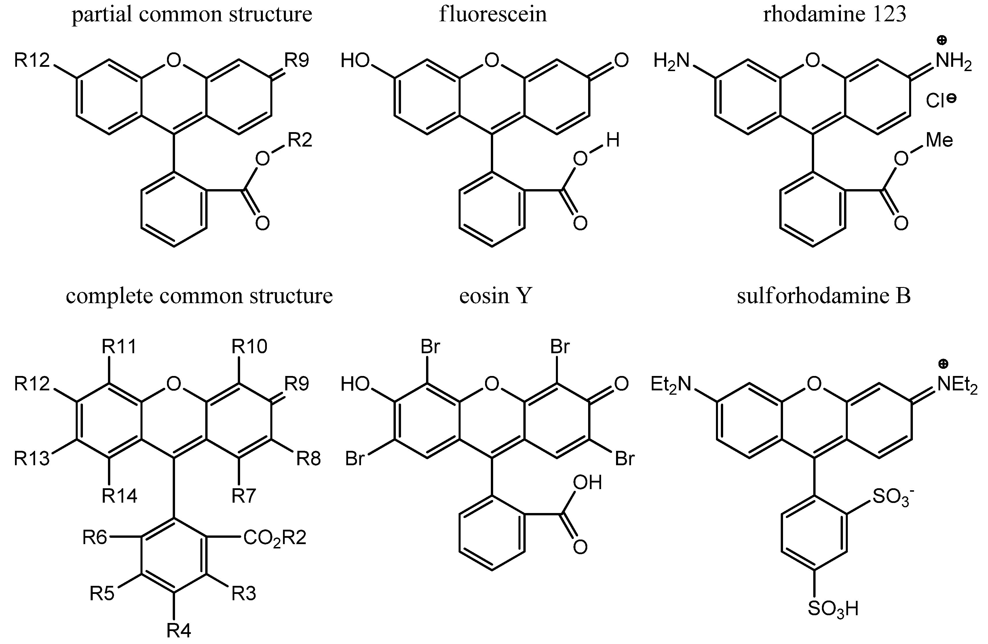
3. Common Small Molecule Fluorophores
4. Historical Perspective: the Origin of Fluorescein

| Name | R2 | R9 | R12 |
|---|---|---|---|
| fluorescein acid | H | O | OH |
| fluorescein sodium | Na | O | ONa |
| rhodamine 123 | Me | NH2+ | NH2 |

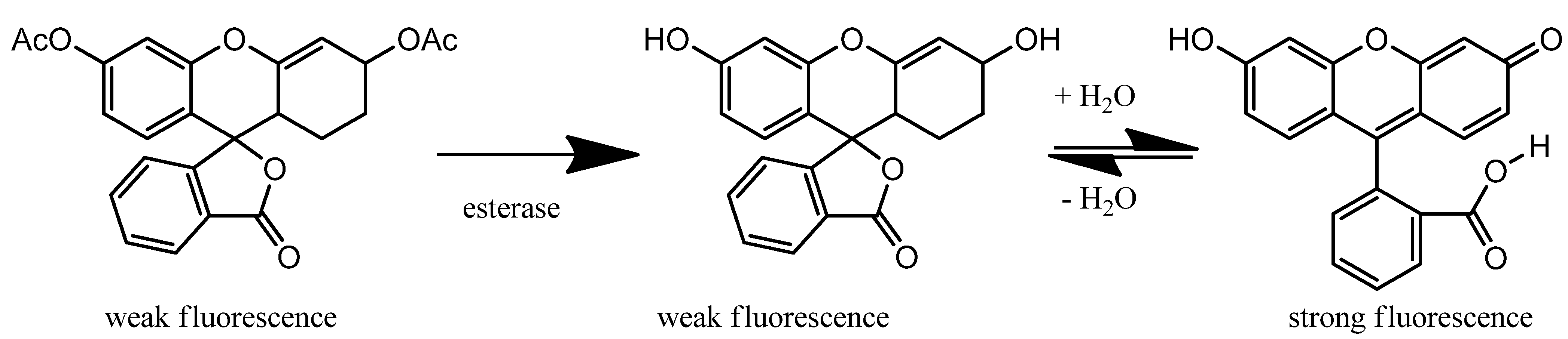
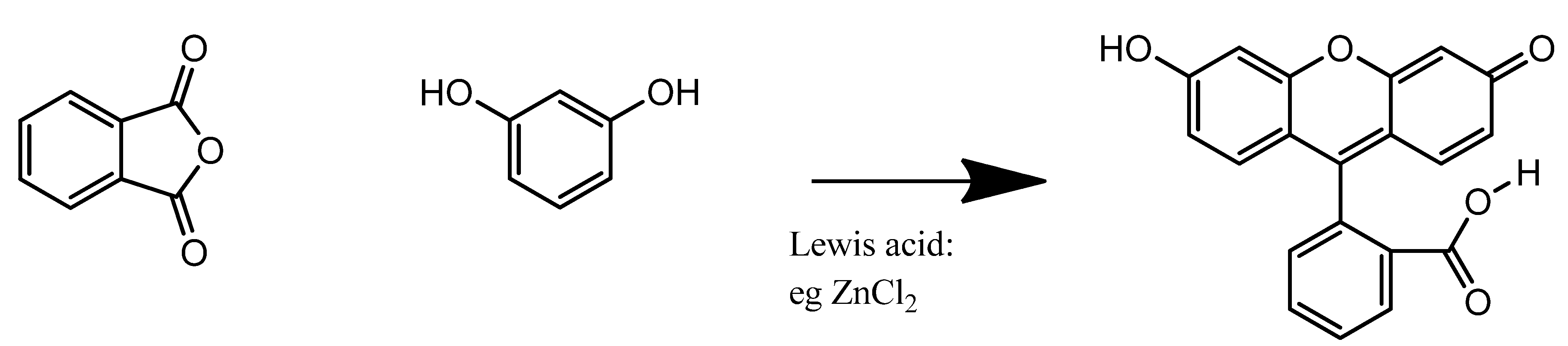
5. Synthesis

6. Eosinophilic / Anionic Stains
| Name | R2 | R5 | R6 | R8=R13 | R9 | R10=R11 | R12 |
|---|---|---|---|---|---|---|---|
| fluorescein | H | H | H | H | O | H | OH |
| eosin Y | H | H | H | Br | O | Br | OH |
| eosin B | H | H | H | NO2 | O | Br | OH |
| carboxyfluorescein | H | CO2H | H | H | O | H | OH |
| erythrosine | H | H | H | I | O | I | OH |
| ΦF | ex/em nm pH 7.4 | anion : dianion : trianion pH 7.4 | dye MW | clog P | clogD pH7.4 | Uses | |
|---|---|---|---|---|---|---|---|
| fluorescein [28,29] | 0.80 | 494/521 | 61:39:0 | 332 | 3.0 | −0.5 | Intravital eye stain |
| eosin Y [29] | 0.20 | 525/545 | 1:99:0 | 648 | 6.2 | 0.9 | H&E stain |
| eosin B | Cell stain | ||||||
| carboxyfluorescein [30] | 0.91 | 492/517 | 0:61:39 | 376 | 2.7 | −4.0 | Membrane impermeant |
| erythrosine [28,29] | 0.02 | 521/534 | 1:99:0 | 880 | 6.8 | 1.8 | Food colour, dental, radiopaque |
| Name | R2 | R5 | R6 | R8=R13 | R9 | R10=R11 | R12 |
|---|---|---|---|---|---|---|---|
| fluorescein | H | H | H | H | O | H | OH |
| fluorescein butyl ester | (CH2)3CH3 | H | H | H | O | H | OH |
| fluorescein butyl ether | H | H | H | H | O | H | O(CH2)3CH3 |
| anthofluorescein | H | H | H | H | O | H | C6H4OH |
| Zinpyr-1 | H | H | H | Cl | O | bipyridyl | OH |
| ΦF | ex/em nm pH 7.4 | anion : dianion : trianion pH 7.4 | dye MW | clog P | clogD pH7.4 | Uses | |
|---|---|---|---|---|---|---|---|
| fluorescein butyl ester [27] | 0.80 | 455/525 | 90:0:0 | 388 | 4.7 | 3.7 | rare |
| fluorescein butyl ether [27] | 0.39 | 455/525 | 100:0:0 | 388 | 4.5 | 1.2 | rare |
| anthofluorescein [31] | 0.33 | 478/538 | 99:1:0 | 408 | 4.7 | 1.3 | rare |
| Zn-Zinpyr-1 complex [32] | 0.87 | 507/525 | 1:99:0 | 824 | 1.3 | 1.0 | in vivo zinc |


7. Rhodamine
| Name | R2 | R5 | R6 | R8=R13 | R9 | R10=R11 | R12 |
|---|---|---|---|---|---|---|---|
| rhodamine 123 | CH3 | H | H | H | +NH2.Cl− | H | NH2 |
| rhodamine B | H | H | H | H | HN+(CH2CH3)2.Cl− | H | HN(CH2CH3)2 |
| rhodamine 6G | CH2CH3 | H | H | CH3 | HN+(CH2CH3).Cl− | H | HNCH2CH3 |
| rhodamine 110 | H | H | H | H | +NH2.Cl− | H | NH2 |
| rhodamine 19 | H | H | H | CH3 | HN+(CH2CH3).Cl− | H | HN(CH2CH3)2 |
| ΦF |  (ns) (ns) | ex/em nm pH 7.4 | ionisation neutral: anion : dianion : trianion pH 7.4 | dye MW | clog P | clogD pH7.4 | |
|---|---|---|---|---|---|---|---|
| rhodamine 123 [41] | 0.90 | 3.6 | 511/534 | 1:0:0:0 | 344 | 2.9 | 2.4 |
| rhodamine B [40,41] | 0.53 | 1.9 | 553/572 | 1:2:0:0 | 442 | 2.3 | 2.3 |
| rhodamine 6G [41,44] | 0.95 | 3.9 | 526/555 | 95:0:0:0 | 444 | 5.4 | 5.3 |
| rhodamine 110 | - | - | 500/525 | 4:1:0:0 | 330 | −0.1 | −0.2 |
| rhodamine 101 [41] | 0.96 | 4.1 | 560/589 | 1:0:0:0 | 491 | 2.6 | 2.6 |
| rhodamine 19 [41] | 0.95 | 4.2 | 535/546 [45] | 9:91:0:0 | 415 | 1.1 | 1.5 |
8. Clinical and Preclinical Applications
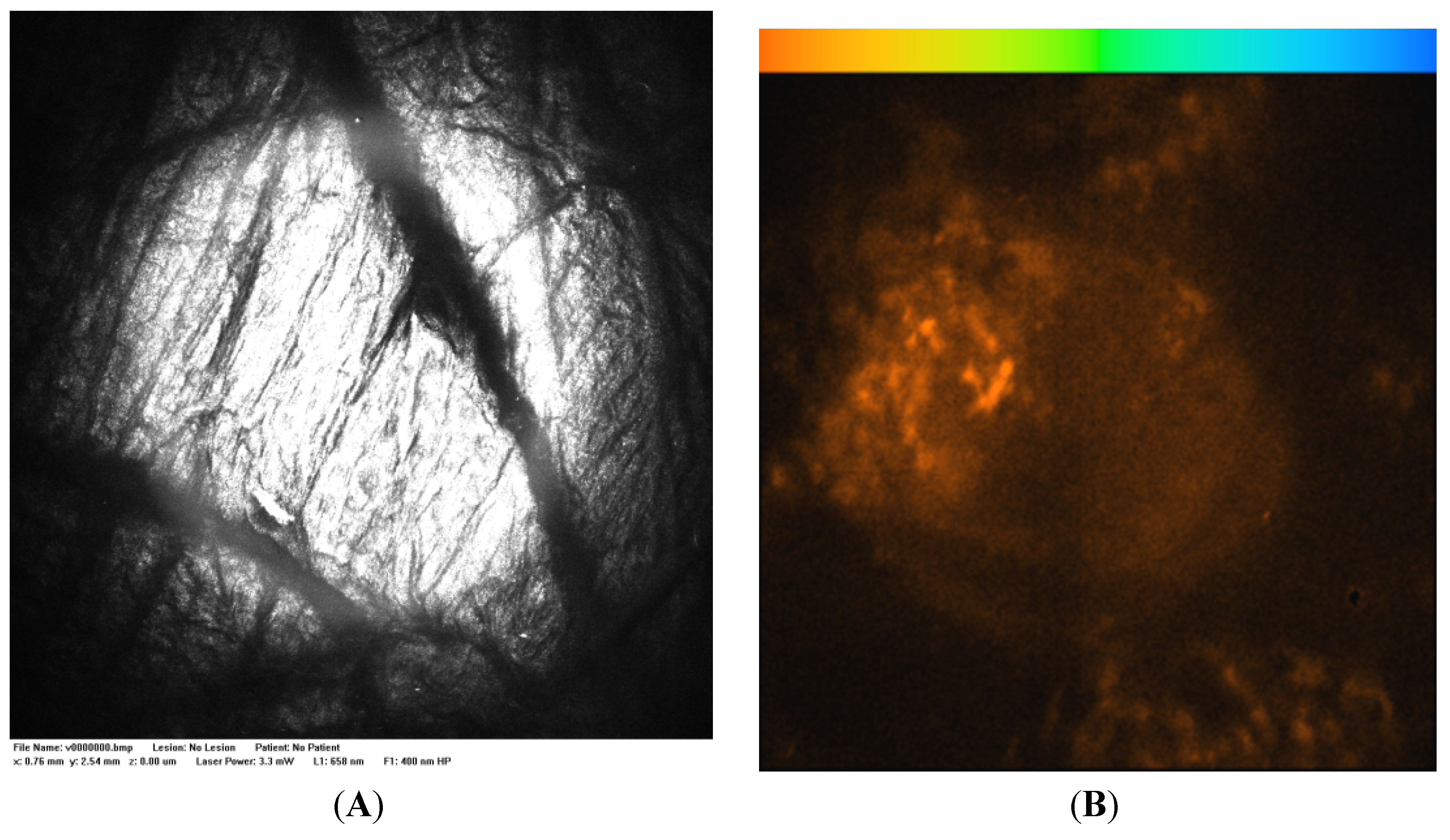
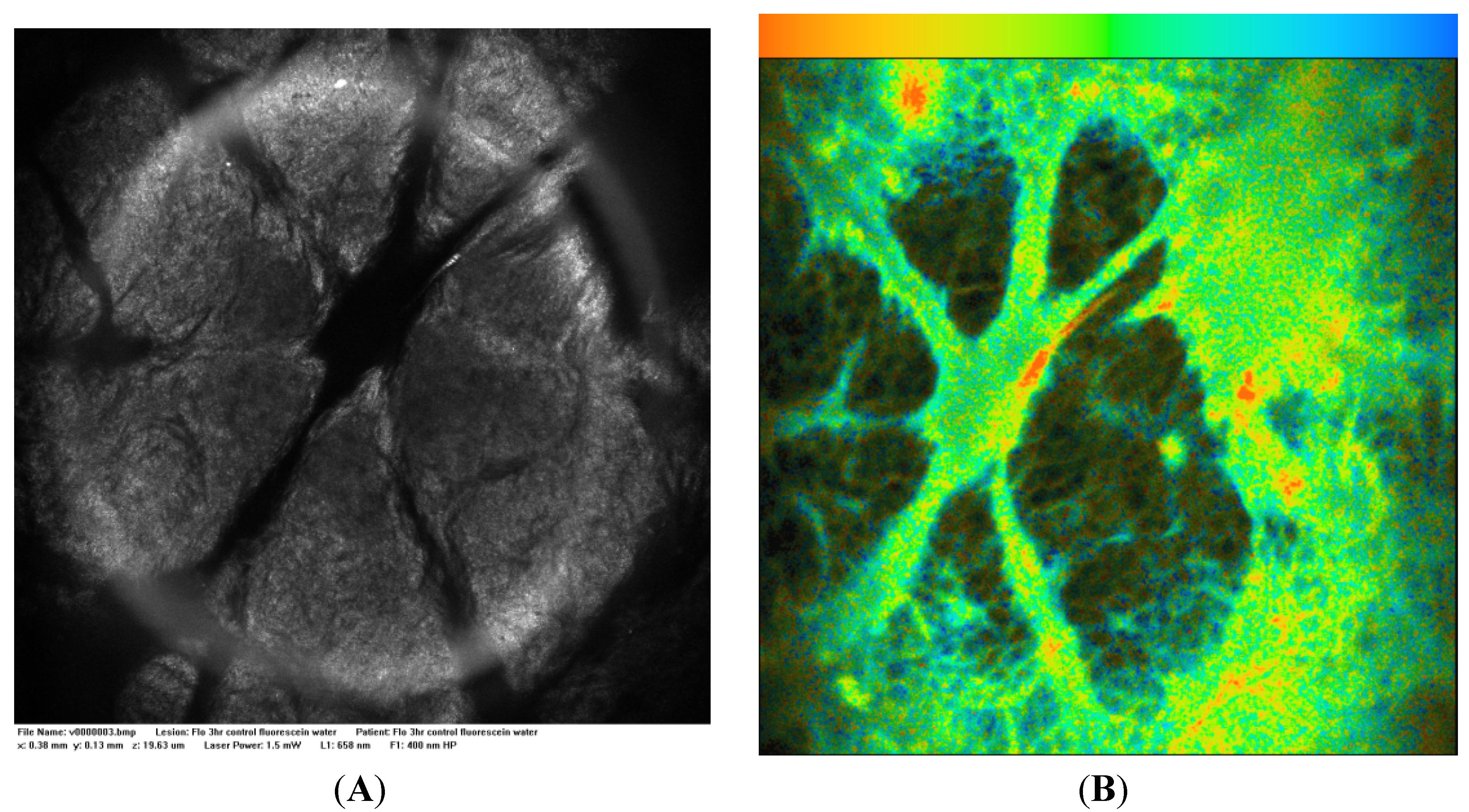
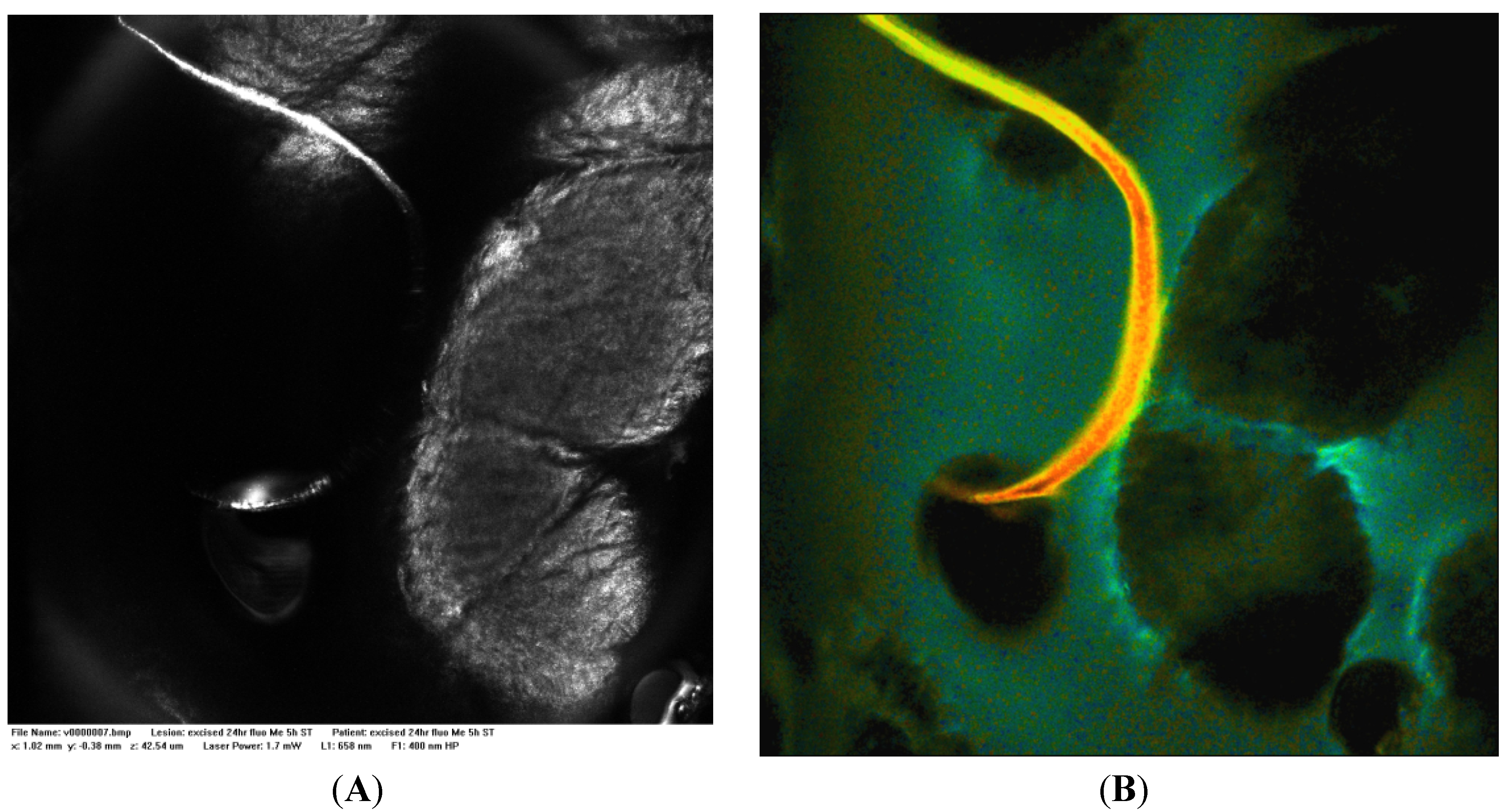
8.1. Fluorescence Endomicroscopy
8.2. Dermatology
9. Advanced Imaging Techniques & Use of FLIM
10. Formulation
11. Conclusion
Acknowledgements
Conflict of Interest
References and Notes
- Lin, L.L.; Grice, J.E.; Butler, M.K.; Zvyagin, A.V.; Becker, W.; Robertson, T.A.; Soyer, H.P.; Roberts, M.S.; Prow, T.W. Time-correlated single photon counting for simultaneous monitoring of zinc oxide nanoparticles and NAD(P)H in intact and barrier-disrupted volunteer skin. Pharm. Res. 2011, 28, 2920–2930. [Google Scholar] [CrossRef]
- Schaafsma, B.E.; Mieog, J.S.; Hutteman, M.; van der Vorst, J.R.; Kuppen, P.J.; Löwik, C.W.; Frangioni, J.V.; van de Velde, C.J.; Vahrmeijer, A.L. The clinical use of indocyanine green as a near-infrared fluorescent contrast agent for image-guided oncologic surgery. J. Surg. Oncol. 2011, 104, 323–332. [Google Scholar] [CrossRef]
- van den Berg, N.S.; van Leeuwen, F.W.; van der Poel, H.G. Fluorescence guidance in urologic surgery. Curr. Opin. Urol. 2012, 22, 109–120. [Google Scholar] [CrossRef]
- Meisel, P.; Kocher, T. Photodynamic therapy for periodontal diseases: State of the art. J. Photochem. Photobiol. B 2005, 79, 159–170. [Google Scholar] [CrossRef]
- Roberts, M.S.; Dancik, Y.; Prow, T.W.; Thorling, C.A.; Lin, L.L.; Grice, J.E.; Robertson, T.A.; Konig, K.; Becker, W. Non-invasive imaging of skin physiology and percutaneous penetration using fluorescence spectral and lifetime imaging with multiphoton and confocal microscopy. Eur. J. Pharm. Biopharm. 2011, 77, 469–488. [Google Scholar] [CrossRef]
- Galletly, N.P.; McGinty, J.; Dunsby, C.; Teixeira, F.; Requejo-Isidro, J.; Munro, I.; Elson, D.S.; Neil, M.A.; Chu, A.C.; French, P.M.; Stamp, G.W. Fluorescence lifetime imaging distinguishes basal cell carcinoma from surrounding uninvolved skin. Brit. J. Dermatol. 2008, 159, 152–161. [Google Scholar] [CrossRef]
- Tanter, M.; Touboul, D.; Gennisson, J.-L.; Bercoff, J.; Fink, M. High-resolution quantitative imaging of cornea elasticity using supersonic shear imaging. IEEE Trans. Med. Imaging 2009, 28, 1881–1893. [Google Scholar] [CrossRef]
- Wollensak, G.; Iomdina, E. Long-term biomechanical properties of rabbit cornea after photodynamic collagen crosslinking. Acta Ophthalmol. 2009, 87, 48–51. [Google Scholar] [CrossRef]
- Sondergaard, A.P.; Hjortdal, J.; Breitenbach, T.; Ivarsen, A. Corneal Distribution of Riboflavin Prior to Collagen Cross-Linking. Curr. Eye Res. 2010, 35, 116–121. [Google Scholar] [CrossRef]
- Roberts, M.S.; Roberts, M.J.; Robertson, T.A.; Sanchez, W.; Thörling, C.; Zou, Y.; Zhao, X.; Becker, W.; Zvyagin, A.V. In vitro and in vivo imaging of xenobiotic transport in human skin and in the rat liver. J. Biophotonics 2008, 1, 478–493. [Google Scholar] [CrossRef]
- Aihara, H.; Tajiri, H.; Suzuki, T. Application of Autofluorescence Endoscopy for Colorectal Cancer Screening: Rationale and an Update. Gastroent. Res. Pract. 2012, 2012, 5. [Google Scholar]
- Lam, S.; MacAulay, C.; Hung, J.; LeRiche, J.; Profio, A.E.; Palcic, B. Detection of dysplasia and carcinoma in situ with a lung imaging fluorescence endoscope device. J. Thorac. Cardiov. Sur. 1993, 105, 1035–1040. [Google Scholar]
- Harris, F.; Pierpoint, L. Photodynamic therapy based on 5-aminolevulinic acid and its use as an antimicrobial Agent. Med. Res. Rev. 2012, 32, 1292–1327. [Google Scholar] [CrossRef]
- Stummer, W.; Pichlmeier, U.; Meinel, T.; Wiestler, O.D.; Zanella, F.; Reulen, H.-J.; Oppel, F.; Brune, A.; Lanksch, W.; Woiciechowsky, C.; et al. Fluorescence-guided surgery with 5-aminolevulinic acid for resection of malignant glioma: a randomized controlled multicenter phase III trial. Lancet Oncol. 2006, 7, 392–401. [Google Scholar] [CrossRef]
- Ris, H.B.; Altermatt, H.J.; Stewart, C.M.; Schaffner, T.; Wang, Q.; Lim, C.K.; Bonnett, R.; Althaus, U. Photodynamic therapy with m-tetrahydroxyphenylchlorin in vivo: Optimization of the therapeutic index. Int. J. Cancer 1993, 55, 245–249. [Google Scholar]
- Marcu, L.; Hartl, B.A. Fluorescence lifetime spectroscopy and imaging in neurosurgery. IEEE J. Sel. Top. Quantum Electron. 2012, 18, 1465–1477. [Google Scholar] [CrossRef]
- Thekkek, N.; Richards-Kortum, R. Optical imaging for cervical cancer detection: solutions for a continuing global problem. Nat. Rev. Cancer 2008, 8, 725–731. [Google Scholar] [CrossRef]
- Baeyer, A. Phthalic acid-phenol compounds. Justus Liebigs Ann. Chem. 1876; 183, 1–74. [Google Scholar]
- Scifinder, version 2007.1; Chemical Abstracts Service: Columbus, OH, USA, 2007; (accessed 10 October 2012). Identifies: (i) 'fluorescein' as [2321–07–5] alternate [518-45-6]: 3',6'-dihydroxy-spiro[isobenzofuran-1(3H),9'-[9H]xanthen]-3-one; Fluorescein (8CI); 3,6-Dihydroxyspiro[xanthene-9,3'-phthalide]; 3',6'-Dihydroxyfluoran; 3',6'-Fluorandiol; 9-(o-Carboxyphenyl)-6-hydroxy-3-isoxanthenone; Benzoic acid, 2-(6-hydroxy-3-oxo-3H-xanthen-9-yl)-; C.I. 45350:1; C.I. Solvent Yellow 94; D and C Yellow No. 7; D&C Yellow No. 7; Fluorescein acid; Japan Yellow 201; Japan Yellow No. 201; NSC 667256; Resorcinolphthalein; Solvent Yellow 94; Yellow fluorescein; (ii) 'uranin' as [518-47-8]: 3',6'-dihydroxy-spiro[isobenzofuran-1(3H),9'-[9H]xanthen]-3-one, sodium salt (1:2); Fluorescein, disodium salt (8CI); Fluorin (6CI,7CI); Spiro[isobenzofuran-1(3H),9'-[9H]xanthen]-3-one, 3',6'-dihydroxy-, disodium salt (9CI); 11824 Yellow; 12417 Yellow; 3058 Uranine; 5(6)-Carboxyfluorescein sodium; ABCO Uranine; Abbey Uranine; Acid Yellow 73; Aizen Uranine; Basacid Yellow 226; Basovit Yellow 227; C.I. 45350; C.I. Acid Yellow 73; Calcocid Uranine B 4315; Certiqual Fluoresceine; Cogilor Yellow 111.10; D & C Yellow 8; D and C Yellow No. 8; D&C Yellow No. 8; D&C Yellow No. 8–307225; Dinacid Florescence Uranine; Dinacid Florescinece Uranine; Disodium fluorescein; Fluo-rectal; Fluor-I-Strip; Fluorescein LT; Fluorescein Sodium B.P; Fluorescein disodium; Fluorescein sodium; Fluorescein sodium salt; Fluoresceine DTD 842; Fluoresceine Extra 019187; Fluorescite; Fluoresein; Flurenate; Ful-Glo; Furanium; Hidacid Uranine; Japan Yellow 202(1); Neelicol Fluorescein; Neelicol Uranine; Obiturine; Resorcinol phthalein sodium; Sicomet Uranin S 45350; Sicomet Uranine S 45350; Simacid Fluoresceine LT; Simacid Fluoresceine Sodium; Sodium fluorescein; Sodium fluoresceinate; Soluble Fluorescein; Soluble Fluoresceine BPS; Triacid Fluoresceine; Urane U 307027; Uranin; Uranin A; Uranin Conc; Uranin S; Uranine; Uranine A; Uranine A extra; Uranine O; Uranine SS; Uranine SSO; Uranine WSS; Victacid Uranine; Victacid Uranine VDC; Yellow 202–1; Yellow No. 202–1.
- Babbey, C.M.; Ryan, J.C.; Gill, E.M.; Ghabril, M.S.; Burch, C.R.; Paulman, A.; Dunn, K.W. Quantitative intravital microscopy of hepatic transport. IntraVital 2012, 1, 44–53. [Google Scholar] [CrossRef]
- Chen, X.; Pradhan, T.; Wang, F.; Kim, J.S.; Yoon, J. Fluorescent Chemosensors Based on Spiroring-Opening of Xanthenes and Related Derivatives. Chem. Rev. 2012, 112, 1910–1956. [Google Scholar] [CrossRef]
- Baranyai, E. Chemistry of staining in histology. Chem. Aust. 2012, 26–29. [Google Scholar]
- Gareau, D.; Bar, A.; Snaveley, N.; Lee, K.; Chen, N.; Swanson, N.; Simpson, E.; Jacques, S. Tri-modal confocal mosaics detect residual invasive squamous cell carcinoma in Mohs surgical excisions. J. Biomed. Opt. 2012, 17, 066018. [Google Scholar] [CrossRef]
- Adur, J.; Pelegati, V.B.; de, T.A.A.; Baratti, M.O.; Almeida, D.B.; Andrade, L.A.L.A.; Bottcher-Luiz, F.; Carvalho, H.F.; Cesar, C.L. Optical biomarkers of serous and mucinous human ovarian tumor assessed with nonlinear optics microscopies. PLoS One 2012, 7, e47007. [Google Scholar] [CrossRef]
- Wallace, M.B.; Meining, A.; Canto, M.I.; Fockens, P.; Miehlke, S.; Roesch, T.; Lightdale, C.J.; Pohl, H.; Carr-Locke, D.; Lohr, M.; et al. The safety of intravenous fluorescein for confocal laser endomicroscopy in the gastrointestinal tract. Aliment. Pharmacol. Ther. 2010, 31, 548–552. [Google Scholar] [CrossRef]
- Slyusareva, E.A.; Tomilin, F.N.; Sizykh, A.G.; Tankevich, E.Y.; Kuzubov, A.A.; Ovchinnikov, S.G. The effect of halogen substitution on the structure and electronic spectra of fluorone dyes. Opt. Spectrosc. 2012, 112, 671–678. [Google Scholar] [CrossRef]
- Song, A.; Zhang, J.; Zhang, M.; Shen, T.; Tang, J. Spectral properties and structure of fluorescein and its alkyl derivatives in micelles. Colloids Surf. A 2000, 167, 253–262. [Google Scholar] [CrossRef]
- Doukas, A.G.; Junnarkar, M.R.; Alfano, R.R.; Callender, R.H.; Kakitani, T.; Honig, B. Fluorescence quantum yield of visual pigments: Evidence for subpicosecond isomerization rates. Proc. Natl. Acad. Sci. USA 1984, 81, 4790–4794. [Google Scholar] [CrossRef]
- Fleming, G.R.; Knight, A.W.E.; Morris, J.M.; Morrison, R.J.S.; Robinson, G.W. Picosecond fluorescence studies of xanthene dyes. J. Am. Chem. Soc. 1977, 99, 4306–4311. [Google Scholar] [CrossRef]
- Mineno, T.; Ueno, T.; Urano, Y.; Kojima, H.; Nagano, T. Creation of superior carboxyfluorescein dyes by blocking donor-excited photoinduced electron transfer. Org. Lett. 2006, 8, 5963–5966. [Google Scholar] [CrossRef]
- Yusop, R.M.; Unciti-Broceta, A.; Bradley, M. A highly sensitive fluorescent viscosity sensor. Bioorg. Med. Chem. Lett. 2012, 22, 5780–5783. [Google Scholar] [CrossRef]
- Walkup, G.K.; Burdette, S.C.; Lippard, S.J.; Tsien, R.Y. A New Cell-Permeable Fluorescent Probe for Zn2+. J. Am. Chem. Soc. 2000, 122, 5644–5645. [Google Scholar]
- Burdette, S.C.; Frederickson, C.J.; Bu, W.; Lippard, S.J. ZP4, an improved neuronal Zn2+ sensor of the zinpyr family. J. Am. Chem. Soc. 2003, 125, 1778–1787. [Google Scholar] [CrossRef]
- Zhang, Q.; Grice, J.E.; Li, P.; Jepps, O.G.; Wang, G.-J.; Roberts, M.S. Skin Solubility Determines Maximum Transepidermal Flux for Similar Size Molecules. Pharm. Res. 2009, 26, 1974–1985. [Google Scholar] [CrossRef]
- Thorling, C.A.; Dancik, Y.; Hupple, C.W.; Medley, G.; Liu, X.; Zvyagin, A.V.; Robertson, T.A.; Burczynski, F.J.; Roberts, M.S. Multiphoton microscopy and fluorescence lifetime imaging provide a novel method in studying drug distribution and metabolism in the rat liver in vivo. J. Biomed. Opt. 2011, 16, 086013/086011–086013/086017. [Google Scholar]
- Liu, X.; Thorling, C.A.; Jin, L.; Roberts, M.S. Intravital multiphoton imaging of rhodamine 123 in the rat liver after intravenous dosing. IntraVital 2012, 1, 30–31. [Google Scholar]
- Lee, P.J.; Langer, R.; Shastri, V.P. Novel microemulsion enhancer formulation for simultaneous transdermal delivery of hydrophilic and hydrophobic drugs. Pharm. Res. 2003, 20, 264–269. [Google Scholar] [CrossRef]
- Okuda, T.; Yoshioka, H.; Kato, A. Fluorescence-guided surgery for glioblastoma multiforme using high-dose fluorescein sodium with excitation and barrier filters. J. Clin. Neurosci. 2012, 19, 1719–1722. [Google Scholar] [CrossRef]
- van Dam, G.M.; Themelis, G.; Crane, L.M.; Harlaar, N.J.; Pleijhuis, R.G.; Kelder, W.; Sarantopoulos, A.; de Jong, J.S.; Arts, H.J.; van der Zee, A.G.; et al. Intraoperative tumor-specific fluorescence imaging in ovarian cancer by folate receptor-α targeting: First in-human results. Nat. Med. 2011, 17, 1315–1319. [Google Scholar] [CrossRef]
- Beija, M.; Afonso, C.A.M.; Martinho, J.M.G. Synthesis and applications of Rhodamine derivatives as fluorescent probes. Chem. Soc. Rev. 2009, 38, 2410–2433. [Google Scholar] [CrossRef]
- Savarese, M.; Aliberti, A.; De, S.I.; Battista, E.; Causa, F.; Netti, P.A.; Rega, N. Fluorescence lifetimes and quantum yields of rhodamine derivatives: New insights from theory and experiment. J. Phys. Chem. A 2012, 116, 7491–7497. [Google Scholar] [CrossRef]
- Guldbrand, S.; Simonsson, C.; Goksoer, M.; Smedh, M.; Ericson, M.B. Two-photon fluorescence correlation microscopy combined with measurements of point spread function; investigations made in human skin. Opt. Express 2010, 18, 15289–15302. [Google Scholar] [CrossRef]
- Maurin, M.; Stephan, O.; Vial, J.-C.; Marder, S.R.; van, d.S.B. Deep in vivo two-photon imaging of blood vessels with a new dye encapsulated in pluronic nanomicelles. J. Biomed. Opt. 2011, 16, 036001. [Google Scholar] [CrossRef]
- Lee, M.; Grissom, C.B. Design, Synthesis, and Characterization of Fluorescent Cobalamin Analogues with High Quantum Efficiencies. Org. Lett. 2009, 11, 2499–2502. [Google Scholar] [CrossRef]
- Kucki, M.; Fuhrmann-Lieker, T. Staining diatoms with rhodamine dyes: Control of emission colour in photonic biocomposites. J. R. Soc. Interface 2012, 9, 727–733. [Google Scholar] [CrossRef]
- Taruttis, A.; Ntziachristos, V. Translational optical imaging. AJR Am. J. Roentgenol. 2012, 199, 263–271. [Google Scholar] [CrossRef]
- Goetz, M.; Wang, T.D. Molecular imaging in gastrointestinal endoscopy. Gastroenterology 2010, 138, 828–833. [Google Scholar] [CrossRef]
- Sanai, N.; Eschbacher, J.; Hattendorf, G.; Coons, S.W.; Preul, M.C.; Smith, K.A.; Nakaji, P.; Spetzler, R.F. Intraoperative confocal microscopy for brain tumors: A feasibility analysis in humans. Neurosurgery 2011, 68, 282–290; discussion 290. [Google Scholar]
- Astner, S.; Dietterle, S.; Otberg, N.; Rowert-Huber, H.-J.; Stockfleth, E.; Lademann, J. Clinical applicability of in vivo fluorescence confocal microscopy for noninvasive diagnosis and therapeutic monitoring of nonmelanoma skin cancer. J. Biomed. Opt. 2008, 13, 014003. [Google Scholar] [CrossRef]
- Saldua, M.A.; Olsovsky, C.A.; Callaway, E.S.; Chapkin, R.S.; Maitland, K.C. Imaging inflammation in mouse colon using a rapid stage-scanning confocal fluorescence microscope. J. Biomed. Opt. 2012, 17, 016006. [Google Scholar] [CrossRef]
- Jonak, C.; Skvara, H.; Kunstfeld, R.; Trautinger, F.; Schmid, J.A. Intradermal Indocyanine green for in vivo fluorescence laser scanning microscopy of human skin: A pilot study. PLoS One 2011, 6, e23972. [Google Scholar]
- Dinish, U.S.; Fu, C.Y.; Ng, B.K.; Chow, T.H.; Murukeshan, V.M.; Seah, L.K.; Tan, S.K. A fluorescence lifetime imaging microscopy (FLIM) system for the characterization of haematoxylin and eosin stained sample. Proc. SPIE 2008, 6859, 68590C/68591–68590C/68599. [Google Scholar]
- Conklin, M.W.; Provenzano, P.P.; Eliceiri, K.W.; Sullivan, R.; Keely, P.J. Fluorescence lifetime imaging of endogenous fluorophores in histopathology sections reveals differences between normal and tumor epithelium in carcinoma in situ of the breast. Cell Biochem. Biophys. 2009, 53, 145–157. [Google Scholar] [CrossRef]
- Seo, H.W.; Kim, G.H.; Kim, D.Y.; Yoon, S.M.; Kwon, J.S.; Kang, W.S.; Lee, B.; Kim, J.H.; Min, B.H.; Kim, M.S. Microemulsion of erythromycine for transdermal drug delivery. J. Appl. Polym. Sci. 2013, 128, 4277–4282. [Google Scholar] [CrossRef]
- Auner, B.G.; Valenta, C.; Hadgraft, J. Influence of phloretin and 6-ketocholestanol on the skin permeation of sodium-fluorescein. J. Control. Release 2003, 89, 321–328. [Google Scholar] [CrossRef]
- Zambito, Y.; Uccello-Barretta, G.; Zaino, C.; Balzano, F.; Di, C.G. Novel transmucosal absorption enhancers obtained by aminoalkylation of chitosan. Eur. J. Pharm. Sci. 2006, 29, 460–469. [Google Scholar]
© 2013 by the authors; licensee MDPI, Basel, Switzerland. This article is an open access article distributed under the terms and conditions of the Creative Commons Attribution license (http://creativecommons.org/licenses/by/3.0/).
Share and Cite
Robertson, T.A.; Bunel, F.; Roberts, M.S. Fluorescein Derivatives in Intravital Fluorescence Imaging. Cells 2013, 2, 591-606. https://doi.org/10.3390/cells2030591
Robertson TA, Bunel F, Roberts MS. Fluorescein Derivatives in Intravital Fluorescence Imaging. Cells. 2013; 2(3):591-606. https://doi.org/10.3390/cells2030591
Chicago/Turabian StyleRobertson, Thomas A., Florestan Bunel, and Michael S. Roberts. 2013. "Fluorescein Derivatives in Intravital Fluorescence Imaging" Cells 2, no. 3: 591-606. https://doi.org/10.3390/cells2030591
APA StyleRobertson, T. A., Bunel, F., & Roberts, M. S. (2013). Fluorescein Derivatives in Intravital Fluorescence Imaging. Cells, 2(3), 591-606. https://doi.org/10.3390/cells2030591