Article

Manganese Suppresses the Haploinsufficiency of Heterozygous trpy1Δ/TRPY1 Saccharomyces cerevisiae Cells and Stimulates the TRPY1-Dependent Release of Vacuolar Ca2+ under H2O2 Stress

Department of Organic Chemistry, Biochemistry and Catalysis, Faculty of Chemistry, University of Bucharest, Sos. Panduri 90-92, 050663 Bucharest, Romania
*
Author to whom correspondence should be addressed.
Cells 2019, 8(2), 79; https://doi.org/10.3390/cells8020079
Submission received: 17 December 2018 / Revised: 9 January 2019 / Accepted: 18 January 2019 / Published: 22 January 2019
(This article belongs to the Special Issue TRP Channels in Health and Disease)

Abstract

:
Transient potential receptor (TRP) channels are conserved cation channels found in most eukaryotes, known to sense a variety of chemical, thermal or mechanical stimuli. The Saccharomyces cerevisiae TRPY1 is a TRP channel with vacuolar localization involved in the cellular response to hyperosmotic shock and oxidative stress. In this study, we found that S. cerevisiae diploid cells with heterozygous deletion in TRPY1 gene are haploinsufficient when grown in synthetic media deficient in essential metal ions and that this growth defect is alleviated by non-toxic Mn2+ surplus. Using cells expressing the Ca2+-sensitive photoprotein aequorin we found that Mn2+ augmented the Ca2+ flux into the cytosol under oxidative stress, but not under hyperosmotic shock, a trait that was absent in the diploid cells with homozygous deletion of TRPY1 gene. TRPY1 activation under oxidative stress was diminished in cells devoid of Smf1 (the Mn2+-high-affinity plasma membrane transporter) but it was clearly augmented in cells lacking Pmr1 (the endoplasmic reticulum (ER)/Golgi located ATPase responsible for Mn2+ detoxification via excretory pathway). Taken together, these observations lead to the conclusion that increased levels of intracytosolic Mn2+ activate TRPY1 in the response to oxidative stress.

1. Introduction

Living cells are continuously exposed to various changes that threaten the dynamic equilibrium associated with the steady state of homeostatic balance. Such changes—often induced by stress agents—need to be sensed and signaled by cell components which belong to intricate networks responsible for homeostatic regulation. Calcium is a secondary messenger used by all eukaryotes—animal, plants, microorganisms—to connect various stimuli or stresses to their corresponding cellular responders. The budding yeast Saccharomyces cerevisiae has been constantly used as a model eukaryote to study the calcium-dependent response to various types of external stresses, which include salt [1], hypotonic [2,3], hypertonic [1,4,5], salicylate [6], alkaline [7], cold [8], ethanol [9,10], drugs [11] antifungals [12,13,14,15,16], electric [17] oxidative [18,19,20] or heavy metal [8,20,21,22] insults. The S. cerevisiae cells respond to such stresses by a sudden increase in cytosolic Ca2+—denoted henceforth [Ca2+]cyt—following the stimulus-dependent opening of Ca2+ channels situated in the plasma membrane and/or in internal compartments. Abrupt increase in [Ca2+]cyt represents a versatile and universally used mechanism which triggers either cell survival/adaptation or cell death [23]. In S. cerevisiae the stress-dependent rise in [Ca2+]cyt can be a consequence of Ca2+ influx via the Cch1/Mid1 channel on the plasma membrane [1,2] release of vacuolar Ca2+ into the cytosol through the vacuole-located Ca2+ channel TRPY1 [4,24], or both [19,20]. After delivering the message, the normal very low level of [Ca2+]cyt is restored through the action of Ca2+ pumps and exchangers [25]. Thus, the Ca2+-ATPase Pmc1 [26] and a vacuolar Ca2+/H+ exchanger Vcx1 [27,28] independently transport [Ca2+]cyt into the vacuole, while Pmr1, the secretory Ca2+-ATPase, pumps [Ca2+]cyt into endoplasmic reticulum (ER) and Golgi along with Ca2+ extrusion from the cell [29,30].
In S. cerevisiae, TRPY1 is almost exclusively localized at the vacuolar membrane [4], playing an important role in adaptation to environmental stresses [4,19,20,21]. Initially named Yvc1, TRPY1 is encoded by TRPY1 gene (systematic gene name, YOR087W) and it is the only member of the TRP (Transient Receptor Potential) superfamily of cationic channels expressed in S. cerevisiae [31]. TRP channels are conserved cation channels found in most eukaryotes, known to sense chemical, thermal, or mechanical stimuli in animals [32]. In yeast, TRPY1 is the main channel responsible for of [Ca2+]cyt elevation under hyperosmotic shock [4,31], when calcium accrues predominantly from vacuolar stores [4]. This behavior can be explained by the mechano-sensitive traits of TRPY1: under hypertonic conditions water evacuates passively from the cytoplasm and then from the vacuole causing deformation of the vacuolar membrane and consequently the opening of the TRPY1 channel, with the release of vacuolar Ca2+ [5,33]. In contrast, under alkaline stress, the elevated [Ca2+]cyt has its origin exclusively from the cell’s exterior, with the Cch1/Mid1 channel solely responsible for the majority of Ca2+ entry, and with no contribution of vacuolar Ca2+ [7]. In between these two situations, oxidative stress triggers [Ca2+]cyt waves which pool both external and vacuolar Ca2+ [19]. TRPY1 is necessary for attaining a maximum level of [Ca2+]cyt under oxidative stress and TRPY1 depends on [Ca2+]cyt elevation for maximal gating, in a process known as Ca2+-induced Ca2+ release [34].
TRPY1 gene is not essential for survival and the knockout mutant cells trpy1Δ have no clear growth defects under various stresses. Rather, it was shown that trpy1Δ cells are slightly more resistant to the oxidative stress imposed by exogenous hydrogen peroxide or tert-butylhydroperoxide [19] and Cu2+ [20] but also less fit under high Cd2+ [21] or tunicamycin-induced ER-stress in Ca2+-depleted medium [31]. In contrast, cells overexpressing the TRPY1 gene are hypersensitive to surplus Ca2+ [4] or oxidative stress [19]. Also, it was revealed in a wide-scale survey that heterozygous trpy1Δ/TRPY1 diploid cells are less fit under nutrient limiting conditions than the wild-type TRPY1/TRPY1 ([35], Supplementary material). Haploinsufficiency occurs when the heterozygous mutation of a gene in a diploid organism results in a reduction of the corresponding gene product which can be correlated with negative alterations of the wild-type phenotype. In this study, we performed a chemical screen and found that non-toxic concentrations of Mn2+ alleviated the trpy1Δ/TRPY1 haploinsufficiency observed by us in minimal growth medium containing half of the recommended amount of essential metal ions, probably by stimulating the TRPY1-mediated Ca2+ release into the cytosol.

2. Materials and Methods

2.1. Yeast Strains and Growth Media

The S. cerevisiae diploid strains used in this study were isogenic with the “wild-type” (WT) parental strain BY4743 (MATa/α; his3Δ1/his3Δ1; leu2Δ0/leu2Δ0; met15Δ0/MET15; LYS2/lys2Δ0; ura3Δ0/ura3Δ0), a S288C-based yeast strain [36]. The strains harbored either heterozygous (BY4732, orf::kanMX4/ORF) or homozygous (BY4732, orf::kanMX4/orf::kanMX4) knockout mutations of individual gene open reading frames (ORF). The heterozygous knockout mutants are referred to in the text as orfΔ/ORF and were cch1Δ/CCH1, mid1Δ/MID1, pmc1Δ/PMC1, pmr1Δ/PMR1, vcx1Δ/VCX1, trpy1Δ/TRPY1. The homozygous knockout mutants are referred to in the text as orfΔ/orfΔ and were trpy1Δ/trpy1Δ, smf1Δ/smf1Δ, and pmr1Δ/pmr1Δ. The strains were obtained from EUROSCARF (European S. cerevisiae Archive for Functional Analysis, www.euroscarf.de) and were propagated, grown, and maintained in YPD medium (1% yeast extract, 2% polypeptone, 2% glucose) or SD (0.17% yeast nitrogen base without amino acids, 0.5% (NH4)2SO4, 2% glucose, supplemented with the necessary amino acids) [37]. The strains transformed with the plasmids harboring apo-aequorin cDNA [38] were selected and maintained on SD lacking uracil (SD-Ura). Minimal defined media (MM) were prepared adding individual components as described [37] using ultrapure reagents (Merck, Darmstadt, Germany) and contained 1 mM Ca2+, 0.25 µM Cu2+, 2 µM Mn2+, 2 µM Fe3+ and 2 µM Zn2+. Low-metal minimal defined medium (LMeMM) had 0.5 mM Ca2+, 0.1 µM Cu2+, 1 µM Mn2+, 1 µM Fe3+ and 1 µM Zn2+, corresponding roughly to half of the amount of essential metals recommended [37]. The concentrations of metals in MM and LMeMM were confirmed by inductively coupled plasma with mass spectrometry (ICP-MS, Perkin-Elmer ELAN DRC-e, Concord, ON, Canada) against Multielement ICP Calibration Standard 3, matrix 5% HNO3 (Perkin-Elmer Pure Plus, Shelton, CT, USA). All synthetic media had their pH adjusted to 6.5. For solid media, 2% agar was used. For growth improvement, all the synthetic media were supplemented with an extra 20 mg/L leucine [39]. All chemicals, including media reagents were from Merck (Darmstadt, Germany),

2.2. Plasmid and Yeast Transformation

For heterologous expression of aequorin, yeast cells were transformed with the multicopy URA3-based plasmid pYX212-cytAEQ harboring the apo-aequorin cDNA under the control of the strong TPI (triosephosphate isomerase) yeast promoter [40]. Plasmid pYX212-cytAEQ was a generous gift from Martegani and Tisi (University of Milano-Bicocca, Milan, Italy). Yeast transformation [41] was performed using S.c. EasyComp™ Transformation Kit (Invitrogen, Carlsbad, CA, USA) following manufacturer’s indications.

2.3. Yeast Cell Growth Assay

2.3.1. Growth in Liquid Media

Unless otherwise specified, cells were incubated at 30 °C under agitation (200 rpm). Yeast strains were pre-grown overnight in MM then diluted (1/20) in fresh MM and grown for 2 h. Cells were harvested by centrifugation, washed with ice-cold water, and resuspended in liquid LMeMM at density which corresponded to optical density measured at 600 nm (OD600) = 0.05. Strain growth in liquid LMeMM was monitored in time by measuring OD600 recorded in a plate reader equipped with thermostat and shaker (Varioskan, Thermo Fisher Scientific, Vantaa, Finland). Relative growth was expressed as the ratio between OD600 recorded at time t and OD600 recorded at time 0. For screening of chemicals against trpy1Δ/TRPY1 haploinsufficiency (HIP), cells shifted to LMeMM (OD600 = 0.05) were incubated for 2 h before chemicals were added from concentrated sterile stocks. Cell growth (%) was determined 24 h from chemical addition and calculated relatively to growth (OD600) of WT strain, no added chemicals. Chemicals used were CuCl2, FeCl2, MnCl2, ZnCl2, ascorbate, ethylene glycol-bis(2-aminoethylether)-N,N,N′,N′-tetraacetic acid (EGTA), GdCl3, glutathione (GSH), indole and were all of high-grade purity.

2.3.2. Growth on Solid Media

For dilution plate assay, exponentially growing cells were 10-fold serially diluted in a 48-well microtiter plate and stamped on agar plates using a pin replicator (approximately 4 μL/spot). Plates were photographed after incubation at 30 °C for 3 days.

2.4. TRPY1 Gene Expression by Quantitative Reverse Transcriptase-Polymerase Chain Reaction (qRT-PCR)

Wild-type cells BY4743 (TRPY1/TRPY1), heterozygous (trpy1Δ/TRPY1), and homozygous (trpy1Δ/trpy1Δ) diploid cells from overnight pre-cultures were inoculated at OD600 = 0.1 in MM or LMeMM and grown to OD600 = 0.5 before Mn2+ was added to final concentration 10 µM, then incubated for 2 additional hours before being harvested for total ribonucleic acid (RNA) isolation. Total RNA was isolated using the RiboPure™ RNA Purification Kit for yeast (Ambion™, Thermo Fischer Scientific, Vilnius, Lithuania) following the manufacturer’s instructions. Approximately 500 ng of total RNA was transcribed into cDNA using GoScript™ Reverse Transcription System (Promega, Madison, WI, USA). Finally, a total of 10 ng cDNA was used for each qRT-PCR done with the GoTaq® qPCR Master Mix (Promega). Each reaction was performed in triplicate using MyiQ Single-Color Real-Time PCR Detection System (BioRad, Hercules, CA, USA). Expression of TRPY1 mRNA was normalized to the relative expression of ACT1 in each sample. The qRT-PCR cycling conditions were 95 °C for 1 min, and 40 cycles of 95 °C for 10 s, 59 °C for 10 s, 72 °C for 12 s. The primers used for amplification of cDNA were: TRPY1-F: 5′-AGATTCTCAG GGTTACGTTA, TRPY1-R: 5′-CAATATGGAATACCACTCAC; ACT1-F: 5′-GGTTGCTGCTTTGGTTATTG, ACT1-R: 5′-CAATTGGGTAACGTAAAGTC.

2.5. Assay of Cell Mn2+

Measurements of cell total manganese content were done on cells grown in LMeMM medium to an OD600 of 1.0. Cells were harvested in triplicate samples, washed two times in ice-cold 10 mM 2-(N-morpholino) ethanesulfonic acid (MES)-Tris buffer (pH 6.0). Cells were finally suspended in deionized water (OD600 = 10) and used for manganese and cell protein assay. Manganese analysis was done by ICP-MS after digestion of cells with 65% ultrapure HNO3 (Merck, Darmstadt, Germany). The metal cellular content was normalized to total cellular proteins, as described [42]. Total cellular manganese was expressed as nanomoles of metal per mg cell protein.

2.6. Detection of [Ca2+]cyt by Aequorin Bioluminescence Assay

Cells transformed with pYX212-cytAEQ were maintained on SD-Ura selective medium and prepared for Ca2+ dependent luminescence detection as described [43] with slight modifications. Overnight pre-cultures of cells expressing apo-aequorin were washed and suspended (OD600 = 0.5) in LMeMM-Ura then incubated to late exponential phase (OD600 = 1, 4–6 h). For pre-incubation with Mn2+, MnCl2 was added to the desired concentration and cells were grown for an additional 2 h. Cells were harvested by centrifugation and resuspened (to OD600 = 10) in LMeMM-Ura in which the corresponding Mn2+ concentration was maintained. To reconstitute functional aequorin, native coelenterazine was added to the cell suspension (from a methanol stock, 20 µM final concentration) and the cells were incubated for 2 h at 30 °C in the dark. The excess coelenterazine was removed by centrifugation. The cells (approximately 107 cells/determination) were finally resuspended in LMeMM-Ura with corresponding Mn2+ concentration and transferred to the luminometer tube. A cellular luminescence baseline was determined for each strain by approximately one minute of recordings at 1/s intervals. After ensuring a stable signal, chemicals tested were injected from sterile stocks to give the final concentrations indicated, and the Ca2+-dependent light emission was monitored in a single tube luminometer (Turner Biosystems, 20n/20, Sunnyvale, CA, USA). The light emission was measured at 1 s intervals and expressed as relative luminescence units (RLU). To ensure that the total reconstituted aequorin was not limiting in our assay, at the end of each experiment aequorin activity was checked by lysing cells with 1% Triton X-100 with 5 mM CaCl2 and only the cells with considerable residual luminescence were considered. Relative luminescence emission was normalized to an aequorin content giving a total light emission of 106 RLUs in 10 min after lysing cells with 1% Triton X-100. The relative luminescence maximum (RLM) was the average of the RLUs flanking the maximum value minus the average luminescence baseline recorded before cells were exposed to the stimulus (10 values on each side), all normalized as described above.

2.7. Statistics

All experiments were repeated, independently, in three biological replicates at least. For each individual experiment, values were expressed as the mean ± standard error of the mean (SEM). For aequorin luminescence determinations, traces represent the mean ± SEM from three independent transformants. The numerical data were examined by Student t test or by analysis of variance with multiple comparisons (ANOVA) using the statistical software Prism version 6.05 for Windows (GraphPad Software, La Jolla, CA, USA). The differences were considered to be significant when p < 0.05. One sample t test was used for the statistical analysis of each strain/condition compared with a strain/condition considered as reference. Asterisks indicate the level of significance: * p < 0.05, ** p < 0.01, and *** p < 0.001.

3. Results

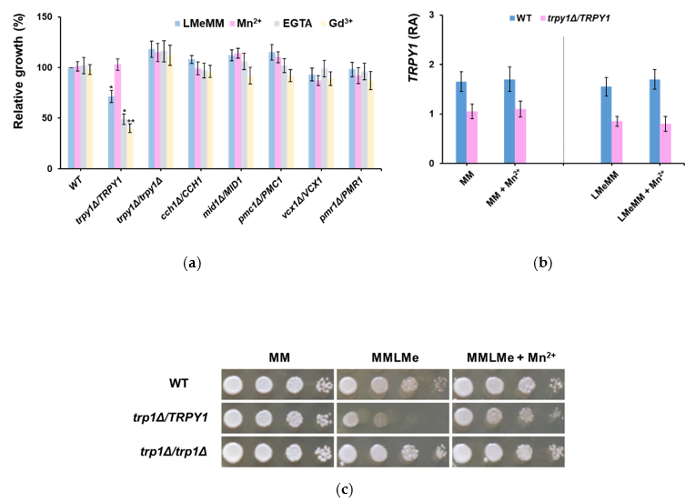
3.1. Haploinsufficiency of Yeast Strain Heterozyous for TRPY1 Is Alleviated by Mn2+

To highlight new aspects related to TRPY1 function in yeast cells, the main target of our study was to identify conditions which interfere with TRPY1 activity. In this direction, haploinsufficiency is a genetic trait which can be very useful in the attempts to identify small molecules which influence the behavior of functional proteins [44]. A genome-wide survey had already pinpointed the heterozygous trpy1Δ/TRPY1 as possibly less fit under nutrient limiting conditions ([35], Supplementary material). We noticed that the growth of the heterozygous trpy1Δ/TRPY1 diploid mutant was not significantly different from the growth of the wild-type diploid when the two strains were incubated in YPD, SD (data not shown) or MM medium (Figure 1a), but trpy1Δ/TRPY1 cells exhibited somehow slower growth (p < 0.001) in minimal synthetic medium LMeMM which had approximately half of the amount of essential metals recommended [37] (Figure 1b).
The haploinsufficiency in LMeMM was noted only for TRPY1; no similar phenotype was recorded for heterozygous strains with mutations in the genes which encode the other transporters known to participate in regulating [Ca2+]cyt, e.g., CCH1, MID1, PMC1 or VCX1 (Figure 2a, dark blue bars).
To identify compounds which potentially interact with TRPY1 activity we screened for conditions which may alleviate or augment the haploinsufficient phenotype observed. The tested substances are presented in Table 1. These substances were added to the LMeMM at the point where the heterozygous trpy1Δ/TRPY1 diploid cells were in the early log phase of growth (OD600 = 0.1) and the effect on growth was determined 24 h after chemical addition. We tested the effect of adding physiological concentrations of the metals initially depleted in LMeMM (i.e., Ca2+, Cu2+, Fe3+, Mn2+, Zn2+) but also of glutathione and indole, which had been reported to interact with TRPY1 [45,46]. As glutathione is a universal intracellular antioxidant, we also tested an exogenous antioxidant, i.e., ascorbate. EGTA was chosen as a chelator of Ca2+ in the growth medium, while Gd3+ was tested as a blocker of the Ca2+ channels. The results showing the effect of the tested compounds on trpy1Δ/TRPY1 haploinsufficiency in LMeMM are included in Supplementary Files, Figure S1.
Out of the compounds tested, only Mn2+ alleviated the trpy1Δ/TRPY1 haploinsufficiency observed in LMeMM. In contrast, EGTA and Gd2+ augmented the LMeMM-associated growth defect (Figure 2a). The level of TRPY1 gene expression was lower in trpy1Δ/TRPY1 compared with wild-type, but this level was not significantly influenced by surplus Mn2+ (Figure 2b), suggesting that Mn2+ acts—directly or indirectly—by activating the TRPY1 channel. The trpy1Δ/TRPY1 haploinsufficiency was also noted on solid LMeMM. In contrast, the trpy1Δ/trpy1Δ growth was not affected (Figure 2c).

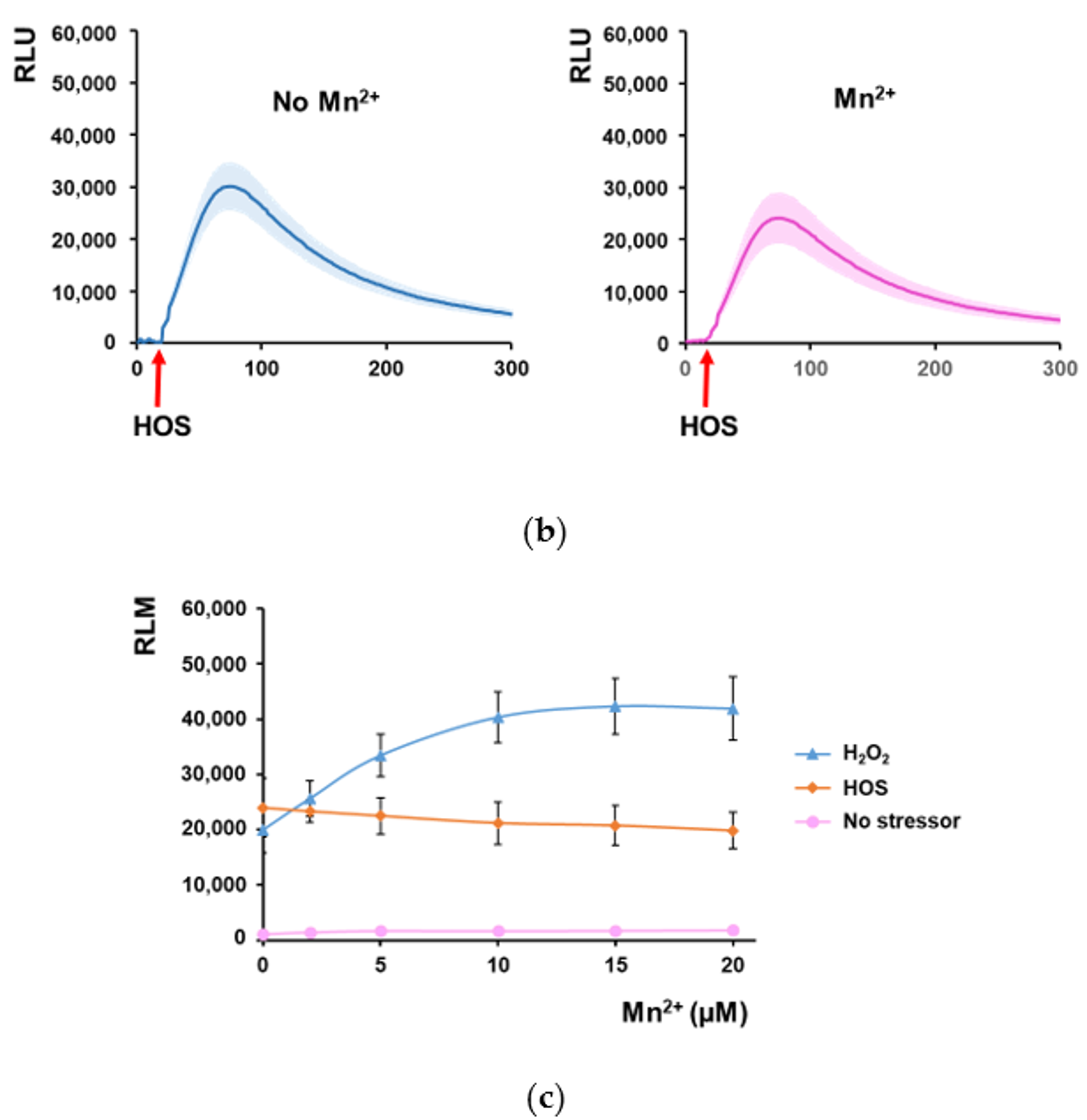
3.2. Mn2+ Potentiates the Increase of [Ca2+]cyt under Oxidative Stress in Strain trpy1Δ/TRPY1

The observation that both EGTA (calcium chelator) and Gd3+ (inhibitor of Ca2+ transport across plasma membrane) augmented the LMeMM-related haploinsufficiency of the trpy1Δ/TRPY1 strain prompted the idea that preventing Ca2+ entry into the cell is deleterious to trpy1Δ/TRPY1, while the observed opposite action of Mn2+ may be the result of Mn2+-related activation of the extant TRPY1 that would compensate the heterozygous loss of TRPY1. To check this possibility, we used cells expressing aequorin, a system suitable for detecting transient modifications in the [Ca2+]cyt [38]. For this purpose, trpy1Δ/TRPY1 cells were transformed with a plasmid harboring the cDNA of the luminescent Ca2+ reporter apo-aequorin under the control of a constitutive promoter, which afforded abundant transgenic protein within the cytosol [40]. The cells expressing apo-aequorin were pre-treated with the cofactor coelenterazine to reconstitute the functional aequorin, and then the cells were exposed to various stimuli directly in the luminometer tube. It was noted that while Mn2+ alone failed to induce any increase in the luminescence of the reconstituted aequorin (data not shown), cell pre-incubation with 10 µM Mn2+ significantly increased the [Ca2+]cyt elevation induced by H2O2 exposure (Figure 3a). Remarkably, pre-incubation with Mn2+ did not augment the cell luminescence when aequorin-expressing trpy1Δ/TRPY1 cells were exposed to hyperosmotic shock (HOS, Figure 3b,c). Surplus Mn2+ reached maximum stimulating activity on trpy1Δ/TRPY1 cells exposed to H2O2 at 10 µM (Figure 3c), a non-toxic concentration to both WT and trpy1 mutants.

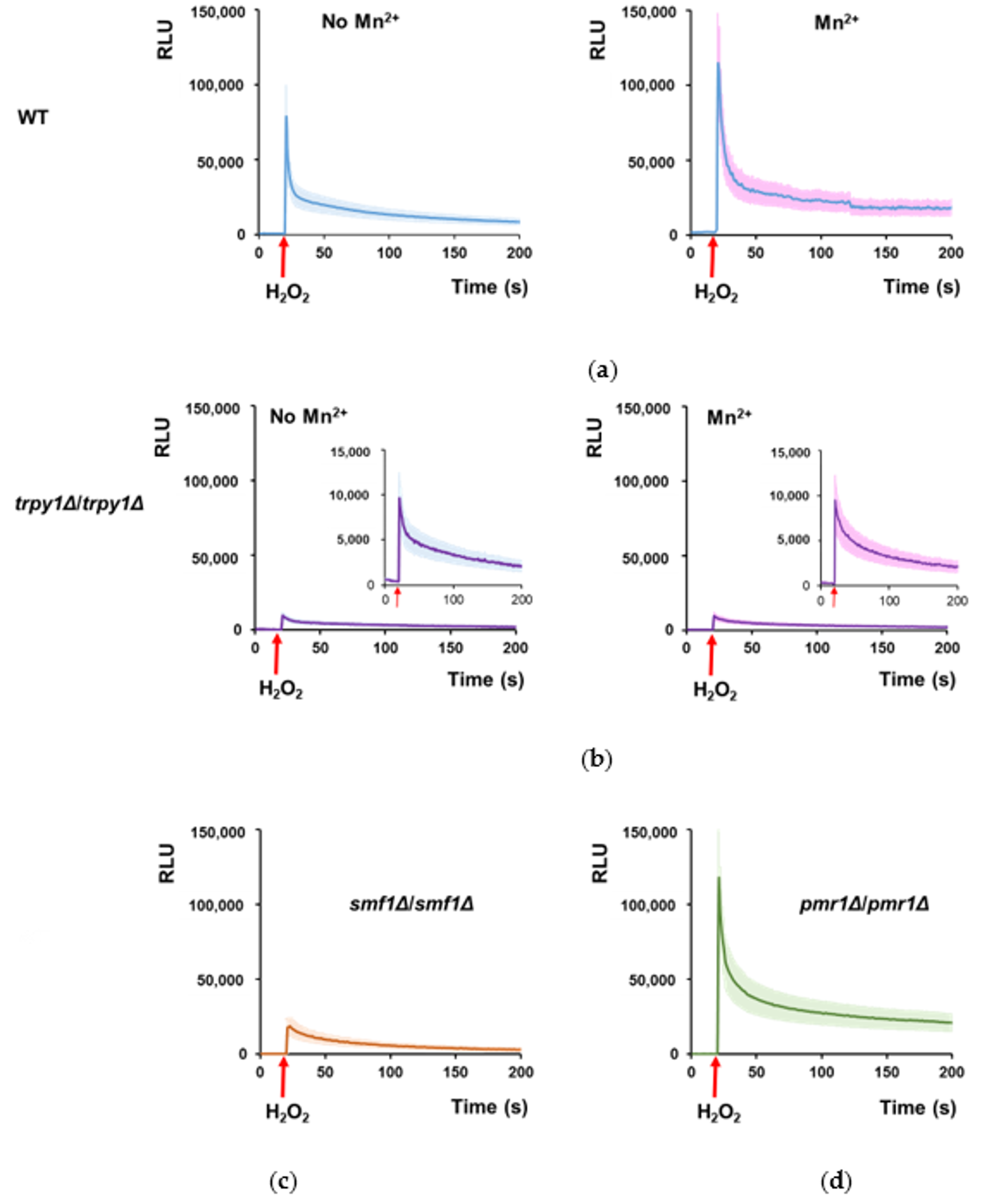
3.3. Mn2+ Stimulates TRPY1 to Release Ca2+ into the Cytosol under H2O2 Stress

Furthermore, we wondered whether Mn2+ influence on elevating [Ca2+]cyt under H2O2 was the result of TRPY1 stimulation by Mn2+. To test this possibility, we determined the effect of Mn2+ on the Ca2+-mediated response to H2O2 of cells completely lacking TRPY1. It was noticed that in WT cells expressing reconstituted aequorin, the H2O2-induced Ca2+-dependent luminescence was significantly increased by cell pre-incubation with 10 µM Mn2+, indicating that in the case of WT cells too, Mn2+ potentiates the Ca2+-dependent response to oxidative stress (Figure 4a). In contrast, homozygous knockout mutant trpy1Δ/trpy1Δ exhibited much lower H2O2-luminescence (Figure 4b, left), which was not altered by pre-incubation with 10 µM Mn2+ (Figure 4b, right). This observation suggested that Mn2+ augments the H2O2-induced [Ca2+]cyt elevation by activating TRPY1, a phenotype clearly absent in the trpy1Δ/trpy1Δ homozygous knockout mutant (Figure 4b).
If Mn2+ were required for TRPY1 activation under oxidative stress, it would be expected that cells with low cytosolic Mn2+ would be less responsive in terms of increasing the [Ca2+]cyt under oxidative stress. Mn2+ is an essential metal which is carried into the yeast cell by the divalent metal ion transporter Smf1, known to have high affinity for Mn2+ [47]. It was noted indeed that the homozygous knockout mutant smf1Δ/smf1Δ expressing reconstituted aequorin exhibited a significantly lower luminescence trace when exposed to H2O2 than WT (Figure 4c). In this line of evidence, the pmr1Δ/pmr1Δ cells expressing reconstituted aequorin responded strongly to H2O2 (in media not supplemented with Mn2+) with a luminescence curve (Figure 4d) which was not significantly different from that obtained from WT cells preincubated with 10 µM Mn2+ (Figure 4a, right). PMR1 encodes the major Golgi/ER membrane P-type ATPase ion pump responsible for transporting Ca2+ and Mn2+ into the Golgi apparatus [48] providing a major route for cellular detoxification of Mn2+ via the secretory pathway vesicles [49]. It was shown that cells knockout for PMR1 gene have the intracellular Mn2+ levels considerably higher than the WT cells [50], a fact that may account for the stronger response of pmr1Δ/pmr1Δ cells (Figure 4d) compared to WT (Figure 4a, left). In this line of evidence, we found that pmr1Δ/pmr1Δ cells had significantly (p < 0.05) more cellular Mn2+ than the WT, while smf1Δ/smf1Δ cell had significantly (p < 0.05) less cellular Mn2+ than the WT (Table 2).
The influence of Mn2+ on the RLM recorded under oxidative stress for various strains which expressed reconstituted aequorin was also determined (Figure 5), revealing that Mn2+ significantly increased the RLM of WT cells exposed to H2O2. RLM determined for trpy1Δ/trpy1Δ was significantly low and was not augmented by Mn2+, indicating the necessity of functional TRPY1 for Mn2+ action. RLM for smf1Δ/smf1Δ cells expressing reconstituted aequorin was also low under H2O2 exposure, indicating that the lack of the high-affinity Mn2+ transporter is associated with cytosolic Mn2+ concentration (Table2) which is too low for an efficient activation of TRPY1. In fact, smf1Δ/smf1Δ attained responses similar to WT only at higher Mn2+ supplementation (Figure 5, grey line), when Mn2+ cell content was high enough (Table 2) for efficient TRPY1 activation. In contrast to smf1Δ/smf1Δ strain, pmr1Δ/pmr1Δ expressing reconstituted aequorin attained high RLM upon H2O2 exposure which was not significantly augmented by surplus Mn2+, suggesting that the intrinsic high level of cytosolic Mn2+ associated with PMR1 knockout [50] is sufficient for attaining efficient activation of TRPY1 (Table2). Moreover, it was noted that when applying Mn2+ concentrations higher than 20 µM the maximum response of pmr1Δ/pmr1Δ to H2O2 started to decline (Figure 5, yellow line) probably due to the hypersensitivity of this strain to Mn2+ [50].

4. Discussion

TRPY1 of S. cerevisiae is a key component in releasing vacuolar Ca2+ into the cytosol for the Ca2+-dependent activation of mechanisms involved in the cell response to hyperosmotic [4] and oxidative stress [19]. Starting from the observation that Mn2+ alleviated the haploinsufficiency exhibited by the heterozygous trpy1Δ/TRPY1 strain in synthetic media deficient in essential metals (LMeMM) we found that Mn2+ differentially stimulated TRPY1 to release Ca2+ from the vacuole under H2O2 exposure, but not under hyperosmotic shock. Mn2+ alone does not induce [Ca2+]cyt elevation—neither under low (0.05–1 mM) nor under high (2–10 mM, lethal) surplus [21, unpublished observations]. The Mn2+ concentrations found to augment the H2O2-induced stimulation of TRPY1 were within the physiological limits (10–50 µM) and far below the concentration that would induce a hyperosmotic shock, explaining why the TRPY1 was not extra stimulated by Mn2+ under hyperosmotic stress (Figure 3b). It was shown that the release of vacuolar Ca2+ via TRPY1 can be stimulated by Ca2+ from outside the cell as well as that released from the vacuole by TRPY1 itself in a positive feedback, a process known as Ca2+-induced Ca2+ release [34,51]. In this regard, Mn2+ surplus could stimulate TRPY1 similarly to Ca2+. Mn2+ is an essential cation which strongly resembles Ca2+ not only in ionic radius but also in its affinity to oxygen-containing ligands, a trait which sometimes makes Mn2+ a good substitute of Ca2+ [52]; this would explain why other essential cations tested (Cu2+, Fe3+, Zn2+) failed to alleviate the haploinsufficiency showed by trpy1Δ/TRPY1 strain. That TRPY1 haploinsufficiency in LMeMM is rescued by Mn2+ can be explained in three ways: (1) the supplemental Mn2+ simply counteracts the deficiency of essential metals of the LMeMM, providing the necessary amount of cations (albeit surrogate in certain cases) that support cell fitness; (2) Mn2+ stimulates TRPY1 activity by increasing the Ca2+ release to the cytososl, and consequently by stimulating other components involved in maintaining the cell fitness; (3) Mn2+ generates reactive oxygen species (ROS) by a Fenton-like reaction augmenting the oxidative stress and indirectly stimulating TRPY1. The fact that surplus Mn2+ augments the TRPY1-related increase in [Ca2+]cyt under oxidative stress clearly correlates with the Mn2+ cytosolic level, as the strain lacking the high-affinity plasma membrane Mn2+ transporter Smf1 exhibited only a modest increase in [Ca2+]cyt under H2O2, when compared with WT (Figure 4c). In this line of evidence, it was shown that a haploid smf1Δ was sensitive to H2O2 [20] probably by not attaining the optimum TRPY1 activation for adaptation to oxidative stress. On the other hand, it had been shown that deletion of PMR1—which leads to increased cytosolic Mn2+—suppresses the sensitivity of superoxide dismutase (SOD) mutants to superoxide-generating drugs due to the Mn2+ capacity to scavenge superoxide ions [50]. In the light of our findings, it is also possible that the high cytosolic Mn2+ in cells devoid of Pmr1 rescue the SOD mutants from ROS attack not due to the scavenger traits of Mn2+, but through TRPY1 activation. Whether Mn2+ rescues the haploinsufficient trpy1Δ/TRPY1 by neutralizing ROS or by directly stimulating TRPY1 are issues to be addressed in the future; nevertheless, the observation that well-known antioxidants such as ascorbate or glutathione did not rescue the trpy1Δ/TRPY1 haploinsufficiency rather supports the latter hypothesis. An open question remains: why is the homozygous trpy1Δ/trpy1Δ apparently more fit than the heterozygous trpy1Δ/TRPY1. The calcium-mediated responses to environmental insults are diverse: depending on the intensity or duration of the [Ca2+]cyt waves, the cell can be led towards adaptation, survival (sometimes with growth arrest or slow growth) or death [19,20,21,23]. The trpy1Δ/trpy1Δ cells are probably more fit because they are never “bothered” by periodic nuisance caused by Ca2+ release from the vacuole; on the other hand, trpy1Δ/TRPY1 cells need to find the right balance in Ca2+ gating while depending on only one gene copy, and sometimes extra help—external Ca2+ carried by Cch1/Mid1, mechanical force [34,51] or even surplus Mn2+—may contribute to find the most suitable path to be followed.

Supplementary Materials

The following are available online. Figure S1: Effect of various substances on the haploinsufficiency of the heterozygous trpy1Δ/TRPY1.

Author Contributions

Conceptualization, I.C.F.; methodology, L.L.R., C.V.P. and I.C.F.; validation, L.L.R., C.V.P. and I.C.F.; formal analysis, I.N. and I.C.F.; investigation, L.L.R., C.V.P. and I.N.; resources, I.C.F.; data curation, L.L.R. and I.C.F.; writing—original draft preparation, I.C.F.; writing—review and editing, I.C.F.; supervision, I.C.F.; project administration, L.L.R.; funding acquisition, I.C.F.

Funding

This research was funded by the EEA Financial Mechanism 2009-2014, grant number 21 SEE/30.06.2014.

Acknowledgments

We thank Enzo Martegani and Renata Tisi (from University of Milano-Bicocca, Milan, Italy) for providing the plasmid pYX212-cytAEQ and Andrei F. Danet for technical support. We thank Aurora D. Neagoe for ICP-MS analysis.

Conflicts of Interest

The authors declare no conflict of interest.

References

  1. Matsumoto, T.K.; Ellsmore, A.J.; Cessna, S.G.; Low, P.S.; Pardo, J.M.; Bressan, R.A.; Hasegawa, P.M. An osmotically induced cytosolic Ca2+ transient activates calcineurin signaling to mediate ion homeostasis and salt tolerance of Saccharomyces cerevisiae. J. Biol. Chem. 2002, 277, 33075–33080. [Google Scholar] [CrossRef] [PubMed]
  2. Batiza, A.F.; Schulz, T.; Masson, P.H. Yeast respond to hypotonic shock with a calcium pulse. J. Biol. Chem. 1996, 271, 23357–23362. [Google Scholar] [CrossRef] [PubMed]
  3. Rigamonti, M.; Groppi, S.; Belotti, F.; Ambrosini, R.; Filippi, G.; Martegani, E.; Tisi, R. Hypotonic stress-induced calcium signaling in Saccharomyces cerevisiae involves TRP-like transporters on the endoplasmic reticulum membrane. Cell Calcium 2015, 57, 57–68. [Google Scholar] [CrossRef] [PubMed]
  4. Denis, V.; Cyert, M.S. Internal Ca2+ release in yeast is triggered by hypertonic shock and mediated by a TRP channel homologue. J. Cell Biol. 2002, 156, 29–34. [Google Scholar] [CrossRef] [PubMed]
  5. Loukin, S.; Zhou, X.; Kung, C. Saimi, Y. A genome-wide survey suggests an osmoprotective role for vacuolar Ca2+ release in cell wall-compromised yeast. FASEB J. 2008, 22, 2405–2415. [Google Scholar] [CrossRef] [PubMed]
  6. Mori, I.C.; Iida, H.; Tsuji, F.I.; Isobe, M.; Uozumi, N.; Muto, S. Salicylic acid induces a cytosolic Ca2+ elevation in yeast. Biosci. Biotechnol. Biochem. 1998, 62, 986–989. [Google Scholar] [CrossRef]
  7. Viladevall, L.; Serrano, R.; Ruiz, A.; Domenech, G.; Giraldo, J.; Barcelo, A.; Arino, J. Characterization of the calcium-mediated response to alkaline stress in Saccharomyces cerevisiae. J. Biol. Chem. 2004, 279, 43614–43624. [Google Scholar] [CrossRef]
  8. Peiter, E.; Fischer, M.; Sidaway, K.; Roberts, S.K.; Sanders, D. The Saccharomyces cerevisiae Ca2+ channel Cch1pMid1p is essential for tolerance to cold stress and iron toxicity. FEBS Lett. 2005, 579, 5697–5703. [Google Scholar] [CrossRef]
  9. Araki, Y.; Wu, H.; Kitagaki, H.; Akao, T.; Takagi, H.; Shimoi, H. Ethanol stress stimulates the Ca2+-mediated calcineurin/Crz1 pathway in Saccharomyces cerevisiae. J. Biosci. Bioeng. 2009, 107, 1–6. [Google Scholar] [CrossRef]
  10. Courchesne, W.E.; Vlasek, C.; Klukovich, R.; Coffee, S. Ethanol induces calcium influx via the Cch1-Mid1 transporter in Saccharomyces cerevisiae. Arch. Microbiol. 2011, 193, 323–334. [Google Scholar] [CrossRef]
  11. Gupta, S.S.; Ton, V.K.; Beaudry, V.; Rulli, S.; Cunningham, K.; Rao, R. Antifungal activity of amiodarone is mediated by disruption of calcium homeostasis. J. Biol. Chem. 2003, 278, 28831–28839. [Google Scholar] [CrossRef] [PubMed]
  12. Rao, A.; Zhang, Y.Q.; Muend, S.; Rao, R. Mechanism of antifungal activity of terpenoid phenols resembles calcium stress and inhibition of the TOR pathway. Antimicrob. Agents Chemother. 2010, 54, 5062–5069. [Google Scholar] [CrossRef]
  13. Hejchman, E.; Ostrowska, K.; Maciejewska, D.; Kossakowski, J.; Courchesne, W.E. Synthesis and antifungal activity of derivatives of 2- and 3-benzofurancarboxylic acids. J. Pharmacol. Exp. Ther. 2012, 343, 380–388. [Google Scholar] [CrossRef]
  14. Roberts, S.K.; McAinsh, M.; Widdicks, L. Cch1p mediates Ca2+ influx to protect Saccharomyces cerevisiae against eugenol toxicity. PLoS ONE 2012, 7, e43989. [Google Scholar] [CrossRef] [PubMed]
  15. Roberts, S.K.; McAinsh, M.; Cantopher, H.; Sandison, S. Calcium dependence of eugenol tolerance and toxicity in Saccharomyces cerevisiae. PLoS ONE 2014, 9, e102712. [Google Scholar] [CrossRef] [PubMed]
  16. Popa, C.V.; Lungu, L.; Cristache, L.F.; Ciuculescu, C.; Danet, A.F.; Farcasanu, I.C. Heat shock, visible light or high calcium augment the cytotoxic effects of Ailanthus altissima (Swingle) leaf extracts against Saccharomyces cerevisiae cells. Nat. Prod. Res. 2015, 29, 1744–1747. [Google Scholar] [CrossRef]
  17. Vilanova, C.; Hueso, A.; Palanca, C.; Marco, G.; Pitarch, M.; Otero, E.; Crespo, J.; Szablowski, J.; Rivera, S.; Domínguez-Escribà, L.; et al. Aequorin-expressing yeast emits light under electric control. J. Biotechnol. 2011, 152, 93–95. [Google Scholar] [CrossRef] [PubMed] [Green Version]
  18. Pinontoan, R.; Krystofova, S.; Kawano, T.; Mori, I.C.; Tsuji, F.I.; Iida, H.; Muto, S. Phenylethylamine induces an increase in cytosolic Ca2+ in yeast. Biosci. Biotechnol. Biosci. 2002, 66, 1069–1074. [Google Scholar] [CrossRef]
  19. Popa, C.V.; Dumitru, I.; Ruta, L.L.; Danet, A.F.; Farcasanu, I.C. Exogenous oxidative stress induces Ca2+ release in the yeast Saccharomyces cerevisiae. FEBS J. 2010, 277, 4027–4038. [Google Scholar] [CrossRef]
  20. Ruta, L.L.; Popa, C.V.; Nicolau, I.; Farcasanu, I.C. Calcium signaling and copper toxicity in Saccharomyces cerevisiae cells. Environ. Sci. Pollut. Res. Int. 2016, 23, 24514–24526. [Google Scholar] [CrossRef]
  21. Ruta, L.L.; Popa, V.C.; Nicolau, I.; Danet, A.F.; Iordache, V.; Neagoe, A.D.; Farcasanu, I.C. Calcium signaling mediates the response to cadmium toxicity in Saccharomyces cerevisiae cells. FEBS Lett. 2014, 588, 3202–3212. [Google Scholar] [CrossRef] [PubMed]
  22. Ene, C.D.; Ruta, L.L.; Nicolau, I.; Popa, C.V.; Iordache, V.; Neagoe, A.D.; Farcasanu, I.C. Interaction between lanthanide ions and Saccharomyces cerevisiae cells. J. Biol. Inorg. Chem. 2015, 20, 1097–1107. [Google Scholar] [CrossRef]
  23. Bootman, M.D.; Berridge, M.J.; Putney, J.W.; Llewelyn Roderick, H. Calcium Signaling; Cold Spring Harbor Laboratory Press: Cold Spring Harbor, NY, USA, 2011; 449p, ISBN 978-0-87969-903-1. [Google Scholar]
  24. Palmer, C.P.; Zhou, X.; Lin, J.; Loukin, S.H.; Kung, C.; Saimi, Y. A TRP homolog in Saccharomyces cerevisiae forms an intracellular Ca2+-permeable channel in the yeast vacuolar membrane. Proc. Natl. Acad. Sci. USA 2001, 98, 7801–7805. [Google Scholar] [CrossRef] [PubMed]
  25. Cunningham, K.W. Acidic calcium stores of Saccharomyces cerevisiae. Cell Calcium 2011, 50, 129–138. [Google Scholar] [CrossRef] [PubMed]
  26. Cunningham, K.W.; Fink, G.R. Calcineurin-dependent growth control in Saccharomyces cerevisiae mutants lacking PMC1, a homolog of plasma membrane Ca2+ ATPases. J. Cell Biol. 1994, 124, 351–363. [Google Scholar] [CrossRef]
  27. Cunningham, K.W.; Fink, G.R. Calcineurin inhibits VCX1-dependent H+/Ca2+ exchange and induces Ca2+ ATPases in Saccharomyces cerevisiae. Mol. Cell Biol. 1996, 16, 2226–2237. [Google Scholar] [CrossRef]
  28. Miseta, A.; Kellermayer, R.; Aiello, D.P.; Fu, L.; Bedwell, D.M. The vacuolar Ca2+/H+ exchanger Vcx1p/Hum1p tightly controls cytosolic Ca2+ levels in S. cerevisiae. FEBS Lett. 1999, 451, 132–136. [Google Scholar] [CrossRef]
  29. Sorin, A.; Rosas, G.; Rao, R. PMR1, a Ca2+-ATPase in yeast Golgi, has properties distinct from sarco/endoplasmic reticulum and plasma membrane calcium pumps. J. Biol. Chem. 1997, 272, 9895–9901. [Google Scholar] [CrossRef]
  30. Strayle, J.; Pozzan, T.; Rudolph, H.K. Steady-state free Ca2+ in the yeast endoplasmic reticulum reaches only 10 mM and is mainly controlled by the secretory pathway pump Pmr1. EMBO J. 1999, 18, 4733–4743. [Google Scholar] [CrossRef]
  31. Hamamoto, S.; Mori, Y.; Yabe, I.; Uozumi, N. In vitro and in vivo characterization of modulation of the vacuolar cation channel TRPY1 from Saccharomyces cerevisiae. FEBS J. 2018, 285, 1146–1161. [Google Scholar] [CrossRef]
  32. Clapham, D.E. TRP channels as cellular sensors. Nature 2003, 426, 517–524. [Google Scholar] [CrossRef] [PubMed]
  33. Zhou, X.L.; Batiza, A.F.; Loukin, S.H.; Palmer, C.P.; Kung, C.; Saimi, Y. The transient receptor potential channel on the yeast vacuole is mechanosensitive. Proc. Natl. Acad. Sci. USA 2003, 100, 7105–7110. [Google Scholar] [CrossRef] [PubMed] [Green Version]
  34. Chang, Y.; Schlenstedt, G.; Flockerzi, V.; Beck, A. Properties of the intracellular transient receptor potential (TRP) channel in yeast, Yvc1. FEBS Lett. 2010, 584, 2028–2032. [Google Scholar] [CrossRef] [PubMed]
  35. Pir, P.; Gutteridge, A.; Wu, J.; Rash, B.; Kell, D.B.; Zhang, N.; Oliver, S.G. The genetic control of growth rate: A systems biology study in yeast. BMC Syst. Biol. 2012, 6, 4. [Google Scholar] [CrossRef] [PubMed]
  36. Brachmann, C.B.; Davies, A.; Cost, G.J.; Caputo, E.; Li, J.; Hieter, P.; Boeke, J.D. Designer deletion strains derived from Saccharomyces cerevisiae S288C: A useful set of strains and plasmids for PCR-mediated gene disruption and other applications. Yeast 1998, 14, 115–132. [Google Scholar] [CrossRef]
  37. Sherman, F. Getting started with yeast. Methods Enzymol. 2002, 350, 3–41. [Google Scholar] [PubMed]
  38. Nakajima-Shimada, J.; Iida, H.; Tsuji, F.I.; Anraku, Y. Monitoring of intracellular calcium in Saccharomyces cerevisiae with an apoaequorine cDNA expression system. Proc. Natl. Acad. Sci. USA 1991, 88, 6878–6882. [Google Scholar] [CrossRef] [PubMed]
  39. Cohen, R.; Engelberg, D. Commonly used Saccharomyces cerevisiae strains (e.g. BY4741, W303) are growth sensitive on synthetic complete medium due to poor leucine uptake. FEMS Microbiol. Lett. 2007, 273, 239–243. [Google Scholar] [CrossRef] [PubMed]
  40. Tisi, R.; Baldassa, S.; Belotti, F.; Martegani, E. Phospholipase C is required for glucose-induced calcium influx in budding yeast. FEBS Lett. 2002, 520, 133–138. [Google Scholar] [CrossRef] [Green Version]
  41. Dohmen, R.J.; Strasser, A.W.M.; Honer, C.B.; Hollenberg, C.P. An efficient transformation procedure enabling long-term storage of competent cells of various yeast genera. Yeast 1991, 7, 691–692. [Google Scholar] [CrossRef]
  42. Ruta, L.L.; Kissen, R.; Nicolau, I.; Neagoe, A.D.; Petrescu, A.J.; Bones, A.M.; Farcasanu, I.C. Heavy metal accumulation by Saccharomyces cerevisiae cells armed with metal binding hexapeptides targeted to the inner face of the plasma membrane. Appl. Microbiol. Biotechnol. 2017, 101, 5749–5763. [Google Scholar] [CrossRef] [PubMed]
  43. Tisi, R.; Martegani, E.; Brandão, R.L. Monitoring yeast intracellular Ca2+ levels using an in vivo bioluminescence assay. Cold Spring Harb. Protoc. 2015, 2015, 210–213. [Google Scholar] [CrossRef] [PubMed]
  44. Petrovic, K.; Pfeifer, M.; Parker, C.N.; Schuierer, S.; Tallarico, J.; Hoepfner, D.; Movva, N.R.; Scheel, G.; Helliwell, S.B. Two low complexity ultra-high throughput methods to identify diverse chemically bioactive molecules using Saccharomyces cerevisiae. Microbiol. Res. 2017, 199, 10–18. [Google Scholar] [CrossRef] [PubMed]
  45. Chandel, A.; Das, K.K.; Bachhawat, A.K. Glutathione depletion activates the yeast vacuolar transient receptor potential channel, Yvc1p, by reversible glutathionylation of specific cysteines. Mol. Biol. Cell 2016, 27, 3913–3925. [Google Scholar] [CrossRef] [PubMed] [Green Version]
  46. John Haynes, W.; Zhou, X.L.; Su, Z.W.; Loukin, S.H.; Saimi, Y.; Kung, C. Indole and other aromatic compounds activate the yeast TRPY1channel. FEBS Lett. 2008, 582, 1514–1518. [Google Scholar] [CrossRef] [PubMed]
  47. Supek, F.; Supekova, L.; Nelson, H.; Nelson, N. A yeast manganese transporter related to the macrophage protein involved in conferring resistance to mycobacteria. Proc. Natl. Acad. Sci. USA 1996, 93, 5105–5110. [Google Scholar] [CrossRef] [PubMed]
  48. Dürr, G.; Strayle, J.; Plemper, R.; Elbs, S.; Klee, S.K.; Catty, P.; Wolf, D.H.; Rudolph, H.K. The medial-Golgi ion pump Pmr1 supplies the yeast secretory pathway with Ca2+ and Mn2+ required for glycosylation, sorting, and endoplasmic reticulum-associated protein degradation. Mol. Biol. Cell 1998, 9, 1149–1162. [Google Scholar] [CrossRef]
  49. Mandal, D.; Woolf, T.B.; Rao, R. Manganese selectivity of Pmr1, the yeast secretory pathway ion pump, is defined by residue Gln783 in transmembrane segment 6. Residue Asp778 is essential for cation transport. J. Biol. Chem. 2000, 275, 23933–23938. [Google Scholar] [CrossRef]
  50. Lapinskas, P.J.; Cunningham, K.W.; Liu, X.F.; Fink, G.R.; Culotta, V.C. Mutations in PMR1 suppress oxidative damage in yeast cells lacking superoxide dismutase. Mol. Cell. Biol. 1995, 15, 1382–1388. [Google Scholar] [CrossRef]
  51. Su, Z.; Zhou, X.; Loukin, S.H.; Saimi, Y.; Kung, C. Mechanical force and cytoplasmic Ca2+ activate yeast TRPY1 in parallel. J. Membr. Biol. 2009, 227, 141–150. [Google Scholar] [CrossRef]
  52. Loukin, S.; Kung, C. Manganese effectively supports yeast cell-cycle progression in place of calcium. J. Cell Biol. 1995, 131, 1025–1037. [Google Scholar] [CrossRef] [PubMed] [Green Version]
Figure 1. Growth of heterozygous trpy1Δ/TRPY1. Isogenic diploid strains WT (BY4743, TRPY1/TRPY1), trpy1Δ/TRPY1 and trpy1Δ/trpy1Δ were shifted at time 0 to (a) minimal medium, MM or (b) minimum medium with low metal content, LMeMM, as described in Materials and Methods section. Growth was determined spectrophotometrically at 600 nm as the ratio between OD600 at time t and OD600 at time 0 for each individual strain.
Cells 08 00079 g001
Figure 2. Haploinsufficiency of heterozygous trpy1Δ/TRPY1. (a) Mn2+ alleviates trpy1Δ/TRPY1 haploinsufficiency in LMeMM. Diploid strains were shifted to LMeMM (final OD600 = 0.05) and grown for 2 h before MnCl2 (10 µM), EGTA (0.5 mM) or GdCl3 (50 µM) were added from concentrated stocks. Cell growth was recorded spectrophotometrically 24 h after the addition of the chemicals and normalized (%) to the growth of WT in the absence of chemicals. One sample t test compared WT in the absence of chemicals. * p < 0.05; ** p < 0.01. (b) Relative abundance (RA) of TRPY1 mRNA in WT (TRPY1/TRPY1) and heterozygous trpy1Δ/TRPY1. Analysis of transcript abundance was done by qRT-PCR as described in Materials and Methods section. Expression of TRPY1 mRNA was normalized to the relative expression of ACT1 in each sample. (c) Heterozygous trpy1Δ/TRPY1 exhibits haploinsufficiency in LMeMM, but not in normal MM. Cells in log phase (OD600 ~ 0.5) were 10-fold serially diluted (left to right) in a 48-well microtiter plate and stamped on the agar plates using a pin replicator (approximately 4 µL/spot). Plates were photographed after 3 days’ incubation at 30 °C.
Cells 08 00079 g002
Figure 3. In trpy1Δ/TRPY1 cells, Mn2+ pre-incubation stimulates the increase of [Ca2+]cyt under H2O2 stress but not under hyperosmotic shock. Heterozygous trpy1Δ/TRPY1 cells expressing reconstituted aequorin were pre-grown in LMeMM-Ura without or with 10 µM surplus Mn2+ before being exposed to (a) oxidative stress (5 mM H2O2) or (b) hyperosmotic stress (HOS, 0.8 M NaCl). [Ca2+]cyt-dependent aequorin luminescence was recorded on samples of approximately 107 cells. The arrow indicates the time when the stressor was added. The luminescence traces represent the mean ± SEM from 3 independent transformants. RLU, relative luminescence units. (c) Effect of pre-incubation with various concentrations of Mn2+ on the maximum intensity of the Ca2+-dependent aequorin luminescence recorded under 5 mM H2O2, or 0.8 M NaCl (HOS). The relative maximum luminescence (RLM) was calculated as described in Materials and Methods. Bars represent the mean ± SEM from 3 independent transformants.
Cells 08 00079 g003aCells 08 00079 g003b
Figure 4. Variation of [Ca2+]cyt in response to H2O2 exposure depends on Mn2+ cellular load. Diploid cells expressing reconstituted aequorin were pre-grown in LMeMM-Ura with or without 10 µM surplus Mn2+ before being exposed to oxidative stress (5 mM H2O2) as described in Materials and Methods. [Ca2+]cyt-dependent aequorin luminescence was recorded on samples of approximately 107 cells. The arrow indicates the time when the stressor (H2O2) was added. The luminescence traces represent the mean ± SEM from 3 independent transformants. (a) WT (BY4743). (b) trpy1Δ/trpy1Δ; insets: same representation at lower scale. (c) smf1Δ/smf1Δ. (d) pmr1Δ/pmr1Δ. RLU, relative luminescence units.
Cells 08 00079 g004
Figure 5. Effect of Mn2+ pre-incubation on the maximum intensity of the Ca2+-dependent aequorin luminescence recorded for various strains under H2O2 stress. The relative maximum luminescence (RLM) was calculated as described in Materials and Methods. Diploid cells expressing reconstituted aequorin were pre-grown in LMeMM-Ura with or without surplus Mn2+ before being exposed to oxidative stress (5 mM H2O2) directly in the luminometer tube. [Ca2+]cyt-dependent aequorin luminescence was recorded on samples of approximately 107 cells. Bars represent the mean ± SEM from 3 independent transformants.
Cells 08 00079 g005
Table 1. Substances screened for the capacity to alleviate trpy1Δ/TRPY1 haploinsufficiency in LMeMM.
Substance Tested 1Concentration RangeEffect on trpy1Δ/TRPY1 Haploinsufficiency
CaCl22–10 mMNo
CuCl20.5–50 µMNo
FeCl31–50 µMNo
MnCl21–50 µMAlleviation
ZnCl21–50 µMNo
EGTA0.1–2 mMAugmentation
GdCl30.1–1 mMAugmentation
Ascorbate1–10 mMNo
Glutathione 21–10 mMNo
Indole 31–10 mMNo
1 The quantitative results are presented in Supplementary Files, Figure S1. 2 Glutathione depletion leads to TRPY1 activation [45]. 3 Indole activates TRPY1 under hyperosmotic stress [46].
Table 2. Manganese content (pmoles/mg cell protein) of diploid yeast cells grown in LMeMM supplemented or not with Mn2+.
StrainSurplus Mn2+
010 µM50 µM
WT0.12 ± 0.120.64 ± 0.20.92 ± 0.3
trpy1Δ/TRPY10.11 ± 0.140.72 ± 0.10.98 ± 0.2
trpy1Δ/trpy1Δ0.12 ± 0.20.7 ± 0.20.84 ± 0.2
smf1Δ/smf1Δ0.01 ± 0.0140.1 ± 0.20.72 ± 0.2
pmr1Δ/pmr1Δ0.7 ± 0.220.84 ± 0.28.4 ± 1.2

