Article

Genome-Wide Identification and Characterization of Xyloglucan Endotransglycosylase/Hydrolase in Ananas comosus during Development

1
Agricultural Big-Data Research Center and College of Plant Protection, Shandong Agricultural University, Taian 271018, China
2
Center for Genomics and Biotechnology, Haixia Institute of Science and Technology, Fujian Provincial Key Laboratory of Haixia Applied Plant Systems Biology, Fujian Agriculture and Forestry University, Fuzhou 350002, China
*
Author to whom correspondence should be addressed.
Genes 2019, 10(7), 537; https://doi.org/10.3390/genes10070537
Submission received: 26 April 2019 / Revised: 5 July 2019 / Accepted: 10 July 2019 / Published: 16 July 2019
(This article belongs to the Special Issue Genetics, genomics, and evolution of CAM photosynthesis)

Abstract

:
Xyloglucan endotransglycosylase/hydrolase (XTH) is a cell-wall-modifying enzyme participating in diverse cell morphogenetic processes and adaptation to stress. In this study, 48 XTH genes were identified from two pineapple (Ananas comosus) cultivars (‘F153’ and ‘MD2’) and designated Ac(F153)XTH1 to -24 and Ac(MD2)XTH1 to -24 based on their orthology with Arabidopsis thaliana genes. Endoglucanase family 16 members were identified in addition to XTHs of glycoside hydrolase family 16. Phylogenetic analysis clustered the XTHs into three major groups (Group I/II, III and Ancestral Group) and Group III was subdivided into Group IIIA and Group IIIB. Similar gene structure and motif number were observed within a group. Two highly conserved domains, glycosyl hydrolase family 16 (GH16-XET) and xyloglucan endotransglycosylase C-terminus (C-XET), were detected by multiple sequences alignment of all XTHs. Segmental replication were detected in the two cultivars, with only the paralogous pair Ac(F153)XTH7-Ac(F153)XTH18 presented in ‘F153’ prior to genomic expansion. Transcriptomic analysis indicated that XTHs were involved in the regulation of fruit ripening and crassulacean acid metabolism with tissue specificity and quantitative real-time PCR analysis suggested that Ac(MD2)XTH18 was involved in root growth. The results enhance our understanding of XTHs in the plant kingdom and provide a basis for further studies of functional diversity in A. comosus.

1. Introduction

Pineapple (Ananas comosus), a member of the Bromeliaceae family, is cultivated widely in tropical and subtropical regions and is renowned for its nutritional and medicinal values [1]. Given its status as a herbaceous perennial monocotyledon with classical crassulacean acid metabolism (CAM), the pineapple genome was sequenced in 2015 as a model plant [2]. The genome of the cultivar ‘MD2’, which is the predominant pineapple cultivar grown worldwide by virtue of its fruit flesh flavor and commercial value, and the cultivar ‘F153’ has been sequenced [3]. Pineapple genome sequencing has provided valuable information for further research for crop improvement [4,5].
Xyloglucan endotransglycosylase/hydrolase (XTH) participates in diverse physiological processes, especially cell elongation and stress resistance [6]. XTH is a cell-wall-modifying enzyme encoded by a multigene, which belongs to a subfamily of the glycoside hydrolase family 16 (GH16) [7,8]. Generally, XTH proteins perform two diverse catalytic activities: xyloglucan endohydrolase (XEH) and xyloglucan endotransglycosylase (XET) [9,10]. XET activity is characterized by the no-hydrolytic cleavage and rejoining of xyloglucan (XyG) chains, whereas XTH activity irreversibly cleaves hydrolytic XyG chains to promote the expansion, degradation, remediation, and morphogenesis of the cell wall [6,9]. To date, the majority of identified XTH proteins show XET activity [6]. XTH proteins share the conserved glycosyl hydrolase family 16 domain (GH16_XET) with a specific EXDXE motif likely to be the catalytic site for both XET and XEH activities. XTH proteins also contain the significant xyloglucan endotransglycosylase C-terminus domain (C-XET), which distinguishes XTH proteins from other GH16 subfamilies [6,11,12]. The XTH gene subfamily was originally divided into three major groups, of which Group III was subdivided into subgroups IIIA and IIIB [10,13]. With the expansion of XTH observations, more detailed clade and subclade groupings (Group I/II, IIIA, IIIB, and an Ancestral Group) were applied to different species on the basis of sequence similarity [10,11]. Interestingly, XTH genes predominantly display XET activity in Group I/ II and IIIB, whereas XEH activity is characteristic of Group IIIA [13]. In Arabidopsis thaliana, Group IIIA genes are ubiquitous and dispensable in plant growth [14].
An increasing number of XTHs have been identified using publicly available datasets [15]. For instance, 33, 29, 25, 56, 61, and 24 potential XTH members have been defined in A. thaliana, Oryza sativa, Solanum lycopersicum, Nicotiana tabacum, Glycine max, and Hordeum vulgare, respectively [5,16,17,18,19,20]. In A. thaliana, XTHs show distinct and diverse organ-specific expression patterns. Five genes (AtXTH-1, -21, -22, -30, and -33) were expressed preferentially in green siliques, whereas two genes (AtXTH-24 and -32) were expressed primarily in stems [16]. In O. sativa, seven root-specific XTH genes (OsXTH1, -2, -4, -13, -15, -16, and -25) were predominantly expressed in roots of 14-d-old seedling, whereas no expression was detected in other tissues [17]. XTH proteins modify the complex structure of lignin and cellulose in a variety of developmental processes, such as root formation, flower generation, and fruit softening [5,21,22]. Numerous XTHs have been detected in root elongation zones and trichoblasts of diverse vascular plants [23]. Compared with other tissues in Dianthus caryophyllus, DcXTH2 and DcXTH3 transcripts were markedly accumulated in petals and showed XET activity during flower opening stages [22]. SlXTH5 detected at multiple stages during tomato fruit’s expansion and displayed XET activity in concordance with results for apple, kiwifruit, and strawberry [24,25]. Thus, XTH proteins play a critical role during fruit growth and ripening. In addition, XTHs show abnormal expression under abiotic stress. The XTH gene CaXTH3 of Capsicum annuum showed a high expression level in transgenic A. thaliana lines and conferred enhanced salt and drought tolerance [26]. AtXTH14, -15, and -31 showed remarkably low expression under aluminum treatment in A. thaliana roots, especially AtXTH31 [27]. In contrast, MtXTH3 was strongly up-regulated by a higher NaCl concentration in Medicago truncatula [28]. Thus, XTHs possess considerable spatial and temporal specificity, and respond to a variety of environmental stimuli for adaptation cell wall enzyme activities.
Additional potential members of the XTH gene family can be identified by genome-wide analysis using the published genome resources in silico. Systematic identification and characterization of XTHs in pineapple have received only limited attention to date [15]. In this study, we conducted a comprehensive analysis, including the classification, evolutionary relationships, and expression patterns of XTHs to determine whether pineapple XTHs participate in the CAM pathway and perform important functions in the leaf and root. This research provides novel insights into the functional characteristics of XTHs in pineapple and will facilitate further study of the regulatory mechanism at different developmental stages.

2. Materials and Methods

2.1. Dataset Compilation and Identification of the XTH Gene Family

The protein sequences of A. comosus (L.) Merry two cultivars: ‘F153’ and ‘MD2’ were downloaded from the National Center for Biotechnology Information database (accession numbers are GCA-001540865.1 and GCA-001661175.1, respectively) [29]. The protein sequences for A. thaliana were obtained from The Arabidopsis Information Resource [30]. The protein ID of XTHs in A. thaliana as reference sequences collected from a former publication [9].
Two Hidden Markov Models (HMMs) were downloaded from Pfam and used to scan XTH sequences using the default E-value in HMMER 3.0 [31,32]. The HMM profile established for AtXTH genes was used to search for candidate XTH family members in pineapple. Potential XTH protein sequences were further detected using BLASp. Candidate genes were filtered and identified using the Conserved Domain Search Service (CD-Search) [33]. The length, molecular weight (MW), and theoretical isoelectric point (PI) of XTHs were characterized with ExPASy [34]. Their single peptide and subcellular localization were predicted by SignalP 4.1 and Plant-mPLoc [35,36].

2.2. Multiple Sequence Alignment and Identification of Motifs

A multiple sequence alignment of the candidate pineapple XTH proteins was generated and obvious features of the sequences were displayed using ClustalX2 with the default options [37]. In addition, motifs were detected using Multiple Expectation Maximization for Motif Elicitation with a motif width of 6–50 residues and a maximum of 10 motifs [38].

2.3. Phylogenetic Tree Analysis and Nomenclature of XTHs

Multiple sequence alignments of the XTH proteins from pineapple and A. thaliana were generated using the ClustalW with default parameters (pairwise alignment with gap opening penalty of 10 and gap extension penalty of 0.1, multiple alignment parameters with gap opening penalty of 10, a gap extension penalty of 0.2, and delay divergent sequences set at 30%) [37]. A phylogenetic tree was constructed using the neighbor-joining algorithm with 1000 bootstrap replications using MEGA7 [39]. All members were numbered sequentially and designated as Ac(F153)XTH or Ac(MD2)XTH based on the genotypic origin of the gene [40].

2.4. Gene Structure Analysis

The gene structures of the candidate XTHs were predicted using the online software Gene Structure Display Server [41]. The complex figure including the phylogenetic tree, gene structure, and motif distribution was arranged correctly using TBtools [42].

2.5. Chromosomal Distribution and Gene Duplication

All Ac(F153)XTH genes were localized on chromosomes based on their physical coordinates using MapChart and Perl script [43]. The Multiple Collinearity Scan toolkit was employed to identify syntenic blocks in the two pineapple genome assemblies and Circos software was used to depict the collinearity relationships [44,45].

2.6. Calculation of Ka/Ks

The synonymous substitution (Ka) and non-synonymous substitution (Ks) of XTH pairs were calculated using Ka/Ks Calculator 2.0 by the Nei and Gojobori (NG) method [46]. Fisher’s exact test was applied to confirm the validity of the ratio. To estimate the selection pressure, a ratio of Ka/Ks greater than one, equal to one, and less than one displayed positive selection, neutral selection, and purity selection respectively. The divergence time (T) was calculated as T = Ks/(2 × 6.1 × 10−9) × 10−6 million years ago (Mya).

2.7. Transcriptome Analysis and Gene Expansion Patterns

Pineapple transcriptome data were downloaded from the Pineapple Genomics Database consisting of data from fruit at five developmental stages, the green leaf tip, and white leaf base tissues. The stages Fruit1 to Fruit 5 were ordered chronologically and represented the entire period of fruit ripening. Leaves were harvested from plants of the cultivar ‘MD2’ at 13 time points over a 24-h period [47]. Daytime is from 6.a.m to 4.p.m. and nighttime is from 4.p.m. to 6.a.m. in this study. The white leaf base comprised non-photosynthetic tissues sensitive to sunlight and the green leaf tip represented photosynthetic tissues. The transcript levels were visualized using R software.

2.8. Experimental Validation of XTH Transcript Levels by RT-qPCR Analysis

The green leaf tip, white leaf base, and root of ‘MD2’ plants were sampled at the Fujian Agriculture and Forestry University. All samples were rapidly frozen in liquid nitrogen then stored at −80 °C. Total RNA was extracted from each sample using a RNA extraction kit (Roche Diagnostics GmbH, Mannheim, Germany), then stored at −80 °C until further analysis. Quantitative real time PCR (RT-qPCR) analysis was performed using the BIO-RAD CFX Connect Real-time PCR Detection System with three biological replicates per sample. The transcript levels were analyzed using the 2−ΔΔCt method and means ± standard errors (SE). The primer sequences used are presented in Table S1.

3. Results

3.1. Identification and Characteristics of XTHs

Twenty-eight potential XTH family members were detected with the two HMM models of the GH16_XET domain with a β-jelly-roll topology and XET_C domain, and 24 proteins were detected by BLASTp in the ‘F153’ reference genome. Candidates were confirmed to contain two highly conserved domains using CD-Search. Redundant proteins were manually removed on account of the absence of characteristic amino acid residues in the C-terminal region or a lack of the conserved motif ExDxE [12]. Finally, 24 candidates were identified in ‘F153’ and were designated Ac(F153)XTH1 to -24 based on homology with the classification of A. thaliana (Figure 1). In the same manner, 24 proteins were identified in ‘MD2’ and designated Ac(MD2)XTH1 to -24 in ‘MD2’. In addition, EG16 homologs, which are related to XTH members of the GH16 family but lack the XET_C extension, were identified in cultivars ‘F153’ and ‘MD2’ [48].
The candidate XTHs exhibited similar properties, including length, molecular weight (MW), isoelectric point (PI), and signal peptide (SP) (Table 1). Comparison of the length of the 48 XTH proteins revealed that Ac(MD2)XTH5 was the largest protein with 575 amino acids, and the smallest one was Ac(MD2)XTH1 with 230 amino acids. The MW ranged from 25.99 kDa to 64.58 kDa and corresponded with the protein length. Owing to the complex amino acid polarity, the PI ranged from 4.68 to 9.53. Subcellular localization prediction revealed that each XTH was localized to the cell wall, and 14 (7/7, ‘F153’/‘MD2’) proteins were targeted in both the cell wall and cytoplasm. The majority of the proteins contained signal peptide sequences (Figure S1).

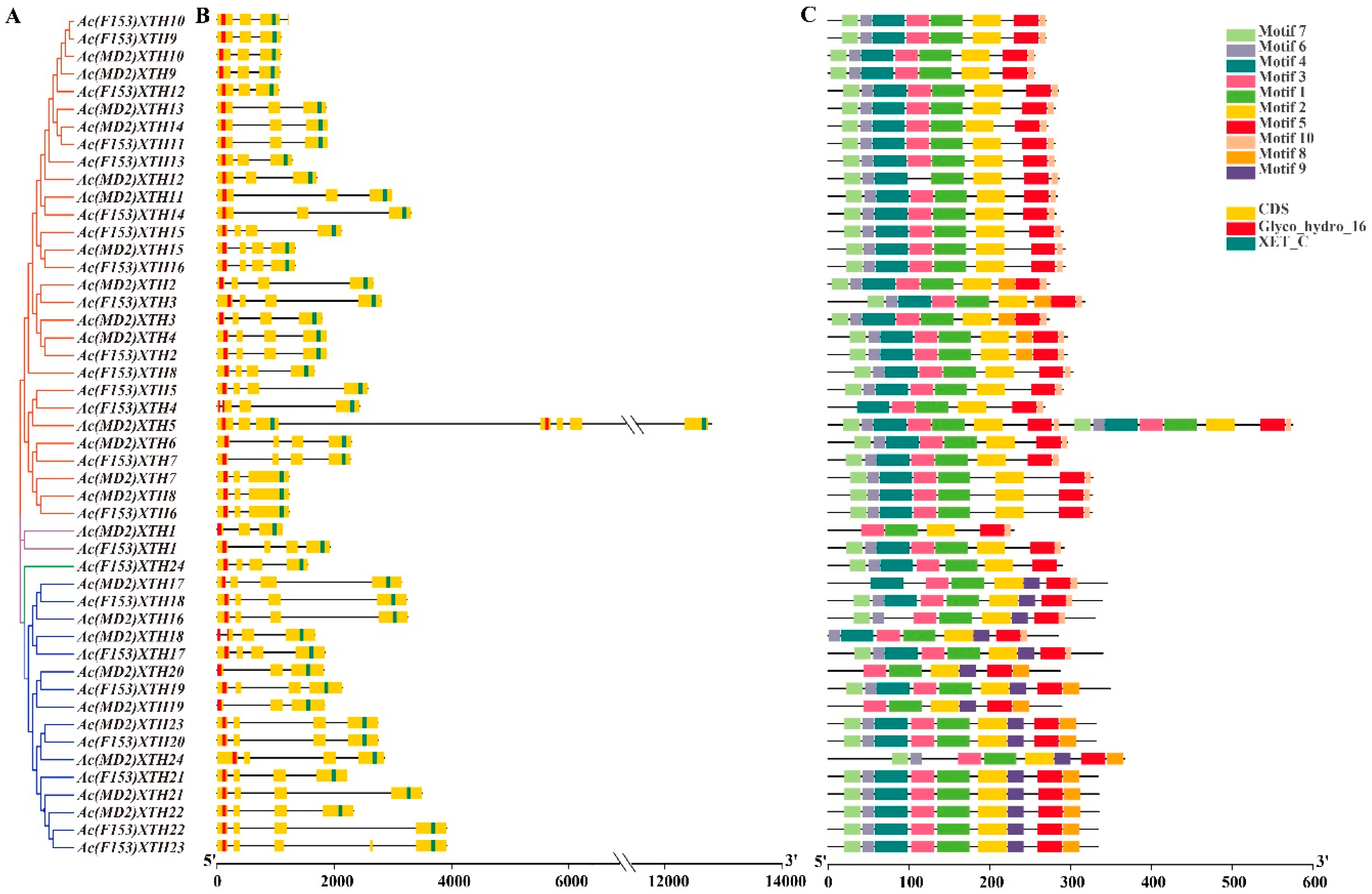
3.2. Phylogenetic Analysis and Classification of XTH Proteins

A phylogenetic tree representing the relationships among 81 (24/24/33) XTHs of ‘F153’, ‘MD2’ and A. thaliana was constructed. The XTHs were clustered into three main groups (Group I/II, III, and Ancestral Group) (Figure 1). The majority of groups comprised the same number of XTHs in ‘F153’ and ‘MD2’. Group III consisted of nine Ac(MD2)XTH, eight Ac(F153)XTH, and seven AtXTH genes, and was further subdivided into Group IIIA and Group IIIB, as described previously by Baumann et al. [10]. Group IIIA contained only Ac(F153)XTH24. The Ancestral Group contained the fewest members group, namely Ac(F153)XTH1 and Ac(MD2)XTH1, whereas Group I/II contained the most members in each cultivar.

3.3. Sequence Alignment of XTHs

Multiple sequence alignment showed that Ac(MD2)XTHs and Ac(F153)XTHs genes shared a highly conserved domain containing the motif ExDxE (Figure S1) [12]. One potential N-linked glycosylation site sharing N(T)-L(K/R/V/T/I)-S(T)-G(N) was located close to catalytic residues in 30 XTHs [49]. In addition, a conserved DWATRGG motif and Cys residues were located in the C-terminal region.

3.4. Gene Structure Analysis and the Pattern of the Motif in XTHs

Highly structural similarity was evident in each phylogenetic group of XTHs. The exon number varied from three to seven in ‘F153’ and ‘MD2’ (Figure S2). Three or four exons were observed in majority of XTHs. Group I/II comprised three or four exons, except that Ac(MD2)XTH5 contained seven 7 exons. Fourteen of the 17 genes in Group III possessed four exons, whereas Ac(MD2)XTH19, Ac(MD2)XTH20 and Ac(F153)XTH23 comprised three, three and five exons, respectively. Ac(MD2)XTH1 with three exons and Ac(F153)XTH1 with four exons were placed in Group IV. Ac(MD2)XTH5 with seven exons was longer than all other members (Figure S2), because it possessed four highly conserved domains: two GH16_XET and two C-XET domains (Figure 2).
Almost all XTHs within the same group shared common motifs (Figure 2). Motif1-2 and motif5 were highly conserved in all XTHs. Motif1-7 and motif 10 were present in Group I/II except that Ac(MD2)XTH12 lacked motif3 and five genes contained the additional motif8. Ac(MD2)XTH5 contained 16 motifs in accordance with its structure. Ac(F153)XTH24 shared motif1-7 in Group IIIA and motifs without rules (6–9 motifs) were presented in Group IIIB. In Group IV, Ac(MD2)XTH1 possessed the fewest motifs and the motif composition of Ac(F153)XTH1 was identical to that of Group I/II.

3.5. Chromosomal Distribution and Syntenic Analysis of XTH Genes

Twenty-four Ac(F153)XTHs genes were unevenly distributed in 14 of 25 linkage groups (LG) in ‘F153’. LG06 contained the most Ac(F153)XTHs genes (Figure 3A). Three linkage groups, consisting of LG03, LG14, and LG15, shared more than two XTH members, whereas only one gene was discovered on each of the remaining chromosomes. All Ac(MD2)XTH genes were mapped on different scaffolds in ‘MD2’ (Figure S4).
To analyze duplication events, we detected syntenic blocks using MCScanX in ‘F153’ and ‘MD2’. Thirteen collinear pairs, including 14 Ac(F153)XTHs and 12 Ac(MD2)XTHs genes, were discovered through synteny analysis (Figure 3B, Table S2). Almost all pairs represented segmental duplication without tandem duplication and were placed in the same phylogenetic group. The gene pair of Ac(F153)XTH6-Ac(MD2)XTH8 was placed in Group I/II and Ac(F153)XTH20-Ac(MD2)XTH23 was placed in Group IIIB, which indicated that XTHs generated multiple segmental duplications have occurred during XTH diversification in pineapple.

3.6. Ka/Ks Analysis of XTH Genes

To assess whether XTH genes had been subject to Darwinian selection, all paralogous XTH pairs were used to calculate Ka/Ks values (Table S3). Thirteen XTH paralogous with high similarity were detected with the Ka/Ks < 1, which suggested that XTHs had undergone strong purifying selection in ‘F153’ and ‘MD2’. Most paralogous showed a relatively recent duplication time with an average value of about 1.7 Mya, except that Ac(F153)XTH18-Ac(MD2)XTH18 and Ac(F153)XTH7-Ac(F153)XTH18 diverged about 388 and 340 Mya, respectively (Table 2).

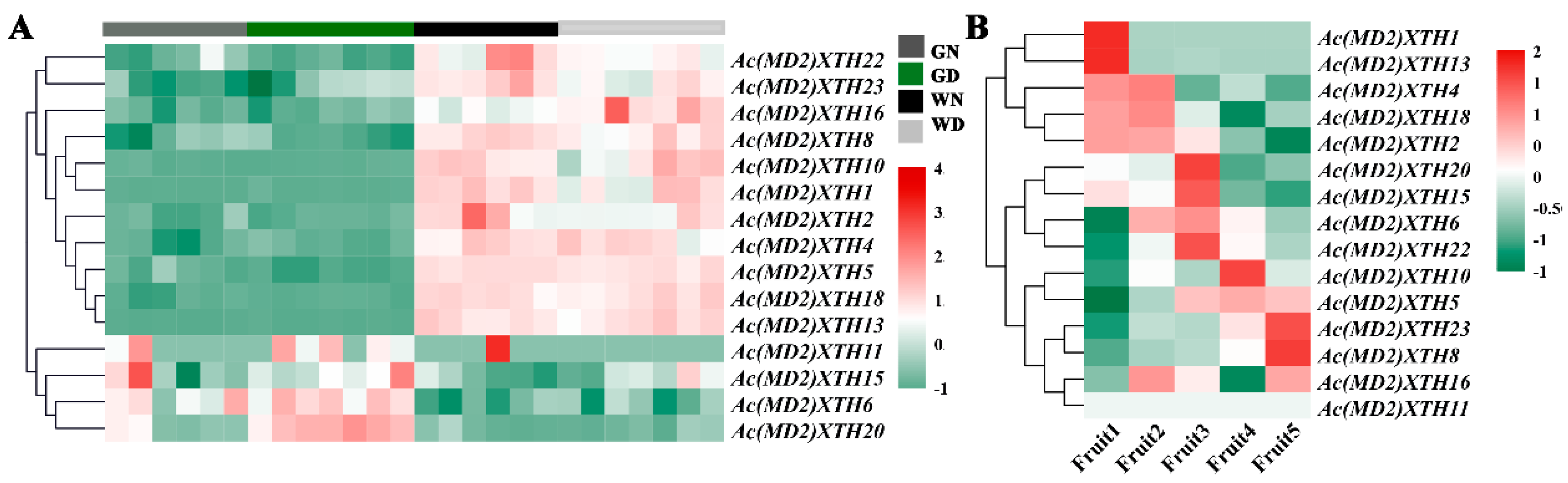
3.7. Differential Expression Profiles of XTHs during Development

Fifteen XTH genes were simultaneously induced in the green leaf tip and white leaf base, which were used to investigate diurnal expression patterns (Figure 4A). The majority of XTHs expressed in the green leaf tip showed transcript levels lower than those detected in the white leaf base or no transcripts were detected. Four genes (27%), comprising Ac(MD2)XTH6, 11, 15, and 20 exhibited higher transcript levels. Interestingly, several XTHs may show a diurnal expression pattern owing to the contrasting transcript levels detected during day and night in each tissues.
Fifteen Ac(MD2)XTHs genes, which were divided into three diverse categories, were detected during fruit ripening (Figure 4B). Five genes showed higher transcript levels at the Fruit1 and/or Fruit2 stages than that at advanced stages of ripening. Ac(MD2)XTH6, 15, 20, and 22 showed normal transcript levels or no significant difference among immature stages, but showed a high transcript level in Fruit3 and subsequently a low transcript level during fruit maturation stages. Six XTHs were showed a low transcript levels at the onset of maturity and subsequently were highly expressed. Notably Ac(MD2)XTH8 and Ac(MD2)XTH23, for which expression increased more than 6-fold and 11-fold, respectively, were indicated to have important roles in fruit ripening.

3.8. RT-qPCR Analysis of XTH Genes in Root and Leaf

Quantitative real-time PCR analysis was used to analyze the expression patterns of 11 selected Ac(MD2)XTH genes in the root and leaf of ‘MD2’. Eight of the 11 Ac(MD2)XTH genes showed differential expression patterns in different tissues, and expression of the remaining three Ac(MD2)XTH genes was not detected (Figure 5, Figure S3). Ac(MD2)XTH15 and Ac(MD2)XTH18 were detected simultaneously in three tissues. Ac(MD2)XTH15 showed the highest relative expression level in the green leaf tip, sequentially in the root, and finally in the white leaf base. Ac(MD2)XTH18 was significantly more highly expressed in the root compared with the other tissues (Figure 5A). Four genes (Ac(MD2)XTH11, 15, 18, and 20) showed higher relative expression levels in the green leaf than those in the white leaf base consistent with the corresponding transcriptome except for Ac(MD2)XTH18 (Figure 5B, Figure S3). Six genes were observed to show a low relative expression level in the white leaf base, of which the highest was Ac(MD2)2XTH13 about 0.12 (Figure S3). The relative expression level of four genes ranged from 0.13 to 49.5 in the green leaf tip. In the root, Ac(MD2)XTH18 showed the highest relative expression level of about 532.5.

4. Discussion

As a vital cell-wall-modifying enzyme, XTH is implicated in the incorporation or hydrolysis of XyG to regulate cell wall remodeling and degradation. In this study, 48 (24/24) non-redundant XTHs were identified in pineapple ‘F153’ and ‘MD2’, respectively. The number of XTHs identified is fewer than that in a previous study because of the redundancy of annotation [15]. The number of XTHs detected in each cultivar is less than that reported in most vascular plant species except Actinidia deliciosa (14 AdXTHs) and Malus sieversii (11 MsXTHs) [24]. The difference may be attributed to fewer duplication events of pineapple compared with most other plant species. Gene duplication provides a source for gene functional diversification and contributes to amplification of the number of members of a gene family. For example, an ancient whole-genome duplication (WGD) with massive gene duplication were occurred in O. sativa and the genome of A. thaliana primarily experienced at least four large-scale duplication events [50,51]. Therefore, the limited number of XTHs in pineapple might reflect that the genome diverged prior to the Poales-specific ρWGD event [2].
The groups retrieved in the phylogenetic tree contained a similar number of XTH genes from each cultivar, which indicated that XTH genes were relatively conserved before the differentiation of cultivars in pineapple. Although differing considerably in MW, PI, and length, the XTHs contained relatively conserved motifs and gene structure in each group, indicating that XTHs of the same group may perform similar functions. In addition, unique traits of XTHs were confirmed in pineapple. For instance, the catalytic sites, D(N)E(L/I/V)DF(Y)EFLG as motif B, were highly conserved in all XTH proteins, especially three absolutely conserved catalytic residues (ExDxE) [12], suggesting that the highly conserved motif could have a similar and conserved function in plant XTHs [13,52]. All proteins were localized to the cell wall, and several proteins were also localized to the cytoplasm. These results indicated that XTH proteins could easily access the substrate and promote catalytic reactions. A signal peptide of about 20–25 nucleotides adjacent to the start codon was present in the majority of XTHs, which suggested that this short sequence of hydrophobic amino acids may be responsible for transmembrane transport of XTH proteins through interaction with subcellular organelle membranes [53]. Motif 9 distinguished Group IIIB from the other groups, and thus might play an important role in the distinctive function of Group IIIB XTHs. Several Cys residues present in the C-XET domain may contribute to the structural stabilization through disulfide bonds [12,54]. Therefore, the conservation of features of the domains, motifs, and gene structure strongly supported the reliability of phylogenetic classification and especially the close evolutionary relationship of two cultivars.
Group I/II was the biggest group of XTHs, whereas Group IIIA was the smallest group in each cultivar, which was consistent with other species [10]. The XEH activity of Group IIIA evolved as a-gain-of function in ancestral XET [6,10]. However, no XTH from ‘MD2’ was placed in Group IIIA. Archetypal XTH from Tropaeolum majus, Vigna angularis, and A. thaliana in Group IIIA only showed demonstrable XEH activity [14,55,56]. Hence, we speculated that Ac(F153)XTH24 in Group IIIA might possess XEH activity.
Twenty-six of the 48 XTHs identified were involved in the segmental duplication events without tandem duplication, which implied that segmental replication events played an important role in XTH gene family expansion and greatly drove the evolution of XTHs in pineapple. Tandem duplication events are undoubtedly crucial for genome expansion [57]. Each gene of the Ac(F153)XTH7-Ac(F153)XTH8 pair was linked and connected with Ac(MD2)XTH18. This result indicated that the three XTHs were highly conserved and experienced translocation or segmental replication in ‘F153’ first, and subsequently evolved in parallel in the two cultivars [58]. The paralogous pairs Ac(F153)XTH7-Ac(F153)XTH18 and Ac(F153)XTH18-Ac(MD2)XTH18, which were indicated to have diverged more than 300 Mya, arose before divergence of the Poales [59,60,61]. All XTHs were indicated to have undergone strong purifying selection suggesting that XTHs have evolved slowly between cultivars [62].
Expression of XTHs varies under exposure to stress and shows tissue, organ, and temporal specificity [4,22,25]. The contrasting expression patterns in the green leaf tip and white leaf base was suggestive of tissue specificity of XTHs in pineapple. Pineapple fixes carbon dioxide nocturnally by activity of CAM-related enzymes and is stored rapidly as malic acid in the vacuole [2,63]. Ac(MD2)XTH15, Ac(MD2)XTH11, Ac(MD2)XTH6, and Ac(MD2)XTH20 showed higher expression levels in photosynthetic tissues and may be putative CAM-related genes that enhance the efficiency of water use by the CAM pathway. Several XTHs participated in fruit ripening, such as in apple and kiwifruit [24]. Together with fruit ripening, continuously fluctuating expression patterns with gradual decline or accumulated increment indicated that XTHs showed polygenic interaction and temporal expression during fruit development stages. In addition, the RT-qPCR results were highly consistent with the transcriptomic data. The results revealed that the expression pattern of Ac(MD2)XTH15 differed from that of Ac(MD2)XTH18 in three tissues consistent with placement of the two genes in different phylogenetic groups. An extremely high expression level of Ac(MD2)XTH18 in the root at noon suggested this gene may be involved in root growth during photosynthesis. These results indicated that several XTHs are involved in photosynthesis with tissue specificity as putative CAM-related enzymes.

Supplementary Materials

The following are available online at https://www.mdpi.com/2073-4425/10/7/537/s1, Supplement File 1: Figure S1: Multiple amino acid sequences alignment of XTH proteins in ‘F153’ and ‘MD2’. The XTHs protein sequences were multi-aligned using the ClustalW2 program. Two typical motifs and signal peptides are indicated in black line and letters. The arrow indicates the N-linked glycosylation site. Supplement File 2: Figure S2: Gene structure of XTHs in ‘F153’ and ‘MD2’. Supplement File 3: Figure S3: The relative expression level in the green leaf tip and white leaf base of ‘MD2’ by RT-qPCR. WB and GT represented white leaf base and the green leaf tip. (A) The expression levels of Ac(MD2)XTH18 and Ac(MD2)XTH15 in two tissues; (B) Different expression profiles of genes in the white leaf base. Supplement File 4: Figure S4: The position of Ac(MD2)XTHs on scaffold. Supplement File 5: Table S1: Primer sequence used in RT-qPCR. Supplement File 6: Table S2. The position information of XTHs in ‘F153’ and ‘MD2’ Supplement File 7: Table S3. The ratio of Ka/Ks in XTH gene pairs based on p-value < 0.05. Supplement File 8: 48 protein sequences in ‘F153’ and ‘MD2’. Supplement File 9: The FASTA format file of XTHs’ multiple sequence alignment. Supplement File 10: The FASTA format file of EG16’s multiple sequence alignment. Supplement File 11: The result file of EG16’s domain analysis by CD-search.

Author Contributions

Conceptualization, Q.L. and L.Y.; data curation, L.Y.; formal analysis, Q.L., R.Z., F.G., and Y.C.; funding acquisition, L.Y.; investigation, C.Y. and Q.J.; methodology, Q.L.; resources, H.L.; software, Q.L.; supervision, L.Y.; writing—original draft, Q.L.; Writing—review & editing, Q.L., L.Y., H.L, and X.W.

Funding

This work was supported by the Foundation of Shandong Province Modern Agricultural Technology System Innovation Team (SDAIT-25-02 to L.Y.).

Conflicts of Interest

The authors declare no conflict of interest.

References

  1. Detoni, A.M.; Carvalho, S.L.C.; Hoshino, A.T.; Pastorio, M.A.; Schmidt, M.A.H.; Menezes Junior, A.O.; Androcioli, H.G. First report of Chrysodeixis includens (Walker, [1858]) (Lepidotera: Noctuidae) Injurious to Pineapple (Ananas comosus L.) (Bromeliaceae) in Brazil. Braz. J. Biol. 2018, 78, 796–798. [Google Scholar] [CrossRef] [PubMed]
  2. Ming, R.; VanBuren, R.; Wai, C.M.; Tang, H.; Schatz, M.C.; Bowers, J.E.; Lyons, E.; Wang, M.L.; Chen, J.; Biggers, E.; et al. The pineapple genome and the evolution of CAM photosynthesis. Nat. Genet. 2015, 47, 1435–1442. [Google Scholar] [CrossRef] [PubMed] [Green Version]
  3. Žemlička, L.; Fodran, P.; Kolek, E.; Prónayová, N. Analysis of natural aroma and flavor of MD2 pineapple variety (Ananas comosus [L.] Merr.). Acta Chim. Slovaca 2013, 6, 123–128. [Google Scholar] [CrossRef]
  4. Iurlaro, A.; De Caroli, M.; Sabella, E.; De Pascali, M.; Rampino, P.; De Bellis, L.; Perrotta, C.; Dalessandro, G.; Piro, G.; Fry, S.C.; et al. Drought and Heat Differentially Affect XTH Expression and XET Activity and Action in 3-Day-Old Seedlings of Durum Wheat Cultivars with Different Stress Susceptibility. Front. Plant Sci. 2016, 7, 1686. [Google Scholar] [CrossRef] [PubMed]
  5. Miedes, E.; Lorences, E.P. Xyloglucan endotransglucosylase/hydrolases (XTHs) during tomato fruit growth and ripening. J. Plant Physiol. 2009, 166, 489–498. [Google Scholar] [CrossRef] [PubMed]
  6. Eklof, J.M.; Brumer, H. The XTH gene family: An update on enzyme structure, function, and phylogeny in xyloglucan remodeling. Plant Physiol. 2010, 153, 456–466. [Google Scholar] [CrossRef] [PubMed]
  7. Fry, S.C.; Smith, R.C.; Renwick, K.F.; Martin, D.J.; Hodge, S.K.; Matthews, K.J. Xyloglucan endotransglycosylase, a new wall-loosening enzyme activity from plants. Biochem. J. 1992, 282, 821–828. [Google Scholar] [CrossRef] [PubMed] [Green Version]
  8. Strohmeier, M.; Hrmova, M.; Fischer, M.; Harvey, A.J.; Fincher, G.B.; Pleiss, J. Molecular modeling of family GH16 glycoside hydrolases: Potential roles for xyloglucan transglucosylases/hydrolases in cell wall modification in the poaceae. Protein Sci. 2004, 13, 3200–3213. [Google Scholar] [CrossRef] [PubMed]
  9. Rose, J.K.; Braam, J.; Fry, S.C.; Nishitani, K. The XTH family of enzymes involved in xyloglucan endotransglucosylation and endohydrolysis: Current perspectives and a new unifying nomenclature. Plant Cell Physiol. 2002, 43, 1421–1435. [Google Scholar] [CrossRef]
  10. Baumann, M.J.; Eklof, J.M.; Michel, G.; Kallas, A.M.; Teeri, T.T.; Czjzek, M.; Brumer, H., 3rd. Structural evidence for the evolution of xyloglucanase activity from xyloglucan endo-transglycosylases: Biological implications for cell wall metabolism. Plant Cell 2007, 19, 1947–1963. [Google Scholar] [CrossRef]
  11. Michailidis, G.; Argiriou, A.; Darzentas, N.; Tsaftaris, A. Analysis of xyloglucan endotransglycosylase/hydrolase (XTH) genes from allotetraploid (Gossypium hirsutum) cotton and its diploid progenitors expressed during fiber elongation. J. Plant Physiol. 2009, 166, 403–416. [Google Scholar] [CrossRef] [PubMed]
  12. Johansson, P.; Denman, S.; Brumer, H.; Kallas, A.M.; Henriksson, H.; Bergfors, T.; Teeri, T.T.; Jones, T.A. Crystallization and preliminary X-ray analysis of a xyloglucan endotransglycosylase from Populus tremula x tremuloides. Acta Crystallogr. D Biol. Crystallogr. 2003, 59, 535–537. [Google Scholar] [CrossRef] [PubMed]
  13. Campbell, P.; Braam, J. Xyloglucan endotransglycosylases: Diversity of genes, enzymes and potential wall-modifying functions. Trends Plant Sci. 1999, 4, 361–366. [Google Scholar] [CrossRef]
  14. Kaewthai, N.; Gendre, D.; Eklof, J.M.; Ibatullin, F.M.; Ezcurra, I.; Bhalerao, R.P.; Brumer, H. Group III-A XTH genes of Arabidopsis encode predominant xyloglucan endohydrolases that are dispensable for normal growth. Plant Physiol. 2013, 161, 440–454. [Google Scholar] [CrossRef] [PubMed]
  15. Behar, H.; Graham, S.W.; Brumer, H. Comprehensive cross-genome survey and phylogeny of glycoside hydrolase family 16 members reveals the evolutionary origin of EG16 and XTH proteins in plant lineages. Plant J. 2018, 95, 1114–1128. [Google Scholar] [CrossRef] [PubMed]
  16. Yokoyama, R.; Nishitani, K. A comprehensive expression analysis of all members of a gene family encoding cell-wall enzymes allowed us to predict cis-regulatory regions involved in cell-wall construction in specific organs of Arabidopsis. Plant Cell Physiol. 2001, 42, 1025–1033. [Google Scholar] [CrossRef] [PubMed]
  17. Yokoyama, R.; Rose, J.K.; Nishitani, K. A surprising diversity and abundance of xyloglucan endotransglucosylase/hydrolases in rice. Classification and expression analysis. Plant Physiol. 2004, 134, 1088–1099. [Google Scholar] [CrossRef] [PubMed]
  18. Wang, M.; Xu, Z.; Ding, A.; Kong, Y. Genome-Wide Identification and Expression Profiling Analysis of the Xyloglucan Endotransglucosylase/Hydrolase Gene Family in Tobacco (Nicotiana tabacum L.). Genes 2018, 9, 273. [Google Scholar] [CrossRef] [PubMed]
  19. Song, L.; Valliyodan, B.; Prince, S.; Wan, J.; Nguyen, H.T. Characterization of the XTH Gene Family: New Insight to the Roles in Soybean Flooding Tolerance. Int. J. Mol. Sci. 2018, 19, 2705. [Google Scholar] [CrossRef] [PubMed]
  20. Fu, M.M.; Liu, C.; Wu, F. Genome-Wide Identification, Characterization and Expression Analysis of Xyloglucan Endotransglucosylase/Hydrolase Genes Family in Barley (Hordeum vulgare). Molecules 2019, 24, 1935. [Google Scholar] [CrossRef]
  21. Thompson, J.E.; Fry, S.C. Restructuring of wall-bound xyloglucan by transglycosylation in living plant cells. Plant J. 2001, 26, 23–34. [Google Scholar] [CrossRef] [PubMed]
  22. Harada, T.; Torii, Y.; Morita, S.; Onodera, R.; Hara, Y.; Yokoyama, R.; Nishitani, K.; Satoh, S. Cloning, characterization, and expression of xyloglucan endotransglucosylase/hydrolase and expansin genes associated with petal growth and development during carnation flower opening. J. Exp. Bot. 2011, 62, 815–823. [Google Scholar] [CrossRef] [PubMed]
  23. Vissenberg, K.; Van Sandt, V.; Fry, S.C.; Verbelen, J.P. Xyloglucan endotransglucosylase action is high in the root elongation zone and in the trichoblasts of all vascular plants from Selaginella to Zea mays. J. Exp. Bot. 2003, 54, 335–344. [Google Scholar] [CrossRef] [PubMed]
  24. Atkinson, R.G.; Johnston, S.L.; Yauk, Y.-K.; Sharma, N.N.; Schröder, R. Analysis of xyloglucan endotransglucosylase/hydrolase (XTH) gene families in kiwifruit and apple. Postharvest Biol. Technol. 2009, 51, 149–157. [Google Scholar] [CrossRef]
  25. Opazo, M.C.; Figueroa, C.R.; Henriquez, J.; Herrera, R.; Bruno, C.; Valenzuela, P.D.; Moya-Leon, M.A. Characterization of two divergent cDNAs encoding xyloglucan endotransglycosylase/hydrolase (XTH) expressed in Fragaria chiloensis fruit. Plant Sci. 2010, 179, 479–488. [Google Scholar] [CrossRef] [PubMed]
  26. Cho, S.K.; Kim, J.E.; Park, J.A.; Eom, T.J.; Kim, W.T. Constitutive expression of abiotic stress-inducible hot pepper CaXTH3, which encodes a xyloglucan endotransglucosylase/hydrolase homolog, improves drought and salt tolerance in transgenic Arabidopsis plants. FEBS Lett. 2006, 580, 3136–3144. [Google Scholar] [CrossRef] [PubMed]
  27. Zhu, X.F.; Shi, Y.Z.; Lei, G.J.; Fry, S.C.; Zhang, B.C.; Zhou, Y.H.; Braam, J.; Jiang, T.; Xu, X.Y.; Mao, C.Z.; et al. XTH31, encoding an in vitro XEH/XET-active enzyme, regulates aluminum sensitivity by modulating in vivo XET action, cell wall xyloglucan content, and aluminum binding capacity in Arabidopsis. Plant Cell 2012, 24, 4731–4747. [Google Scholar] [CrossRef] [PubMed]
  28. Xuan, Y.; Zhou, Z.S.; Li, H.B.; Yang, Z.M. Identification of a group of XTHs genes responding to heavy metal mercury, salinity and drought stresses in Medicago truncatula. Ecotoxicol. Environ. Saf. 2016, 132, 153–163. [Google Scholar] [CrossRef]
  29. Sayers, E.W.; Agarwala, R.; Bolton, E.E.; Brister, J.R.; Canese, K.; Clark, K.; Connor, R.; Fiorini, N.; Funk, K.; Hefferon, T.; et al. Database resources of the National Center for Biotechnology Information. Nucleic Acids Res. 2019, 47, D23–D28. [Google Scholar] [CrossRef]
  30. Lamesch, P.; Berardini, T.Z.; Li, D.; Swarbreck, D.; Wilks, C.; Sasidharan, R.; Muller, R.; Dreher, K.; Alexander, D.L.; Garcia-Hernandez, M.; et al. The Arabidopsis Information Resource (TAIR): Improved gene annotation and new tools. Nucleic Acids Res. 2012, 40, D1202–D1210. [Google Scholar] [CrossRef]
  31. Potter, S.C.; Luciani, A.; Eddy, S.R.; Park, Y.; Lopez, R.; Finn, R.D. HMMER web server: 2018 update. Nucleic Acids Res. 2018, 46, W200–W204. [Google Scholar] [CrossRef] [PubMed] [Green Version]
  32. El-Gebali, S.; Mistry, J.; Bateman, A.; Eddy, S.R.; Luciani, A.; Potter, S.C.; Qureshi, M.; Richardson, L.J.; Salazar, G.A.; Smart, A.; et al. The Pfam protein families database in 2019. Nucleic Acids Res. 2019, 47, D427–D432. [Google Scholar] [CrossRef] [PubMed]
  33. Marchler-Bauer, A.; Bryant, S.H. CD-Search: Protein domain annotations on the fly. Nucleic Acids Res. 2004, 32, W327–W331. [Google Scholar] [CrossRef] [PubMed]
  34. Wilkins, M.R.; Gasteiger, E.; Bairoch, A.; Sanchez, J.C.; Williams, K.L.; Appel, R.D.; Hochstrasser, D.F. Protein identification and analysis tools in the ExPASy server. Methods Mol. Biol. 1999, 112, 531–552. [Google Scholar] [PubMed]
  35. Petersen, T.N.; Brunak, S.; von Heijne, G.; Nielsen, H. SignalP 4.0: Discriminating signal peptides from transmembrane regions. Nat. Methods 2011, 8, 785–786. [Google Scholar] [CrossRef] [PubMed]
  36. Chou, K.-C.; Shen, H.-B. Cell-PLoc 2.0: An improved package of web-servers for predicting subcellular localization of proteins in various organisms. Nat. Sci. 2010, 2, 1090–1103. [Google Scholar] [CrossRef]
  37. Larkin, M.A.; Blackshields, G.; Brown, N.P.; Chenna, R.; McGettigan, P.A.; McWilliam, H.; Valentin, F.; Wallace, I.M.; Wilm, A.; Lopez, R.; et al. Clustal W and Clustal X version 2.0. Bioinformatics 2007, 23, 2947–2948. [Google Scholar] [CrossRef] [Green Version]
  38. Bailey, T.L.; Johnson, J.; Grant, C.E.; Noble, W.S. The MEME Suite. Nucleic Acids Res. 2015, 43, W39–W49. [Google Scholar] [CrossRef] [PubMed] [Green Version]
  39. Kumar, S.; Stecher, G.; Tamura, K. MEGA7: Molecular Evolutionary Genetics Analysis Version 7.0 for Bigger Datasets. Mol. Biol. Evol. 2016, 33, 1870–1874. [Google Scholar] [CrossRef] [Green Version]
  40. Henrissat, B.; Teeri, T.T.; Warren, R.A.J. A scheme for designating enzymes that hydrolyse the polysaccharides in the cell walls of plants. FEBS Lett. 1998, 425, 352–354. [Google Scholar] [CrossRef]
  41. Hu, B.; Jin, J.; Guo, A.Y.; Zhang, H.; Luo, J.; Gao, G. GSDS 2.0: An upgraded gene feature visualization server. Bioinformatics 2015, 31, 1296–1297. [Google Scholar] [CrossRef] [PubMed]
  42. Chen, C.; Xia, R.; Chen, H.; He, Y. TBtools, a Toolkit for Biologists integrating various biological data handling tools with a user-friendly interface. BioRxiv 2018, 289660. [Google Scholar]
  43. Voorrips, R.E. MapChart: Software for the graphical presentation of linkage maps and QTLs. J. Hered. 2002, 93, 77–78. [Google Scholar] [CrossRef] [PubMed]
  44. Wang, Y.; Tang, H.; Debarry, J.D.; Tan, X.; Li, J.; Wang, X.; Lee, T.H.; Jin, H.; Marler, B.; Guo, H.; et al. MCScanX: A toolkit for detection and evolutionary analysis of gene synteny and collinearity. Nucleic Acids Res. 2012, 40, e49. [Google Scholar] [CrossRef] [PubMed]
  45. Krzywinski, M.; Schein, J.; Birol, I.; Connors, J.; Gascoyne, R.; Horsman, D.; Jones, S.J.; Marra, M.A. Circos: An information aesthetic for comparative genomics. Genome Res. 2009, 19, 1639–1645. [Google Scholar] [CrossRef] [Green Version]
  46. Wang, D.; Zhang, Y.; Zhang, Z.; Zhu, J.; Yu, J. KaKs_Calculator 2.0: A toolkit incorporating γ-series methods and sliding window strategies. Genom. Proteom. Bioinform. 2010, 8, 77–80. [Google Scholar] [CrossRef]
  47. Xu, H.; Yu, Q.; Shi, Y.; Hua, X.; Tang, H.; Yang, L.; Ming, R.; Zhang, J. PGD: Pineapple Genomics Database. Hortic. Res. 2018, 5, 66. [Google Scholar] [CrossRef] [Green Version]
  48. McGregor, N.; Yin, V.; Tung, C.C.; Van Petegem, F.; Brumer, H. Crystallographic insight into the evolutionary origins of xyloglucan endotransglycosylases and endohydrolases. Plant J. 2017, 89, 651–670. [Google Scholar] [CrossRef]
  49. Kallas, A.M.; Piens, K.; Denman, S.E.; Henriksson, H.; Faldt, J.; Johansson, P.; Brumer, H.; Teeri, T.T. Enzymatic properties of native and deglycosylated hybrid aspen (Populus tremulaxtremuloides) xyloglucan endotransglycosylase 16A expressed in Pichia pastoris. Biochem. J. 2005, 390, 105–113. [Google Scholar] [CrossRef]
  50. Thiel, T.; Graner, A.; Waugh, R.; Grosse, I.; Close, T.J.; Stein, N. Evidence and evolutionary analysis of ancient whole-genome duplication in barley predating the divergence from rice. BMC Evol. Biol. 2009, 9, 209. [Google Scholar] [CrossRef]
  51. Vision, T.J.; Brown, D.G.; Tanksley, S.D. The origins of genomic duplications in Arabidopsis. Science 2000, 290, 2114–2117. [Google Scholar] [CrossRef] [PubMed]
  52. Mark, P.; Baumann, M.J.; Eklof, J.M.; Gullfot, F.; Michel, G.; Kallas, A.M.; Teeri, T.T.; Brumer, H.; Czjzek, M. Analysis of nasturtium TmNXG1 complexes by crystallography and molecular dynamics provides detailed insight into substrate recognition by family GH16 xyloglucan endo-transglycosylases and endo-hydrolases. Proteins 2009, 75, 820–836. [Google Scholar] [CrossRef] [PubMed]
  53. Weihofen, A.; Binns, K.; Lemberg, M.K.; Ashman, K.; Martoglio, B. Identification of signal peptide peptidase, a presenilin-type aspartic protease. Science 2002, 296, 2215–2218. [Google Scholar] [CrossRef]
  54. Saura-Valls, M.; Faure, R.; Ragas, S.; Piens, K.; Brumer, H.; Teeri, T.T.; Cottaz, S.; Driguez, H.; Planas, A. Kinetic analysis using low-molecular mass xyloglucan oligosaccharides defines the catalytic mechanism of a Populus xyloglucan endotransglycosylase. Biochem. J. 2006, 395, 99–106. [Google Scholar] [CrossRef] [PubMed]
  55. Fanutti, C.; Gidley, M.J.; Reid, J.S. Action of a pure xyloglucan endo-transglycosylase (formerly called xyloglucan-specific endo-(1-4)-β-D-glucanase) from the cotyledons of germinated nasturtium seeds. Plant J. 1993, 3, 691–700. [Google Scholar] [CrossRef] [PubMed]
  56. Tabuchi, A.; Mori, H.; Kamisaka, S.; Hoson, T. A new type of endo-xyloglucan transferase devoted to xyloglucan hydrolysis in the cell wall of azuki bean epicotyls. Plant Cell Physiol. 2001, 42, 154–161. [Google Scholar] [CrossRef] [PubMed]
  57. Marburger, S.; Alexandrou, M.A.; Taggart, J.B.; Creer, S.; Carvalho, G.; Oliveira, C.; Taylor, M.I. Whole genome duplication and transposable element proliferation drive genome expansion in Corydoradinae catfishes. Proc. Biol. Sci. 2018, 285, 20172732. [Google Scholar] [CrossRef] [Green Version]
  58. Flavell, R.B.; O’Dell, M.; Hutchinson, J. Nucleotide sequence organization in plant chromosomes and evidence for sequence translocation during evolution. Cold Spring Harb. Symp. Quant. Biol. 1981, 45, 501–508. [Google Scholar] [CrossRef] [PubMed]
  59. Bremer, K. Gondwanan evolution of the grass alliance of families (Poales). Evolution 2002, 56, 1374–1387. [Google Scholar] [CrossRef]
  60. Linder, H.; Rudall, P. Evolutionary History of Poales. Annu. Rev. Ecol. Evol. Syst. 2005, 36, 107–124. [Google Scholar] [CrossRef]
  61. Puranik, S.; Sahu, P.P.; Mandal, S.N.; B, V.S.; Parida, S.K.; Prasad, M. Comprehensive genome-wide survey, genomic constitution and expression profiling of the NAC transcription factor family in foxtail millet (Setaria italica L.). PLoS ONE 2013, 8, e64594. [Google Scholar] [CrossRef] [PubMed]
  62. Benderoth, M.; Textor, S.; Windsor, A.J.; Mitchell-Olds, T.; Gershenzon, J.; Kroymann, J. Positive selection driving diversification in plant secondary metabolism. Proc. Natl. Acad. Sci. USA 2006, 103, 9118–9123. [Google Scholar] [CrossRef] [PubMed] [Green Version]
  63. Rainha, N.; Medeiros, V.P.; Camara, M.; Faustino, H.; Leite, J.P.; Barreto Mdo, C.; Cruz, C.; Pacheco, C.A.; Ponte, D.; Bernardes da Silva, A. Plasticity of crassulacean acid metabolism at subtropical latitudes: A pineapple case study. Physiol. Plant. 2016, 156, 29–39. [Google Scholar] [CrossRef] [PubMed]
Figure 1. The classification of XTH genes in ‘F153’, ‘MD2’ and A. thaliana. Four colorful braches with red, green, blue, and purple were showed Group I/II, IIIA, IIIB and Ancestral Group, respectively. Circles of different color represented the kinds of species (‘MD2’ with blue circles, ‘F153’ with purple circles and A. thaliana with black circles).
Genes 10 00537 g001
Figure 2. Phylogenetic relationships, gene structure, and motifs distribution of XTHs. (A) The phylogenetic tree was highlighted by different colors with Group I/II, IIIA, IIIB and Ancestral Group; (B) Gene structure and conserved domains. Yellow boxes and black lines represented exons and introns, respectively. The conserved domains were highlighted by red and blue strips; (C) The motif distribution. Motif1-10 in different colorful boxes.
Genes 10 00537 g002
Figure 3. The chromosome distribution and synteny analysis of XTHs in pineapple. (A) There were 24 Ac(F153)XTHs on each chromosome in ‘F153’. Each pillar represented a chromosome and the scale bar was set in mega base (Mb). The gene names were shown on each chromosome with red; (B) Syntenic relationships among ‘F153’ and ‘MD2’. The links represented different gene replications across cultivars or chromosomes. ‘F153’ marked by the purple arcs and ‘MD2’ marked by the orange arcs.
Genes 10 00537 g003
Figure 4. The expression patterns during development in ‘MD2’. (A) The expression in the green leaf tip and white leaf base during thirteen time points over a 24-h period. GN was displayed green leaf tip during nighttime, GD was displayed green leaf tip during daytime, WN was displayed white leaf base during nighttime, WD was displayed white leaf base during daytime; (B) The expression levels during fruit development. Fruit1, Fruit 2 and Fruit 3 were indicated immature stages, Fruit 4 and Fruit5 displayed maturity stages.
Genes 10 00537 g004
Figure 5. The relative expression level in different tissues of ‘MD2’ by RT-qPCR. WB, GT, and R represented white leaf base, the green leaf tip, and root, respectively. (A) The expression levels in the green leaf tip, white leaf base, and (or) root; (B) Expression levels of several Ac(MD2)XTH genes in the green leaf tip and root.
Genes 10 00537 g005
Table 1. The physicochemical properties of XTHs from ‘F153’ and ‘MD2’.
NameTranscript IDLengthMW (kDa)PISPCatalytic SiteSubcellular Localization
Ac(F153)XTH1XP_020091206.129232.785.3520DELDFEFLGCell wall
Ac(F153)XTH2XP_020111283.129634.175.9820DEIDFEFLGCell wall Cytoplasm
Ac(F153)XTH3XP_020087864.131837.297.59DEIDFEFLGCell wall Cytoplasm
Ac(F153)XTH4XP_020102739.126830.715.96DELDFEFLGCell wall
Ac(F153)XTH5XP_020102738.129133.336.0624DELDFEFLGCell wall
Ac(F153)XTH6XP_020109096.132735.985.0724NEFDFEFLGCell wall
Ac(F153)XTH7XP_020084286.128531.655.5327DEVDFEFLGCell wall
Ac(F153)XTH8XP_020104828.130335.628.81DEIDFEFLGCell wall
Ac(F153)XTH9XP_020089756.127030.566.120DEIDFEFLGCell wall Cytoplasm
Ac(F153)XTH10XP_020090359.127030.636.4320DEIDFEFLGCell wall Cytoplasm
Ac(F153)XTH11XP_020110218.128131.544.6921DEIDFEFLGCell wall Cytoplasm
Ac(F153)XTH12XP_020102740.128531.584.7524DEIDFEFLGCell wall Cytoplasm
Ac(F153)XTH13XP_020091231.128131.465.7123DEIDFEFLGCell wall Cytoplasm
Ac(F153)XTH14XP_020090869.128232.155.4125DEVDFEFLGCell wall
Ac(F153)XTH15XP_020097886.129133.334.826DEIDYEFLGCell wall
Ac(F153)XTH16XP_020100605.129332.899.0420NEVDFEFLGCell wall
Ac(F153)XTH17XP_020106929.134038.925.9528DELDFEFLGCell wall
Ac(F153)XTH18XP_020104936.133938.686.5626DELDFEFLGCell wall
Ac(F153)XTH19XP_020092864.134939.418.6923DELDFEFLGCell wall
Ac(F153)XTH20XP_020094226.133137.685.7920DELDFEFLGCell wall
Ac(F153)XTH21XP_020085280.133437.635.9319DELDFEFLGCell wall
Ac(F153)XTH22XP_020085278.133437.655.9319DELDFEFLGCell wall
Ac(F153)XTH23XP_020085279.133437.655.9319DELDFEFLGCell wall
Ac(F153)XTH24XP_020112380.129033.239.5318DEVDIEFLGCell wall
Ac(MD2)XTH1OAY79161.123025.995.36DELDFEFLGCell wall
Ac(MD2)XTH2OAY64709.127432.157.63DEIDFEFLGCell wall Cytoplasm
Ac(MD2)XTH3OAY72845.127431.835.98DEIDFEFLGCell wall Cytoplasm
Ac(MD2)XTH4OAY65283.129634.175.9820DEIDFEFLGCell wall Cytoplasm
Ac(MD2)XTH5OAY76125.157564.585.2624DELDFEFLGCell wall Cytoplasm
Ac(MD2)XTH6OAY78767.129632.875.738DEVDFEFLGCell wall
Ac(MD2)XTH7OAY70160.132836.16524NEFDFEFLGCell wall
Ac(MD2)XTH8OAY63484.132735.985.0724NEFDFEFLGCell wall
Ac(MD2)XTH9OAY62696.125629.066.1DEIDFEFLGCell wall Cytoplasm
Ac(MD2)XTH10OAY67076.125629.066.1DEIDFEFLGCell wall Cytoplasm
Ac(MD2)XTH11OAY62698.128432.345.4127DEVDFEFLGCell wall
Ac(MD2)XTH12OAY79036.128632.285.9725DEIDFEFLGCell wall
Ac(MD2)XTH13OAY76653.128131.574.6921DEIDFEFLGCell wall Cytoplasm
Ac(MD2)XTH14OAY71418.127230.484.6821DEIDFEFLGCell wall
Ac(MD2)XTH15OAY73488.129332.899.0420NEVDFEFLGCell wall
Ac(MD2)XTH16OAY70295.133037.646.626DELDFEFLGCell wall
Ac(MD2)XTH17OAY81259.134538.765.63DELDFEFLGCell wall
Ac(MD2)XTH18OAY66122.128532.606.22DELDFEFLGCell wall
Ac(MD2)XTH19OAY83621.128932.558.57DELDFEFLGCell wall
Ac(MD2)XTH20OAY70631.128732.538.26DELDFEFLGCell wall
Ac(MD2)XTH21OAY84325.133537.796.1719DELDFEFLGCell wall
Ac(MD2)XTH22OAY70279.133537.806.1719DELDFEFLGCell wall
Ac(MD2)XTH23OAY81925.133137.685.9520DELDFEFLGCell wall
Ac(MD2)XTH24OAY65080.136741.618.54DELDFEFLGCell wall
MW: molecular weight; PI: isoelectric point; SP: Signal Peptide.
Table 2. Ka/Ks analysis and estimated divergence time of XTHs.
Collinear XTH PairsKaKsKa/Ksp-Value (Fisher)Duplication Time (Mya)
Ac(F153)XTH7-Ac(MD2)XTH60.0030770.030040.1024420.0030982.46
Ac(F153)XTH21-Ac(MD2)XTH220.0039690.0249980.1587560.0084382.05
Ac(F153)XTH21-Ac(MD2)XTH210.0066280.0292150.2268770.0118962.39
Ac(F153)XTH3-Ac(MD2)XTH20.001550.0114510.1353220.1078580.94
Ac(F153)XTH1-Ac(MD2)XTH10.0083360.0102610.8124540.3773770.84
Ac(F153)XTH19-Ac(MD2)XTH190.0077050.0194140.3968730.1507221.59
Ac(F153)XTH7-Ac(MD2)XTH120.0620470.0719250.8626660.6305055.90
Ac(F153)XTH20-Ac(MD2)XTH240.0013240.0084820.156110.1300650.70
Ac(F153)XTH16-Ac(MD2)XTH15NA0.0051030NA0.41
Ac(F153)XTH18-Ac(MD2)XTH180.2657294.642750.0572351.19E−40380.55
Ac(F153)XTH17-Ac(MD2)XTH18NANANANANA
Ac(F153)XTH7-Ac(F153)XTH180.3119194.152390.0751181.28E−41340.36
Ac(F153)XTH6-Ac(MD2)XTH18NANANANANA

