Brief Report

The Risk Genes for Neuropsychiatric Disorders negr1 and opcml Are Expressed throughout Zebrafish Brain Development

1
Department of Cellular, Computational and Integrative Biology, CIBIO, University of Trento, 38123 Trento, Italy
2
Molecular Cardiology, Department of Internal Medicine II, University of Ulm, 89081 Ulm, Germany
*
Author to whom correspondence should be addressed.
Genes 2024, 15(3), 363; https://doi.org/10.3390/genes15030363
Submission received: 2 February 2024 / Revised: 11 March 2024 / Accepted: 12 March 2024 / Published: 14 March 2024
(This article belongs to the Special Issue The Genetic Basis of Mental Disorders)

Abstract

:
The immunoglobulin LAMP/OBCAM/NTM (IgLON) family of cell adhesion molecules comprises five members known for their involvement in establishing neural circuit connectivity, fine-tuning, and maintenance. Mutations in IgLON genes result in alterations in these processes and can lead to neuropsychiatric disorders. The two IgLON family members NEGR1 and OPCML share common links with several of them, such as schizophrenia, autism, and major depressive disorder. However, the onset and the underlying molecular mechanisms have remained largely unresolved, hampering progress in developing therapies. NEGR1 and OPCML are evolutionarily conserved in teleosts like the zebrafish (Danio rerio), which is excellently suited for disease modelling and large-scale screening for disease-ameliorating compounds. To explore the potential applicability of zebrafish for extending our knowledge on NEGR1- and OPCML-linked disorders and to develop new therapeutic strategies, we investigated the spatio-temporal expression of the two genes during early stages of development. negr1 and opcml are expressed maternally and subsequently in partially distinct domains of conserved brain regions. Other areas of expression in zebrafish have not been reported in mammals to date. Our results indicate that NEGR1 and OPCML may play roles in neural circuit development and function at stages earlier than previously anticipated. A detailed functional analysis of the two genes based on our findings could contribute to understanding the mechanistic basis of related psychiatric disorders.

1. Introduction

The immunoglobulin LAMP/OBCAM/NTM (IgLON) protein family comprises the Opioid Binding Protein/cell Adhesion Molecule-Like (OPCML), previously named OBCAM or IgLON1 [1], Neurotrimin (NTM or IgLON2) [2], Limbic System-Associated Protein (LSAMP or IgLON3) [3], Neural Growth Regulator 1 (NEGR1, KILON or IgLON4) [4], and IgLON5 [5] (Table 1). All proteins are characterised by three Ig domains and a glycosylphosphatidylinositol (GPI) anchor [6]. IgLON neural adhesion protein family members form both homo- and heterodimers and have diverse roles in neural development, neurite outgrowth, neuronal arborisation, axon fasciculation, and synapse formation, including plasticity [4,7,8,9,10]. In addition, IgLON genes are tumour suppressors in a number of non-neural organs and tissue types [6,11,12,13,14,15].
Among the five IgLON family members, both NEGR1 and OPCML have been linked to major depressive disorder [16,17,18,19,20], schizophrenia [21,22,23,24,25,26], autism [27,28], anorexia nervosa [29,30], and Alzheimer’s disease [31,32]. Other pathologies and disorders ranging from dyslexia to Huntington’s disease and obesity were attributed to alterations in one gene or the other [33]. These findings suggest a role of NEGR1 and OPCML in partially overlapping brain areas. In vitro studies and loss-of-function investigations in mammalian animal systems have started to shed light on the mechanisms causing the human phenotypes [22,33,34,35]. However, much of the cellular and molecular basis underlying the genes’ role in these disorders in vivo has remained elusive. Moreover, available studies have focused on rather late stages of nervous system development and adulthood and the onset of the neuropsychiatric disorders linked to NEGR1 and OPCML has not been defined [4,10,36,37,38]. For instance, a detailed early gene expression analysis could reveal insights into the time window in which the genes begin to exert their function. Such studies can contribute to developing therapeutic options other than symptomatic treatments.
IgLON protein-encoding genes are evolutionarily conserved from arthropods to teleosts and mammals, including humans [39] (Table 1). The zebrafish (Danio rerio) genome contains all five family members, which, however, have not been studied extensively to date. The zebrafish is an excellent model for investigating gene functions in vivo and complements the methodologies used in other vertebrate model systems. Especially at early stages of development, the combination of transgenesis for the fluorescent visualisation of proteins and cells in normal and genetically manipulated transparent embryos and time-lapse analysis facilitates exploring gene functions under physiological conditions. Moreover, the abundance of ex utero developing small-size embryos allows for large-scale screening of compounds to elucidate the molecular underpinnings of diseases and to identify new therapeutic targets as well as ameliorating substances [40,41]. Drug discovery is further aided by the increasing number of robust behaviour test systems in particular for neuropsychiatric disorders [42].
Herein, we report the spatio-temporal expression of negr1 and opcml during zebrafish embryonic development. We discovered that transcripts of both genes are maternally provided. Subsequently, negr1 and opcml are expressed in brain regions similar to mammals, as well as in neural circuits not described in other vertebrates to date. Moreover, comparing negr1 and opcml expression domains to each other at various stages of neural circuit formation reveals partial overlaps. Our analysis can serve as a starting point for functional in vivo studies to disentangle the involvement of IgLONs in common and distinct types of neuropsychiatric disorders.

2. Materials and Methods

2.1. Animals

Adult AB/TL wild-type zebrafish were kept under standard conditions of 13/11 h light/dark cycles at 28 °C [43]. Embryos and larvae were kept at 28 °C in a dark incubator. To prevent pigmentation, 1-Phenyl-2-thiourea (PTU, 0.003% final concentration) was added at 24 h post fertilisation (hpf). Zebrafish were used under the approval of the Animal Welfare Body (OPBA, Organismo Per il Benessere Animale) of the University of Trento and the Italian Ministero della Salute (Project Number 151/2019-PR).

2.2. Whole-Mount In Situ Hybridisation

RNA from zebrafish embryos and larvae at different stages was extracted using standard methods (TRIzol, Life Technology Corporation, Carlsbad, CA, USA) and reverse-transcribed using a reverse transcriptase (Super Script II, Life Technology Corporation, Carlsbad, CA, USA). The cDNA was used as a template for a PCR (initiation: 95 °C 5 min; 34 cycles of 95 °C 30 s, 60 °C 1 min, 72 °C 30 s, and finally 72 °C 5 min), using the following primers: 5′-GACGAGGGCGTCTACACCTG-3′ and 5′-ACACACCCTCGCTTTCCCAA-3′ for negr1 and 5′-CATCCTCTTCACGGGCAATG-3′ and 5′-CTGAGGAGCGACAGGGTTAA-3′ for opcml. The PCR products (1027 bp for negr1 and 811 bp for opcml) were purified with Qiagen PCR purification kit and cloned into the pCRII-TOPO vector with the TOPO cloning kit (Invitrogen). Plasmids were linearised and antisense and sense probes for in situ hybridisation were transcribed using the T3/T7 Polymerase (Thermo Scientific, Life Technology Corporation, Carlsbad, CA, USA) and digoxigenin/fluorescein RNA labelling kits (Roche, Basel, Switzerland).
In situ hybridisation was performed according to standard procedures [44]. In short, zebrafish embryos and larvae were fixed with 4% PFA at different stages of development and stored in 100% Methanol at −20 °C for at least 24 h. Rehydration and permeabilisation in ProteinaseK were followed by refixation in 4% PFA and digoxigenin-labelled RNA probe incubation in a water bath at 65 °C overnight. After thorough washing, incubation with anti-dig FAB fragments at 4 °C overnight was performed. On the last day, washing was followed by a colorimetric reaction using BM Purple AP Substrate (Roche) according to standard procedures [44]. Stained larvae were embedded in glycerol and imaged using a Zeiss Axio Imager M2 microscope in brightfield mode using a 10 and 20× objective. For the sections, larvae were embedded in 5% agarose and sectioned using a Leica VT 1200 Vibratome.

2.3. PCR

PCR on cDNA of 16-cell stage embryos was performed (initiation: 95 °C 5 min; 34 cycles of 95 °C 30 s, 60 °C 1 min, 72 °C 30 s, and finally 72 °C 5 min) using the following primers: 5′-ATGGTGTGCAAGCCACTGGA-3′ and 5′-ACGGTTCAACCATGCTCCTT-3′ for negr1 and 5′-CATCCTCTTCACGGGCAATG-3′ and 5′-CTGAGGAGCGACAGGGTTAA-3′ for opcml. The expected bands of 399 bp and 811 bp, respectively were purified with a QIAex Purification Kit (Qiagen, Hong Kong, China) and sequenced.

3. Results

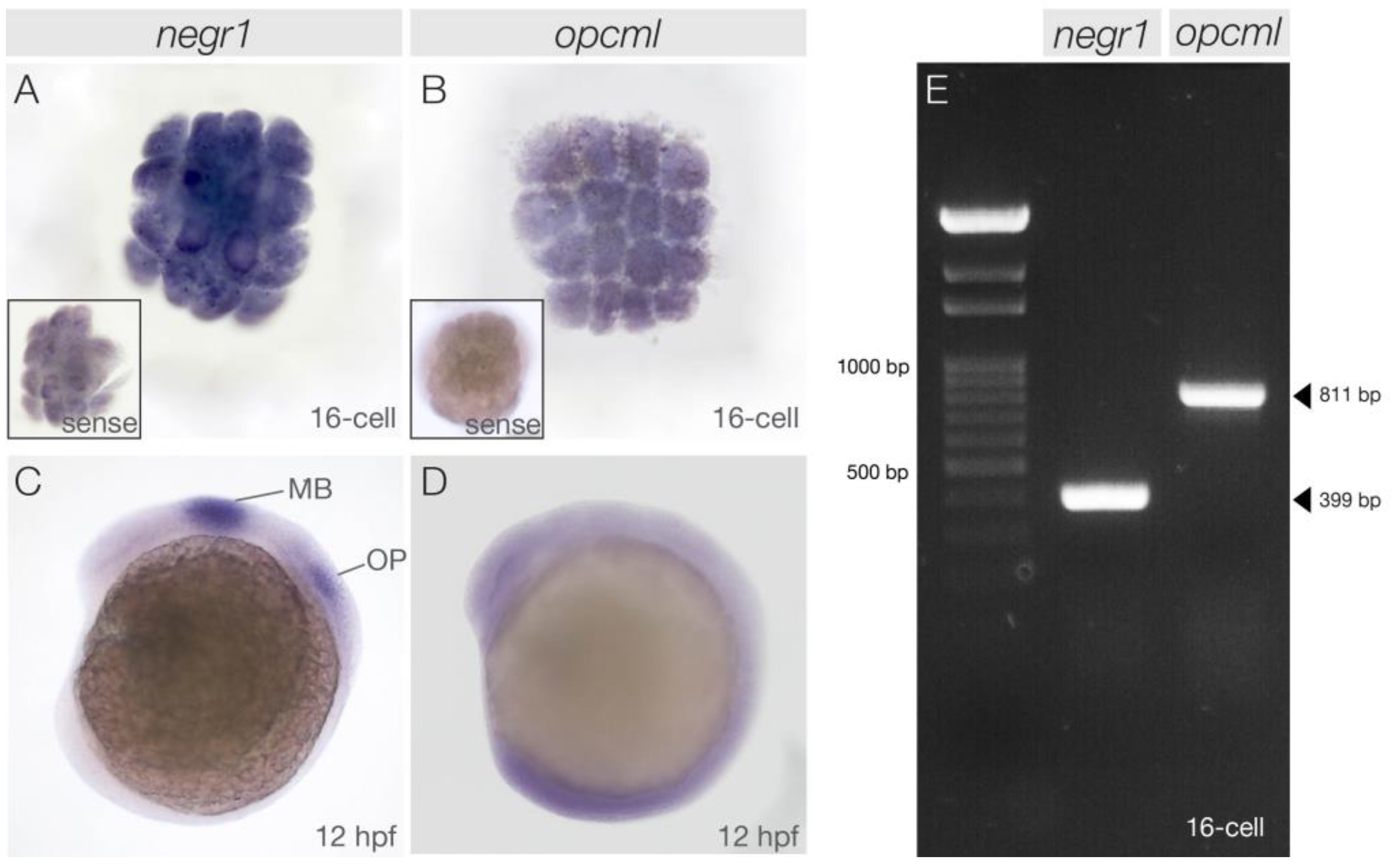
3.1. negr1 and opcml Transcripts Are Maternally Deposited in the Early Embryo

To assess the temporal and spatial expression of negr1 and opcml during embryonic development, we performed whole-mount in situ hybridisation between the 16-cell stage and 5 days post fertilisation (dpf). In the fertilised egg, many maternal gene transcripts are present to facilitate the earliest events in development. Only after a process called midblastula transition (MBT), which in zebrafish occurs at the 512-cell stage (ca. 2 h post fertilisation (hpf)), do the embryos activate transcription [45].
Unexpectedly, given the genes’ reported functions at late stages of development and adulthood in mammals, we revealed ubiquitously distributed maternal mRNA transcripts of both genes already at the 16-cell stage (Figure 1A,B). To confirm this discovery, we additionally performed PCR on retrotranscribed RNA extracted from embryos at the 16-cell stage. Sequence analysis of the resulting RT-PCR products validated the presence of the transcript at this stage of development for both negr1 and opcml (Figure 1E). At 12 h post fertilisation (hpf), strong and specific expression was detected for negr1 in the midbrain and the otic placodes (Figure 1C). opcml showed weak ubiquitous staining with increased intensity in the caudal part of the embryo (Figure 1D).

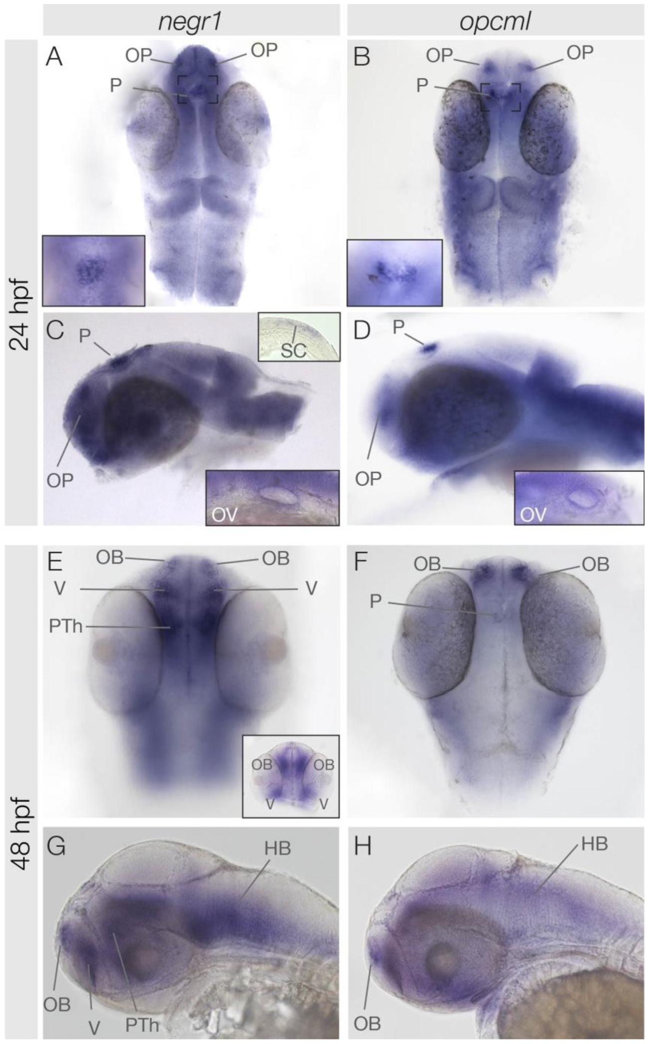
3.2. Similarities and Differences in negr1 and opcml Expression at 24 hpf and 48 hpf

At 24 hpf, both negr1 and opcml exhibited rather weak ubiquitous expression in the central nervous system with several exceptions of distinct expression (Figure 2A–D). Both gene transcripts are present in the olfactory placodes and the pineal gland. Here, the expression appears complementary. negr1 is expressed in the centrally located cells of the pineal gland, while opcml is present in the outer pineal cells (Figure 2A,B insets). Additionally, the otic vesicles exhibited expression of both negr1 and opcml (Figure 2C,D insets). Unlike opcml, negr1 is also expressed in the most caudal part of the spinal cord (Figure 2C upper inset). By 48 hpf, both negr1 and opcml remain expressed in the olfactory bulb and in the hindbrain (Figure 2E–H). In the pineal, opcml continues to be expressed, although the expression appears to be weaker in comparison to earlier stages (Figure 2E–H). Conversely, negr1 starts to be expressed in the ventral telencephalon and the pre-thalamus at this stage (Figure 2E,G and inset in Figure 2E).

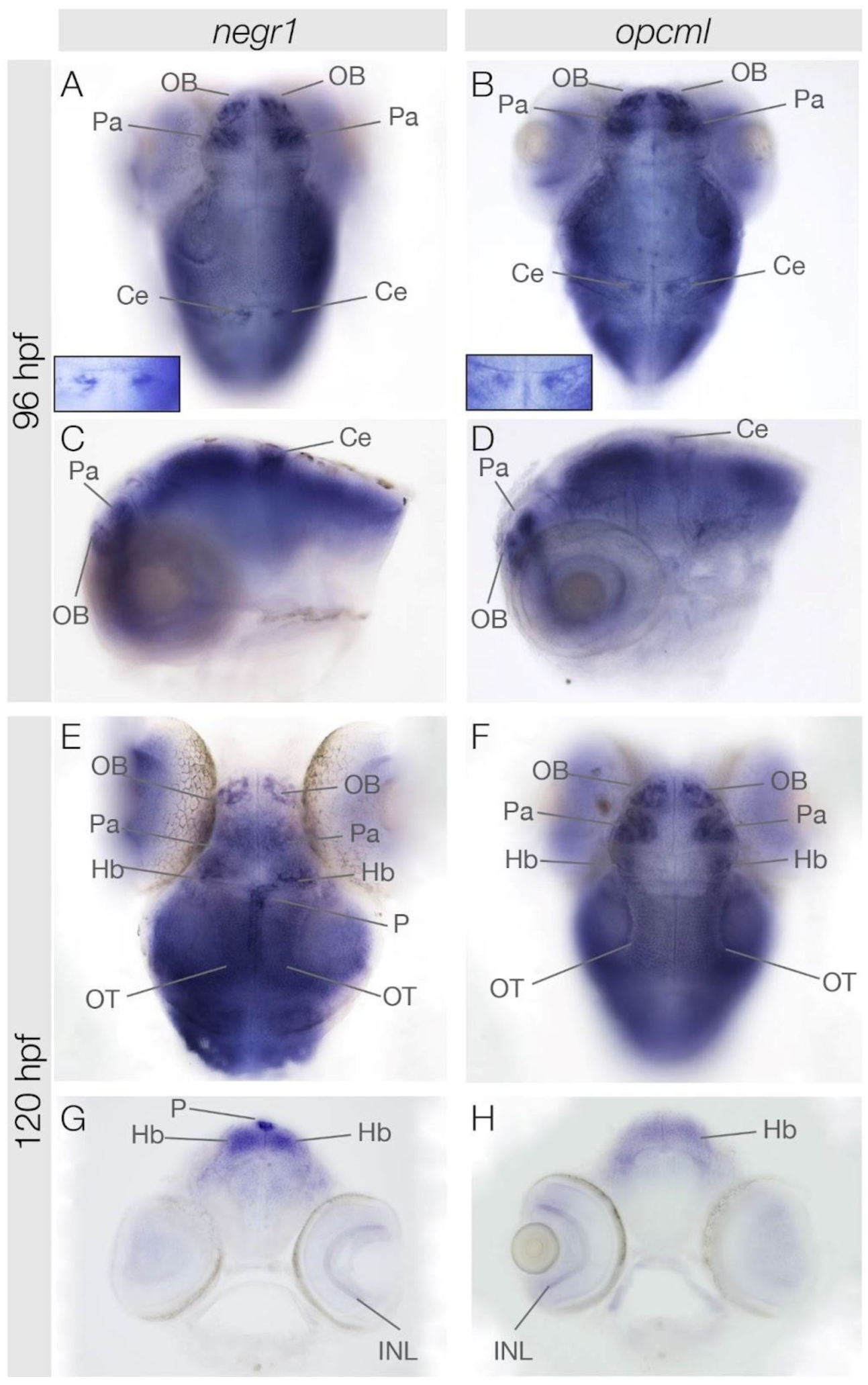
3.3. negr1 and opcml Expression in the Brain at 96 hpf and 120 hpf

negr1 and opcml continued to be expressed in the olfactory bulb at 4 and 5 dpf (Figure 3). In addition, both genes started to be expressed in the pallium area of the telencephalon. Moreover, we observed transient negr1 and opcml expression in a few cells of the cerebellum, which likely correspond to a subpopulation of Purkinje cells at 4 dpf (Figure 3A,B inset). At 5 dpf, both genes were expressed bilaterally and symmetrically in habenular neurons (Figure 3E,F), while only negr1 started to be expressed again in cells of the pineal gland (Figure 3E–H). Additionally, negr1 and opcml were both expressed in the inner nuclear layer of the retina (Figure 3G,H).

4. Discussion

Our present analysis of the two IgLON family members negr1 and opcml reveals new insights into their spatio-temporal expression dynamics in the developing vertebrate embryo. Initially, both genes are provided maternally in zebrafish. The importance of maternal gene function in neural network formation is well established and evolutionarily conserved in vertebrates and invertebrates [46,47,48]. In mice and rats, NEGR1 and OPCML are needed mainly for neuronal outgrowth, dendritic arborisation, and synapse formation, and it may be interesting to investigate their potential involvement in earlier developmental processes. Furthermore, a maternal function of these genes might account for differences in the severity or onset of NEGR1- or OPCML-linked psychiatric disorders. At subsequent stages of development, both genes are expressed in discrete, partially overlapping domains of the zebrafish brain. Available gene expression data in mammals focused on late developmental stages and adult tissues [36,37,49]. Nevertheless, our data suggest that various expression domains appear conserved between zebrafish and mammals. Notably, some of these domains have been linked to neuropsychiatric disorders which both genes have been implicated in, such as major depressive disorder (MDD), schizophrenia [27,28], and autism spectrum disorder (ASD) [27,28,33]. For instance, negr1 and opcml are strongly expressed in the zebrafish pallium, which has been proposed to harbour structures homologous to the hippocampus and amygdala of mammals [50] involved in ASD and schizophrenia [51]. NEGR1−/− mice exhibit a reduced volume of brain regions, including the hippocampus. Specifically, the parvalbumin-positive interneurons were significantly reduced [34,38]. In OPCML-deficient mice, the hippocampal area develops largely normally, but alterations in hippocampus-dependent spatial learning and memory were observed [52]. In primary hippocampal neurons, the absence of OPCML caused increased numbers of filopodia-like spines and fewer mature spines and neurons, which might explain the behavioural phenotypes [52].
Moreover, we found negr1 and opcml expression in restricted subpopulations of cells in the zebrafish cerebellum, which by position correspond to the Purkinje cells in line with the expression profile found in rodents [53]. The function of these GABAergic projection neurons has also been linked to ASD [54,55]. negr1 and opcml expression in zebrafish is also evident in the dorsal diencephalon. Here, the genes are found both in the medially located pineal gland and in the left and right adjacent habenulae. Interestingly, the pineal gland exhibits complementary expression of the two genes: negr1-expressing cells are centrally located while opcml-positive cells are located mainly in the outer layer of the pineal gland. These two layers have distinct functions, as the outer layer cells are typically active in periods of darkness [56]. In addition, the regulation of negr1 expression seems to fluctuate over time: the expression is stronger early in development, decreases subsequently, and increases again at 5 dpf. In contrast, opcml expression in the pineal gland is strong at 24 hpf and decreases in the course of development. We did not observe any transcripts in zebrafish parapineal cells, which are known to influence the neurogenesis of the left habenula, causing the left–right asymmetric formation of neuronal subpopulations [57,58]. negr1 and opcml expression in the zebrafish habenulae is symmetric and begins at developmental stages after habenular neuron differentiation [59,60,61]. The habenular neurotransmitter system has been connected to autism and schizophrenia [42,62]. It has also become a major focus for the treatment of MDD. Indeed, patients not responding to conventional pharmacological treatments often benefit from deep brain stimulation to transiently inactivate the lateral habenulae [63,64]. negr1 and opcml expression in the mammalian pineal gland or the habenulae has not been reported to date. It may be revealing to re-analyse the genes in greater detail and to also include earlier stages of brain development. The habenulae relay sensory information such as visual and olfactory input to mid- and hindbrain areas [65,66,67,68]. We found that negr1 and opcml are strongly expressed in the zebrafish olfactory bulb throughout development. This resembles gene expression in the mammalian olfactory system [36,37,49]. Olfaction has recently become a focus in the field of ASD research as it is involved in social behaviour [69]. However, a link between smell and neuropsychiatric disorders in mice mutant for NEGR1 or OPCML remains to be explored.
Our detailed expression study extends and refines currently available expression data on zebrafish negr1 and opcml [44]. It should encourage future research to, for instance, analyse mammalian NEGR1 and OPCML expression and function in brain areas and at developmental time points other than those described to date. A particular area of interest may be to investigate a maternal contribution of the two genes and their expression and function in the habenular neural circuit. These studies could reveal new insights into the onset of neuropsychiatric disorders, their severity and the brain area(s) affected. A zebrafish knock-out for negr1, which did not result in overt morphological alterations, has been reported in the framework of a large schizophrenia study [70]. In the same study, an opcml mutant was generated, which, however, was not further described. For both gene knock-outs, no functional or behavioural investigations are available. Our detailed expression analysis should provide an excellent starting point for such in vivo studies, for instance to determine the precise temporal role of the genes during neural network formation and function. This in turn provides a platform for expression profiling to unravel the molecular network downstream NEGR1 and OPCML for the identification of suitable therapeutic targets. Furthermore, it aids screening for disorder-ameliorating compounds, which subsequently can be tested in mammalian models. Combining the advantages of available resources to gain deeper insights into the mechanisms underlying NEGR1 and OPCML function in health and disease is a pivotal prerequisite for developing therapies.

Author Contributions

Conceptualization, M.C. and G.P.; methodology, M.C.; validation, J.H. and M.C.; formal analysis, J.H. and I.S.; investigation, J.H., I.S. and S.S.; resources, M.C.; data curation, J.H., I.S. and M.C.; writing—original draft preparation, J.H. and M.C; writing—review and editing, J.H. and M.C.; visualization, J.H.; supervision, M.C., A.B. and J.H.; project administration, M.C.; funding acquisition, A.B., J.H., G.P. and M.C. All authors have read and agreed to the published version of the manuscript.

Funding

J.H. was funded by Wenner-Gren Foundations (WGF2021-0026), A.B. was funded by Caritro Bando 2020 per progetti presentati da giovani ricercatori post-doc #10868, and G.P. was funded by Fondazione Cariplo (2019-3415).

Institutional Review Board Statement

The study was conducted according to the guidelines of the Declaration of Helsinki and according to European and Italian law, D.Lgs. 26/2014, authorization 151/2019-PR to M. Carl.

Informed Consent Statement

Not applicable.

Data Availability Statement

Data supporting reported results can be requested.

Acknowledgments

We are grateful to Yuri Bozzi for his support and we would like to thank Ilaria Mazzeo and Sergio Robbiati from the Model Organism Facility, Department of CIBIO, University of Trento and Giorgina Scarduelli and Michela Roccuzzo from the Advanced Imaging Core Facility, Department of CIBIO, University of Trento for their help with fish care and image acquisition, respectively. We are grateful to the Department of Cellular, Computational and Integrative Biology, CIBIO, University of Trento for financial support.

Conflicts of Interest

The authors declare no conflicts of interest. The funders had no role in the design of the study; in the collection, analyses, or interpretation of data; in the writing of the manuscript; or in the decision to publish the results.

References

  1. Schofield, P.R.; McFarland, K.C.; Hayflick, J.S.; Wilcox, J.N.; Cho, T.M.; Roy, S.; Lee, N.M.; Loh, H.H.; Seeburg, P.H. Molecular Characterization of a New Immunoglobulin Superfamily Protein with Potential Roles in Opioid Binding and Cell Contact. EMBO J. 1989, 8, 489–495. [Google Scholar] [CrossRef] [PubMed]
  2. Struyk, A.; Canoll, P.; Wolfgang, M.; Rosen, C.; D’Eustachio, P.; Salzer, J. Cloning of Neurotrimin Defines a New Subfamily of Differentially Expressed Neural Cell Adhesion Molecules. J. Neurosci. 1995, 15, 2141–2156. [Google Scholar] [CrossRef] [PubMed]
  3. Horton, H.; Levitt, P. A Unique Membrane Protein Is Expressed on Early Developing Limbic System Axons and Cortical Targets. J. Neurosci. 1988, 8, 4653–4661. [Google Scholar] [CrossRef] [PubMed]
  4. Funatsu, N.; Miyata, S.; Kumanogoh, H.; Shigeta, M.; Hamada, K.; Endo, Y.; Sokawa, Y.; Maekawa, S. Characterization of a Novel Rat Brain Glycosylphosphatidylinositol-Anchored Protein (Kilon), a Member of the IgLON Cell Adhesion Molecule Family. J. Biol. Chem. 1999, 274, 8224–8230. [Google Scholar] [CrossRef] [PubMed]
  5. Grimwood, J.; Gordon, L.A.; Olsen, A.; Terry, A.; Schmutz, J.; Lamerdin, J.; Hellsten, U.; Goodstein, D.; Couronne, O.; Tran-Gyamfi, M.; et al. The DNA Sequence and Biology of Human Chromosome 19. Nature 2004, 428, 529–535. [Google Scholar] [CrossRef] [PubMed]
  6. Sellar, G.C.; Watt, K.P.; Rabiasz, G.J.; Stronach, E.A.; Li, L.; Miller, E.P.; Massie, C.E.; Miller, J.; Contreras-Moreira, B.; Scott, D.; et al. OPCML at 11q25 Is Epigenetically Inactivated and Has Tumor-Suppressor Function in Epithelial Ovarian Cancer. Nat. Genet. 2003, 34, 337–343. [Google Scholar] [CrossRef] [PubMed]
  7. Gil, O.D.; Zanazzi, G.; Struyk, A.F.; Salzer, J.L. Neurotrimin Mediates Bifunctional Effects on Neurite Outgrowth via Homophilic and Heterophilic Interactions. J. Neurosci. 1998, 18, 9312–9325. [Google Scholar] [CrossRef] [PubMed]
  8. Hashimoto, T.; Yamada, M.; Maekawa, S.; Nakashima, T.; Miyata, S. IgLON Cell Adhesion Molecule Kilon Is a Crucial Modulator for Synapse Number in Hippocampal Neurons. Brain Res. 2008, 1224, 1–11. [Google Scholar] [CrossRef]
  9. Sanz, R.; Ferraro, G.B.; Fournier, A.E. IgLON Cell Adhesion Molecules Are Shed from the Cell Surface of Cortical Neurons to Promote Neuronal Growth. J. Biol. Chem. 2015, 290, 4330–4342. [Google Scholar] [CrossRef]
  10. Schäfer, M.; Bräuer, A.U.; Savaskan, N.E.; Rathjen, F.G.; Brümmendorf, T. Neurotractin/Kilon Promotes Neurite Outgrowth and Is Expressed on Reactive Astrocytes after Entorhinal Cortex Lesion. Mol. Cell. Neurosci. 2005, 29, 580–590. [Google Scholar] [CrossRef]
  11. Cui, Y.; Ying, Y.; van Hasselt, A.; Ng, K.M.; Yu, J.; Zhang, Q.; Jin, J.; Liu, D.; Rhim, J.S.; Rha, S.Y.; et al. OPCML Is a Broad Tumor Suppressor for Multiple Carcinomas and Lymphomas with Frequently Epigenetic Inactivation. PLoS ONE 2008, 3, e2990. [Google Scholar] [CrossRef]
  12. Kim, H.; Hwang, J.-S.; Lee, B.; Hong, J.; Lee, S. Newly Identified Cancer-Associated Role of Human Neuronal Growth Regulator 1 (NEGR1). J. Cancer 2014, 5, 598–608. [Google Scholar] [CrossRef] [PubMed]
  13. Ntougkos, E.; Rush, R.; Scott, D.; Frankenberg, T.; Gabra, H.; Smyth, J.F.; Sellar, G.C. The IgLON Family in Epithelial Ovarian Cancer: Expression Profiles and Clinicopathologic Correlates. Clin. Cancer Res. 2005, 11, 5764–5768. [Google Scholar] [CrossRef] [PubMed]
  14. Reed, J.E.; Dunn, J.R.; du Plessis, D.G.; Shaw, E.J.; Reeves, P.; Gee, A.L.; Warnke, P.C.; Sellar, G.C.; Moss, D.J.; Walker, C. Expression of Cellular Adhesion Molecule “OPCML” Is down-Regulated in Gliomas and Other Brain Tumours. Neuropathol. Appl. Neurobiol. 2007, 33, 77–85. [Google Scholar] [CrossRef] [PubMed]
  15. Pischedda, F.; Ghirelli, A.; Tripathi, V.; Piccoli, G. Negr1-Derived Peptides Trigger ALK Degradation and Halt Neuroblastoma Progression In Vitro and In Vivo. Pharmaceutics 2023, 15, 2307. [Google Scholar] [CrossRef] [PubMed]
  16. Hyde, C.L.; Nagle, M.W.; Tian, C.; Chen, X.; Paciga, S.A.; Wendland, J.R.; Tung, J.Y.; Hinds, D.A.; Perlis, R.H.; Winslow, A.R. Identification of 15 Genetic Loci Associated with Risk of Major Depression in Individuals of European Descent. Nat. Genet. 2016, 48, 1031–1036. [Google Scholar] [CrossRef] [PubMed]
  17. Wray, N.R.; Ripke, S.; Mattheisen, M.; Trzaskowski, M.; Byrne, E.M.; Abdellaoui, A.; Adams, M.J.; Agerbo, E.; Air, T.M.; Andlauer, T.M.F.; et al. Genome-Wide Association Analyses Identify 44 Risk Variants and Refine the Genetic Architecture of Major Depression. Nat. Genet. 2018, 50, 668–681. [Google Scholar] [CrossRef]
  18. Levey, D.F.; Stein, M.B.; Wendt, F.R.; Pathak, G.A.; Zhou, H.; Aslan, M.; Quaden, R.; Harrington, K.M.; Nuñez, Y.Z.; Overstreet, C.; et al. Bi-Ancestral Depression GWAS in the Million Veteran Program and Meta-Analysis in >1.2 Million Individuals Highlight New Therapeutic Directions. Nat. Neurosci. 2021, 24, 954–963. [Google Scholar] [CrossRef]
  19. Howard, D.M.; Adams, M.J.; Clarke, T.-K.; Hafferty, J.D.; Gibson, J.; Shirali, M.; Coleman, J.R.I.; Hagenaars, S.P.; Ward, J.; Wigmore, E.M.; et al. Genome-Wide Meta-Analysis of Depression Identifies 102 Independent Variants and Highlights the Importance of the Prefrontal Brain Regions. Nat. Neurosci. 2019, 22, 343–352. [Google Scholar] [CrossRef]
  20. Schol-Gelok, S.; Janssens, A.C.J.W.; Tiemeier, H.; Liu, F.; Lopez-Leon, S.; Zorkoltseva, I.V.; Axenovich, T.I.; van Swieten, J.C.; Uitterlinden, A.G.; Hofman, A.; et al. A Genome-Wide Screen for Depression in Two Independent Dutch Populations. Biol. Psychiatry 2010, 68, 187–196. [Google Scholar] [CrossRef]
  21. Ding, H.; Ouyang, M.; Wang, J.; Xie, M.; Huang, Y.; Yuan, F.; Jia, Y.; Zhang, X.; Liu, N.; Zhang, N. Shared Genetics between Classes of Obesity and Psychiatric Disorders: A Large-Scale Genome-Wide Cross-Trait Analysis. J. Psychosom. Res. 2022, 162, 111032. [Google Scholar] [CrossRef] [PubMed]
  22. Zhang, Q.; Liu, C.; Li, Q.; Li, J.; Wu, Y.; Liu, J. MicroRNA-25–5p Counteracts Oxidized LDL-Induced Pathological Changes by Targeting Neuronal Growth Regulator 1 (NEGR1) in Human Brain Micro-Vessel Endothelial Cells. Biochimie 2019, 165, 141–149. [Google Scholar] [CrossRef] [PubMed]
  23. Athanasiu, L.; Mattingsdal, M.; Kähler, A.K.; Brown, A.; Gustafsson, O.; Agartz, I.; Giegling, I.; Muglia, P.; Cichon, S.; Rietschel, M.; et al. Gene Variants Associated with Schizophrenia in a Norwegian Genome-Wide Study Are Replicated in a Large European Cohort. J. Psychiatr. Res. 2010, 44, 748–753. [Google Scholar] [CrossRef] [PubMed]
  24. Panichareon, B.; Nakayama, K.; Thurakitwannakarn, W.; Iwamoto, S.; Sukhumsirichart, W. OPCML Gene as a Schizophrenia Susceptibility Locus in Thai Population. J. Mol. Neurosci. 2012, 46, 373–377. [Google Scholar] [CrossRef] [PubMed]
  25. O’Donovan, M.C.; Craddock, N.; Norton, N.; Williams, H.; Peirce, T.; Moskvina, V.; Nikolov, I.; Hamshere, M.; Carroll, L.; Georgieva, L.; et al. Identification of Loci Associated with Schizophrenia by Genome-Wide Association and Follow-Up. Nat. Genet. 2008, 40, 1053–1055. [Google Scholar] [CrossRef]
  26. Pardiñas, A.F.; Holmans, P.; Pocklington, A.J.; Escott-Price, V.; Ripke, S.; Carrera, N.; Legge, S.E.; Bishop, S.; Cameron, D.; Hamshere, M.L.; et al. Common Schizophrenia Alleles Are Enriched in Mutation-Intolerant Genes and in Regions under Strong Background Selection. Nat. Genet. 2018, 50, 381–389. [Google Scholar] [CrossRef] [PubMed]
  27. Minhas, H.M.; Pescosolido, M.F.; Schwede, M.; Piasecka, J.; Gaitanis, J.; Tantravahi, U.; Morrow, E.M. An Unbalanced Translocation Involving Loss of 10q26.2 and Gain of 11q25 in a Pedigree with Autism Spectrum Disorder and Cerebellar Juvenile Pilocytic Astrocytoma. Am. J. Med. Genet. A 2013, 161, 787–791. [Google Scholar] [CrossRef]
  28. Szczurkowska, J.; Pischedda, F.; Pinto, B.; Managò, F.; Haas, C.A.; Summa, M.; Bertorelli, R.; Papaleo, F.; Schäfer, M.K.; Piccoli, G.; et al. NEGR1 and FGFR2 Cooperatively Regulate Cortical Development and Core Behaviours Related to Autism Disorders in Mice. Brain 2018, 141, 2772–2794. [Google Scholar] [CrossRef]
  29. Steiger, H.; Booij, L.; Thaler, L.; St-Hilaire, A.; Israël, M.; Casey, K.F.; Oliverio, S.; Crescenzi, O.; Lee, V.; Turecki, G.; et al. DNA Methylation in People with Anorexia Nervosa: Epigenome-Wide Patterns in Actively Ill, Long-Term Remitted, and Healthy-Eater Women. World J. Biol. Psychiatry 2023, 24, 254–259. [Google Scholar] [CrossRef]
  30. Huckins, L.M.; Hatzikotoulas, K.; Southam, L.; Thornton, L.M.; Steinberg, J.; Aguilera-McKay, F.; Treasure, J.; Schmidt, U.; Gunasinghe, C.; Romero, A.; et al. Investigation of Common, Low-Frequency and Rare Genome-Wide Variation in Anorexia Nervosa. Mol. Psychiatry 2018, 23, 1169–1180. [Google Scholar] [CrossRef]
  31. Raghavan, N.S.; Brickman, A.M.; Andrews, H.; Manly, J.J.; Schupf, N.; Lantigua, R.; Wolock, C.J.; Kamalakaran, S.; Petrovski, S.; Tosto, G.; et al. Whole-exome Sequencing in 20,197 Persons for Rare Variants in Alzheimer’s Disease. Ann. Clin. Transl. Neurol. 2018, 5, 832–842. [Google Scholar] [CrossRef]
  32. Liu, F.; Arias-Vásquez, A.; Sleegers, K.; Aulchenko, Y.S.; Kayser, M.; Sanchez-Juan, P.; Feng, B.-J.; Bertoli-Avella, A.M.; van Swieten, J.; Axenovich, T.I.; et al. A Genomewide Screen for Late-Onset Alzheimer Disease in a Genetically Isolated Dutch Population. Am. J. Hum. Genet. 2007, 81, 17–31. [Google Scholar] [CrossRef] [PubMed]
  33. Salluzzo, M.; Vianello, C.; Abdullatef, S.; Rimondini, R.; Piccoli, G.; Carboni, L. The Role of IgLON Cell Adhesion Molecules in Neurodegenerative Diseases. Genes 2023, 14, 1886. [Google Scholar] [CrossRef] [PubMed]
  34. Noh, K.; Park, J.-C.; Han, J.-S.; Lee, S.J. From Bound Cells Comes a Sound Mind: The Role of Neuronal Growth Regulator 1 in Psychiatric Disorders. Exp. Neurobiol. 2020, 29, 1–10. [Google Scholar] [CrossRef] [PubMed]
  35. Pischedda, F.; Piccoli, G. The IgLON Family Member Negr1 Promotes Neuronal Arborization Acting as Soluble Factor via FGFR2. Front. Mol. Neurosci. 2016, 8, 89. [Google Scholar] [CrossRef] [PubMed]
  36. Vanaveski, T.; Singh, K.; Narvik, J.; Eskla, K.-L.; Visnapuu, T.; Heinla, I.; Jayaram, M.; Innos, J.; Lilleväli, K.; Philips, M.-A.; et al. Promoter-Specific Expression and Genomic Structure of IgLON Family Genes in Mouse. Front. Neurosci. 2017, 11, 38. [Google Scholar] [CrossRef] [PubMed]
  37. Hachisuka, A.; Nakajima, O.; Yamazaki, T.; Sawada, J. Developmental Expression of Opioid-Binding Cell Adhesion Molecule (OBCAM) in Rat Brain. Dev. Brain Res. 2000, 122, 183–191. [Google Scholar] [CrossRef]
  38. Singh, K.; Jayaram, M.; Kaare, M.; Leidmaa, E.; Jagomäe, T.; Heinla, I.; Hickey, M.A.; Kaasik, A.; Schäfer, M.K.; Innos, J.; et al. Neural Cell Adhesion Molecule Negr1 Deficiency in Mouse Results in Structural Brain Endophenotypes and Behavioral Deviations Related to Psychiatric Disorders. Sci. Rep. 2019, 9, 5457. [Google Scholar] [CrossRef]
  39. Kubick, N.; Brösamle, D.; Mickael, M.-E. Molecular Evolution and Functional Divergence of the IgLON Family. Evol. Bioinform. 2018, 14, 117693431877508. [Google Scholar] [CrossRef]
  40. Choi, T.-Y.; Choi, T.-I.; Lee, Y.-R.; Choe, S.-K.; Kim, C.-H. Zebrafish as an Animal Model for Biomedical Research. Exp. Mol. Med. 2021, 53, 310–317. [Google Scholar] [CrossRef]
  41. Patton, E.E.; Zon, L.I.; Langenau, D.M. Zebrafish Disease Models in Drug Discovery: From Preclinical Modelling to Clinical Trials. Nat. Rev. Drug Discov. 2021, 20, 611–628. [Google Scholar] [CrossRef] [PubMed]
  42. Bühler, A.; Carl, M. Zebrafish Tools for Deciphering Habenular Network-Linked Mental Disorders. Biomolecules 2021, 11, 324. [Google Scholar] [CrossRef] [PubMed]
  43. McNabb, A.; Scott, K.; von Ochsenstein, E.; Seufert, K.; Carl, M. Don’t Be Afraid to Set Up Your Fish Facility. Zebrafish 2012, 9, 120–125. [Google Scholar] [CrossRef] [PubMed]
  44. Thisse, C.; Thisse, B. High-Resolution in Situ Hybridization to Whole-Mount Zebrafish Embryos. Nat. Protoc. 2008, 3, 59–69. [Google Scholar] [CrossRef]
  45. Kane, D.A.; Kimmel, C.B. The Zebrafish Midblastula Transition. Development 1993, 119, 447–456. [Google Scholar] [CrossRef] [PubMed]
  46. Mardirossian, S.; Rampon, C.; Salvert, D.; Fort, P.; Sarda, N. Impaired Hippocampal Plasticity and Altered Neurogenesis in Adult Ube3a Maternal Deficient Mouse Model for Angelman Syndrome. Exp. Neurol. 2009, 220, 341–348. [Google Scholar] [CrossRef]
  47. Campinho, M.A.; Saraiva, J.; Florindo, C.; Power, D.M. Maternal Thyroid Hormones Are Essential for Neural Development in Zebrafish. Mol. Endocrinol. 2014, 28, 1136–1149. [Google Scholar] [CrossRef] [PubMed]
  48. Jiménez, F.; Campos-Ortega, J.A. Maternal Effects of Zygotic Mutants Affecting Early Neurogenesis in Drosophila. Wilehm Roux Arch. Dev. Biol. 1982, 191, 191–201. [Google Scholar] [CrossRef]
  49. Lee, A.W.S.; Hengstler, H.; Schwald, K.; Berriel-Diaz, M.; Loreth, D.; Kirsch, M.; Kretz, O.; Haas, C.A.; de Angelis, M.H.; Herzig, S.; et al. Functional Inactivation of the Genome-Wide Association Study Obesity Gene Neuronal Growth Regulator 1 in Mice Causes a Body Mass Phenotype. PLoS ONE 2012, 7, e41537. [Google Scholar] [CrossRef]
  50. Langova, V.; Vales, K.; Horka, P.; Horacek, J. The Role of Zebrafish and Laboratory Rodents in Schizophrenia Research. Front. Psychiatry 2020, 11, 703. [Google Scholar] [CrossRef]
  51. Tayanloo-Beik, A.; Hamidpour, S.K.; Abedi, M.; Shojaei, H.; Tavirani, M.R.; Namazi, N.; Larijani, B.; Arjmand, B. Zebrafish Modeling of Autism Spectrum Disorders, Current Status and Future Prospective. Front. Psychiatry 2022, 13, 911770. [Google Scholar] [CrossRef] [PubMed]
  52. Zhang, Z.; Ye, M.; Li, Q.; You, Y.; Yu, H.; Ma, Y.; Mei, L.; Sun, X.; Wang, L.; Yue, W.; et al. The Schizophrenia Susceptibility Gene OPCML Regulates Spine Maturation and Cognitive Behaviors through Eph-Cofilin Signaling. Cell Rep. 2019, 29, 49–61.e7. [Google Scholar] [CrossRef] [PubMed]
  53. Jagomäe, T.; Singh, K.; Philips, M.-A.; Jayaram, M.; Seppa, K.; Tekko, T.; Gilbert, S.F.; Vasar, E.; Lilleväli, K. Alternative Promoter Use Governs the Expression of IgLON Cell Adhesion Molecules in Histogenetic Fields of the Embryonic Mouse Brain. Int. J. Mol. Sci. 2021, 22, 6955. [Google Scholar] [CrossRef] [PubMed]
  54. Sudarov, A. Defining the Role of Cerebellar Purkinje Cells in Autism Spectrum Disorders. Cerebellum 2013, 12, 950–955. [Google Scholar] [CrossRef] [PubMed]
  55. Tsai, P.T.; Hull, C.; Chu, Y.; Greene-Colozzi, E.; Sadowski, A.R.; Leech, J.M.; Steinberg, J.; Crawley, J.N.; Regehr, W.G.; Sahin, M. Autistic-like Behaviour and Cerebellar Dysfunction in Purkinje Cell Tsc1 Mutant Mice. Nature 2012, 488, 647–651. [Google Scholar] [CrossRef] [PubMed]
  56. Sapède, D.; Chaigne, C.; Blader, P.; Cau, E. Functional Heterogeneity in the Pineal Projection Neurons of Zebrafish. Mol. Cell. Neurosci. 2020, 103, 103468. [Google Scholar] [CrossRef] [PubMed]
  57. Concha, M.L.; Russell, C.; Regan, J.C.; Tawk, M.; Sidi, S.; Gilmour, D.T.; Kapsimali, M.; Sumoy, L.; Goldstone, K.; Amaya, E.; et al. Local Tissue Interactions across the Dorsal Midline of the Forebrain Establish CNS Laterality. Neuron 2003, 39, 423–438. [Google Scholar] [CrossRef]
  58. Gamse, J.T.; Thisse, C.; Thisse, B.; Halpern, M.E. The Parapineal Mediates Left-Right Asymmetry in the Zebrafish Diencephalon. Development 2003, 130, 1059–1068. [Google Scholar] [CrossRef]
  59. Beretta, C.A.; Dross, N.; Guglielmi, L.; Bankhead, P.; Soulika, M.; Gutierrez-Triana, J.A.; Paolini, A.; Poggi, L.; Falk, J.; Ryu, S.; et al. Early Commissural Diencephalic Neurons Control Habenular Axon Extension and Targeting. Curr. Biol. 2017, 27, 270–278. [Google Scholar] [CrossRef]
  60. Hüsken, U.; Stickney, H.L.; Gestri, G.; Bianco, I.H.; Faro, A.; Young, R.M.; Roussigne, M.; Hawkins, T.A.; Beretta, C.A.; Brinkmann, I.; et al. Tcf7l2 Is Required for Left-Right Asymmetric Differentiation of Habenular Neurons. Curr. Biol. 2014, 24, 2217–2227. [Google Scholar] [CrossRef]
  61. Aizawa, H.; Goto, M.; Sato, T.; Okamoto, H. Temporally Regulated Asymmetric Neurogenesis Causes Left-Right Difference in the Zebrafish Habenular Structures. Dev. Cell 2007, 12, 87–98. [Google Scholar] [CrossRef] [PubMed]
  62. Hu, H.; Cui, Y.; Yang, Y. Circuits and Functions of the Lateral Habenula in Health and in Disease. Nat. Rev. Neurosci. 2020, 21, 277–295. [Google Scholar] [CrossRef] [PubMed]
  63. Sartorius, A.; Henn, F.A. Deep Brain Stimulation of the Lateral Habenula in Treatment Resistant Major Depression. Med. Hypotheses 2007, 69, 1305–1308. [Google Scholar] [CrossRef] [PubMed]
  64. Kiening, K.; Sartorius, A. A New Translational Target for Deep Brain Stimulation to Treat Depression. EMBO Mol. Med. 2013, 5, 1151–1153. [Google Scholar] [CrossRef] [PubMed]
  65. Dreosti, E.; Vendrell Llopis, N.; Carl, M.; Yaksi, E.; Wilson, S.W. Left-Right Asymmetry Is Required for the Habenulae to Respond to Both Visual and Olfactory Stimuli. Curr. Biol. 2014, 24, 440–445. [Google Scholar] [CrossRef] [PubMed]
  66. Aizawa, H.; Amo, R.; Okamoto, H. Phylogeny and Ontogeny of the Habenular Structure. Front. Neurosci. 2011, 5, 138. [Google Scholar] [CrossRef] [PubMed]
  67. Beretta, C.A.; Dross, N.; Guiterrez-Triana, J.A.; Ryu, S.; Carl, M. Habenula Circuit Development: Past, Present, and Future. Front. Neurosci. 2012, 6, 51. [Google Scholar] [CrossRef] [PubMed]
  68. Bianco, I.H.; Wilson, S.W. The Habenular Nuclei: A Conserved Asymmetric Relay Station in the Vertebrate Brain. Philos. Trans. R. Soc. B Biol. Sci. 2009, 364, 1005–1020. [Google Scholar] [CrossRef]
  69. Barros, F.; Figueiredo, C.; Costa, A.; Soares, S.C. Sensory Processing in the Autism Spectrum: The Role of Attention to Detail and Somatic Trait Anxiety in the Olfactory Perception of the General Population. J. Autism Dev. Disord. 2021, 51, 2338–2353. [Google Scholar] [CrossRef]
  70. Thyme, S.B.; Pieper, L.M.; Li, E.H.; Pandey, S.; Wang, Y.; Morris, N.S.; Sha, C.; Choi, J.W.; Herrera, K.J.; Soucy, E.R.; et al. Phenotypic Landscape of Schizophrenia-Associated Genes Defines Candidates and Their Shared Functions. Cell 2019, 177, 478–491.e20. [Google Scholar] [CrossRef]
Figure 1. negr1 and opcml expression at early developmental stages. 16-cell stage zebrafish embryos expressing negr1 (A) and opcml (B); sense probe stains are shown in the insets. (C,D) show lateral views of 12 hpf old embryos. negr1 is expressed in the midbrain and the otic placodes (C), while opcml exhibits ubiquitous expression (D). RT-PCR corroborates the presence of negr1 and opcml transcripts at the 16-cell stage (E). MB, midbrain; OP, otic placodes.
Genes 15 00363 g001
Figure 2. Expression patterns of negr1 and opcml at 24 hpf and 48 hpf. Dorsal (A,B,E,F) and lateral (C,D,G,H) views of the head region of zebrafish embryos at stages indicated. Insets show the pineal gland (A,B), the otic vesicle (C,D), the spinal cord (upper inset in C), and a frontal view (E). HB, hindbrain; OB, olfactory bulb; OP, olfactory placode; OV, otic vesicle; PTh, pre-thalamus; P, pineal gland; SC, spinal cord; V, ventral telencephalon.
Genes 15 00363 g002
Figure 3. Expression patterns of negr1 and opcml at 96 hpf and 120 hpf. Dorsal (A,B,E,F) and lateral (C,D) views of zebrafish larvae focussed on the head at stages indicated. Transversal sections were carried out at the level of the pineal organ (G,H). Ce, cerebellum (inset in (A,B)); Hb, habenula; INL, inner nuclear layer; OB, olfactory bulb; Pa, pallium; P, pineal gland; OT, optic tectum.
Genes 15 00363 g003
Table 1. Gene and protein similarity for negr1 and opcml in zebrafish and mammals, including previous gene names.
Human (Homo sapiens)
NEGR1
(IgLON4, KILON, NTRA)
Mice (Mus musculus)
Negr1
(Ntra, neurotractin)
Rat (Rattus norvegicus)
Negr1
Zebrafish (Danio rerio)
negr1
64.1% similarity (bp sequence)
78.1% similarity (aa sequence)
61.7% similarity (bp sequence)
75.8% similarity (aa sequence)
63% similarity (bp sequence)
75.8% similarity (aa sequence)
Human (Homo sapiens)
OPCML
(IGLON1, OBCAM, OPCM)
Mice (Mus musculus)
Opcml
(Obcam)
Rat (Rattus norvegicus)
Opcml
Zebrafish (Danio rerio)
opcml
58.7% similarity (bp sequence)
78.3% similarity (aa sequence)
61.4% similarity (bp sequence)
77.8% similarity (aa sequence)
62.2% similarity (bp sequence)
77.8% similarity (aa sequence)
Disclaimer/Publisher’s Note: The statements, opinions and data contained in all publications are solely those of the individual author(s) and contributor(s) and not of MDPI and/or the editor(s). MDPI and/or the editor(s) disclaim responsibility for any injury to people or property resulting from any ideas, methods, instructions or products referred to in the content.

