
Resolving Discrepancies in Idylla BRAF Mutational Assay Results Using Targeted Next-Generation Sequencing

Department of Pathology and Laboratory Medicine, University of Rochester Medical Center, Rochester, NY 14642, USA
*
Author to whom correspondence should be addressed.
Genes 2024, 15(5), 527; https://doi.org/10.3390/genes15050527
Submission received: 20 March 2024
/
Revised: 15 April 2024
/
Accepted: 20 April 2024
/
Published: 23 April 2024
(This article belongs to the Special Issue Precision Medicine and Genetics)
Abstract
:BRAF mutation identification is important for the diagnosis and treatment of several tumor types, both solid and hematologic. Rapid identification of BRAF mutations is required to determine eligibility for targeted BRAF inhibitor therapy. The Idylla BRAF mutation assay is a rapid, multiplex allele-specific PCR test designed to detect the most common oncogenic BRAF V600 mutations in formalin-fixed paraffin-embedded (FFPE) tissue samples. Here, we describe the validation of the Idylla BRAF mutation assay in our laboratory. During routine clinical practice, we noticed cases in which BRAF V600 mutations were identified with unusual amplification curves, with three cases displaying a delayed amplification within a double amplification pattern and two false-positive calls. We therefore initiated a quality improvement effort to systematically and retrospectively evaluate next-generation sequencing (NGS)-tested cases with BRAF mutations identified within five amino acids of BRAF codon V600 and did not identify additional false-positive cases. We hypothesize that late amplification in a double amplification pattern may represent non-specific amplification, whereas cases displaying single delayed amplification curves may stem from the presence of either non-V600 variants, very low-level V600 variants, cytosine deamination artifacts, and/or non-specific amplification by an allele-specific PCR primer. Regardless, we recommend that Idylla BRAF cases with non-classical amplification curves undergo reflex NGS testing. These findings are likely relevant for other Idylla assays interrogating hotspot mutations in genes such as EGFR, IDH1/2, KRAS, and NRAS.
1. Introduction
Detection of BRAF mutations is clinically important in several tumor types. For instance, nearly 50% of UV exposure-related melanomas contain BRAF mutations, with p.V600E (i.e., a valine to glutamic acid substitution) being the most common (>90% of all BRAF mutations) [1,2]. Other solid tumors that commonly harbor BRAF V600E mutations include papillary thyroid cancer, serous ovarian cancer, and colorectal cancer [3], to list a few, while BRAF V600E-mutant hematologic malignancies include hairy cell leukemia (HCL) [4,5] and histiocytic neoplasms, such as Langerhans cell histiocytosis (LCH) [3]. As such, the rapid identification of BRAF mutational status is useful for diagnostic purposes and/or to determine eligibility for FDA-approved targeted BRAF inhibitors, such as vemurafenib, dabrafenib, or encorafenib [6,7,8].
Compared to next-generation sequencing (NGS), the turn-around time (TAT) for rapid multiplex polymerase chain reaction (PCR) is a mere 1.5–3 h, enabling faster mutation identification for initiation of targeted therapy. The Idylla BRAF mutation assay is a multiplex PCR-based assay [9] designed to detect the most common oncogenic BRAF V600 mutations (denoted as V600E/E2/D or V600K/R/M) in purified DNA and formalin-fixed paraffin-embedded (FFPE) specimens. Previous studies have established that a subset of BRAF V600-bearing tumor samples contain additional oncogenic or likely oncogenic variants in other genes [10]. However, the specificity of the Idylla BRAF assay for detecting BRAF V600 variants has not yet been rigorously studied.
We validated the Idylla BRAF mutation assay in our laboratory and have performed >200 Idylla BRAF assays over the past 30 months. During routine clinical use, we noticed a few cases in which BRAF V600 mutations were identified by the Idylla assay but were later found to be discordant with NGS results, including three cases of delayed amplification within a double amplification pattern and two false-positive calls. This prompted us to initiate a quality improvement effort to systematically and retrospectively evaluate cases in which NGS identified BRAF mutations within five amino acids upstream or downstream of codon V600 by the Idylla assay. In nine such FFPE specimens identified, the Idylla test resulted in no false-positive calls. We hypothesize that late amplification in a double amplification pattern likely represents non-specific amplification, whereas cases displaying single delayed amplification curves may stem from the presence of either non-V600 variants, very low-level V600 variants, cytosine deamination artifacts in FFPE specimens, and/or non-specific amplification by an allele-specific PCR primer. Regardless, we recommend that Idylla BRAF cases with single delayed amplification curves (or even uncertain cases of late amplification with double amplification) undergo reflex NGS testing for further evaluation.
2. Materials and Methods
2.1. Validation of the Idylla BRAF Mutation Assay
The Idylla BRAF mutation assay (Biocartis, Inc., Mechelen, Belgium) is a cartridge-based, automated, rapid multiplex RT-qPCR assay designed to qualitatively detect the V600E/E2/D and V600K/R/M mutations in BRAF codon 600 [9]. Automation begins with the FFPE slide(s) and enables nucleic acid extraction, reverse transcription, amplification, and variant detection. However, testing can also be started using previously purified DNA. Each cartridge is intended for one-time use with a single sample. Briefly, the purified DNA or FFPE sample is inserted into the cartridge and the cartridge is placed into the lysis chamber within the Idylla system. Within the lysis chamber, various chemicals and reagents, heat, and high-intensity focused ultrasound enable de-paraffinization and cell lysis. Nucleic acids are purified and concentrated on a solid support and subsequently eluted. After this, they are transferred to the PCR chambers for multiplex RT-qPCR using TaqMan PCR and fluorescence-based detection.
For the purpose of validation, we used previously extracted DNA from clinical samples, CAP proficiency samples, and commercially available reference samples. In total, 14 clinical samples, two commercially available reference samples, and three CAP proficiency samples were tested at 50 ng DNA. All samples had been tested for BRAF mutational status via previously validated assays. The reference materials contained BRAF mutations at variant allele frequencies (VAFs) confirmed by digital droplet PCR (ddPCR) by the manufacturer.
In addition, 18 clinical formalin-fixed, paraffin-embedded (FFPE) samples were obtained; of these, 12 were known to be positive for BRAF V600E/E2/D mutations, while the remaining 6 samples were BRAF wild type. Specimens included lymph node, lung, pleural fluid, skin, brain, and colon tissue. In all cases, FFPE tissue (10 μm thick) was directly scraped off slides and sandwiched between two pieces of moistened (with H2O) 0.5 cm2 Whatman paper and then placed into the cartridge with tweezers. qPCR curves for all reactions were visually observed using the Idylla Explore web-based interface.
The two commercially available samples were serially diluted with Hapmap normal control DNA NA12878 to attain different VAFs. All runs were performed by directly pipetting extracted nucleic acid into the cartridge. Initial validation runs used a minimum of 5 μL input volume. No maximum volume has been established. The assay was performed at the input levels of 12.5 ng, 25 ng, and 50 ng. The commercial samples were tested to determine the lowest permissible input amount.
All samples were tested for assay performance (nucleic acid variant concordance and tissue variant concordance), limit of detection, input limit, precision and reproducibility, and quality control metrics.
2.2. Assessment of BRAF Mutational Status in Routine Clinical Samples
In addition to rapid multiplex PCR, orthogonal targeted DNA-based NGS was performed using ThermoFisher’s 35-gene Oncomine Focus Assay (OFA), as previously described [11]. For the purpose of quality assessment, additional cases were identified retrospectively based on NGS results that showed BRAF mutations within five amino acids of codon V600. The potential diagnostic and therapeutic significance of the discrepant results were assessed.
3. Results
3.1. Validation of the Idylla BRAF Mutation Assay
The BioCartis Idylla BRAF mutation assay is a rapid testing modality for the qualitative assessment of the most common mutations in codon 600 of the BRAF oncogene; it has an impressive TAT of approximately 90 min and a technologist hands-on time of fewer than two minutes. To complement our in-house targeted NGS panels, we validated the Idylla BRAF mutation assay using purified DNA from a total of 19 samples, including 14 clinical samples, three CAP proficiency samples, and two commercially available reference samples. All samples had been tested for BRAF mutational status via previously validated assays. The reference samples contained BRAF mutations at variant allele frequencies (VAFs) confirmed by digital droplet (dd) PCR by the manufacturer. The clinical samples were orthogonally tested using NGS. Using the Idylla BRAF assay, across all sample types, 12 samples were identified to have mutations in BRAF V600E/E2/D, three possessed mutations in BRAF V600K/R/M, and four samples were found to be BRAF wild type.
Assay Performance: Four of the clinical samples were negative for mutations in BRAF, and the remainder of the samples contained one BRAF variant covered by the assay. The commercial reference standard HD705 (Horizon Discovery, Shanghai, China) contained BRAF c.1799T > A, p.Val600Glu (p.V600E) at a VAF of 5%, and the standard HD123 contained BRAF c.1798_1799GtdelinsAA, p.Val600Lys (p.V600K) at a 5% VAF. The Idylla BRAF assay produced negative calls or expected variant calls in all cases (Supplementary Table S1).
Additionally, 18 clinical FFPE samples, including 12 BRAF-mutation positive and six BRAF wild type, were directly tested (i.e., without prior DNA purification) during the validation process (Supplementary Table S2). The Idylla assay was able to identify all variants correctly in all BRAF-mutant samples. The Idylla assay also yielded the same negative results as the reference method for the BRAF V600 mutation negative samples. Two of the 18 samples were also re-checked with the respective purified DNA form. The result of the direct FFPE sample input was identical to that obtained on the extracted DNA sample. When considering these findings, the clinical sensitivity and specificity of the BRAF mutation Idylla assay were 100%.
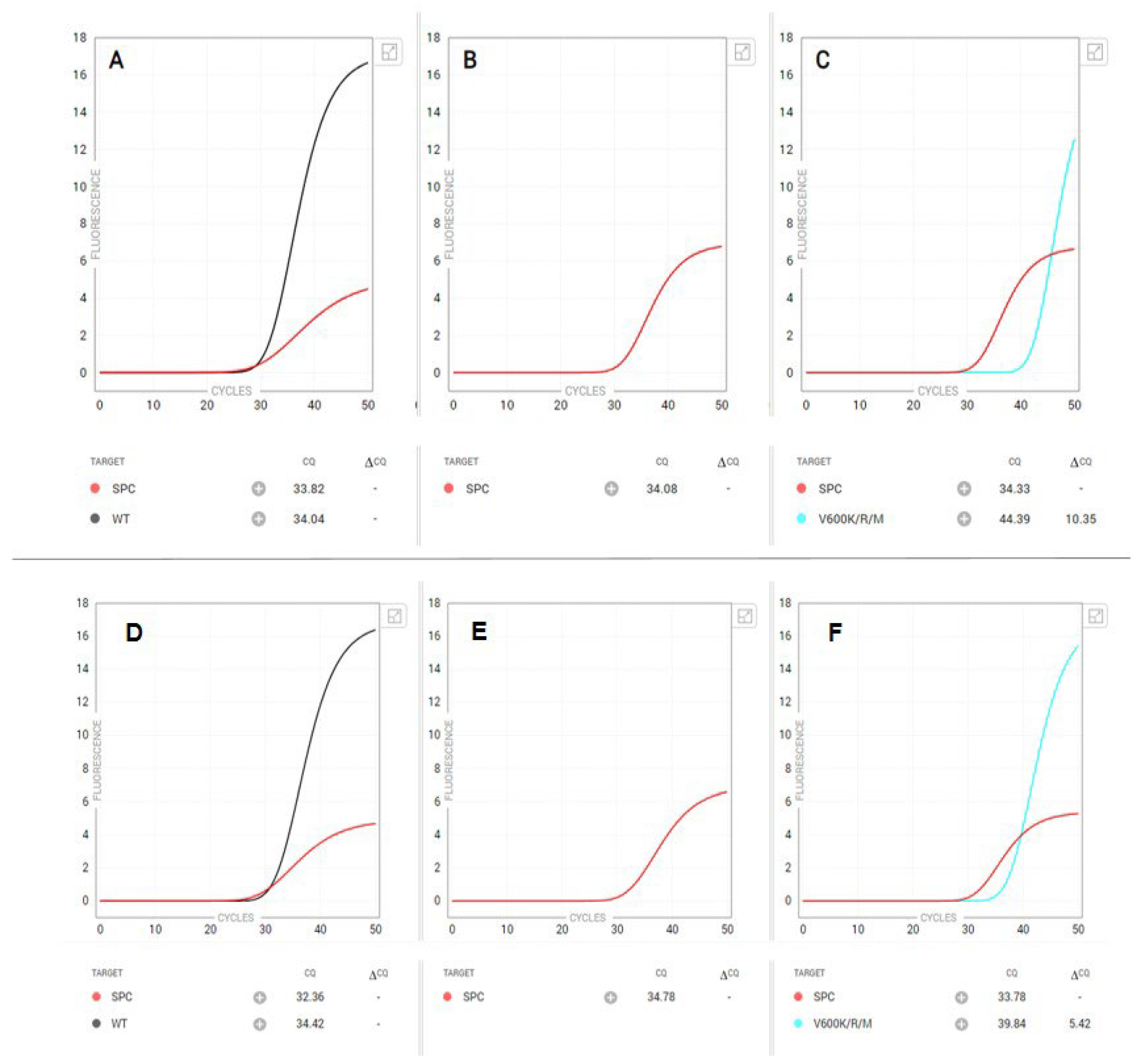
A classic BRAF V600-positive amplification curve is depicted in Figure 1 (top panel), in which a V600E/E2/D mutation was detected by the Idylla assay. This corresponded to the NGS results, which revealed a BRAF c.1799T > A, p.Val600Glu mutation with a VAF of 53%. Figure 1 (bottom panel) highlights another classic case, in which a BRAF V600 K/R/M was detected by the Idylla BRAF assay. NGS later revealed two BRAF mutations: BRAF c.1798_1799delGTinsAG, p.Val600Arg (VAF: 43%) and BRAF c.910G > A, p.Glu304Lys (VAF: 40%) and a sub-clonal CTNNB1 c.133T > C, p.Ser45Pro mutation with a VAF of 3%, which was consistent with the Idylla BRAF test result.
Limit of Detection: Two commercial standard DNA samples were serially diluted with Hapmap normal control DNA NA12878 to attain different VAFs. The HD705 contained BRAF c.1799T > A, p.Val600Glu at a 5% VAF. The HD123 contained BRAF c.1798_1799GTdelinsAA, p.Val600Lys at a 5% VAF. Idylla was able to correctly call the BRAF V600E and V600K variants at VAFs as low as 1.25% with 50 ng input DNA (Supplementary Table S3). Thus, the limit of detection was determined to be 1.25% VAF and above for BRAF V600E and V600K variants at an input of 50 ng DNA.
Input Limit: We tested two commercial BRAF standard DNA mixtures to determine the lowest permissible input amount. The aforementioned VAFs were noted. The assay was performed at three input levels (12.5 ng, 25 ng, and 50 ng DNA). The BRAF V600E and V600K variants were successfully detected at all three input amounts (Supplementary Table S4).
We further established the lowest permissible DNA input for variant detection at 1.25% VAF. The lowest DNA input required for the Idylla BRAF cartridge to detect both BRAF variants at 1.25% VAF was 50 ng (Supplementary Table S4). Taken together, the required input for the assay is at least 50 ng DNA to ensure variants with the lowest VAFs will be detected. Should a specimen contain a low tumor cellularity percentage, macro-dissection may be performed to enrich the tumor. Lower inputs may be attempted in cases of limited nucleic acid; however, the limit of detection will be compromised. Negative results in such cases should be considered uninformative.
Precision and Reproducibility: The assay was performed using three BRAF-positive and three BRAF-negative clinical samples. We assessed inter-technologist reproducibility by testing each sample on different days by two technologists. Intra-instrument reproducibility was assessed by testing each sample using different instruments on different days. In all cases, results were concordant and the range of ΔCq values between triplicate runs was ≤1.0 cycles (Supplementary Table S5). Thus, the assay was deemed to be highly reproducible.
Quality Control Metrics: Metrics for determining sufficient DNA input (valid assay) were established by the manufacturer. When the console displays a “MUTATION DETECTED IN BRAF CODON 600” result, in case of a valid assay, the variant is either BRAF V600E/E2/D or V600K/R/M. Both the PCRs for wild type BRAF and the V600E/E2/D or V600K/R/M variants will have generated valid curves, indicating that a mutation has been detected in codon 600 of the BRAF gene.
Conversely, when the console shows a “NO MUTATION DETECTED” result, only the PCR for wild type BRAF has generated a valid curve, and no mutations in BRAF V600E/E2/D or V600K/R/M have been detected within the validated ΔCq range. The result is dependent on the integrity of the specimen DNA, the percentage of mutant sequences present in the specimen, the absence of inhibiting substances, and the presence of sufficient amplifiable DNA.
In the case of a limited DNA sample, a BRAF-negative sample will be marked with V600K/R/M mutation <5% and may not be detected. Should the console display “INSUFFICIENT DNA INPUT,” it indicates that the DNA amount in the sample is out of range (too low or too high) to ascertain a reliable genotype call. Repeating the assay with a new cartridge, using more sample input but not exceeding the maximum allowed amount of sample, may rectify this issue.
An INVALID result may be reported in the following scenarios: in the presence of inhibitors in the sample, with severe DNA fragmentation potentially caused by a long formalin fixation time, with incorrect placement of a sample in the cartridge, if the sample volume is out of range, if no sample is added, if cartridges are incorrectly stored, with cartridges that have exceeded the in-use period after removal from the pouch, or with cartridge malfunction.
3.2. Discrepant Idylla V600-Positive Amplification Profiles and Corresponding NGS Data
Since its validation and implementation in the summer of 2021, our laboratory has successfully performed over 200 Idylla BRAF mutational assays on DNA or FFPE slides from various tissues. During the course of routine clinical practice, we also noticed cases in which BRAF V600 mutations were identified by the Idylla assay but were later found to be discordant with NGS results. We describe our observations below.
3.3. Unusual “Double Amplification” Idylla V600 Amplification Curves and NGS Results
In one case (Figure 2—top panel), a V600 K/R/M mutation was detected by the Idylla BRAF assay; however, late amplification was also noticed within the V600 E/E2/D channel. Subsequent NGS revealed a BRAF c.1798_1799delGTinsAA, p.Val600Lys mutation at a VAF of 35%, consistent with the Idylla BRAF V600 K/R/M result. Manual inspection of the sequencing reads did not identify an additional BRAF V600 variant above the background level.
Similarly, in another case (Figure 2—bottom panel), a BRAF V600 E/E2/D mutation was identified by the Idylla BRAF assay as depicted by early amplification; however, late amplification was also observed in the BRAF V600 K/R/M channel. NGS revealed a BRAF c.1799T > A, p.Val600Glu mutation at a VAF of 40% (consistent with the Idylla V600 E/E2/D result) and a small, sub-clonal MAP2K1 c.371C > T, p.Pro124Leu variant at a VAF of 5%. Again, manual inspection of the sequencing reads did not identify an additional BRAF V600 variant above the background level. We also identified an additional case with a BRAF c.1799T > A, p.Val600Glu mutation at a VAF of 26% (consistent with the observed Idylla V600 E/E2/D result), with additional mutations detected in ALK, ERBB3, and PIK3CA.
3.4. False-Positive Idylla BRAF V600 Results
Additionally, we observed two false-positive calls by the Idylla BRAF mutation assay. Figure 3 (top panel) depicts a case with substantially delayed amplification curve in the V600K/R/M channel. NGS on this FFPE specimen identified a BRAF c.1789_1790delCTinsTC (p.Leu597Ser) variant at a VAF of 29%, along with mutations in ALK and FGFR3. Figure 3 (bottom panel) depicts an additional case in which a BRAF V600 K/R/M was identified with late normal amplification. NGS on this specimen identified a BRAF c.1797_1798insACA (p.Thr599dup) variant at a VAF of 53%.Manual inspection of the sequencing reads did not identify an additional BRAF V600 variant above the background level in either case.
3.5. Idylla BRAF Testing of NGS-Detected Non-V600 BRAF Variants
Given the discrepancies and false-positive Idylla results, we initiated an institutional quality improvement effort to systematically and retrospectively evaluate NGS-tested FFPE specimens that were positive for non-V600 BRAF variants within five amino acids of codon V600 (n = 9). Of the nine tested specimens with non-V600 BRAF variants, none showed any amplification (Figures S1–S9). Thus, only specimens with BRAF c.1789_1790delCTinsTC (p.L597S) and c.1797_98insACA (p.T599dup) variants led to false-positive V600 K/R/M amplification curves.
3.6. Potential Clinical Significance of Non-V600 BRAF Variants in Idylla BRAF V600-Positive Cases
The Idylla BRAF assay is a rapid test requiring minimal technologist hands-on time. However, it remains unclear how frequently BRAF V600 mutation-bearing tumors harbor additional BRAF variants, which could potentially alter their sensitivity to BRAF inhibitor therapy. To address this issue, we reviewed all in-house Idylla BRAF V600-positive specimens for which we had corresponding NGS data. As described above, by NGS we identified several samples bearing non-V600 BRAF variants, which, as expected, proved negative by the Idylla BRAF V600 assay (see Figures S1–S9). However, some of these variants, such as L597R and L597V, are known to be sensitive to BRAF inhibitors [12,13]; thus, their detection would be of both diagnostic and therapeutic importance.
We searched for specimens bearing two BRAF variants, V600 and non-V600. Of 311 BRAF V600 mutation-positive specimens, we only discovered two that also harbored a second BRAF variant. One of these cases proved to be the aforementioned Idylla BRAF V600 mutation-positive specimen (Figure 1—bottom panel) harboring BRAF c.1798_1799delGTinsAG, p.Val600Arg, and BRAF c.910G > A, p.Glu304Lys variants at a VAF of 43% and 40%, respectively. Using our in-house NGS platforms, the amplicon-based Oncomine Focus Assay (OFA) (ThermoFisher, Inc., Waltham, MA, USA) and Illumina TruSight myeloid assay, we could not determine if these two variants were on the same allele (i.e., in cis) or on different alleles (i.e., in trans), although their VAFs were sufficiently close for either possibility to be plausible.
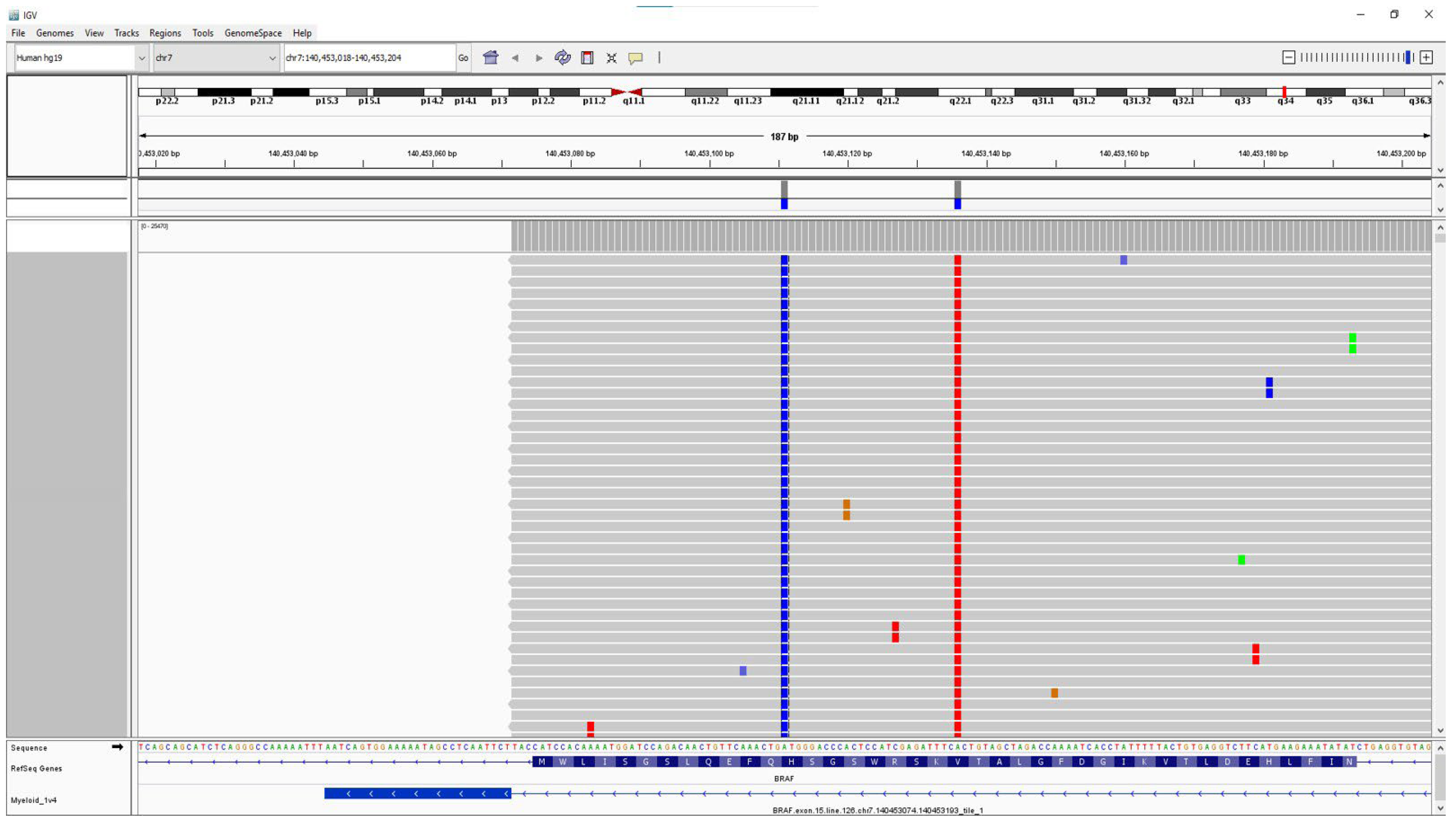
However, we identified one sample from a patient with HCL that was BRAF V600 E/E2/D-positive by the Idylla BRAF assay, which harbored BRAF c.1799T > A (p.Val600Glu) and BRAF c.1824T > G (p.His608Gln) variants in cis at VAFs of 14%, as identified by NGS (Figure 4). Of note, both variants are within a region that encodes the protein kinase domain of the B-raf protein. Thus, in a very small subset of cases, both BRAF V600 and non-V600 variants can occur on the same allele and may have an influence on the efficacy of BRAF V600 mutation-targeted inhibitor therapy.
4. Discussion
Identification of BRAF mutations is of major importance in the diagnosis of several tumor types, both solid and hematologic. As such, the rapid identification of BRAF mutational status is required to determine eligibility for targeted BRAF inhibition therapy. In this study, we describe the validation of the Idylla BRAF mutation assay and report several cases in which BRAF V600 mutations were identified by the Idylla assay but were later found to be discordant with NGS results. We thus initiated an institutional quality improvement effort to resolve these discrepancies.
We validated the Idylla BRAF mutation assay using previously extracted DNA from 19 samples, including 14 clinical samples, three CAP proficiency samples, and two commercially available reference samples. All samples had been tested for BRAF mutational status via previously validated assays and were tested for assay performance (nucleic acid variant concordance and tissue variant concordance), limit of detection, input limit, precision and reproducibility, and quality control metrics. The clinical samples were orthogonally tested using NGS. Across all sample types, 12 samples were found to have mutations in BRAF V600E/E2/D, three possessed mutations in BRAF V600K/R/M, and four samples were found to be BRAF wild type. Overall, we found the clinical sensitivity and specificity of the BRAF mutation Idylla assay to be 100%. Concordant with our validation results, other studies have also shown the Idylla BRAF mutation assay to possess high sensitivity, specificity, and remarkable concordance to existing reference methods [9,14].
During the course of routine clinical practice, however, we also noticed two cases in which BRAF V600 mutations were identified by the Idylla assay but were later found to be discordant with NGS results (Figure 2). In both cases, we observed double amplification, with early normal amplification in one channel but delayed amplification in a separate channel. NGS, however, only identified a single BRAF V600 variant in all three cases. We surmise that late amplification in a double amplification pattern likely represents non-specific amplification and may not necessarily warrant confirmatory NGS testing.
Reciprocally, the presence of non-V600 BRAF variants (as identified by NGS) should not lead to an unusual Idylla V600 amplification profile, and even if two BRAF variants are present in cis (i.e., affecting the same allele), the Idylla amplification curves should not be affected by the proximity of the second variant. As mentioned prior, we identified a sample from a patient with HCL that was BRAF V600 E/E2/D-positive by the Idylla BRAF assay and harbored BRAF c.1799T > A (p.Val600Glu) and BRAF c.1824T > G (p.His608Gln) variants in cis at VAFs of 14%, as identified by NGS (Figure 4). In patients with HCL who develop resistance to first-line therapy with the purine analog 2-chloro-2′-deoxyadenosine [2-CdA] (cladribine), the use of BRAF inhibitor therapy is the standard of care. BRAF H608Q has not been biochemically characterized and is considered a variant of unknown significance due to its absence from the gnomAD population database [15]. Of note, the expression of an adjacent variant, BRAF Q609H, results in similar cell proliferation and viability levels as wild type B-raf in cell culture [16] and is thus predicted to have no effect on B-raf protein function. Consequently, BRAF H608Q may also be a neutral variant in itself. Yet, its presence in cis may affect the binding of allosteric BRAF inhibitors to V600E-mutant BRAF molecules, rendering them less effective, as has been demonstrated prior in a similar double BRAF mutation-bearing case [17] and which may be predictable using molecular dynamics simulations [18]. In such rare cases, frequent follow-up testing for minimal residual disease testing by highly sensitive BRAF assays may be warranted.
Additionally, we observed two false-positive calls by the Idylla assay (Figure 3), in which subsequent confirmatory NGS revealed non-V600 BRAF variants. This prompted us to initiate an institutional quality improvement effort to evaluate systematically and retrospectively NGS-tested cases within five amino acids of BRAF codon V600. Of the nine cases identified, none showed any amplification, confirming the overall high fidelity of the Idylla BRAF assay. We hypothesize that samples with single delayed amplification curves are due to the presence of either non-V600 variants, very low-level V600 variants, cytosine deamination artifacts, and/or non-specific amplification by an allele-specific PCR primer. Regardless, we recommend these cases undergo reflex NGS testing for further evaluation.
Cytosine deamination, in which C/G → T/A, is a possible consequence of histological formalin fixation [19]. Tissue preservation is enabled through formalin cross-linkage with uncharged reactive amino acids; however, rarely, such as in cases of prolonged fixation or exposure to cold ischemia, this preservation process also causes nucleic acid fragmentation [20,21]. In-vivo, this error is repaired by intracellular enzymes, such as uracil DNA glycosylase (UDG)/uracil-N-glycosylase (UNG) and 5-methylcytosine DNA glycosylase [22]. When interpreting NGS data derived from FFPE specimens, low-frequency mutations may reflect such deamination artifacts [19]. We posit whether the two false-positive cases we initially observed may stem from cytosine deamination artifacts consequent to formalin fixation.
Kim et al. report that deamination artifacts may result from poor fixation methods with prolonged exposure, acidic or basic pH, and/or delayed fixation [19]. To mitigate deamination-induced alterations, they implemented UDG pre-treatment, which only affected these changes minimally [19]. Prentice et al. observed no statistical difference in sequencing performance and the number of mutational calls between samples that were pre-treated with UNG versus those that did not undergo pre-treatment until the 48 h fixation mark [21]. After 48 h, they observed a significant increase in deamination artifacts, suggesting that these events may be triggered by a prolonged fixation time [21]. Berra and colleagues support UDG pre-treatment of FFPE specimens intended for NGS, noting a medium reduction in transitions of 80% with sequence artifacts presenting at a VAF < 10% after pre-treatment [22]. Methylated CpG sequences, however, remain despite UDG pre-treatment [19,21].
At our institution, since the fixation times for specimens vary, we are unsure whether this may have caused the false-positive results we observed with the Idylla BRAF assay. Interestingly, enhanced DNA integrity has been observed in cold-formalin (4 °C) fixed tissues, which may circumvent the need to monitor formalin fixation times across several specimen types [23]. Although NGS is generally considered the gold standard of mutational analysis, the Idylla BRAF mutation assay possesses a number of advantages. Unlike NGS, the Idylla BRAF mutation assay is a rapid multiplex RT-qPCR with an impressive TAT of approximately 90 min and a technologist hands-on time of fewer than three minutes. In the case of NGS, low sample volume and the specifics of testing typically require sample batching, with prolonged turnaround times (12–15 days). Unlike the Idylla assay, NGS is also highly complex (with a complicated bioinformatics pipeline) and labor-intensive, requiring highly skilled personnel, precluding its wide use and availability at smaller hospitals. Thus, the Idylla assay serves as an apt, cost-effective alternative for prompt BRAF mutation identification to enable targeted therapy.
Our study has several limitations. Our sample size was too limited to achieve general conclusions and our observations were descriptive. Additionally, we were not able to control for variables, such as fixation times, which may affect Idylla and NGS assays differentially. Regardless, we recommend that Idylla BRAF cases with single delayed amplification curves (or even uncertain cases of late amplification with double amplification) undergo reflex NGS testing for further evaluation. More generally, given their potential diagnostic and therapeutic significance, Idylla amplification curves of all BRAF variants should be inspected and carefully assessed prior to result reporting.
Supplementary Materials
The following supporting information can be downloaded at: https://www.mdpi.com/article/10.3390/genes15050527/s1. Figures S1–S9: Idylla amplification profiles of NGS-tested specimens with non-V600 BRAF variants that were ±5 amino acids of the BRAF V600 codon. Table S1: Nucleic Acid Variant Concordance; Table S2: Tissue Variant Concordance; Table S3: Limit of Detection; Table S4: Input Limitation for 1.25% and 5% VAF; Table S5: Reproducibility.
Author Contributions
This study was designed by Z.N.O. with input from G.V.G. and A.N.J. All validation experiments were carried out by H.L. The manuscript was written by G.V.G. and Z.N.O. with input from A.N.J. All authors have read and agreed to the published version of the manuscript.
Funding
This research received no external funding.
Institutional Review Board Statement
This manuscript contains secondary research on data or specimens for which no consent is required.
Informed Consent Statement
Not applicable.
Data Availability Statement
No new data were created or analyzed in this study. Data sharing is not applicable to this article.
Acknowledgments
We thank Daniel Bach, E. Adele Blake, John Fitzsimmons, Danielle Hewitt, and Veronica Marisa for performing the Idylla BRAF assays; Saide Nur Okutan and Cynthia Tang for performing NGS testing; and William Crowe for bioinformatics assistance.
Conflicts of Interest
The authors declare no conflict of interest.
References
- Ascierto, P.A.; Kirkwood, J.M.; Grob, J.J.; Simeone, E.; Grimaldi, A.M.; Maio, M.; Palmieri, G.; Testori, A.; Marincola, F.M.; Mozzillo, N. The role of BRAF V600 mutation in melanoma. J. Transl. Med. 2012, 10, 85. [Google Scholar] [CrossRef] [PubMed]
- Yang, T.T.; Yu, S.; Ke, C.K.; Cheng, S.T. The Genomic Landscape of Melanoma and Its Therapeutic Implications. Genes 2023, 14, 1021. [Google Scholar] [CrossRef] [PubMed]
- Cohn, A.L.; Day, B.M.; Abhyankar, S.; McKenna, E.; Riehl, T.; Puzanov, I. BRAF(V600) mutations in solid tumors, other than metastatic melanoma and papillary thyroid cancer, or multiple myeloma: A screening study. Onco Targets Ther. 2017, 10, 965–971. [Google Scholar] [CrossRef] [PubMed]
- Maitre, E.; Macro, M.; Troussard, X. Hairy cell leukaemia with unusual BRAF mutations. J. Cell Mol. Med. 2023, 27, 2626–2630. [Google Scholar] [CrossRef] [PubMed]
- Yap, J.; Yuan, J.; Ng, W.H.; Chen, G.B.; Sim, Y.R.M.; Goh, K.C.; Teo, J.; Lim, T.Y.H.; Goay, S.M.; Teo, J.H.J.; et al. BRAF(V600E) mutation together with loss of Trp53 or pTEN drives the origination of hairy cell leukemia from B-lymphocytes. Mol. Cancer 2023, 22, 125. [Google Scholar] [CrossRef] [PubMed]
- Tafinlar (Dabrafenib); Package Insert; Novartis: Basel, Switzerland, 2013.
- ZELBORAF (Vemurafenib); Package Insert; Genetech: South San Francisco, CA, USA, 2011.
- BRAFTOVI (Encorafenib); Package Insert; Pfizer: Hong Kong, China, 2018.
- Schiefer, A.I.; Parlow, L.; Gabler, L.; Mesteri, I.; Koperek, O.; von Deimling, A.; Streubel, B.; Preusser, M.; Lehmann, A.; Kellner, U.; et al. Multicenter Evaluation of a Novel Automated Rapid Detection System of BRAF Status in Formalin-Fixed, Paraffin-Embedded Tissues. J. Mol. Diagn. 2016, 18, 370–377. [Google Scholar] [CrossRef] [PubMed]
- Nkosi, D.; Casler, V.L.; Syposs, C.R.; Oltvai, Z.N. Utility of Select Gene Mutation Detection in Tumors by the Idylla Rapid Multiplex PCR Platform in Comparison to Next-Generation Sequencing. Genes 2022, 13, 799. [Google Scholar] [CrossRef] [PubMed]
- Cox, A.J.; Crowe, W.E.; Yang, Q.; Zhang, B.; Oltvai, Z.N.; Liao, X. Clinicopathologic and Molecular Characterization of Anorectal Neuroendocrine Carcinomas Reveals Human Papillomavirus, p53, and c-Myc as Alternative Mechanisms of Carcinogenesis. Mod. Pathol. 2023, 36, 100295. [Google Scholar] [CrossRef] [PubMed]
- Bahadoran, P.; Allegra, M.; Le Duff, F.; Long-Mira, E.; Hofman, P.; Giacchero, D.; Passeron, T.; Lacour, J.P.; Ballotti, R. Major clinical response to a BRAF inhibitor in a patient with a BRAF L597R-mutated melanoma. J. Clin. Oncol. 2013, 31, e324–e326. [Google Scholar] [CrossRef] [PubMed]
- Johnson, D.B.; Zhao, F.; Noel, M.; Riely, G.J.; Mitchell, E.P.; Wright, J.J.; Chen, H.X.; Gray, R.J.; Li, S.; McShane, L.M.; et al. Trametinib Activity in Patients with Solid Tumors and Lymphomas Harboring BRAF Non-V600 Mutations or Fusions: Results from NCI-MATCH (EAY131). Clin. Cancer Res. 2020, 26, 1812–1819. [Google Scholar] [CrossRef] [PubMed]
- Janku, F.; Claes, B.; Huang, H.J.; Falchook, G.S.; Devogelaere, B.; Kockx, M.; Bempt, I.V.; Reijans, M.; Naing, A.; Fu, S.; et al. BRAF mutation testing with a rapid, fully integrated molecular diagnostics system. Oncotarget 2015, 6, 26886–26894. [Google Scholar] [CrossRef] [PubMed]
- Gudmundsson, S.; Singer-Berk, M.; Watts, N.A.; Phu, W.; Goodrich, J.K.; Solomonson, M.; Genome Aggregation Database, C.; Rehm, H.L.; MacArthur, D.G.; O‘Donnell-Luria, A. Variant interpretation using population databases: Lessons from gnomAD. Hum. Mutat. 2022, 43, 1012–1030. [Google Scholar] [CrossRef] [PubMed]
- Ng, P.K.; Li, J.; Jeong, K.J.; Shao, S.; Chen, H.; Tsang, Y.H.; Sengupta, S.; Wang, Z.; Bhavana, V.H.; Tran, R.; et al. Systematic Functional Annotation of Somatic Mutations in Cancer. Cancer Cell 2018, 33, 450–462.e10. [Google Scholar] [CrossRef] [PubMed]
- Choi, J.; Landrette, S.F.; Wang, T.; Evans, P.; Bacchiocchi, A.; Bjornson, R.; Cheng, E.; Stiegler, A.L.; Gathiaka, S.; Acevedo, O.; et al. Identification of PLX4032-resistance mechanisms and implications for novel RAF inhibitors. Pigment. Cell Melanoma Res. 2014, 27, 253–262. [Google Scholar] [CrossRef] [PubMed]
- Niu, Y.; Zhang, Y.; Yao, X. Resistance mechanism of the oncogenic beta3-alphaC deletion mutation in BRAF kinase to dabrafenib and vemurafenib revealed by molecular dynamics simulations and binding free energy calculations. Chem. Biol. Drug Des. 2019, 93, 177–187. [Google Scholar] [CrossRef] [PubMed]
- Kim, S.; Park, C.; Ji, Y.; Kim, D.G.; Bae, H.; van Vrancken, M.; Kim, D.H.; Kim, K.M. Deamination Effects in Formalin-Fixed, Paraffin-Embedded Tissue Samples in the Era of Precision Medicine. J. Mol. Diagn. 2017, 19, 137–146. [Google Scholar] [CrossRef] [PubMed]
- Howat, W.J.; Wilson, B.A. Tissue fixation and the effect of molecular fixatives on downstream staining procedures. Methods 2014, 70, 12–19. [Google Scholar] [CrossRef] [PubMed]
- Prentice, L.M.; Miller, R.R.; Knaggs, J.; Mazloomian, A.; Aguirre Hernandez, R.; Franchini, P.; Parsa, K.; Tessier-Cloutier, B.; Lapuk, A.; Huntsman, D.; et al. Formalin fixation increases deamination mutation signature but should not lead to false positive mutations in clinical practice. PLoS ONE 2018, 13, e0196434. [Google Scholar] [CrossRef]
- Berra, C.M.; Torrezan, G.T.; de Paula, C.A.; Hsieh, R.; Lourenço, S.V.; Carraro, D.M. Use of uracil-DNA glycosylase enzyme to reduce DNA-related artifacts from formalin-fixed and paraffin-embedded tissues in diagnostic routine. Appl. Cancer Res. 2019, 39, 7. [Google Scholar] [CrossRef]
- Berrino, E.; Annaratone, L.; Miglio, U.; Maldi, E.; Piccinelli, C.; Peano, E.; Balmativola, D.; Cassoni, P.; Pisacane, A.; Sarotto, I.; et al. Cold Formalin Fixation Guarantees DNA Integrity in Formalin Fixed Paraffin Embedded Tissues: Premises for a Better Quality of Diagnostic and Experimental Pathology With a Specific Impact on Breast Cancer. Front. Oncol. 2020, 10, 173. [Google Scholar] [CrossRef] [PubMed]
Figure 1.
Idylla BRAF classic PCR amplification curves for (top) BRAF V600E/E2/D and (bottom) V600K/R/M variants. The top panel displays (A) the PCR amplification curve for wild type BRAF, (B) a positive result for a BRAF V600E/E2/D mutation, and (C) a negative result for a K/R/M variant. Subsequently performed NGS on this specimen identified a BRAF c.1799T > A (p.Val600Glu) variant at a VAF of 53%. The bottom panel displays (D) the PCR amplification curve for wild type BRAF, (E) a negative result for a V600E/E2/D variant, and (F) a positive result for a BRAF V600K/R/M variant. Confirmatory NGS identified a BRAF c.1798_1799delGTinsAG (p.Val600Arg) variant at a VAF of 43%.

Figure 2.
Unusual Idylla BRAF PCR amplification curves. The top panel shows (A) the PCR amplification curve for wild type BRAF, (B) late amplification for a V600E/E2/D variant, and (C) early amplification yielding a positive result for a BRAF V600K/R/M variant. NGS on the same specimen identified a BRAF c.1798_1799delGTinsAA (p.Val600Lys) variant at a VAF of 35%. The bottom panel displays (D) the PCR amplification curve for wild type BRAF, (E) early amplification yielding a positive result for a BRAF V600E/E2/D variant, and (F) late amplification in the V600K/R/M channel. NGS revealed a BRAF c.1799T > A, p.Val600Glu mutation at a VAF of 40% (consistent with the Idylla V600 E/E2/D result) and an additional MAP2K1 variant.

Figure 3.
False-positive Idylla BRAF PCR amplification curves. The top panel shows (A) the PCR amplification curve for wild-type BRAF, (B) a negative result for a BRAF V600E/E2/D variant, and (C) a substantially delayed amplification curve in the V600K/R/M channel. NGS on this FFPE specimen identified a BRAF c.1789_1790delCTinsTC (p.Leu597Ser) variant at a VAF of 29%, along with mutations in ALK and FGFR3. The bottom panel displays (D) the PCR amplification curve for wild type BRAF, (E) a negative result for a BRAF V600E/E2/D variant, and (F) late normal amplification in the V600K/R/M channel. NGS on the same FFPE specimen identified a BRAF c.1797_1798insACA (p.Thr599dup) variant at a VAF of 53%.

Figure 4.
Integrated Genome Viewer (IGV) screenshot. IGV screenshot of a case of hairy cell leukemia (HCL) that proved positive for two BRAF variants, c.1799T > A (p.Val600Glu)(red) and c.1824T > G (p.His608Gln)(blue), in cis at VAFs of 14%.
Figure 4.
Integrated Genome Viewer (IGV) screenshot. IGV screenshot of a case of hairy cell leukemia (HCL) that proved positive for two BRAF variants, c.1799T > A (p.Val600Glu)(red) and c.1824T > G (p.His608Gln)(blue), in cis at VAFs of 14%.

Disclaimer/Publisher’s Note: The statements, opinions and data contained in all publications are solely those of the individual author(s) and contributor(s) and not of MDPI and/or the editor(s). MDPI and/or the editor(s) disclaim responsibility for any injury to people or property resulting from any ideas, methods, instructions or products referred to in the content. |
© 2024 by the authors. Licensee MDPI, Basel, Switzerland. This article is an open access article distributed under the terms and conditions of the Creative Commons Attribution (CC BY) license (https://creativecommons.org/licenses/by/4.0/).
Share and Cite
MDPI and ACS Style
George, G.V.; Liu, H.; Jajosky, A.N.; Oltvai, Z.N. Resolving Discrepancies in Idylla BRAF Mutational Assay Results Using Targeted Next-Generation Sequencing. Genes 2024, 15, 527. https://doi.org/10.3390/genes15050527
AMA Style
George GV, Liu H, Jajosky AN, Oltvai ZN. Resolving Discrepancies in Idylla BRAF Mutational Assay Results Using Targeted Next-Generation Sequencing. Genes. 2024; 15(5):527. https://doi.org/10.3390/genes15050527
Chicago/Turabian StyleGeorge, Giby V., Huijie Liu, Audrey N. Jajosky, and Zoltán N. Oltvai. 2024. "Resolving Discrepancies in Idylla BRAF Mutational Assay Results Using Targeted Next-Generation Sequencing" Genes 15, no. 5: 527. https://doi.org/10.3390/genes15050527
Note that from the first issue of 2016, this journal uses article numbers instead of page numbers. See further details here.