Aerosol Microbiome over the Mediterranean Sea Diversity and Abundance
by
 , , and
Esra Mescioglu
1,* ,
,
Eyal Rahav
2,
Natalia Belkin
2,
Peng Xian
3,
Jordan M. Eizenga
4,
Ania Vichik
2,
Barak Herut
2 and
Adina Paytan
5 1
Earth and Planetary Science, University of California, Santa Cruz, CA 95060, USA
2
Israel Oceanographic and Limnological Research, National Institute of Oceanography, Haifa 3108000, Israel
3
Marine Meteorology Division, Naval Research Laboratory, 7 Grace Hopper Avenue, Monterey, CA 93940, USA
4
Biomolecular Engineering, University of California, Santa Cruz, CA 95060, USA
5
Institute of Marine Science, University of California, Santa Cruz, CA 95060, USA
*
Author to whom correspondence should be addressed.
Atmosphere 2019, 10(8), 440; https://doi.org/10.3390/atmos10080440
Submission received: 18 June 2019
/
Revised: 20 July 2019
/
Accepted: 24 July 2019
/
Published: 1 August 2019
(This article belongs to the Special Issue Detection and Monitoring of Bioaerosols)
Abstract
:Prokaryotic microbes can become aerosolized and deposited into new environments located thousands of kilometers away from their place of origin. The Mediterranean Sea is an oligotrophic to ultra-oligotrophic marginal sea, which neighbors northern Africa (a major source of natural aerosols) and Europe (a source of mostly anthropogenic aerosols). Previous studies demonstrated that airborne bacteria deposited during dust events over the Mediterranean Sea may significantly alter the ecology and function of the surface seawater layer, yet little is known about their abundance and diversity during ‘background’ non-storm conditions. Here, we describe the abundance and genetic diversity of airborne bacteria in 16 air samples collected over an East-West transect of the entire Mediterranean Sea during non-storm conditions in April 2011. The results show that airborne bacteria represent diverse groups with the most abundant bacteria from the Firmicutes (Bacilli and Clostridia) and Proteobacteria (Alphaproteobacteria, Betaproteobacteria, and Gammaproteobacteria) phyla. Most of the bacteria in our samples have previously been observed in the air at other open ocean locations, in the air over the Mediterranean Sea during dust storms, and in the Mediterranean seawater. Airborne bacterial abundance ranged from 0.7 × 10⁴ to 2.5 × 10⁴ cells m−3 air, similar to abundances at other oceanic regimes. Our results demonstrate that airborne bacterial diversity is positively correlated with the mineral dust content in the aerosols and was spatially separated between major basins of the Mediterranean Sea. To our knowledge, this is the first comprehensive biogeographical dataset to assess the diversity and abundance of airborne microbes over the Mediterranean Sea. Our results shed light on the spatiotemporal distribution of airborne microbes and may have implications for dispersal and distribution of microbes (biogeography) in the ocean.
1. Introduction
Prokaryotic microorganisms are found in the air over the global ocean in substantial numbers, with a median abundance of 6.7 × 10³ m−3 air [1] and are referred to as ‘airborne microbes’. These airborne microbes can originate from both land [2,3] and the ocean [4,5]. Upon aerosolization, wind can transport microbes over great distances, including across large ocean basins and seas [6,7,8,9,10]. The residence time of microbes in the air can reach up to seven days [1], which enables them to cross thousands of kilometers. Airborne, microbes can be exposed to atmospheric oxidant gases [11] and meteorological factors, like variable temperatures and UV radiation [12], that can cause cell damage and reduce their viability. However, up to ~20% of these airborne microbes remain viable during atmospheric transport [13], and this has important implications for receiving ecosystems. Airborne microbes are deposited with dry (aerosol particles) or wet (rain) atmospheric deposition back onto Earth’s surface, including the surface of the ocean [9,14,15].
Airborne microbes include a diverse array of organisms, and their deposition can impact human health through spreading infectious diseases [16], agriculture through dispersal of plant pathogens [17], and ecosystem productivity and function through introduction of new organisms [18]. Recently, it was shown that abundance of microbes in outdoor air can be influenced by seasons, with Bragoszewska and Pastuszka [12] reporting highest abundance in spring and Kaarakainen et al. [19] reporting highest abundance in the summer. Interestingly, certain bacterial species, like Streptomyces and Cladosporium, have stronger temperature and seasonal variation than other species, like Penicillium and Aspergillus [19]. The diversity of microbes in outdoor air has also been shown to vary between environments [19], yet there are only a few studies investigating airborne microbes over the ocean. The few studies conducted so far indicate that airborne microbes are found ubiquitously over marine environments, but their abundance, diversity, and the factors driving their diversity are still poorly studied [3,20].
The Mediterranean Sea (MS) is an ideal marine environment to study airborne microbes. The MS is a low-nutrient low-chlorophyll (LNLC) ecosystem [21,22], and the surrounding landmasses provide ample aerosols: The densely populated land to the north is a source of anthropogenic aerosols, and the arid land to the south is a source of mineral dust [20]. The effects of the high atmospheric deposition in this basin (1–50 g dust m−2 y−1 [23]) has been studied extensively and shown to be important chemically, providing limiting micro (e.g., Fe, Zn) and macro (e.g., N, P) nutrients to the MS [24,25,26,27]. These leached nutrients support primary production in the mixed layer of the MS [28] and can stimulate N2 fixation, which may induce further primary production [28,29]. In addition to leached nutrients and trace metals, atmospheric deposition has been shown to add viable microbes to the MS [30]. These airborne microbes can fix N2 and utilize organic carbon (i.e., leucine) in seawater after deposition [30]. Therefore, airborne microbes may have an important contribution to the ecology of MS waters, with ecological implications for other LNLC settings receiving high atmospheric deposition, such as the North Atlantic Ocean.
Most studies investigating airborne microbes over the MS have focused on determining their diversity and abundance during dust storm events [20]. These studies showed that during storm events, airborne microbial abundance increases, and diversity is dependent on source [31,32]. However, it is also important to evaluate these variables during background conditions (clear days), which are far more common than dust storms events. Understanding background conditions may help identify what is unique about storm events that have resulted in measurable changes in the receiving water following deposition events [20]. Moreover, identifying airborne microbes and the factors driving their diversity over the ocean during background conditions may further our understanding of the mechanisms of bioaerosol dispersion, with possible implications for biogeography.
In this study, we analyzed aerosol samples collected at all major basins of the MS (Levantine, Ionian Sea, Tyrrhenian Sea, Algero-Provencal basin, Alboran Sea) during “normal” background non-dust-storm conditions in April 2011 (spring). We analyzed the microbial diversity using 16S rRNA sequencing and microbial abundance using microscopy. Due to the proximity of the MS to terrestrial sources of aerosols, we hypothesized that we would find a high number of airborne bacteria comprising a diverse community. We further hypothesized that this community would encompass both marine and terrestrial microbes.
2. Methods and Materials
2.1. Sampling
Samples were collected aboard the R/V Meteor (cruise M84/3) during an east to west transect in the MS from 6th to 28th April 2011 (Figure 1). Aerosols were collected in all major basins onto Whatman 41 filters for 24 h using a high-volume sampler pumping air at 42 m3 h−1 [30]. The sampler was positioned at the front of the ship (to reduce collection of ship emissions) and samples were processed in an aerosol designated laboratory. Volumes of air pumped, and the start and end coordinates of sample collection were recorded (Supplementary Table S1). The filters were frozen and kept at −80 °C until processing.
2.2. Aerosol Optical Depth
To assemble additional information about the aerosols present over the MS at the time of sample collection, we used a global 1 × 1 degree and six-hourly 550 nm aerosol optical depth (AOD, an approximate measure of total atmospheric column of aerosol mass) reanalysis product that was developed and validated at the Naval Research Laboratory, CA, USA [33]. The core model of this aerosol reanalysis product is the Navy Aerosol Analysis and Prediction System (NAAPS), which characterizes anthropogenic and biogenic fine aerosol species (ABF, including pollutions from industry, fossil fuel and biofuel, and organic aerosols), dust, biomass-burning smoke and sea salt aerosols. The reanalyzed aerosol fields were obtained by running NAAPS with the assimilation of quality-controlled retrievals of AOD from moderate resolution imaging spectroradiometer (MODIS) on Terra and Aqua and the multi-angle imaging spectro radiometer (MISR) on Terra [34,35,36]. The fine and coarse mode AOD at 550 nm from the reanalysis is shown to have good agreement with the ground-based global scale sun photometer Aerosol Robotic Network (AERONET) observations regionally and seasonally [33]. Speciated AOD data were extracted (with the nearest neighbor method) from the NAAPS reanalysis along the ship track for the study period. Correlational relationships were analyzed between bacterial abundance and diversity and ABF, dust and total AODs, to compare to studies that have found increased abundances of bacteria associated with elevated pollution [37] and dust [38] levels.
2.3. Air Mass Backward Trajectories
Seventy-two-hour isentropic back trajectories were constructed from the National Oceanic and Atmospheric Administration (NOAA) database using the hybrid single-particle Langrangian integrated trajectories (HYSPLIT) program [39]. Back trajectories for elevations 50, 250, and 500 m were computed using the GPS coordinates of the midpoint between the start and end locations of sampling for each filter (Supplementary Table S1, Supplementary Figure S1). The samples were assigned to one of four origin zones according to the geographic location from which the airmass originated, as determined from the backward trajectory model results (Table 1, Supplemental Figure S1). The four zones are Western Europe (Z1), Eastern Europe (Z2), Mediterranean Sea (Z3), Northern Africa (Z4) (Figure 1). Note that in some cases the airmass crossed more than one zone during collection (Supplemental Figure S1).
2.4. Region and Distance to Land
Samples were grouped according to the location of collection (Figure 1, Table 1) in order to determine if the diversity was influenced by location and if proximal sites had similar diversity. We also measured the distance from the closest landmass, including islands, at five points of sampling (beginning, quarter-point, midpoint, three-quarters point, and end) for each sample, and used the average of the five values as the distance from land in our analysis (Table 1).
2.5. Aluminum
After collection, a subsample of the Whatman 41 filters was dried in a desiccator for 24 h before being reweighed. Filters were digested with hydrogen fluoride (HF) following the procedure of ASTM (1983) [27]. Aluminum (Al) concentrations in the bulk digest were measured on an atomic absorption spectrometer Agilent 280FS AA and graphite furnace Agilent 240Z AA.
2.6. Bacterial Abundance
Subsamples from each of the filters were cut with sterile scissors (3 × 3 cm), placed into 5 mL of sterile MS water, and fixed with ultrapure glutaraldehyde solution (Sigma, St. Louis, MO USA, final concentration 0.02% v:v). The filters were sonicated for 5 min to detach organisms from the filter, stained with SYBR green solution (Applied Biosystems, Foster City, CA USA), and filtered through a 0.2 µm polycarbonate filter (PALL). The filters were placed on a microscope slide, and bacterial cells were enumerated using epifluorescence microscopy (Olympus BH12). The values were normalized to the area of the whole filter (17 × 23 cm) and divided by the volume of air pumped during collection to determine the number of cells per m3 of air. SYBR green is a robust bacterial stain [40] used in numerous microbiology studies, including aeromicrobiology studies [1,41]. We used microscopy-grade SYBR, so the introduction of counting errors is unlikely.
2.7. DNA Extraction, Amplification, Sequencing
Subsamples from each of the filters were cut with sterile scissors (2 cm × 2 cm), and total DNA was extracted in triplicates using the phenol chloroform method, modified from Massana et al. [42]. The triplicates were pooled into one sample to ensure enough DNA for sequencing. The DNA was sent to Mr. DNA Molecular Research Laboratories. Polymerase chain reaction (PCR) using primers 515 (forward) and 806 (reverse) to amplify 16S rRNA, with barcodes on the forward primer, were carried out using the HotStarTaq Plus master mix kit (Qiagen, Valencia, CA USA). The conditions of the protocol were as following: 94 °C for 3 min, 28 cycles of 94 °C for 30 s, 53 °C for 40 s and 72 °C for 1 min, and a final elongation step at 72 °C for 5 min. PCR products were visualized in 2% agarose gel using electrophoresis to confirm successful amplification. The samples were pooled together in equal proportions (based on their molecular weight and DNA concentrations), purified using calibrated Ampure XP beads, and used to prepare libraries using a Nextera DNA sample preparation kit (Illumina, Foster City, CA USA). Libraries were loaded to a 600 cycles v3 reagent cartridge (Illumina) and the sequencing was performed on Miseq (Illumina). DNA extraction and amplification protocols were repeated for blank filters brought onboard the cruise and treated similarly to the samples, and the PCR products were checked by electrophoresis. The electrophoresis visualization showed no amplification bands indicating there was no contamination by the filters (i.e., no microbes present on the blank filters).
2.8. Bioinformatics
Samples were processed using the open-source Quantitative Insights into Microbial Ecology 2 (QIIME 2) pipeline [43]. Sequences were demultiplexed and barcodes were trimmed using the cutadapt plugin [44]. Data were denoised using dada2 [45], sequences were clustered into amplicon sequence variants (ASVs) which can be thought of as 100% operational taxonomic units (OTUs). Taxonomic classifier was trained [46] using Greengenes [47]. Taxonomies were assigned using the Naive Bayes method [48]. Samples were filtered to remove sequences identified as mitochondria and chloroplast. Alpha-diversity metrics, observed OTUs and Shannon’s diversity index [H] [49], beta diversity metrics (weighted UniFrac [50]), and principle coordinate analysis (PCoA) were estimated using q2-diversity after samples were rarefied (subsampled without replacement). The samples were grouped according to the location in which they were collected in the MS (i.e., region) (Figure 1) and the origin of the air mass (Figure 1) to test how abundance, richness, and diversity were influenced by these factors. Weighted UniFrac distances (a quantitative measure of community dissimilarity that incorporates phylogenetic relationships between the bacteria) were used to generate the PCoA plots. Associations between regions of sample collection and UniFrac were tested using PERMANOVA [51] to investigate whether microbial communities in samples within a region (e.g., Eastern MS) were more similar to each other than they were to samples from the other regions (e.g., Central MS and Western MS). We also tested for any association between geographical distances and community dissimilarity (weighted UniFrac) using the Mantel test. To simplify visualization of relative abundance, we clustered bacteria into two categories based on their relative abundance in our samples: (1) “Common” bacteria (families that made up more than 5% of at least 1 sample) (Table 2), and (2) “rare” bacteria which did not meet the 5% relative abundance threshold (Table 3).
3. Results and Discussion
3.1. Aerosol Origin and Chemical Properties
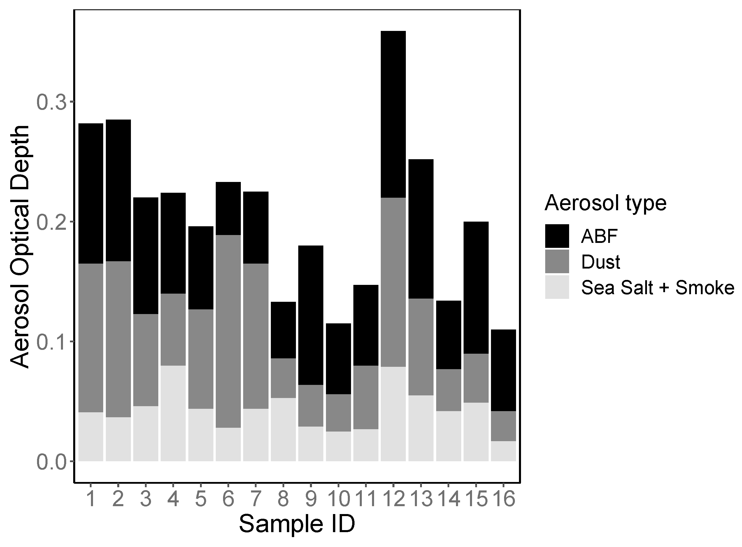
The aerosol optical depth (AOD) data derived from the Navy Aerosol Analysis and Prediction System (NAAPS) AOD reanalysis, as described in the methods section are shown in Figure 2. Total AOD, which includes mineral dust, anthropogenic and biogenic fine aerosol species (ABF), smoke, and sea salt, during the cruise ranged from 0.11 to 0.36, with the lowest values measured during collection of sample 16 and the highest measured during collection of sample 12, both collected from the Western MS (Figure 1 and Figure 2, Table 1). ABF and mineral dust were the main contributors to the total AOD during our study, together comprising between 60% and 88% of total AOD. Smoke and sea salt estimates from the NAAPS model were both relatively low in concentration and evenly distributed in all the samples. Smoke and sea salt contributed only to a small portion of total AOD during our sampling period, and thus were not included in further analysis.
The average AOD fraction, based on NAAPS reanalysis, attributed to dust in the MS during the month of April for 2003–2018 was on the order of 0.1–0.2 with a decreasing gradient from the south (closer to the African continent, the main aerosol source) to the north, and generally decreasing from east to west. However, this long-term average for April likely included some dust-storm events. During our sampling (April 2011) dust AOD on April 7–8, (samples 1 and 2), April 12–13 (samples 6 and 7) and April 18 (sample 9) was relatively high compared to other days (Figure 2). However, dust AOD for these days was still low compared to dust contribution to AOD during storms, which can frequently exceed 1.0 [52,53]. From the low-level wind and the movement of dust plumes based on NAAPS reanalysis and NOAA HYSPLIT back trajectories (Supplementary Figure S1), dust detected at the location of the ship on April 7–8 (samples 1 and 2) likely originated from Turkey. The April 12–13 (sample 6 and 7) dust peak observed is related to a dust storm that occurred in the northwest of Africa on April 5 (with maximum dust AOD around 2.0) [52]. NAAPS reanalysis shows that the dust plume originating from this storm moved northwest and reached 60° N on April 9 and then moved southeastward and reached the location of the ship on April 12. After this long-range transport, dust AOD was much weaker when it arrived at the MS (0.14). As this air mass traveled over the European continent, it mixed with anthropogenic aerosols (ABF). Throughout the cruise, ABF ranged from 0.04 to 0.14, with the lowest ABF AOD during collection of sample 6, in the central MS, and the highest during collection of sample 12 in the western MS (Table 1, Figure 2).
Aluminum (Al) concentration, a proxy for mineral dust [27], ranged between 41 and 661 ng m−3 air, and was highest on April 13 during collection of sample 7, which occurred when the dust storm originating from northwest Africa arrived in the MS. Overall, Al measurements were positively correlated with total AOD (Spearman correlation: = 0.694, p = 0.004), and especially with the AOD fraction attributed to dust (Spearman correlation: = 0.834, p < 0.0001) (Table 1). There was also a significant positive correlation of Al concentrations and longitude, with more Al in samples collected in the air above the Eastern MS than above the Western MS (Spearman correlation: = 0.597, p = 0.017) (Table 1).
Overall, the aerosol concentration in the air during our sampling campaign (background non-dust-storm period), particularly the mineral dust (as derived from the dust fraction of AOD and Al), were within the range of previously measured values in days without dust storms and about an order of magnitude lower than values recorded during dust storm event in the region [52,53].
3.2. Airborne Bacterial Abundance
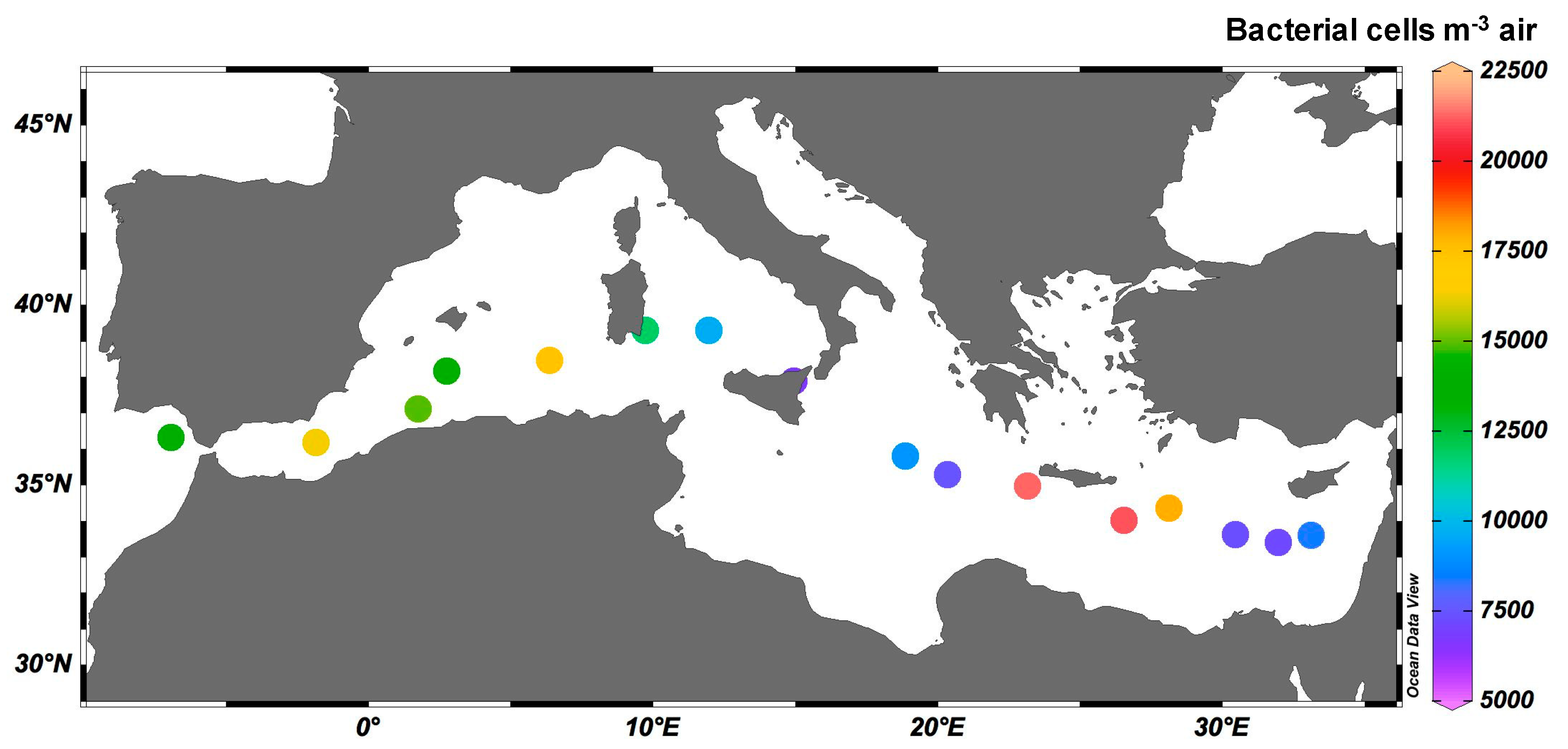
Bacterial abundances in our samples ranged from 103 to 104 cells m−3 air (Figure 3). The highest abundance of bacteria was measured in sample 6 in the Central MS (2.12 × 10⁴ cells m−3 air), near the island of Crete, during the arrival of the tail of the dust storm that originated from North Africa. The lowest abundances were measured in the Central and Eastern Mediterranean (6.64 × 10³ to 7.17 × 10³ cells m−3 air) in samples 9 and 4, respectively. Bacterial abundances in aerosols collected over the MS were in agreement with previous studies from the eastern Mediterranean coast [10] and the Atlantic, Pacific, and Indian ocean basins [1], yet were lower than those reported in the East China Sea [54] and the Red Sea [55]. Rahav et al. [10] measured the abundance of airborne prokaryotes at a coastal site located at the easternmost MS during 34 sampling events (between 2015 and 2018) and found that abundances were positively correlated to the concentration of aerosols in the air (mg m−3 air). Here, however, we did not find such a correlation, likely because the range of concentrations during non-dust-storm conditions, represented by our samples, was relatively small in comparison to previous studies. Mayol et al. [1] measured bacterial abundances in the Atlantic, Indian, and Pacific Ocean basins and found that sites closer to land (including islands) had significantly higher numbers of airborne microbes (normalized to the aerosol mass) than those further away from landmasses. This was also not observed in the MS, possibly because the MS is surrounded by land, and all sampling sites are relatively close to land when compared to samples obtained in the open ocean by Mayol et al. [1].
3.3. Airborne Microbiome above the MS
Fifty-nine unique families of bacteria were found in the samples collected during this study. The relative abundance was used to group bacteria into two categories: “Common” (Table 2) or “Rare” (Table 3). Families that had a relative abundance of 5% or greater in at least one of our samples were considered “Common”, and families that did not meet the 5% threshold were considered “Rare”. Common bacteria in our samples belonged to five phyla: Actinobacteria (three families), Bacteroidetes (two families), Firmicutes (eight families), Proteobacteria (eleven families), and Deinococcus-Thermus (one family) (Table 2). These bacteria are of variable gram stains, have diverse oxygen requirements, spore formation, and come from many different habitats (Supplementary Table S2). Five bacteria in our samples, Chitinophagaceae sediminibacterium, Clostridiaceae SMB53, Veillonellaceae spp., Moraxellaceae acinetobacterlwoffii, and Sinobacteraceae spp., had not previously been reported in aerosol samples. All other organisms have previously been identified in airborne bacterial studies in different locations around the world (Supplementary Table S2) and may represent the consortium of bacteria that are more likely to be aerosolized, transported long distance, and hence dispersed over large areas.
The relative abundances of the “Common” orders of bacteria in each region of the MS (Eastern, Central, Western) are shown in a bar plot (Figure 4), with rare bacteria (constituting less than 5% of all samples) grouped into “other”. The Eastern MS had a higher relative abundance of Bacillales, Salinisphaerales, and Enterobacteriales, and lower relative abundances of Clostridiales and Saprospirales than the Western and Central regions (Figure 4). The most abundant bacteria found over the MS were Firmicutes (Bacilli and Clostridia) and Proteobacteria (Alphaproteobacteria, Betaproteobacteria, and Gammaproteobacteria) (Figure 4). The Firmicutes and Proteobacteria families we found over the MS have previously been isolated from variable habitats, including soil, plant microbiota, aquatic (including marine) and thermal environments, and human and animal microbiota (Figure 4, Supplementary Table S2). This suggests that the bacterial community of the MS air during non-storm conditions are not tied to one habitat source. The organisms that were significantly more abundant during higher concentrations of dust (Bacillaceae, Paenibacillaceae) are both from the Bacillales order and are terrestrial microbes, commonly found in soil and plant microbiomes (Supplementary Table S2). This is consistent with data from coastal Mediterranean aerosol studies conducted during dust storms [3,31,32,57,58], which also reported the presence of Bacillaceae in the air during storm events. Certain bacteria were more abundant in samples with high concentrations of ABF (Chitinophagaceae, Staphylococcaceae, Planococcaceae, Turicibacteraceae). However, these organisms are found in a wide array of habitats, and thus implication of their association to high concentrations of ABF is not as clear.
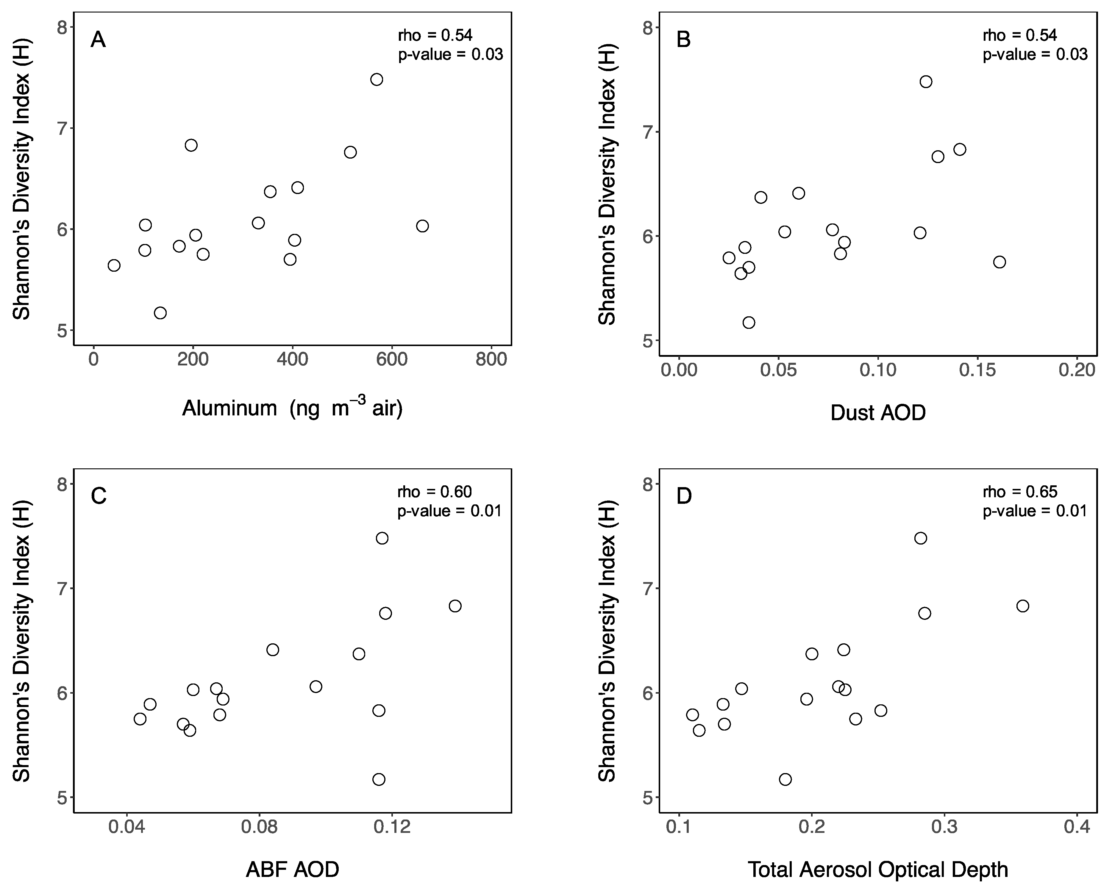
To quantitatively assess the diversity and estimate the differences in airborne bacterial communities over the MS, we report microbial community richness, expressed as the number of unique OTUs observed, and diversity expressed as Shannon’s diversity index (H), estimated from the abundance of bacteria in each sample (Table 1). Observed OTUs corresponds to the number of unique bacteria in each sample, whereas H is a commonly used quantitative measure of diversity [49]. The abundance of observed OTUs ranged from 66 (sample 9) to 241 (sample 1) (Table 1). Observed OTUs varied significantly between the three regions of the MS: Eastern (median = 141), Central (median = 93), and Western (median = 82) (Kruskal–Wallis pairwise test: H = 6.732, df = 2, p = 0.034) (Table 1) and correlated positively to Al concentration (Spearman correlation: = 0.549, p = 0.028), mineral dust AOD (Spearman correlation: = 0.68, p = 0.003), ABF AOD (Spearman correlation: = 0.538, p = 0.031), and total AOD (Spearman correlation: = 0.70, p = 0.002) (Table 1). The diversity ranged from 5.17 (sample 9) to 7.48 (sample 1) (Table 1). H values were positively correlated to mineral dust (Spearman correlation: = 0.547, p = 0.028), ABF (Spearman correlation: = 0.599, p = 0.014), and total AOD concentrations (Spearman correlation: = 0.653, p = 0.007) (Figure 5). Prokaryotic communities from samples within the Eastern MS were significantly more similar to each other than samples from the Western MS (PERMANOVA: F = 1.83, p = 0.009) (Figure 6A). Moreover, distance to land, including islands, was positively correlated to community similarity (Spearman: = 0.377, p = 0.009) (Table 1, Figure 6B), even though the bacterial abundance did not correlate to distance to shore.
Previous studies conducted during different dust storms have shown that the origin and atmospheric route of airmass influences bacterial community composition [30,54]. Our findings show that, during non-dust-storm events, neither bacterial richness nor diversity are influenced by the origin of the airmass. This is likely because during intense dust events copious amounts of desert topsoil from different locations are transported and these topsoil particles have distinct microbial communities [32,60]. Our study took place during non-storm conditions over the ocean, and hence terrestrial origin signatures were less pronounced. Instead, we show that airborne bacterial richness and diversity varied by geographic location over the MS (not the origin of the airmass) (Table 1) and correlated to the concentration of Al (Table 1) (as well as dust AOD, ABF AOD, and total AOD; Table 1, Figure 2). Similarly, the diversity of airborne microbes over the MS increased with increasing concentration of Al, mineral dust AOD, ABF AOD, and total AOD measurements (Table 1, Figure 5).
Microbes in the air are predominantly associated with particles, hence when there are more particles in the air, it is likely to find more bacteria. Interestingly, it has been suggested that crevices in particles maintain local humidity due to moisture adsorption to particles and provide shelter from UV, thus protecting airborne microbes from desiccation and exposure to damaging UV radiation, two elements reducing survival of bacteria in the atmosphere [61,62,63]. Dust may also increase the survival potential of airborne bacteria because dust particles can scatter light and UV radiation, reducing exposure. Thus, high Al, which indicates more mineral dust particles (Figure 5), may protect airborne microbes and results in an increased diversity of airborne microbes (Figure 5). This may also be because there are more unique OTUs when there are more mineral dust particles in the air since mineral dust has higher microbial diversity compared to anthropogenic pollutant sources. Alternatively, the chance of encountering more unique OTUs in the air may increase when there are more dust particles in the air because sampling, DNA extraction, PCR, and sequencing methods are better at detecting “Rare” OTUs under such conditions. The correlation between mineral dust and diversity suggests that the microbiome of the air will become more diverse with increased desertification and related dust input to the atmosphere due to projected changes in climate.
Our samples contained a high percentage (44%) of bacteria that are also found in MS surface water [61]. Additionally, we found that weighted UniFrac, (a measure of beta diversity) positively correlated to distance from land, including islands, regardless of the landmass type (island, populated, un-populated, desert or vegetated) (Table 1, Figure 6B). Although this correlation was rather weak, it may be explained by samples far from land containing a higher proportion of marine prokaryotes, in agreement with Mayol et al. [1]. Waves and bubble bursting in the sea surface result in the aerosolization and transportation of microbes [64]. Indeed, other open ocean aerosol studies have also identified marine bacteria in aerosols [1,54]. Our study demonstrates that aerosolization can be a mechanism for long-distance dispersal for marine bacteria [54,64]. This can have ecological implications for receiving ecosystems and may impact the biogeography of various strains. Airborne microbes can change the community structures of environments into which they are deposited [15,65]. Furthermore, bacteriophages associated with marine bacteria can also be transported to new environments and spread viral infections [15,66]. Therefore, airborne microbes and viruses may impact both microbial community structure and microbial production and should be further studied.
The average number of OTUs in our aerosol samples collected during springtime was similar to the number of OTUs in the Norwegian Sea and the Western Pacific in the summer and lower than OTUs in the Northern and Western Pacific Ocean in the fall [56]. Since seasonality impacts airborne bacterial abundance [12,19] and community composition [56], spatiotemporal variability of airborne microbes should be studied during other seasons to assess interannual variability in this region. The number of observed OTUs during non-storm conditions was lower than those measured in coastal cities in the Mediterranean during dust storms [31,58]. This is likely due to the positive correlation between the concentrations of various aerosol constituents (mineral dust and ABF) and the number of OTUs as observed in our study (Figure 5) and previous studies [31].
To our knowledge, there are only two other studies of airborne microbes in the MS during non-storm conditions [10,32]. The study site of Gat et al. [32] was done at a coastal city in Israel (~10 Km away from the shoreline) and the study site of Rahav et al. [10] was at the rooftop of a building directly next to the ocean. Thus, these studies represent different ecological systems than the open ocean. However, all of the organisms that were prominent during clear non-storm days in Gat et al. [32] were also found in our samples (aside from Dermabacteraceae [Actinobacteria]) indicating that they are commonly found in the air over both the land and the water in this region. Several other studies have reported on the airborne bacterial communities during dust storms in the MS and found organisms that were also present in our samples (Table 2) [3,31,32,57,58], suggesting that some organisms previously assumed as being associated with dust-events exist over the MS during non-storm conditions as well. Some organisms observed during dust storms, however, were absent during non-storm conditions [3,31,32,57,58], particularly many that are ubiquitously found in soils [60].
There are only a few studies which have identified and reported airborne microbial diversity in open ocean settings. However, the few reports cover diverse ocean basins, including the East China Sea [54], Caribbean Sea [8], Norwegian Sea [56], Atlantic Ocean [1,8], Pacific Ocean [1,56], and the Indian Ocean [1]. All these studies identified organisms at the family level, except for Mayol et al. [1], which identified organisms at the class level. We compared the microbes found in our study to organisms found in other open ocean studies (Table 2 and Table 3) and found that 44% of the most common bacteria in our study were also reported in other open ocean aerosol studies at the family level. We also found that 80% of the bacteria at the class level were found in aerosols in other marine studies. Of the rare bacteria (<5% in our samples), 16% were reported in other open ocean aerosol studies at the family level and 60% at the class level (Table 2).
When compared to airborne bacteria in samples collected on the Mediterranean coast [3,31,32,57,58] (Table 2 and Table 3), 48% of the common bacteria and 13% of the rare bacteria found in our study were also reported in these studies at the family level. Highly abundant families (Bacillaceae, Sphingomonadaceae, and Pseudomonadaceae) were also found in the air over other marine environments [1,8,56] at the class level, suggesting that these organisms are commonly dispersed via airmasses. If these organisms are viable upon deposition and have a cosmopolitan distribution throughout the oceans, it could be inferred that airmasses are a vehicle of biogeographical distribution.
Mediterranean seawater samples contained the Bacillaceae family, as well as nine other bacterial families from the Proteobacteria (Bradyrhizobiaceae, Rhodobacteraceae, Rhodospirillaceae, Sphingomonadaceae, Comamonadaceae, Enterobacteriaceae, Pseudomonadaceae, Vibrionaceae) and Deinococcus-Thermus (Thermaceae) phyla [59]. Vibrionaceae and Alteromonadaceae families, which were present in our as well as other studies, have most commonly been found in the sea surface microlayer [67], the ~100 µm surface layer of the ocean where there is a dynamic exchange between the sea and air [68]. Overall, 44% of common airborne bacteria and 16% of rare airborne bacteria in our study were previously reported to be found in the MS surface water [61] (Table 2 and Table 3). The large proportion of organisms being found in both air and water as opposed to air only suggests that the bacterial exchange between sea and air during ‘normal’ atmospheric conditions is an important process that can influence the community structure of both environments.
Current data show a wide range of biogeochemical responses related to atmospheric deposition events in LNLC areas [20]. However, the specific contribution of airborne microbes to the changes documented in these studies is typically not considered [3,33]. To predict the future of LNLC regions and how they will contribute to global biogeochemical cycles, it is imperative to understand how atmospheric deposition impacts these regions [20], and to specifically determine the contribution of airborne microbes to these impacts.
The prevalence and importance of airborne microbes is clear, but key methods in aeromicrobiology have not yet been standardized (sample collection, quantification). We used filters to collect aerosols, but different studies have used other techniques, such as liquid impingement [69,70,71] or electrostatic precipitation [72,73,74]. Similarly, we measured bacterial abundance directly from filters after sonication to promote detachment from the filter, while others used different methods, such as qPCR [54], culture-based methods [3] and flow cytometry [10]. As a result, it is difficult to reliably compare results between studies, even if the sampling site and environmental conditions are similar. These issues merit further research and would provide meaningful advancements to the field.
4. Conclusions
Our results show that a diverse array of microbes is present in the air over the MS, with abundances similar to those over other ocean settings. We found that the diversity of the airborne microbes over the MS during non-dust-storm conditions is influenced by aerosol content (mineral dust as well as polluted aerosols) in the air, with higher diversity linked to increases in particle numbers. Our results also show high percentages of marine bacteria in the air, indicating that there is a significant exchange of microbes between the sea surface and the air, even during background non-storm conditions. We also note that several groups of bacteria are more commonly found in the air, hence these groups may be readily dispersed by air movement with implications to their biogeography. Since desertification may increase with climate change, more particles will be introduced to the air, increasing the abundance and diversity of airborne microbes. This may have a significant impact on the microbial communities and biogeochemical cycles of oceans, particularly in regions that are subject to high rates of atmospheric deposition.
Supplementary Materials
The following are available online at https://www.mdpi.com/2073-4433/10/8/440/s1, Figure S1: Backward trajectories constructed using NOAA HYPSPLIT MODEL for each sample, Table S1: Metadata of Samples, Table S2: Detailed Description of Common Bacteria.
Author Contributions
Project administration and design A.P. and E.R; Writing—original draft, E.M.; Writing—review and editing, E.M., A.P., E.R., P.X., N.B., J.M.E., A.V. and B.H.
Funding
This research was funded by the Israel Science Foundation (grant 1211/17) to B.H and E.R. E.M was supported by the NSF GRFP.
Acknowledgments
The authors would like to thank Katie Roberts for her assistance.
Conflicts of Interest
The authors declare no conflict of interest.
References
- Mayol, E.; Arrieta, J.M.; Jiménez, M.A.; Martínez-Asensio, A.; Garcias-Bonet, N.; Dachs, J.; González-Gaya, B.; Royer, S.J.; Benítez-Barrios, V.M.; Fraile-Nuez, E.; et al. Long-range transport of airborne microbes over the global tropical and subtropical ocean. Nat. Commun. 2017, 8, 1–8. [Google Scholar] [CrossRef] [PubMed]
- Torsvik, V.; Øvreås, L.; Thingstad, T.F. Prokaryotic diversity—Magnitude dynamics, and controlling factors. Science 2002, 296, 1064–1066. [Google Scholar] [CrossRef] [PubMed]
- Griffin, D.W.; Kubilay, N.; Kocak, M.; Gray, M.A.; Borden, T.C.; Shinn, E.A. Airborne desert dust and aeromicrobiology over the Turkish Mediterranean coastline. Atmos. Environ. 2007, 41, 4050–4062. [Google Scholar] [CrossRef]
- Prospero, J.M.; Charlson, R.J.; Mohnen, V.; Jaenicke, R.; Delany, A.C.; Moyers, J.; Zoller, W.; Rahn, K. The atmospheric aerosol system—An overview. Rev. Geophys. Space Phys. 1983, 21, 1607–1629. [Google Scholar] [CrossRef]
- Huebert, B.J. Sea Salt Aerosols. Encyclopedia of Earth System Science; Nierenberg, W.A., Ed.; Academic Press: San Diego, CA, USA, 1992; Volume 4, pp. 63–68. [Google Scholar]
- Bovallius, A.; Bucht, B.; Roffey, R.; Anas, P. Long-range air transmission of bacteria. Appl. Environ. Microbiol. 1978, 35, 1231–1232. [Google Scholar] [PubMed]
- Prospero, J.M.; Blades, E.; Mathison, G.; Naidu, R. Interhemispheric transport of viable fungi and bacteria from Africa to the Caribbean with soil dust. Aerobiologia 2005, 21, 1–19. [Google Scholar] [CrossRef] [Green Version]
- Griffin, D.; Kellogg, C.A.; Garrison, V.; Lisle, J.; Borden, T.; Shinn, E. Atmospheric microbiology in the northern Caribbean during African dust events. Aerobiologia 2003, 19, 143–157. [Google Scholar] [CrossRef]
- Kellogg, C.A.; Griffin, D.W. Aerobiology and the global transport of desert dust. Trends Ecol. Evol. 2006, 21, 638–644. [Google Scholar] [CrossRef]
- Rahav, E.; Belkin, N.; Paytan, A.; Herut, B. The Relationship between Air-Mass Trajectories and the Abundance of Dust-Borne Prokaryotes at the SE Mediterranean Sea. Atmosphere 2019, 10, 280. [Google Scholar] [CrossRef]
- Estillore, A.D.; Trueblood, J.V.; Grassian, V.H. Atmospheric chemistry of bioaerosols: Heterogeneous and multiphase reactions with atmospheric oxidants and other trace gases. Chem. Sci. 2016, 7, 6604–6616. [Google Scholar] [CrossRef]
- Brągoszewska, E.; Pastuszka, J.S. Influence of meteorological factors on the level and characteristics of culturable bacteria in the air in Gliwice, Upper Silesia (Poland). Aerobiologia 2016, 34, 241–255. [Google Scholar] [CrossRef] [PubMed]
- Womack, A.M.; Bohannan, B.J.M.; Green, J.L. Biodiversity and biogeography of the atmosphere. Philos. Trans. R. Soc. Lond. B Biol. Sci. 2010, 365, 3645–3653. [Google Scholar] [CrossRef] [PubMed] [Green Version]
- Caliz, J.; Triadó-Margarit, X.; Camarero, L.; Casamayor, E.O. A long-term survey unveils strong seasonal patterns in the airborne microbiome coupled to atmospheric circulations. Proc. Natl. Acad. Sci. USA 2018, 115, 12229–12234. [Google Scholar] [CrossRef] [PubMed]
- Reche, I.; D’Orta, G.; Mladenov, N.; Winget, D.M.; Suttle, C.A. Deposition rates of viruses and bacteria above the atmospheric boundary layer. ISME J. 2018, 12, 1154–1162. [Google Scholar] [CrossRef] [PubMed] [Green Version]
- Brodie, E.L.; DeSantis, T.Z.; Parker, J.P.M.; Zubietta, I.X.; Piceno, Y.M.; Andersen, G.L. Urban aerosols harbor diverse and dynamic bacterial populations. Proc. Natl. Acad. Sci. USA 2007, 104, 299–304. [Google Scholar] [CrossRef] [PubMed] [Green Version]
- Brown, J.K.M.; Hovmoller, M.S. Aerial dispersal of pathogens on the global and continental scales and its impact on plant disease. Science 2002, 297, 537–541. [Google Scholar] [CrossRef]
- Weir-Brush, J.; Garrison, V.H.; Smith, G.W.; Shinn, E.A. The Relationship Between Gorgonian Coral (Cnidaria: Gorgonacea) Diseases and African Dust Storms. Aerobiologia 2004, 20, 119–126. [Google Scholar] [CrossRef]
- Kaarakainen, P.; Meklin, T.; Rintala, H.; Hyvärinen, A.; Kärkkäinen, P.; Vepsäläinen, A.; Hirvonen, M.-R.; Nevalainen, A. Seasonal Variation in Airborne Microbial Concentrations and Diversity at Landfill, Urban and Rural Sites. Clean 2008, 36, 556–563. [Google Scholar] [CrossRef]
- Guieu, C.; Aumont, O.; Paytan, A.; Bopp, L.; Law, C.S.; Mahowald, N.; Achterberg, E.P.; Marañón, E.; Salihoglu, B.; Crise, A.; et al. Global biogeochemical cycles deposition to Low Nutrient Low Chlorophyll regions. Global Biogeochem. Cycles 2014, 28, 1179–1198. [Google Scholar] [CrossRef]
- Siokou-Frangou, I.; Christaki, U.; Mazzocchi, M.G.; Montresor, M.; Ribera d’Alcalá, M.; Vaqué, D.; Zingone, A. Plankton in the open Mediterranean Sea: A review. Biogeosciences 2010, 7, 1543–1586. [Google Scholar] [CrossRef]
- Tanhua, T.; Hainbucher, D.; Schroeder, K.; Cardin, V.; Álvarez, M.; Civitarese, G. The Mediterranean Sea system: A review and a nintroduction to the special issue. Ocean Sci. 2013, 9, 789–803. [Google Scholar] [CrossRef]
- Lawrence, C.R.; Neff, J.C. The contemporary physical and chemical flux of aeolian dust: A synthesis of direct measurements of dust deposition. Chem. Geol. 2009, 267, 46–63. [Google Scholar] [CrossRef]
- Loye-Pilot, M.D.; Martin, J.M.; Morelli, J. Atmospheric input of inorganic nitrogen to the Western Mediterranean. Biogeochemistry 1990, 9, 117–134. [Google Scholar] [CrossRef]
- Bergametti, G.; Remoudaki, E.; Losno, R.; Steiner, E.; Chatenet, B.; Buat-Ménard, P. Source, transport and deposition of atmospheric phosphorus over the northwestern Mediterranean. J. Atmos. Chem. 1992, 14, 501–513. [Google Scholar] [CrossRef]
- Quétel, C.R.; Remoudaki, E.; Davies, J.E.; Miquel, J.-C.; Fowler, S.W.; Lambert, C.E.; Bergametti, G.; Buat-Ménard, P. Impact of atmospheric deposition on particulate iron flux and distribution in northwestern Mediterranean waters. Deep Sea Res. 1993, 40, 989–1002. [Google Scholar] [CrossRef]
- Herut, B.; Krom, M.D.D.; Pan, G.; Mortimer, R. Atmospheric input of nitrogen and phosphorus to the Southeast Mediterranean: Sources, fluxes, and possible impact. Limnol. Oceanogr. 1999, 44, 1683–1692. [Google Scholar] [CrossRef] [Green Version]
- Ridame, C.; Guieu, C. Saharan input of phosphorus to the oligotrophic water of the open western Mediterranean. Limnol. Oceanogr. 2002, 47, 856–869. [Google Scholar] [CrossRef]
- Ridame, C.; Guieu, C.; L’Helguen, S. Strong stimulation of N2 fixation to contrasted Saharan dust events in a Low Nutrient-Low Chlorophyll environment: Results from dust addition in large mesocosms. Biogeosciences 2013, 10, 7333–7346. [Google Scholar] [CrossRef]
- Rahav, E.; Ovadia, G.; Paytan, A.; Herut, B. Contribution of airborne microbes to bacterial production and N2 fixation in seawater upon aerosol deposition. Geophys. Res. Lett. 2016, 43, 719–727. [Google Scholar] [CrossRef]
- Mazar, Y.; Cytryn, E.; Erel, Y.; Rudich, Y. Effect of dust storms on the atmospheric microbiome in the Eastern Mediterranean. Environ. Sci. Technol. 2016, 50, 4194–4202. [Google Scholar] [CrossRef]
- Gat, D.; Mazar, Y.; Cytryn, E.; Rudich, Y. Origin-dependent variations in the atmospheric microbiome community in Eastern Mediterranean dust storms. Environ. Sci. Technol. 2017, 51, 6709–6718. [Google Scholar] [CrossRef]
- Lynch, P.; Reid, J.S.; Westphal, D.L.; Zhang, J.; Hogan, T.F.; Hyer, E.J.; Curtis, C.A.; Hegg, D.A.; Shi, Y.; Campbell, J.R.; et al. An 11-year global gridded aerosol optical thickness reanalysis (v1.0) for atmospheric and climate sciences. Geosci. Model Dev. 2016, 9, 1489–1522. [Google Scholar] [CrossRef] [Green Version]
- Zhang, Q.S.; Manche, L.; Xu, R.M.; Krainer, A.R. hnRNP A1 associates with telomere ends and stimulates telomerase activity. RNA 2006, 12, 1116–1128. [Google Scholar] [CrossRef] [PubMed] [Green Version]
- Hyer, E.J.; Reid, J.S.; Zhang, J. An over-land aerosol optical depth data set for data assimilation by filtering, correction, and aggregation of MODIS Collection 5 optical depth retrievals. Atmos. Meas. Tech. 2011, 4, 379–408. [Google Scholar] [CrossRef] [Green Version]
- Shi, Y.; Zhang, J.; Reid, J.S.; Holben, B.; Hyer, E.J.; Curtis, C. An analysis of the collection 5 MODIS over-ocean aerosol optical depth product for its implication in aerosol assimilation. Atmos. Chem. Phys. 2011, 11, 557–565. [Google Scholar] [CrossRef] [Green Version]
- Liu, T.; Chen, A.L.-W.; Zhang, M.; Watson, J.; Chow, J.; Cao, J.; Chen, H.; Wang, W.; Zhang, J.; Zhan, C.; et al. Bioaerosol Concentrations and Size Distributions during the Autumn and Winter Seasons in an Industrial City of Central China. Aerosol Air Qual. Res. 2019, 19, 1095–1104. [Google Scholar] [CrossRef]
- Griffin, D.W.; Westphal, D.L.; Gray, M.A. Airborne microorganisms in the African desert dust corridor over the mid-Atlantic ridge, Ocean Drilling Program, Leg 209. Aerobiologia 2006, 22, 211. [Google Scholar] [CrossRef]
- Draxler, R.R. HYSPLIT_4 User’s Guide, NOAA Technical Memorandum ERL ARL-230; NOAA Air Resources Laboratory: Silver Spring, MD, USA, 1999; Volume 35.
- Clauß, M.; Springoru, A.C.; Hartung, J. Comparison of Different Fluorescence and Non-Fluorescence Staining Techniques for Rapid Detection of Airborne Micro-Organisms Collected on Room Temperature Vulcanizing (RTV) Silicones from Generated Aerosols and from Ambient Air. Aerosol Sci. Technol. 2012, 46, 818–827. [Google Scholar] [CrossRef]
- Mayol, E.; Jiménez, M.A.; Herndl, G.J.; Duarte, C.M.; Arrieta, J.M. Resolving the abundance and air-sea fluxes of airborne microorganisms in the North Atlantic Ocean. Front. Microbial. 2014, 5, 557. [Google Scholar] [CrossRef]
- Massana, R.; Murray, A.E.; Preston, C.M.; Delong, E. Vertical distribution and phylogenetic characterization of marine planktonic Archaea in the Santa Barbara Channel. Appl. Environ. Microbiol. 1997, 63, 50–56. [Google Scholar] [Green Version]
- Bolyen, E.; Rideout, J.R.; Dillon, M.R.; Bokulich, N.A.; Abnet, C.; Al-Ghalith, G.A.; Alexander, H.; Alm, E.J.; Arumugam, M.; Asnicar, F.; et al. Reproducible, interactive, scalable and extensible microbiome data science using QIIME 2. Nat. Biotechnol. 2019. [Google Scholar] [CrossRef]
- Martin, M. CUTADAPT removes adapter sequences from high-throughput sequencing reads. EMBnet. J. 2011, 17, 10–12. [Google Scholar] [CrossRef]
- Callahan, B.J.; McMurdie, P.J.; Rosen, M.J.; Han, A.W.; Johnson, A.J.; Holmes, S.P. DADA2: High-resolution sample inference from Illumina amplicon data. Nat. Methods 2016, 13, 581–583. [Google Scholar] [CrossRef] [Green Version]
- Pedregosa, F.; Varoquaux, G.; Gramfort, A.; Michel, V.; Thirion, B.; Grisel, O.; Blondel, M.; Prettenhofer, P.; Weiss, R.; Dubourg, V.; et al. Scikit-learn: Machine Learning in Python. J. Mach. Learn. Res. 2011, 12, 2825–2830. [Google Scholar]
- DeSantis, T.Z.; Hugenholtz, P.; Larsen, N.; Rojas, M.; Brodie, E.L.; Keller, K.; Huber, T.; Dalevi, D.; Hu, P.; Andersen, G.L. Greengenes, a chimera-checked 16S rRNA gene database and workbench compatible with ARB. Appl. Environ. Microbiol. 2006, 72, 5069–5072. [Google Scholar] [CrossRef]
- Bokulich, N.A.; Kaehler, B.D.; Rideout, J.R.; Dillon, M.; Bolyen, E.; Knight, R.; Huttley, G.A.; Caporaso, G.J. Optimizing taxonomic classification of marker-gene amplicon sequences with QIIME 2’s q2-feature-classifier plugin. Microbiome 2018, 6, 90. [Google Scholar] [CrossRef]
- Shannon, C.E. A mathematical theory of communication. Bell Syst. Tech. J. 1948, 27, 379–423. [Google Scholar] [CrossRef]
- Lozupone, C.A.; Hamady, M.; Kelley, S.T.; Knight, R. Quantitative and qualitative beta diversity measures lead to different insights into factors that structure microbial communities. Appl. Environ. Microbiol. 2007, 73, 1576–1585. [Google Scholar] [CrossRef]
- Anderson, M.J. A new method for non-parametric multivariate analysis of variance. Austral Ecol. 2001, 26, 32–46. [Google Scholar] [CrossRef]
- Huneeus, N.; Schulz, M.; Balkanski, Y.; Griesfeller, J.; Prospero, J.; Kinne, S.; Bauer, S.l.; Boucher, O.; Chin, M.; Dentener, F. Global dust model intercomparison in AeroCom phase I. Atmos. Chem. Phys. 2011, 11, 7781–7816. [Google Scholar] [CrossRef] [Green Version]
- Xian, P.; Reid, J.S.; Hyer, E.J.; Sampson, C.R.; Rubin, J.I.; Ades, M.; Asencio, N.; Basart, S.; Benedetti, A.; Bhattacharjee, P.S. Current State of the global operational aerosol multi-model ensemble: An update from the International Cooperative for Aerosol Prediction (ICAP). Q. J. R. Meteorol. Soc. 2019, 1–34. [Google Scholar] [CrossRef]
- Cho, B.C.; Hwang, C.Y. Prokaryotic abundance and 16S rRNA gene sequences detected in marine aerosols on the East Sea (Korea). FEMS Microbiol. Ecol. 2011, 76, 327–341. [Google Scholar] [CrossRef]
- Yahya, R.Z.; Arrieta, M.J.; Cusack, M.; Duarte, C.M. Airborne Prokaryote and Virus Abundance Over the Red Sea. Front. Microbiol. 2019, 10, 1112. [Google Scholar] [CrossRef]
- Xia, X.; Wang, J.; Ji, J.; Zhang, J.; Chen, L.; Zhang, R. Bacterial Communities in Marine Aerosols Revealed by 454 Pyrosequencing of the 16S rRNA Gene. J. Atmos. Sci. 2015, 72, 2997–3008. [Google Scholar] [CrossRef]
- Rahav, E.; Shun-Yan, C.; Cui, G.; Liu, H.; Tsagakari, T.M.; Giannakourou, A.; Tsiola, A.; Psarra, S.; Lagaria, A.; Mulholland, M.R.; et al. Evaluating the impact of atmospheric depositions on springtime dinitrogen fixation in the Cretan Sea (Eastern Mediterranean)—A mesocosm approach. Front. Mar. Sci. 2016, 3, 1–13. [Google Scholar] [CrossRef]
- Rahav, E.; Paytan, A.; Chien, C.-T.; Ovadia, G.; Katz, T.; Herut, B. The impact of atmospheric dry deposition associated microbes on the southeastern Mediterranean Sea surface water following an intense dust storm. Front. Mar. Sci. 2016, 3, 127. [Google Scholar] [CrossRef]
- Sunagawa, S.; Coelho, P.; Chaffron, S.; Kultima, J.R.; Labadie, K.; Salazar, G.; Djahanschiri, B.; Zeller, G.; Mende, D.R.; Alberti, A. Structure and function of the global ocean microbiome. Science 2015, 348, 1261359. [Google Scholar] [CrossRef] [Green Version]
- Bahram, M.; Hildebrand, F.; Forslund, S.K.; Anderson, J.L.; Soudzilovskaia, N.A.; Bodegom, P.M.; Bengtsson-Palme, J.; Anslan, S.; Coelho, L.P.; Harend, H. Structure and function of the global topsoil microbiome. Nature 2018, 560, 233–237. [Google Scholar] [CrossRef]
- Gorbushina, A.A.; Kort, R.; Schulte, A.; Lazarus, D.; Schnetger, B.; Brumsack, H.J.; Broughton, W.J.; Favet, J. Life in Darwin’s dust: Intercontinental transport and survival of microbes in the nineteenth century. Environ. Microbiol. 2007, 9, 2911–2922. [Google Scholar] [CrossRef]
- Tang, K.; Huang, Z.; Huang, J.; Maki, T.L.; Zhang, S.; Shimizu, A.; Ma, X.; Shi, J.; Bi, J.; Zhou, T.; et al. Characterization of atmospheric bioaerosols along the transport pathway of Asian dust during the Dust-Bioaerosol 2016 Campaign. Atmos. Chem. Phys. 2018, 18, 7131–7148. [Google Scholar] [CrossRef] [Green Version]
- Tong, Y.; Lighthart, B. The Annual Bacterial Particle Concentration and Size Distribution in the Ambient Atmosphere in a Rural Area of the Willamette Valley, Oregon. Aerosol Sci. Technol. 2010, 3, 393–403. [Google Scholar] [CrossRef]
- Rastelli, E.; Corinaldesi, C.; Dell’Anno, A.; Martire, M.L.; Greco, S.; Facchini, M.C.; Rinaldi, M.; O’Dowd, C.; Ceburnis, D.; Danovaro, R. Transfer of labile organic matter and microbes from the ocean surface to the marine aerosol: An experimental approach. Sci. Rep. 2017, 7, 11475. [Google Scholar] [CrossRef]
- Peter, H.; Hörtnagl, P.; Reche, I.; Sommaruga, R. Bacterial diversity and composition during rain events with and without Saharan dust influence reaching a high mountain lake in the Alps. Environ. Microbiol. Rep. 2014, 6, 618–624. [Google Scholar] [CrossRef]
- Sharoni, S.; Trainic, M.; Schatz, D.; Lehahn, Y.; Flores, M.J.; Bidle, K.D.; Ben-Dor, S.; Rudich, Y.; Koren, I.; Vardi, A. Infection of phytoplankton by aerosolized marine viruses. Proc. Natl. Acad. Sci. USA 2015, 112, 6643–6647. [Google Scholar] [CrossRef] [Green Version]
- Cunliffe, M.; Upstill-Goddard, R.C.; Murrell, J.C. Microbiology of aquatic surface microlayers. FEMS Microbiol. Rev. 2010, 35, 233–246. [Google Scholar] [CrossRef]
- Liss, S.N.; Bicho, P.; Saddler, J. Microbiology and biodegradation of resin acids in pulp mill effluents: A minireview. Can. J. Microbiol. 1997, 43, 599–611. [Google Scholar] [CrossRef]
- Fields, N.D.; Oxborrow, G.S.; Puleo, J.R.; Herring, C.M. Evaluation of membrane filter field monitors for microbiologial air sampling. Appl. Microbiol. 1974, 27, 517–520. [Google Scholar]
- Jensen, P.A.; Todd, W.F.; Davis, G.N.; Scarpino, P.V. Evaluation of eight bioaersol samplers challenged with aerosols of free bacteria. Am. Ind. Hyg. Assoc. J. 1992, 53, 660–667. [Google Scholar] [CrossRef]
- Kesavan, J.; Schepers, D.; McFarland, A.R. Sampling and Retention Efficiencies of Batch-Type Liquid-Based Bioaerosol Samplers. Aerosol Sci. Technol. 2010, 44, 817–829. [Google Scholar] [CrossRef]
- Grinshpun, S.A.; Willeke, K.; Ulevicius, V.; Donnelly, J.; Lin, X.; Mainelis, G. Collection of Airborne Microorganisms: Advantages and Disadvantages of Different Methods. J. Aerosol Sci. 1996, 27 (Suppl. 1), S247–S248. [Google Scholar] [CrossRef]
- Mainelis, G.; Grinshpun, S.A.; Willeke, K.; Reponen, T.; Ulevicius, V.; Hintz, P. Collection of Airborne Microorganisms by Electrostatic Precipitation. Aerosol Sci. Tech. 1999, 30, 127–144. [Google Scholar] [CrossRef]
- Mbareche, H.; Veillette, M.; Guillaume, J.B.; Duchaine, C. Bioaerosol Sampler Choice Should Consider Efficiency and Ability of Samplers to Cover Microbial Diversity. Appl. Environ. Microbiol. 2018, 84, e01589-18. [Google Scholar] [CrossRef]
Figure 1.
Map of the sites where aerosols were collected throughout the Mediterranean Sea (MS) in April on the R/V METEOR cruise M84/3, with sample IDs, region of collection (eastern Med = blue, central Med = red, and western Med = black), and airmass origin zones (Z1–Z4) shown.
Figure 1.
Map of the sites where aerosols were collected throughout the Mediterranean Sea (MS) in April on the R/V METEOR cruise M84/3, with sample IDs, region of collection (eastern Med = blue, central Med = red, and western Med = black), and airmass origin zones (Z1–Z4) shown.

Figure 2.
Aerosol optical depth (AOD) from Navy Aerosol Analysis and Prediction System (NAAPS) reanalysis at the time of collection at sample sites. Each bar represents one sample, with the height of each bar corresponding to total AOD. ABF (anthropogenic and biogenic fine aerosol species) in black, mineral dust in dark gray, and sea salt + and smoke in light gray, fractions of the total AOD are shown for each station.

Figure 3.
Spatial distribution of airborne bacterial abundance (cells m−3 air) over the MS during April 2011.
Figure 3.
Spatial distribution of airborne bacterial abundance (cells m−3 air) over the MS during April 2011.

Figure 4.
Relative abundance of prokaryote operational taxonomic units (OTUs) in the different regions of the MS. The colors correspond to different taxonomic orders of prokaryotes, as shown in the detailed legend.
Figure 4.
Relative abundance of prokaryote operational taxonomic units (OTUs) in the different regions of the MS. The colors correspond to different taxonomic orders of prokaryotes, as shown in the detailed legend.

Figure 5.
The relationship between bacterial diversity (Shannon’s diversity index) and atmospheric aerosols variables, (A) aluminum, (B) dust, (C) ABF, (D) total aerosol optical density.
Figure 5.
The relationship between bacterial diversity (Shannon’s diversity index) and atmospheric aerosols variables, (A) aluminum, (B) dust, (C) ABF, (D) total aerosol optical density.

Figure 6.
PCoA showing the differences in beta diversity using weighted UniFrac with (A) shapes representing regions of the MS samples were collected in, and (B) distance (km) between each sample site and closest landmass.
Figure 6.
PCoA showing the differences in beta diversity using weighted UniFrac with (A) shapes representing regions of the MS samples were collected in, and (B) distance (km) between each sample site and closest landmass.


Table 1.
Sample ID, region of the MS samples were collected from, airmass origin of samples based on HYSPLIT back trajectory models (Z1 = Western Europe, Z2 = Eastern Europe, Z3 = Mediterranean Sea, and Z4 = Northern Africa), distance between the sampling site to the closest landmass or island (km), total aerosol optical depth (AOD), aluminum concentrations (ng m−3 air), number of OTUs observed, and Shannon’s Diversity Index (H) are shown for each sample.
| Sample ID | Region | Airmass Origin | Distance from Land (km) | Total AOD | Aluminum (ng m−3 air) | Observed OTUs | Shannon’s Index (H) |
|---|---|---|---|---|---|---|---|
| 1 | Eastern | Z2 | 155 | 0.282 | 569 | 241 | 7.48 |
| 2 | Eastern | Z3 | 154 | 0.285 | 516 | 158 | 6.76 |
| 3 | Eastern | Z3 | 102 | 0.22 | 331 | 100 | 6.06 |
| 4 | Eastern | Z3 | 204 | 0.224 | 410 | 141 | 6.41 |
| 5 | Eastern | Z1 | 156 | 0.196 | 205 | 119 | 5.94 |
| 6 | Central | Z1 | 78 | 0.233 | 220 | 95 | 5.75 |
| 7 | Central | Z1 | 213 | 0.225 | 661 | 96 | 6.03 |
| 8 | Central | Z1 | 288 | 0.133 | 404 | 92 | 5.89 |
| 9 | Central | Z2 | 48 | 0.18 | 134 | 66 | 5.17 |
| 10 | Central | Z2 | 112 | 0.115 | 41 | 76 | 5.64 |
| 11 | Central | Z1 | 49 | 0.147 | 104 | 96 | 6.04 |
| 12 | Western | Z4 | 135 | 0.359 | 196 | 164 | 6.83 |
| 13 | Western | Z4 | 126 | 0.252 | 172 | 82 | 5.83 |
| 14 | Western | Z4 | 56 | 0.134 | 395 | 70 | 5.7 |
| 15 | Western | Z4 | 36 | 0.2 | 355 | 108 | 6.37 |
| 16 | Western | Z1 | 42 | 0.11 | 103 | 75 | 5.79 |
Table 2.
‘Common’ organisms found over the Mediterranean Sea in this study compared to five studies focusing on marine aerosols [1,8,38,55,56], five studies focusing on coastal aerosols [3,31,32,57,58], and six samples from one study focusing on Mediterranean surface seawater [59]. Columns under open ocean studies refer to references [1,8,38,55,56], columns under Mediterranean coastal studies refer to references [3,31,32,57,58], and columns under Mediterranean seawater samples refer to six samples from reference [59].
| Common Bacteria | Open Ocean Studies | Med Coastal Studies | Med Seawater Samples | ||||||||||||||||
|---|---|---|---|---|---|---|---|---|---|---|---|---|---|---|---|---|---|---|---|
| Phyla | Class | Order | Family | 1 | 2 | 3 | 4 | 5 | 1 | 2 | 3 | 4 | 5 | 1 | 2 | 3 | 4 | 5 | 6 |
| Actinobacteria | Actinobacteria | Actinomycetales | Actinomycetaceae | × | |||||||||||||||
| Actinobacteria | Actinobacteria | Actinomycetales | Corynebacteriaceae | × | × | × | |||||||||||||
| Actinobacteria | Actinobacteria | Bifidobacteriales | Bifidobacteriaceae | × | |||||||||||||||
| Bacteroidetes | Flavobacteriia | Flavobacteriales | Weeksellaceae | × | |||||||||||||||
| Bacteroidetes | Saprospirae | Saprospirales | Chitinophagaceae | × | × | × | |||||||||||||
| Firmicutes | Bacilli | Bacillales | Bacillaceae | × | × | × | × | × | × | × | × | × | × | × | × | × | |||
| Firmicutes | Bacilli | Bacillales | Paenibacillaceae | × | × | ||||||||||||||
| Firmicutes | Bacilli | Bacillales | Planococcaceae | × | × | × | |||||||||||||
| Firmicutes | Bacilli | Bacillales | Staphylococcaceae | × | × | × | × | ||||||||||||
| Firmicutes | Bacilli | Turicibacterales | Turicibacteraceae | × | |||||||||||||||
| Firmicutes | Clostridia | Clostridiales | Clostridiaceae | × | × | × | |||||||||||||
| Firmicutes | Clostridia | Clostridiales | Peptostreptococcaceae | ||||||||||||||||
| Firmicutes | Clostridia | Clostridiales | Veillonellaceae | ||||||||||||||||
| Proteobacteria | Alphaproteobacteria | Rhizobiales | Bradyrhizobiaceae | × | × | × | × | × | × | × | |||||||||
| Proteobacteria | Alphaproteobacteria | Rhodobacterales | Rhodobacteraceae | × | × | × | × | × | × | × | × | × | × | ||||||
| Proteobacteria | Alphaproteobacteria | Rhodospirillales | Rhodospirillaceae | × | × | × | × | × | × | × | |||||||||
| Proteobacteria | Alphaproteobacteria | Sphingomonadales | Sphingomonadaceae | × | × | × | × | × | × | × | × | ||||||||
| Proteobacteria | Betaproteobacteria | Burkholderiales | Comamonadaceae | × | × | × | × | × | × | × | |||||||||
| Proteobacteria | Gammaproteobacteria | Enterobacteriales | Enterobacteriaceae | × | × | × | × | × | × | × | × | × | |||||||
| Proteobacteria | Gammaproteobacteria | Pseudomonadales | Moraxellaceae | × | × | × | × | × | |||||||||||
| Proteobacteria | Gammaproteobacteria | Pseudomonadales | Pseudomonadaceae | × | × | × | × | × | × | × | × | × | |||||||
| Proteobacteria | Gammaproteobacteria | Salinisphaerales | Salinisphaeraceae | × | |||||||||||||||
| Proteobacteria | Gammaproteobacteria | Vibrionales | Vibrionaceae | × | × | × | × | × | × | ||||||||||
| Proteobacteria | Gammaproteobacteria | Xanthomonadales | Sinobacteraceae | × | |||||||||||||||
| Deinococcus-Thermus | Deinococci | Thermales | Thermaceae | × | |||||||||||||||
Table 3.
‘Rare’ organisms found over the Mediterranean Sea in this study compared to five studies focusing on marine aerosols [1,8,38,55,56], five studies focusing on coastal aerosols [3,31,32,57,58], and six samples from one study focusing on Mediterranean surface seawater [59]. Columns under open ocean studies refer to references [1,8,38,55,56], columns under Mediterranean coastal studies refer to references [3,31,32,57,58], and columns under Mediterranean seawater samples refer to six samples from reference [59].
| Rare Bacteria | Open Ocean Studies | Med Coastal Studies | Med Seawater Studies | ||||||||||||||||
|---|---|---|---|---|---|---|---|---|---|---|---|---|---|---|---|---|---|---|---|
| Phyla | Class | Order | Family | 1 | 2 | 3 | 4 | 5 | 1 | 2 | 3 | 4 | 5 | 1 | 2 | 3 | 4 | 5 | 6 |
| Actinobacteria | Actinobacteria | Actinomycetales | Dietziaceae | × | |||||||||||||||
| Actinobacteria | Actinobacteria | Actinomycetales | Micrococcaceae | × | × | × | × | × | × | ||||||||||
| Actinobacteria | Rubrobacteria | Rubrobacterales | Rubrobacteraceae | × | × | ||||||||||||||
| Bacteroidetes | BME43 | Unassigned | Unassigned | ||||||||||||||||
| Bacteroidetes | Bacteroidia | Bacteroidales | Porphyromonadaceae | ||||||||||||||||
| Bacteroidetes | Cytophagia | Cytophagales | Amoebophilaceae | × | |||||||||||||||
| Bacteroidetes | Saprospirae | Saprospirales | Undefined | ||||||||||||||||
| Bacteroidetes | Saprospirae | Saprospirales | Saprospiraceae | ||||||||||||||||
| Chlamydiae | Chlamydiia | Chlamydiales | Undefined | ||||||||||||||||
| Chlamydiae | Chlamydiia | Chlamydiales | Rhabdochlamydiaceae | ||||||||||||||||
| Cyanobacteria | 4C0d2 | MLE112 | Unassigned | × | × | × | |||||||||||||
| Firmicutes | Bacilli | Bacillales | Unassigned | × | × | × | × | ||||||||||||
| Firmicutes | Bacilli | Bacillales | Alicyclobacillaceae | × | |||||||||||||||
| Firmicutes | Bacilli | Bacillales | Thermoactinomycetaceae | × | |||||||||||||||
| Firmicutes | Bacilli | Bacillales | Exiguobacteraceae | × | |||||||||||||||
| Firmicutes | Bacilli | Gemellales | Gemellaceae | × | × | ||||||||||||||
| Firmicutes | Bacilli | Lactobacillales | Leuconostocaceae | × | |||||||||||||||
| Firmicutes | Bacilli | Lactobacillales | Streptococcaceae | × | × | ||||||||||||||
| Firmicutes | Clostridia | Clostridiales | Tissierellaceae | ||||||||||||||||
| Proteobacteria | Undefined | Unassigned | Unassigned | ||||||||||||||||
| Proteobacteria | Alphaproteobacteria | Rhizobiales | Hyphomicrobiaceae | × | |||||||||||||||
| Proteobacteria | Alphaproteobacteria | Rhizobiales | Methylobacteriaceae | × | |||||||||||||||
| Proteobacteria | Alphaproteobacteria | Rhizobiales | Methylocystaceae | × | |||||||||||||||
| Proteobacteria | Alphaproteobacteria | Rhizobiales | Phyllobacteriaceae | × | × | × | × | × | × | × | |||||||||
| Proteobacteria | Alphaproteobacteria | Rhodobacterales | Hyphomonadaceae | × | × | ||||||||||||||
| Proteobacteria | Betaproteobacteria | Burkholderiales | Oxalobacteraceae | × | × | × | × | × | × | ||||||||||
| Proteobacteria | Gammaproteobacteria | Alteromonadales | Alteromonadaceae | × | × | × | × | × | × | × | × | ||||||||
| Proteobacteria | Gammaproteobacteria | Oceanospirillales | Halomonadaceae | × | × | ||||||||||||||
| Proteobacteria | Gammaproteobacteria | Pasteurellales | Pasteurellaceae | × | |||||||||||||||
| Spirochaetes | Spirochaetes | Spirochaetales | Spirochaetaceae | ||||||||||||||||
| WPS | Unassigned | Unassigned | Unassigned | ||||||||||||||||
| Deinococcus-Thermus | Deinococci | Deinococcales | Deinococcaceae | ||||||||||||||||
© 2019 by the authors. Licensee MDPI, Basel, Switzerland. This article is an open access article distributed under the terms and conditions of the Creative Commons Attribution (CC BY) license (http://creativecommons.org/licenses/by/4.0/).
Share and Cite
MDPI and ACS Style
Mescioglu, E.; Rahav, E.; Belkin, N.; Xian, P.; Eizenga, J.M.; Vichik, A.; Herut, B.; Paytan, A. Aerosol Microbiome over the Mediterranean Sea Diversity and Abundance. Atmosphere 2019, 10, 440. https://doi.org/10.3390/atmos10080440
AMA Style
Mescioglu E, Rahav E, Belkin N, Xian P, Eizenga JM, Vichik A, Herut B, Paytan A. Aerosol Microbiome over the Mediterranean Sea Diversity and Abundance. Atmosphere. 2019; 10(8):440. https://doi.org/10.3390/atmos10080440
Chicago/Turabian StyleMescioglu, Esra, Eyal Rahav, Natalia Belkin, Peng Xian, Jordan M. Eizenga, Ania Vichik, Barak Herut, and Adina Paytan. 2019. "Aerosol Microbiome over the Mediterranean Sea Diversity and Abundance" Atmosphere 10, no. 8: 440. https://doi.org/10.3390/atmos10080440
Note that from the first issue of 2016, this journal uses article numbers instead of page numbers. See further details here.