
The Dual Lens of Endoscopy and Histology in the Diagnosis and Management of Eosinophilic Gastrointestinal Disorders—A Comprehensive Review
 , ,
, ,
Abstract
:1. Introduction
Pathophysiology of Eosinophilic Inflammation
2. Eosinophilic Esophagitis (EoE)
2.1. Epidemiology, Physiopathology, and Clinical Manifestations
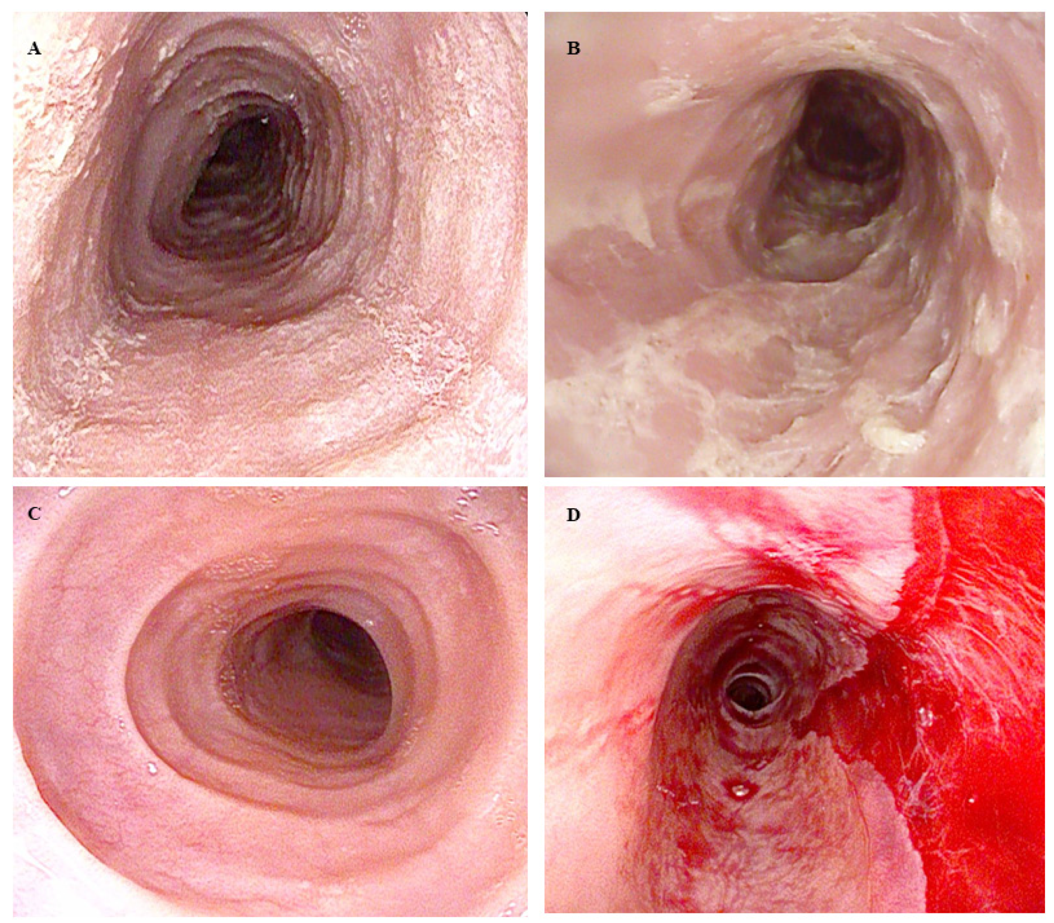
2.2. Endoscopy
2.2.1. EoE Diagnosis
2.2.2. Therapeutic Drug Monitoring
2.2.3. Management of Fibrostenotic Disease
2.3. Histology
2.3.1. Histological Features
2.3.2. Scores Assessing Histology
2.4. Treatment: Target Drugs and Emerging Therapies
3. Eosinophilic Gastritis (EoG) and Enteritis (EoN)
3.1. Epidemiology, Physiopathology, and Clinical Aspects
3.2. Endoscopy
3.2.1. Eosinophilic Gastritis
3.2.2. Eosinophilic Enteritis
3.2.3. Biopsy Sampling
3.3. Histology
3.4. Treatment: Current Drugs and Emerging Novelties
4. Eosinophilic Colitis (EoC)
4.1. Epidemiology, Physiopathology, and Clinical Manifestations
4.2. Endoscopy
4.3. Histology
4.4. Treatment
5. Conclusions and Future Directions
Author Contributions
Funding
Institutional Review Board Statement
Informed Consent Statement
Data Availability Statement
Conflicts of Interest
References
- Gotlib, J. World Health Organization-defined eosinophilic disorders: 2011 update on diagnosis, risk stratification, and management. Am. J. Hematol. 2011, 86, 677–688. [Google Scholar] [CrossRef] [PubMed]
- Collins, M.H.; Capocelli, K.; Yang, G.-Y. Eosinophilic Gastrointestinal Disorders Pathology. Front. Med. 2017, 4, 261. [Google Scholar] [CrossRef] [PubMed]
- Dellon, E.S.; Gonsalves, N.; Abonia, J.P.; Alexander, J.A.; Arva, N.C.; Atkins, D.; Attwood, S.E.; Auth, M.K.; Bailey, D.D.; Biederman, L.; et al. International Consensus Recommendations for Eosinophilic Gastrointestinal Disease Nomenclature. Clin. Gastroenterol. Hepatol. 2022, 20, 2474–2484.e3. [Google Scholar] [CrossRef] [PubMed]
- Steinbach, E.C.; Hernandez, M.; Dellon, E.S. Eosinophilic Esophagitis and the Eosinophilic Gastrointestinal Diseases: Approach to Diagnosis and Management. J. Allergy Clin. Immunol. Pract. 2018, 6, 1483–1495. [Google Scholar] [CrossRef]
- Uppal, V.; Kreiger, P.; Kutsch, E. Eosinophilic Gastroenteritis and Colitis: A Comprehensive Review. Clin. Rev. Allergy Immunol. 2016, 50, 175–188. [Google Scholar] [CrossRef] [PubMed]
- Gonsalves, N. Eosinophilic Gastrointestinal Disorders. Clin. Rev. Allergy Immunol. 2019, 57, 272–285. [Google Scholar] [CrossRef]
- Walker, M.M.; Potter, M.; Talley, N.J. Eosinophilic gastroenteritis and other eosinophilic gut diseases distal to the oesophagus. Lancet Gastroenterol. Hepatol. 2018, 3, 271–280. [Google Scholar] [CrossRef] [PubMed]
- Gómez-Aldana, A.; Jaramillo-Santos, M.; Delgado, A.; Jaramillo, C.; Lúquez-Mindiola, A. Eosinophilic esophagitis: Current concepts in diagnosis and treatment. World J. Gastroenterol. 2019, 25, 4598–4613. [Google Scholar] [CrossRef] [PubMed]
- Hirano, I.; Chan, E.S.; Rank, M.A.; Sharaf, R.N.; Stollman, N.H.; Stukus, D.R.; Wang, K.; Greenhawt, M.; Falck-Ytter, Y.T.; Chachu, K.A.; et al. AGA Institute and the Joint Task Force on Allergy-Immunology Practice Parameters Clinical Guidelines for the Management of Eosinophilic Esophagitis. Gastroenterology 2020, 158, 1776–1786. [Google Scholar] [CrossRef]
- Lwin, T.; Melton, S.D.; Genta, R.M. Eosinophilic gastritis: Histopathological characterization and quantification of the normal gastric eosinophil content. Mod. Pathol. 2011, 24, 556–563. [Google Scholar] [CrossRef]
- Dellon, E.S.; Bortey, E.; Chang, A.T.; Paterson, C.A.; Turner, K.; Genta, R.M. Determination of Optimal Eosinophil Thresholds for Diagnosis of Eosinophilic Gastritis and Duodenitis: A Pooled Analysis of 4 Prospective Studies. Clin. Transl. Gastroenterol. 2023, 15, e00656. [Google Scholar] [CrossRef] [PubMed]
- Dellon, E.S.; Gonsalves, N.; Rothenberg, M.E.; Hirano, I.; Chehade, M.; Peterson, K.A.; Falk, G.W.; Murray, J.A.; Gehman, L.T.; Chang, A.T.; et al. Determination of Biopsy Yield That Optimally Detects Eosinophilic Gastritis and/or Duodenitis in a Randomized Trial of Lirentelimab. Clin. Gastroenterol. Hepatol. 2022, 20, 535–545.e15. [Google Scholar] [CrossRef] [PubMed]
- Collins, M.H. Histopathology Associated with Eosinophilic Gastrointestinal Diseases. Immunol. Allergy Clin. North Am. 2009, 29, 109–117. [Google Scholar] [CrossRef] [PubMed]
- Turner, K.O.; Sinkre, R.A.; Neumann, W.L.; Genta, R.M. Primary Colonic Eosinophilia and Eosinophilic Colitis in Adults. Am. J. Surg. Pathol. 2017, 41, 225–233. [Google Scholar] [CrossRef] [PubMed]
- Mehta, P.; Furuta, G.T. Eosinophils in Gastrointestinal Disorders. Immunol. Allergy Clin. North Am. 2015, 35, 413–437. [Google Scholar] [CrossRef] [PubMed]
- Jung, Y.; Rothenberg, M.E. Roles and Regulation of Gastrointestinal Eosinophils in Immunity and Disease. J. Immunol. 2014, 193, 999–1005. [Google Scholar] [CrossRef] [PubMed]
- Abdala-Valencia, H.; Coden, M.E.; Chiarella, S.E.; Jacobsen, E.A.; Bochner, B.S.; Lee, J.J.; Berdnikovs, S. Shaping eosinophil identity in the tissue contexts of development, homeostasis, and disease. J. Leukoc. Biol. 2018, 104, 95–108. [Google Scholar] [CrossRef]
- Walker, M.M.; Potter, M.D.; Talley, N.J. Eosinophilic colitis and colonic eosinophilia. Curr. Opin. Gastroenterol. 2019, 35, 42–50. [Google Scholar] [CrossRef]
- Marichal, T.; Mesnil, C.; Bureau, F. Homeostatic Eosinophils: Characteristics and Functions. Front. Med. 2017, 4, 101. [Google Scholar] [CrossRef]
- Matsushita, T.; Maruyama, R.; Ishikawa, N.; Harada, Y.; Araki, A.; Chen, D.; Tauchi-Nishi, P.; Yuki, T.; Kinoshita, Y. The Number and Distribution of Eosinophils in the Adult Human Gastrointestinal Tract. Am. J. Surg. Pathol. 2015, 39, 521–527. [Google Scholar] [CrossRef]
- Yantiss, R.K. Eosinophils in the GI tract: How many is too many and what do they mean? Mod. Pathol. 2015, 28 (Suppl. S1), S7–S21. [Google Scholar] [CrossRef] [PubMed]
- Iwata, A.; Toda, Y.; Furuya, H.; Nakajima, H. Group 2 innate lymphoid cells in human asthma. Allergol. Int. 2023, 72, 194–200. [Google Scholar] [CrossRef] [PubMed]
- Awad, H.; Sfaira, A.; Abu Osba, Y.; Shahin, M.; Al-Asa’D, Y.; Isbeih, N.; Hayagneh, W.; Shomaf, M. Mast Cell Numbers in Primary Eosinophilic Colitis Are Significantly Higher Than in Secondary Tissue Eosinophilia and Normal Controls: A Possible Link to Pathogenesis. Iran. J. Immunol. 2021, 18, 220–229. [Google Scholar] [CrossRef] [PubMed]
- Masterson, J.C.; McNamee, E.N.; Fillon, S.A.; Hosford, L.; Harris, R.; Fernando, S.D.; Jedlicka, P.; Iwamoto, R.; Jacobsen, E.; Protheroe, C.; et al. Eosinophil-mediated signalling attenuates inflammatory responses in experimental colitis. Gut 2015, 64, 1236–1247. [Google Scholar] [CrossRef]
- Giudici, G.; Ribaldone, D.G.; Astegiano, M.; Saracco, G.M.; Pellicano, R. Eosinophilic colitis: Clinical review and 2020 update. Minerva Gastroenterol. Dietol. 2020, 66, 157–163. [Google Scholar] [CrossRef]
- Yan, B.M.; A Shaffer, E. Primary eosinophilic disorders of the gastrointestinal tract. Gut 2009, 58, 721–732. [Google Scholar] [CrossRef]
- O’Shea, K.M.; Aceves, S.S.; Dellon, E.S.; Gupta, S.K.; Spergel, J.M.; Furuta, G.T.; Rothenberg, M.E. Pathophysiology of Eosinophilic Esophagitis. Gastroenterology 2018, 154, 333–345. [Google Scholar] [CrossRef] [PubMed]
- Shoda, T.; Wen, T.; Caldwell, J.M.; Morgenstern, N.B.-B.; Osswald, G.A.; Rochman, M.; Mack, L.E.; Felton, J.M.; Abonia, J.P.; Arva, N.C.; et al. Loss of Endothelial TSPAN12 Promotes Fibrostenotic Eosinophilic Esophagitis via Endothelial Cell–Fibroblast Crosstalk. Gastroenterology 2022, 162, 439–453. [Google Scholar] [CrossRef]
- Gonsalves, N.P.; Aceves, S.S. Diagnosis and treatment of eosinophilic esophagitis. J. Allergy Clin. Immunol. 2020, 145, 1–7. [Google Scholar] [CrossRef]
- Attwood, S.E.A.; Smyrk, T.C.; Demeester, T.R.; Jones, J.B. Esophageal eosinophilia with dysphagia. Dig. Dis. Sci. 1993, 38, 109–116. [Google Scholar] [CrossRef]
- Navarro, P.; Arias, Á.; Arias-González, L.; Laserna-Mendieta, E.J.; Ruiz-Ponce, M.; Lucendo, A.J. Systematic review with meta-analysis: The growing incidence and prevalence of eosinophilic oesophagitis in children and adults in population-based studies. Aliment. Pharmacol. Ther. 2019, 49, 1116–1125. [Google Scholar] [CrossRef] [PubMed]
- Reed, C.C.; Tappata, M.; Eluri, S.; Shaheen, N.J.; Dellon, E.S. Combination Therapy with Elimination Diet and Corticosteroids Is Effective for Adults with Eosinophilic Esophagitis. Clin. Gastroenterol. Hepatol. 2019, 17, 2800–2802. [Google Scholar] [CrossRef]
- Alexander, E.S.; Martin, L.J.; Collins, M.H.; Kottyan, L.C.; Sucharew, H.; He, H.; Mukkada, V.A.; Succop, P.A.; Abonia, J.P.; Foote, H.; et al. Twin and family studies reveal strong environmental and weaker genetic cues explaining heritability of eosinophilic esophagitis. J. Allergy Clin. Immunol. 2014, 134, 1084–1092.e1. [Google Scholar] [CrossRef] [PubMed]
- Fogg, M. Pollen and eosinophilic esophagitis. J. Allergy Clin. Immunol. 2003, 112, 796–797. [Google Scholar] [CrossRef]
- Visaggi, P.; Savarino, E.; Del Corso, G.; Hunter, H.; Svizzero, F.B.; Till, S.J.; Dunn, J.; Wong, T.; de Bortoli, N.; Zeki, S. Six-Food Elimination Diet Is Less Effective During Pollen Season in Adults with Eosinophilic Esophagitis Sensitized to Pollens. Am. J. Gastroenterol. 2023, 118, 1957–1962. [Google Scholar] [CrossRef] [PubMed]
- Dellon, E.S.; Peery, A.F.; Shaheen, N.J.; Morgan, D.R.; Hurrell, J.M.; Lash, R.H.; Genta, R.M. Inverse Association of Esophageal Eosinophilia with Helicobacter pylori Based on Analysis of a US Pathology Database. Gastroenterology 2011, 141, 1586–1592. [Google Scholar] [CrossRef]
- Furuta, K.; Adachi, K.; Aimi, M.; Ishimura, N.; Sato, S.; Ishihara, S.; Kinoshita, Y. Case-control study of association of eosinophilic gastrointestinal disorders with Helicobacter pylori infection in Japan. J. Clin. Biochem. Nutr. 2013, 53, 60–62. [Google Scholar] [CrossRef]
- Furuta, G.T.; Katzka, D.A. Eosinophilic Esophagitis. N. Engl. J. Med. 2015, 373, 1640–1648. [Google Scholar] [CrossRef]
- Hirano, I.; Collins, M.H.; Assouline-Dayan, Y.; Evans, L.; Gupta, S.; Schoepfer, A.M.; Straumann, A.; Safroneeva, E.; Grimm, M.; Smith, H.; et al. RPC4046, a Monoclonal Antibody Against IL13, Reduces Histologic and Endoscopic Activity in Patients with Eosinophilic Esophagitis. Gastroenterology 2019, 156, 592–603.e10. [Google Scholar] [CrossRef]
- Dellon, E.S.; Collins, M.H.; Rothenberg, M.E.; Assouline-Dayan, Y.; Evans, L.; Gupta, S.; Schoepfer, A.; Straumann, A.; Safroneeva, E.; Rodriguez, C.; et al. Long-term Efficacy and Tolerability of RPC4046 in an Open-Label Extension Trial of Patients with Eosinophilic Esophagitis. Clin. Gastroenterol. Hepatol. 2021, 19, 473–483.e17. [Google Scholar] [CrossRef]
- Zhang, X.; Cheng, E.; Huo, X.; Yu, C.; Zhang, Q.; Pham, T.H.; Wang, D.H.; Spechler, S.J.; Souza, R.F. Omeprazole Blocks STAT6 Binding to the Eotaxin-3 Promoter in Eosinophilic Esophagitis Cells. PLoS ONE 2012, 7, e50037. [Google Scholar] [CrossRef]
- Cheng, E.; Zhang, X.; Huo, X.; Yu, C.; Zhang, Q.; Wang, D.H.; Spechler, S.J.; Souza, R.F. Omeprazole blocks eotaxin-3 expression by oesophageal squamous cells from patients with eosinophilic oesophagitis and GORD. Gut 2013, 62, 824–832. [Google Scholar] [CrossRef] [PubMed]
- Blanchard, C.; Wang, N.; Stringer, K.F.; Mishra, A.; Fulkerson, P.C.; Abonia, J.P.; Jameson, S.C.; Kirby, C.; Konikoff, M.R.; Collins, M.H.; et al. Eotaxin-3 and a uniquely conserved gene-expression profile in eosinophilic esophagitis. J. Clin. Investig. 2006, 116, 536–547. [Google Scholar] [CrossRef]
- Litosh, V.A.; Rochman, M.; Rymer, J.K.; Porollo, A.; Kottyan, L.C.; Rothenberg, M.E. Calpain-14 and its association with eosinophilic esophagitis. J. Allergy Clin. Immunol. 2017, 139, 1762–1771.e7. [Google Scholar] [CrossRef]
- Kottyan, L.C.; Davis, B.P.; Sherrill, J.D.; Liu, K.; Rochman, M.; Kaufman, K.; Weirauch, M.T.; Vaughn, S.; Lazaro, S.; Rupert, A.M.; et al. Genome-wide association analysis of eosinophilic esophagitis provides insight into the tissue specificity of this allergic disease. Nat. Genet. 2014, 46, 895–900. [Google Scholar] [CrossRef] [PubMed]
- Barchi, A.; Massimino, L.; Mandarino, F.V.; Vespa, E.; Sinagra, E.; Almolla, O.; Passaretti, S.; Fasulo, E.; Parigi, T.L.; Cagliani, S.; et al. Microbiota profiling in esophageal diseases: Novel insights into molecular staining and clinical outcomes. Comput. Struct. Biotechnol. J. 2024, 23, 626–637. [Google Scholar] [CrossRef]
- Mandarino, F.V.; Sinagra, E.; Barchi, A.; Verga, M.C.; Brinch, D.; Raimondo, D.; Danese, S. Gastroparesis: The Complex Interplay with Microbiota and the Role of Exogenous Infections in the Pathogenesis of the Disease. Microorganisms 2023, 11, 1122. [Google Scholar] [CrossRef]
- Salmeri, N.; Sinagra, E.; Dolci, C.; Buzzaccarini, G.; Sozzi, G.; Sutera, M.; Candiani, M.; Ungaro, F.; Massimino, L.; Danese, S.; et al. Microbiota in Irritable Bowel Syndrome and Endometriosis: Birds of a Feather Flock Together—A Review. Microorganisms 2023, 11, 2089. [Google Scholar] [CrossRef] [PubMed]
- Napolitano, M.; Fasulo, E.; Ungaro, F.; Massimino, L.; Sinagra, E.; Danese, S.; Mandarino, F.V. Gut Dysbiosis in Irritable Bowel Syndrome: A Narrative Review on Correlation with Disease Subtypes and Novel Therapeutic Implications. Microorganisms 2023, 11, 2369. [Google Scholar] [CrossRef]
- Massimino, L.; Barchi, A.; Mandarino, F.V.; Spanò, S.; Lamparelli, L.A.; Vespa, E.; Passaretti, S.; Peyrin-Biroulet, L.; Savarino, E.V.; Jairath, V.; et al. A multi-omic analysis reveals the esophageal dysbiosis as the predominant trait of eosinophilic esophagitis. J. Transl. Med. 2023, 21, 46. [Google Scholar] [CrossRef]
- Straumann, A.; Katzka, D.A. Diagnosis and Treatment of Eosinophilic Esophagitis. Gastroenterology 2018, 154, 346–359. [Google Scholar] [CrossRef]
- Schoepfer, A.M.; Safroneeva, E.; Bussmann, C.; Kuchen, T.; Portmann, S.; Simon, H.; Straumann, A. Delay in Diagnosis of Eosinophilic Esophagitis Increases Risk for Stricture Formation in a Time-Dependent Manner. Gastroenterology 2013, 145, 1230–1236.e2. [Google Scholar] [CrossRef]
- Almansa, C.; DeVault, K.R.; Achem, S.R. A Comprehensive Review of Eosinophilic Esophagitis in Adults. J. Clin. Gastroenterol. 2011, 45, 658–664. [Google Scholar] [CrossRef] [PubMed]
- Dellon, E.S.; Gibbs, W.B.; Fritchie, K.J.; Rubinas, T.C.; Wilson, L.A.; Woosley, J.T.; Shaheen, N.J. Clinical, Endoscopic, and Histologic Findings Distinguish Eosinophilic Esophagitis from Gastroesophageal Reflux Disease. Clin. Gastroenterol. Hepatol. 2009, 7, 1305–1313. [Google Scholar] [CrossRef]
- Alexander, R.; Alexander, J.A.; Ravi, K.; Geno, D.; Tholen, C.; Mara, K.; Katzka, D.A. Measurement of Observed Eating Behaviors in Patients with Active and Inactive Eosinophilic Esophagitis. Clin. Gastroenterol. Hepatol. 2019, 17, 2371–2373. [Google Scholar] [CrossRef] [PubMed]
- Straumann, A.; Spichtin, H.P.; Grize, L.; Bucher, K.A.; Beglinger, C.; Simon, H.U. Natural history of primary eosinophilic esophagitis: A follow-up of 30 adult patients for up to 11.5 years. Gastroenterology 2003, 125, 1660–1669. [Google Scholar] [CrossRef] [PubMed]
- Schoepfer, A.; Straumann, A.; Safroneeva, E. Patient-Reported Outcomes in Eosinophilic Esophagitis and Achalasia. Curr. Treat. Options Gastroenterol. 2016, 14, 51–60. [Google Scholar] [CrossRef]
- Hudgens, S.; Evans, C.; Phillips, E.; Hill, M. Psychometric validation of the Dysphagia Symptom Questionnaire in patients with eosinophilic esophagitis treated with budesonide oral suspension. J. Patient Rep. Outcomes. 2017, 1, 1–11. [Google Scholar] [CrossRef]
- Schoepfer, A.M.; Straumann, A.; Panczak, R.; Coslovsky, M.; Kuehni, C.E.; Maurer, E.; Haas, N.A.; Romero, Y.; Hirano, I.; Alexander, J.A.; et al. Development and Validation of a Symptom-Based Activity Index for Adults with Eosinophilic Esophagitis. Gastroenterology 2014, 147, 1255–1266.e21. [Google Scholar] [CrossRef]
- Hirano, I.; Rothenberg, M.E.; Zhang, S.; de Oliveira, C.; Charriez, C.M.; Coyne, K.S.; Bacci, E.D.; Dellon, E.S. Dysphagia Days as an Assessment of Clinical Treatment Outcome in Eosinophilic Esophagitis. Am. J. Gastroenterol. 2023, 118, 744–747. [Google Scholar] [CrossRef]
- Visaggi, P.; Del Corso, G.; Svizzero, F.B.; Ghisa, M.; Bardelli, S.; Venturini, A.; Donati, D.S.; Barberio, B.; Marciano, E.; Bellini, M.; et al. Artificial Intelligence Tools for the Diagnosis of Eosinophilic Esophagitis in Adults Reporting Dysphagia: Development, External Validation, and Software Creation for Point-of-Care Use. J. Allergy Clin. Immunol. Pract. 2023, 12, 1008–1016.e1. [Google Scholar] [CrossRef] [PubMed]
- Hirano, I.; Moy, N.; Heckman, M.G.; Thomas, C.S.; Gonsalves, N.; Achem, S.R. Endoscopic assessment of the oesophageal features of eosinophilic oesophagitis: Validation of a novel classification and grading system. Gut 2013, 62, 489–495. [Google Scholar] [CrossRef] [PubMed]
- Dellon, E.S.; Cotton, C.C.; Gebhart, J.H.; Higgins, L.L.; Beitia, R.; Woosley, J.T.; Shaheen, N.J. Accuracy of the Eosinophilic Esophagitis Endoscopic Reference Score in Diagnosis and Determining Response to Treatment. Clin. Gastroenterol. Hepatol. 2016, 14, 31–39. [Google Scholar] [CrossRef] [PubMed]
- van Rhijn, B.D.; Warners, M.J.; Curvers, W.L.; van Lent, A.U.; Bekkali, N.L.; Takkenberg, R.B.; Kloek, J.J.; Bergman, J.J.G.H.M.; Fockens, P.; Bredenoord, A.J. Evaluating the Endoscopic Reference Score for eosinophilic esophagitis: Moderate to substantial intra- and interobserver reliability. Endoscopy 2014, 46, 1049–1055. [Google Scholar] [CrossRef] [PubMed]
- Müller, S.; Pühl, S.; Vieth, M.; Stolte, M. Analysis of symptoms and endoscopic findings in 117 patients with histological diagnoses of eosinophilic esophagitis. Endoscopy 2007, 39, 339–344. [Google Scholar] [CrossRef] [PubMed]
- Liacouras, C.A.; Furuta, G.T.; Hirano, I.; Atkins, D.; Attwood, S.E.; Bonis, P.A.; Burks, A.W.; Chehade, M.; Collins, M.H.; Dellon, E.S.; et al. Eosinophilic esophagitis: Updated consensus recommendations for children and adults. J. Allergy Clin. Immunol. 2011, 128, 3–20.e6. [Google Scholar] [CrossRef] [PubMed]
- Kim, H.P.; Vance, R.B.; Shaheen, N.J.; Dellon, E.S. The Prevalence and Diagnostic Utility of Endoscopic Features of Eosinophilic Esophagitis: A Meta-analysis. Clin. Gastroenterol. Hepatol. 2012, 10, 988–996.e5. [Google Scholar] [CrossRef] [PubMed]
- Aceves, S.S.; Alexander, J.A.; Baron, T.H.; Bredenoord, A.J.; Day, L.; Dellon, E.S.; Falk, G.W.; Furuta, G.T.; Gonsalves, N.; Hirano, I.; et al. Endoscopic approach to eosinophilic esophagitis: American Society for Gastrointestinal Endoscopy Consensus Conference. Gastrointest. Endosc. 2022, 96, 576–592.e1. [Google Scholar] [CrossRef]
- Salek, J.; Clayton, F.; Vinson, L.; Saffari, H.; Pease, L.F.; Boynton, K.; Fang, J.; Cox, K.; Peterson, K.A. Endoscopic appearance and location dictate diagnostic yield of biopsies in eosinophilic oesophagitis. Aliment. Pharmacol. Ther. 2015, 41, 1288–1295. [Google Scholar] [CrossRef]
- Fujiwara, Y.; Hashimoto, A.; Uemura, R.; Sawada, A.; Otani, K.; Tanaka, F.; Yamagami, H.; Tanigawa, T.; Watanabe, T.; Kabata, D.; et al. Optimal Biopsy Protocol to Evaluate Histological Effectiveness of Proton Pump Inhibitor Therapy in Patients with Eosinophilic Esophagitis. Digestion 2019, 100, 64–71. [Google Scholar] [CrossRef]
- Dhar, A.; Haboubi, H.N.; Attwood, S.E.; Auth, M.K.H.; Dunn, J.M.; Sweis, R.; Morris, D.; Epstein, J.; Novelli, M.R.; Hunter, H.; et al. British Society of Gastroenterology (BSG) and British Society of Paediatric Gastroenterology, Hepatology and Nutrition (BSPGHAN) joint consensus guidelines on the diagnosis and management of eosinophilic oesophagitis in children and adults. Gut 2022, 71, 1459–1487. [Google Scholar] [CrossRef]
- Mulder, D.J.; Gander, S.; Hurlbut, D.J.; Soboleski, D.A.; Smith, R.G.; Justinich, C.J. Multiple squamous hyperplastic-fibrous inflammatory polyps of the oesophagus: A new feature of eosinophilic oesophagitis? J. Clin. Pathol. 2009, 62, 845–846. [Google Scholar] [CrossRef] [PubMed]
- Moawad, F.J.; Robinson, C.L.; Veerappan, G.R.; A Summers, T.; Maydonovitch, C.L.; Wong, R.K. The Tug Sign: An Endoscopic Feature of Eosinophilic Esophagitis. Am. J. Gastroenterol. 2013, 108, 1938–1939. [Google Scholar] [CrossRef]
- Dellon, E.S.; Gebhart, J.H.; Higgins, L.L.; Hathorn, K.E.; Woosley, J.T.; Shaheen, N.J. The esophageal biopsy “pull” sign: A highly specific and treatment-responsive endoscopic finding in eosinophilic esophagitis (with video). Gastrointest. Endosc. 2016, 83, 92–100. [Google Scholar] [CrossRef]
- Ishimura, N.; Sumi, S.; Okada, M.; Izumi, D.; Mikami, H.; Okimoto, E.; Ishikawa, N.; Tamagawa, Y.; Mishiro, T.; Oshima, N.; et al. Ankylosaurus back sign: Novel endoscopic finding in esophageal eosinophilia patients indicating proton pump inhibitor response. Endosc. Int. Open 2018, 06, E165–E172. [Google Scholar] [CrossRef] [PubMed]
- Ayaki, M.; Manabe, N.; Nakamura, J.; Fujita, M.; Kamada, T.; Imamura, K.; Inoue, K.; Haruma, K. The “caterpillar sign”: A novel endoscopic indicator of eosinophilic esophagitis. Esophagus 2021, 18, 156–162. [Google Scholar] [CrossRef]
- Abe, Y.; Sasaki, Y.; Yagi, M.; Mizumoto, N.; Onozato, Y.; Umehara, M.; Ueno, Y. Endoscopic Diagnosis of Eosinophilic Esophagitis: Basics and Recent Advances. Diagnostics 2022, 12, 3202. [Google Scholar] [CrossRef] [PubMed]
- Mandarino, F.V.; Danese, S.; Uraoka, T.; Parra-Blanco, A.; Maeda, Y.; Saito, Y.; Kudo, S.; Bourke, M.J.; Iacucci, M. Precision endoscopy in colorectal polyps’ characterization and planning of endoscopic therapy. Dig. Endosc. 2023. [Google Scholar] [CrossRef] [PubMed]
- Gregory, E.; Gasia, M.F.; Gui, X.; Ghosh, S.; Iacucci, M. High-definition-iSCAN virtual chromoendoscopy has high sensitivity and specificity for the diagnosis of eosinophilic esophagitis. Endosc. Int. Open 2017, 5, E613–E621. [Google Scholar] [CrossRef]
- Tanaka, K.; Rubio, C.A.; Dlugosz, A.; Truskaite, K.; Befrits, R.; Lindberg, G.; Schmidt, P.T. Narrow-band imaging magnifying endoscopy in adult patients with eosinophilic esophagitis/esophageal eosinophilia and lymphocytic esophagitis. Gastrointest. Endosc. 2013, 78, 659–664. [Google Scholar] [CrossRef]
- Ichiya, T.; Tanaka, K.; Rubio, C.A.; Hammar, U.; Schmidt, P.T. Evaluation of narrow-band imaging signs in eosinophilic and lymphocytic esophagitis. Endoscopy 2017, 49, 429–437. [Google Scholar] [CrossRef] [PubMed]
- Abe, Y.; Sasaki, Y.; Kon, T.; Ueno, Y. Linked Color Imaging of Eosinophilic Esophagitis. Intern. Med. 2020, 59, 1573–1574. [Google Scholar] [CrossRef] [PubMed]
- Kumagai, Y.; Kawada, K.; Higashi, M.; Ishiguro, T.; Sobajima, J.; Fukuchi, M.; Ishibashi, K.; Baba, H.; Mochiki, E.; Aida, J.; et al. Endocytoscopic observation of various esophageal lesions at ×600: Can nuclear abnormality be recognized? Dis. Esophagus 2015, 28, 269–275. [Google Scholar] [CrossRef] [PubMed]
- Shimamura, Y.; Goda, K.; Hirooka, S.; Inoue, H. Observation of bilobed nucleus sign by endocytoscopy in eosinophilic esophagitis. Gastrointest. Endosc. 2021, 93, 259–260. [Google Scholar] [CrossRef] [PubMed]
- Römmele, C.; Mendel, R.; Barrett, C.; Kiesl, H.; Rauber, D.; Rückert, T.; Kraus, L.; Heinkele, J.; Dhillon, C.; Grosser, B.; et al. An artificial intelligence algorithm is highly accurate for detecting endoscopic features of eosinophilic esophagitis. Sci. Rep. 2022, 12, 11115. [Google Scholar] [CrossRef] [PubMed]
- Cotton, C.C.; Woosley, J.T.; Moist, S.E.; McGee, S.J.; Iuga, A.; Shaheen, N.J.; Dellon, E.S. Determination of a treatment response threshold for the Eosinophilic Esophagitis Endoscopic Reference Score. Endoscopy 2022, 54, 635–643. [Google Scholar] [CrossRef] [PubMed]
- von Arnim, U.; Biedermann, L.; Aceves, S.S.; Bonis, P.A.; Collins, M.H.; Dellon, E.S.; Furuta, G.T.; Gonsalves, N.; Gupta, S.; Hirano, I.; et al. Monitoring Patients with Eosinophilic Esophagitis in Routine Clinical Practice-International Expert Recommendations. Clin. Gastroenterol. Hepatol. 2022, 21, 2526–2533. [Google Scholar] [CrossRef] [PubMed]
- Lucendo, A.J.; De Rezende, L.C.; Jiménez-Contreras, S.; Yagüe-Compadre, J.L.; González-Cervera, J.; Mota-Huertas, T.; Guagnozzi, D.; Angueira, T.; González-Castillo, S.; Arias, A. Montelukast Was Inefficient in Maintaining Steroid-Induced Remission in Adult Eosinophilic Esophagitis. Dig. Dis. Sci. 2011, 56, 3551–3558. [Google Scholar] [CrossRef]
- Warners, M.J.; Nijhuis, R.A.B.O.; de Wijkerslooth, L.R.H.; Smout, A.J.P.M.; Bredenoord, A.J. The Natural Course of Eosinophilic Esophagitis and Long-Term Consequences of Undiagnosed Disease in a Large Cohort. Am. J. Gastroenterol. 2018, 113, 836–844. [Google Scholar] [CrossRef]
- Muir, A.; Falk, G.W. Eosinophilic Esophagitis. JAMA 2021, 326, 1310–1318. [Google Scholar] [CrossRef]
- Hoffmann, N.V.; Keeley, K.; Wechsler, J.B. Esophageal Distensibility Defines Fibrostenotic Severity in Pediatric Eosinophilic Esophagitis. Clin. Gastroenterol. Hepatol. 2023, 21, 1188–1197.e4. [Google Scholar] [CrossRef] [PubMed]
- Carlson, D.A.; Hirano, I.; Gonsalves, N.; Kahrilas, P.J.; Araujo, I.K.; Yang, M.; Tetreault, M.-P.; Pandolfino, J.E. Composite score of physiomechanical esophageal function using functional lumen imaging probe panometry in eosinophilic esophagitis. Gastrointest. Endosc. 2023, 99, 499–510.e1. [Google Scholar] [CrossRef] [PubMed]
- Lucendo, A.J.; Arias, Á.; Molina-Infante, J.; Arias-González, L. The role of endoscopy in eosinophilic esophagitis: From diagnosis to therapy. Expert Rev. Gastroenterol. Hepatol. 2017, 11, 1135–1149. [Google Scholar] [CrossRef] [PubMed]
- Sgouros, S.N.; Bergele, C.; Mantides, A. Eosinophilic Esophagitis in Adults: What is the Clinical Significance? Endoscopy 2006, 38, 515–520. [Google Scholar] [CrossRef] [PubMed]
- Castillejo, N.; Urmeneta, J.Z.; Virseda, J.M.-P.; Pérez, F.J.; Celaya, F.B. Incidence and characteristics of eosinophilic esophagitis in adults. An. Sist. Sanit. Navar. 2009, 32, 227–234. [Google Scholar] [CrossRef]
- Dougherty, M.; Runge, T.M.; Eluri, S.; Dellon, E.S. Esophageal dilation with either bougie or balloon technique as a treatment for eosinophilic esophagitis: A systematic review and meta-analysis. Gastrointest. Endosc. 2017, 86, 581–591.e3. [Google Scholar] [CrossRef]
- Moawad, F.J.; Molina-Infante, J.; Lucendo, A.J.; Cantrell, S.E.; Tmanova, L.; Douglas, K.M. Systematic review with meta-analysis: Endoscopic dilation is highly effective and safe in children and adults with eosinophilic oesophagitis. Aliment. Pharmacol. Ther. 2017, 46, 96–105. [Google Scholar] [CrossRef]
- Mandarino, F.V.; Barchi, A.; Leone, L.; Fanti, L.; Azzolini, F.; Viale, E.; Esposito, D.; Salmeri, N.; Puccetti, F.; Barbieri, L.; et al. Endoscopic vacuum therapy versus self-expandable metal stent for treatment of anastomotic leaks < 30 mm following oncologic Ivor-Lewis esophagectomy: A matched case–control study. Surg. Endosc. 2023, 37, 7039–7050. [Google Scholar] [CrossRef] [PubMed]
- Mandarino, F.V.; Barchi, A.; D’amico, F.; Fanti, L.; Azzolini, F.; Viale, E.; Esposito, D.; Rosati, R.; Fiorino, G.; Bemelman, W.A.; et al. Endoscopic Vacuum Therapy (EVT) versus Self-Expandable Metal Stent (SEMS) for Anastomotic Leaks after Upper Gastrointestinal Surgery: Systematic Review and Meta-Analysis. Life 2023, 13, 287. [Google Scholar] [CrossRef]
- Dall’oglio, L.; Caldaro, T.; Foschia, F.; Faraci, S.; di Abriola, G.F.; Rea, F.; Romeo, E.; Torroni, F.; Angelino, G.; De Angelis, P. Endoscopic management of esophageal stenosis in children: New and traditional treatments. World J. Gastrointest. Endosc. 2016, 8, 212–219. [Google Scholar] [CrossRef]
- Greenberg, S.; Chang, N.C.; Corder, S.R.; Reed, C.C.; Eluri, S.; Dellon, E.S. Dilation-predominant approach versus routine care in patients with difficult-to-treat eosinophilic esophagitis: A retrospective comparison. Endoscopy 2022, 54, 243–250. [Google Scholar] [CrossRef] [PubMed]
- Collins, M.H. Histopathologic Features of Eosinophilic Esophagitis and Eosinophilic Gastrointestinal Diseases. Gastroenterol. Clin. N. Am. 2014, 43, 257–268. [Google Scholar] [CrossRef] [PubMed]
- Dellon, E.S.; Fritchie, K.J.; Rubinas, T.C.; Woosley, J.T.; Shaheen, N.J. Inter- and Intraobserver Reliability and Validation of a New Method for Determination of Eosinophil Counts in Patients with Esophageal Eosinophilia. Dig. Dis. Sci. 2010, 55, 1940–1949. [Google Scholar] [CrossRef] [PubMed]
- Archila, L.R.; Smith, L.; Sihvo, H.-K.; Koponen, V.; Jenkins, S.M.; O’sullivan, D.M.; Fernandez, M.C.C.; Wang, Y.; Sivasubramaniam, P.; Patil, A.; et al. Performance of an Artificial Intelligence Model for Recognition and Quantitation of Histologic Features of Eosinophilic Esophagitis on Biopsy Samples. Mod. Pathol. 2023, 36, 100285. [Google Scholar] [CrossRef] [PubMed]
- Whitney-Miller, C.L.; Katzka, D.; Furth, E.E. Eosinophilic Esophagitis. Am. J. Clin. Pathol. 2009, 131, 788–792. [Google Scholar] [CrossRef] [PubMed]
- Gill, R.; Durst, P.; Rewalt, M.; Elitsur, Y. Eosinophilic Esophagitis Disease in Children from West Virginia: A Review of the Last Decade (1995–2004). Am. J. Gastroenterol. 2007, 102, 2281–2285. [Google Scholar] [CrossRef] [PubMed]
- Furuta, G.T.; Liacouras, C.A.; Collins, M.H.; Gupta, S.K.; Justinich, C.; Putnam, P.E.; Bonis, P.; Hassall, E.; Straumann, A.; Rothenberg, M.E. Eosinophilic Esophagitis in Children and Adults: A Systematic Review and Consensus Recommendations for Diagnosis and Treatment: Sponsored by the American Gastroenterological Association (AGA) Institute and North American Society of Pediatric Gastroenterology, Hepatology, and Nutrition. Gastroenterology 2007, 133, 1342–1363. [Google Scholar] [CrossRef]
- Abonia, J.P.; Blanchard, C.; Butz, B.B.; Rainey, H.F.; Collins, M.H.; Stringer, K.; Putnam, P.E.; Rothenberg, M.E. Involvement of mast cells in eosinophilic esophagitis. J. Allergy Clin. Immunol. 2010, 126, 140–149. [Google Scholar] [CrossRef]
- Molina–Infante, J.; Ferrando–Lamana, L.; Ripoll, C.; Hernandez–Alonso, M.; Mateos, J.M.; Fernandez–Bermejo, M.; Dueñas, C.; Fernandez–Gonzalez, N.; Quintana, E.M.; Gonzalez–Nuñez, M.A. Esophageal Eosinophilic Infiltration Responds to Proton Pump Inhibition in Most Adults. Clin. Gastroenterol. Hepatol. 2011, 9, 110–117. [Google Scholar] [CrossRef]
- Ngo, P.; Furuta, G.T.; Antonioli, D.A.; Fox, V.L. Eosinophils in the Esophagus—Peptic or Allergic Eosinophilic Esophagitis? Case Series of Three Patients with Esophageal Eosinophilia. Am. J. Gastroenterol. 2006, 101, 1666–1670. [Google Scholar] [CrossRef]
- Collins, M.H. Histopathology of Eosinophilic Esophagitis. Dig. Dis. 2014, 32, 68–73. [Google Scholar] [CrossRef] [PubMed]
- Hiremath, G.; Sun, L.; Collins, M.H.; Bonis, P.A.; Arva, N.C.; Capocelli, K.E.; Chehade, M.; Davis, C.M.; Falk, G.W.; Gonsalves, N.; et al. Esophageal Epithelium and Lamina Propria Are Unevenly Involved in Eosinophilic Esophagitis. Clin. Gastroenterol. Hepatol. 2023, 21, 2807–2816.e3. [Google Scholar] [CrossRef]
- Collins, M.H.; Martin, L.J.; Alexander, E.S.; Boyd, J.T.; Sheridan, R.; He, H.; Pentiuk, S.; Putnam, P.E.; Abonia, J.P.; Mukkada, V.; et al. Newly developed and validated eosinophilic esophagitis histology scoring system and evidence that it outperforms peak eosinophil count for disease diagnosis and monitoring. Dis. Esophagus 2017, 30, 1–8. [Google Scholar] [CrossRef] [PubMed]
- Lin, B.; Rabinowitz, S.; Haseeb, M.; Gupta, R. Usefulness of the Eosinophilic Esophagitis Histologic Scoring System in Distinguishing Active Eosinophilic Esophagitis from Remission and Gastroesophageal Reflux Disease. Gastroenterol. Res. 2021, 14, 220–226. [Google Scholar] [CrossRef]
- Hiremath, G.; Correa, H.; Acra, S.; Dellon, E.S. Correlation of endoscopic signs and mucosal alterations in children with eosinophilic esophagitis. Gastrointest. Endosc. 2020, 91, 785–794.e1. [Google Scholar] [CrossRef] [PubMed]
- Ma, C.; Ma, C.; Jairath, V.; Jairath, V.; Feagan, B.G.; Feagan, B.G.; Guizzetti, L.; Guizzetti, L.; Zou, G.; Zou, G.; et al. Responsiveness of a Histologic Scoring System Compared with Peak Eosinophil Count in Eosinophilic Esophagitis. Am. J. Gastroenterol. 2022, 117, 264–271. [Google Scholar] [CrossRef]
- El Demellawy, D.; Oltean, I.; Hayawi, L.; Agarwal, A.; Webster, R.; de Nanassy, J.; Chernetsova, E. Evaluating the Prognostic Implication of the Collins Histology Scoring System in a Pediatric Eastern Ontario Population with Eosinophilic Esophagitis. Pediatr. Dev. Pathol. 2022, 25, 296–303. [Google Scholar] [CrossRef]
- Alexander, R.G.; Ravi, K.; Collins, M.H.; Lavey, C.J.; Snyder, D.L.; Lennon, R.J.; Kassmeyer, B.A.; Katzka, D.A.; Alexander, J.A. Eosinophilic Esophagitis Histologic Scoring System: Correlation with Histologic, Endoscopic, and Symptomatic Disease and Clinical Use. Dig. Dis. Sci. 2023, 68, 3573–3583. [Google Scholar] [CrossRef]
- Collins, M.H.; Martin, L.J.; Wen, T.; Abonia, J.P.; Putnam, P.E.; Mukkada, V.A.; Rothenberg, M.E. Eosinophilic Esophagitis Histology Remission Score. J. Pediatr. Gastroenterol. Nutr. 2020, 70, 598–603. [Google Scholar] [CrossRef]
- Lucendo, A.J.; Arias, Á.; Molina-Infante, J. Efficacy of Proton Pump Inhibitor Drugs for Inducing Clinical and Histologic Remission in Patients with Symptomatic Esophageal Eosinophilia: A Systematic Review and Meta-Analysis. Clin. Gastroenterol. Hepatol. 2016, 14, 13–22.e1. [Google Scholar] [CrossRef]
- Lucendo, A.J.; Miehlke, S.; Schlag, C.; Vieth, M.; von Arnim, U.; Molina-Infante, J.; Hartmann, D.; Bredenoord, A.J.; de Los Rios, C.C.; Schubert, S.; et al. Efficacy of Budesonide Orodispersible Tablets as Induction Therapy for Eosinophilic Esophagitis in a Randomized Placebo-Controlled Trial. Gastroenterology 2019, 157, 74–86.e15. [Google Scholar] [CrossRef] [PubMed]
- Straumann, A.; Lucendo, A.J.; Miehlke, S.; Vieth, M.; Schlag, C.; Biedermann, L.; Vaquero, C.S.; de Los Rios, C.C.; Schmoecker, C.; Madisch, A.; et al. Budesonide Orodispersible Tablets Maintain Remission in a Randomized, Placebo-Controlled Trial of Patients with Eosinophilic Esophagitis. Gastroenterology 2020, 159, 1672–1685.e5. [Google Scholar] [CrossRef] [PubMed]
- Miehlke, S.; Schlag, C.; Lucendo, A.J.; Biedermann, L.; Vaquero, C.S.; Schmoecker, C.; Hayat, J.; Hruz, P.; Ciriza de Los Rios, C.; Bredenoord, A.J.; et al. Budesonide orodispersible tablets for induction of remission in patients with active eosinophilic oesophagitis: A 6-week open-label trial of the EOS-2 Programme. United Eur. Gastroenterol. J. 2022, 10, 330–343. [Google Scholar] [CrossRef] [PubMed]
- Dellon, E.S.; Lucendo, A.J.; Schlag, C.; Schoepfer, A.M.; Falk, G.W.; Eagle, G.; Nezamis, J.; Comer, G.M.; Knoop, K.; Hirano, I. Fluticasone Propionate Orally Disintegrating Tablet (APT-1011) for Eosinophilic Esophagitis: Randomized Controlled Trial. Clin. Gastroenterol. Hepatol. 2022, 20, 2485–2494.e15. [Google Scholar] [CrossRef] [PubMed]
- Arias, Á.; González-Cervera, J.; Tenias, J.M.; Lucendo, A.J. Efficacy of Dietary Interventions for Inducing Histologic Remission in Patients with Eosinophilic Esophagitis: A Systematic Review and Meta-analysis. Gastroenterology 2014, 146, 1639–1648. [Google Scholar] [CrossRef] [PubMed]
- Kliewer, K.L.; Gonsalves, N.; Dellon, E.S.; Katzka, D.A.; Abonia, J.P.; Aceves, S.S.; Arva, N.C.; Besse, J.A.; Bonis, P.A.; Caldwell, J.M.; et al. One-food versus six-food elimination diet therapy for the treatment of eosinophilic oesophagitis: A multicentre, randomised, open-label trial. Lancet Gastroenterol. Hepatol. 2023, 8, 408–421. [Google Scholar] [CrossRef] [PubMed]
- Dellon, E.S.; Rothenberg, M.E.; Collins, M.H.; Hirano, I.; Chehade, M.; Bredenoord, A.J.; Lucendo, A.J.; Spergel, J.M.; Aceves, S.; Sun, X.; et al. Dupilumab in Adults and Adolescents with Eosinophilic Esophagitis. N. Engl. J. Med. 2022, 387, 2317–2330. [Google Scholar] [CrossRef] [PubMed]
- E Rothenberg, M.; Dellon, E.S.; Collins, M.H.; Hirano, I.; Chehade, M.; Bredenoord, A.J.; Lucendo, A.J.; Spergel, J.M.; Sun, X.; Hamilton, J.D.; et al. Efficacy and safety of dupilumab up to 52 weeks in adults and adolescents with eosinophilic oesophagitis (LIBERTY EoE TREET study): A multicentre, double-blind, randomised, placebo-controlled, phase 3 trial. Lancet Gastroenterol. Hepatol. 2023, 8, 990–1004. [Google Scholar] [CrossRef] [PubMed]
- Spergel, J.M.; Rothenberg, M.E.; Collins, M.H.; Furuta, G.T.; Markowitz, J.E.; Fuchs, G.; O’gorman, M.A.; Abonia, J.P.; Young, J.; Henkel, T.; et al. Reslizumab in children and adolescents with eosinophilic esophagitis: Results of a double-blind, randomized, placebo-controlled trial. J. Allergy Clin. Immunol. 2012, 129, 456–463.e3. [Google Scholar] [CrossRef]
- Dellon, E.S.; Peterson, K.A.; Mitlyng, B.L.; Iuga, A.; Bookhout, C.E.; Cortright, L.M.; Walker, K.B.; Gee, T.S.; McGee, S.J.; Cameron, B.A.; et al. Mepolizumab for treatment of adolescents and adults with eosinophilic oesophagitis: A multicentre, randomised, double-blind, placebo-controlled clinical trial. Gut 2023, 72, 1828–1837. [Google Scholar] [CrossRef]
- Marasco, G.; Visaggi, P.; Vassallo, M.; Fiocca, M.; Cremon, C.; Barbaro, M.R.; De Bortoli, N.; Bellini, M.; Stanghellini, V.; Savarino, E.V.; et al. Current and Novel Therapies for Eosinophilic Gastrointestinal Diseases. Int. J. Mol. Sci. 2023, 24, 15165. [Google Scholar] [CrossRef] [PubMed]
- Pesek, R.D.; Reed, C.C.; Muir, A.B.; Fulkerson, P.C.; Menard-Katcher, C.; Falk, G.W.; Kuhl, J.; Martin, E.K.; Magier, A.Z.; Ahmed, F.; et al. Increasing Rates of Diagnosis, Substantial Co-Occurrence, and Variable Treatment Patterns of Eosinophilic Gastritis, Gastroenteritis, and Colitis Based on 10-Year Data Across a Multicenter Consortium. Am. J. Gastroenterol. 2019, 114, 984–994. [Google Scholar] [CrossRef] [PubMed]
- Jensen, E.T.; Martin, C.F.; Kappelman, M.D.; Dellon, E.S. Prevalence of Eosinophilic Gastritis, Gastroenteritis, and Colitis. J. Pediatr. Gastroenterol. Nutr. 2016, 62, 36–42. [Google Scholar] [CrossRef] [PubMed]
- Mansoor, E.; Saleh, M.A.; Cooper, G.S. Prevalence of Eosinophilic Gastroenteritis and Colitis in a Population-Based Study, from 2012 to 2017. Clin. Gastroenterol. Hepatol. 2017, 15, 1733–1741. [Google Scholar] [CrossRef]
- Mansoor, E.; Cooper, G.S. The 2010–2015 Prevalence of Eosinophilic Esophagitis in the USA: A Population-Based Study. Dig. Dis. Sci. 2016, 61, 2928–2934. [Google Scholar] [CrossRef] [PubMed]
- Dellon, E.S.; Jensen, E.T.; Martin, C.F.; Shaheen, N.J.; Kappelman, M.D. Prevalence of Eosinophilic Esophagitis in the United States. Clin. Gastroenterol. Hepatol. 2014, 12, 589–596.e1. [Google Scholar] [CrossRef] [PubMed]
- Dellon, E.S.; Spergel, J.M. Biologics in eosinophilic gastrointestinal diseases. Ann. Allergy Asthma Immunol. 2023, 130, 21–27. [Google Scholar] [CrossRef]
- de Chambrun, G.P.; Desreumaux, P.; Cortot, A. Eosinophilic Enteritis. Dig. Dis. 2015, 33, 183–189. [Google Scholar] [CrossRef] [PubMed]
- Rothenberg, M.E. Eosinophilic gastrointestinal disorders (EGID). J. Allergy Clin. Immunol. 2004, 113, 11–28. [Google Scholar] [CrossRef]
- Caldwell, J.H.; Tennenbaum, J.I.; Bronstein, H.A. Serum IgE in Eosinophilic Gastroenteritis. N. Engl. J. Med. 1975, 292, 1388–1390. [Google Scholar] [CrossRef]
- Ko, H.M.; A Morotti, R.; Yershov, O.; Chehade, M. Eosinophilic Gastritis in Children: Clinicopathological Correlation, Disease Course, and Response to Therapy. Am. J. Gastroenterol. 2014, 109, 1277–1285. [Google Scholar] [CrossRef] [PubMed]
- Caldwell, J.M.; Collins, M.H.; Stucke, E.M.; Putnam, P.E.; Franciosi, J.P.; Kushner, J.P.; Abonia, J.P.; Rothenberg, M.E. Histologic eosinophilic gastritis is a systemic disorder associated with blood and extragastric eosinophilia, TH2 immunity, and a unique gastric transcriptome. J. Allergy Clin. Immunol. 2014, 134, 1114–1124. [Google Scholar] [CrossRef] [PubMed]
- Papadopoulos, A.A.; Tzathas, C.; Polymeros, D.; Ladas, S.D. Symptomatic eosinophilic gastritis cured with Helicobacter pylori eradication. Gut 2005, 54, 1822. [Google Scholar] [CrossRef] [PubMed]
- Kawaguchi, Y.; Mine, T.; Yasuzaki, H.; Kusakabe, A.; Kawana, I.; Umemura, S. Eosinophilic Gastroenteritis Cured with Helicobacter pylori Eradication. J. Clin. Gastroenterol. 2008, 42, 1063–1064. [Google Scholar] [CrossRef] [PubMed]
- Zhang, L.; Duan, L.; Ding, S.; Lu, J.; Jin, Z.; Cui, R.; McNutt, M.; Wang, A. Eosinophilic gastroenteritis: Clinical manifestations and morphological characteristics, a retrospective study of 42 patients. Scand. J. Gastroenterol. 2011, 46, 1074–1080. [Google Scholar] [CrossRef] [PubMed]
- Bedell, A.; Taft, T.; Craven, M.R.; Guadagnoli, L.; Hirano, I.; Gonsalves, N. Impact on Health-Related Quality of Life in Adults with Eosinophilic Gastritis and Gastroenteritis: A Qualitative Assessment. Dig. Dis. Sci. 2018, 63, 1148–1157. [Google Scholar] [CrossRef] [PubMed]
- Talley, N.J.; Shorter, R.G.; Phillips, S.F.; Zinsmeister, A.R. Eosinophilic gastroenteritis: A clinicopathological study of patients with disease of the mucosa, muscle layer, and subserosal tissues. Gut 1990, 31, 54–58. [Google Scholar] [CrossRef] [PubMed]
- Klein, N.C.M.; Hargrove, R.L.M.; Sleisenger, M.H.M.; Jeffries, G.H.M. Eosinophilic Gastroenteritis. Medicine 1970, 49, 299–320. [Google Scholar] [CrossRef] [PubMed]
- Kinoshita, Y.; Sanuki, T. Review of Non-Eosinophilic Esophagitis-Eosinophilic Gastrointestinal Disease (Non-EoE-EGID) and a Case Series of Twenty-Eight Affected Patients. Biomolecules 2023, 13, 1417. [Google Scholar] [CrossRef]
- Yalon, M.; Amawi, A.D.T.; Kelm, Z.S.; Wells, M.L.; Teo, L.L.; Heiken, J.P.; Sheedy, S.P.; Torbenson, M.S.; Fidler, J.L.; Venkatesh, S.K. Eosinophilic Disorders of the Gastrointestinal Tract and Associated Abdominal Viscera: Imaging Findings and Diagnosis. Radiographics 2022, 42, 1081–1102. [Google Scholar] [CrossRef]
- Huang, X.; Liao, X.; Xiao, Z.; Huang, Z. Halo sign and araneid limb-like sign in eosinophilic enteritis. Lancet Gastroenterol. Hepatol. 2020, 5, 954. [Google Scholar] [CrossRef]
- Zheng, X.; Cheng, J.; Pan, K.; Yang, K.; Wang, H.; Wu, E. Eosinophilic enteritis: CT features. Abdom. Imaging 2008, 33, 191–195. [Google Scholar] [CrossRef]
- Vendrami, C.L.; Kelahan, L.; Escobar, D.J.; Goodhartz, L.; Hammond, N.; Nikolaidis, P.; Yang, G.-Y.; Hirano, I.; Miller, F.H. Imaging Findings of Eosinophilic Gastrointestinal Diseases in Adults. Curr. Probl. Diagn. Radiol. 2023, 52, 139–147. [Google Scholar] [CrossRef] [PubMed]
- Anuradha, C.; Mittal, R.; Yacob, M.; Manipadam, M.T.; Kurian, S.; Eapen, A. Eosinophilic disorders of the gastrointestinal tract: Imaging features. Diagn. Interv. Radiol. 2012, 18, 183–188. [Google Scholar] [CrossRef]
- Janssens, J.; Vanuytsel, T. Non-esophageal eosinophilic gastrointestinal diseases: A narrative review. Acta Gastroenterol Belg. 2023, 86, 449–459. [Google Scholar] [CrossRef] [PubMed]
- Hirano, I.; Collins, M.H.; King, E.; Sun, Q.; Chehade, M.; Abonia, J.P.; Bonis, P.A.; Capocelli, K.E.; Dellon, E.S.; Falk, G.W.; et al. Prospective Endoscopic Activity Assessment for Eosinophilic Gastritis in a Multisite Cohort. Am. J. Gastroenterol. 2022, 117, 413–423. [Google Scholar] [CrossRef]
- Sasaki, Y.; Abe, Y.; Mizumoto, N.; Nomura, E.; Ueno, Y. Small Bowel Endoscopic Features of Eosinophilic Gastroenteritis. Diagnostics 2022, 13, 113. [Google Scholar] [CrossRef] [PubMed]
- Reed, C.C.; Genta, R.M.; Youngblood, B.A.; Wechsler, J.B.; Dellon, E.S. Mast Cell and Eosinophil Counts in Gastric and Duodenal Biopsy Specimens from Patients with and without Eosinophilic Gastroenteritis. Clin. Gastroenterol. Hepatol. 2021, 19, 2102–2111. [Google Scholar] [CrossRef] [PubMed]
- Mizumoto, N.; Sasaki, Y.; Abe, Y.; Yagi, M.; Kon, T.; Onozato, Y.; Sakai, T.; Ito, M.; Umehara, M.; Ueno, Y. Small-bowel Capsule Endoscopic Features in Patients with Eosinophilic Gastroenteritis: Three Case Reports. Intern. Med. 2021, 60, 2961–2965. [Google Scholar] [CrossRef]
- Pesek, R.D.; Reed, C.C.; Collins, M.H.; Muir, A.B.; Fulkerson, P.C.; Menard-Katcher, C.; Falk, G.W.; Kuhl, J.; Magier, A.Z.; Ahmed, F.N.; et al. Association Between Endoscopic and Histologic Findings in a Multicenter Retrospective Cohort of Patients with Non-esophageal Eosinophilic Gastrointestinal Disorders. Dig. Dis. Sci. 2020, 65, 2024–2035. [Google Scholar] [CrossRef]
- Chen, P.H.; Anderson, L.; Zhang, K.; Weiss, G.A. Eosinophilic Gastritis/Gastroenteritis. Curr. Gastroenterol. Rep. 2021, 23, 1–13. [Google Scholar] [CrossRef] [PubMed]
- Brenner, E.J.; Greenberg, S.B.; Chang, N.C.; Corder, S.R.; Cowherd, E.L.; Dellon, E.S. Peripheral eosinophilia and hypoalbuminemia are associated with a higher biopsy diagnostic yield for eosinophilic gastroenteritis. Clin. Res. Hepatol. Gastroenterol. 2021, 45, 101746. [Google Scholar] [CrossRef] [PubMed]
- Dellon, E.S.; Collins, M.H.; Bonis, P.A.; Leung, J.; Capocelli, K.E.; Dohil, R.; Falk, G.W.; Furuta, G.T.; Menard-Katcher, C.; Gupta, S.K.; et al. Substantial Variability in Biopsy Practice Patterns Among Gastroenterologists for Suspected Eosinophilic Gastrointestinal Disorders. Clin. Gastroenterol. Hepatol. 2016, 14, 1842–1844. [Google Scholar] [CrossRef]
- Turner, K.O.D.; Collins, M.H.; Walker, M.M.B.; Genta, R.M.M. Quantification of Mucosal Eosinophils for the Histopathologic Diagnosis of Eosinophilic Gastritis and Duodenitis. Am. J. Surg. Pathol. 2022, 46, 557–566. [Google Scholar] [CrossRef] [PubMed]
- Zhang, M.; Li, Y. Eosinophilic gastroenteritis: A state-of-the-art review. J. Gastroenterol. Hepatol. 2017, 32, 64–72. [Google Scholar] [CrossRef] [PubMed]
- DeBrosse, C.W.; Case, J.W.; Putnam, P.E.; Collins, M.H.; Rothenberg, M.E. Quantity and Distribution of Eosinophils in the Gastrointestinal Tract of Children. Pediatr. Dev. Pathol. 2006, 9, 210–218. [Google Scholar] [CrossRef]
- Silva, J.; Canão, P.; Espinheira, M.C.; Trindade, E.; Carneiro, F.; Dias, J.A. Eosinophils in the gastrointestinal tract: How much is normal? Virchows Arch. 2018, 473, 313–320. [Google Scholar] [CrossRef] [PubMed]
- Wong, G.W.; Lim, K.H.; Wan, W.K.; Low, S.C.; Kong, S.C. Eosinophilic gastroenteritis: Clinical profiles and treatment outcomes, a retrospective study of 18 adult patients in a Singapore Tertiary Hospital. Med. J. Malays. 2015, 70, 232–237. [Google Scholar]
- Kinoshita, Y.; Ishihara, S. Eosinophilic gastroenteritis: Epidemiology, diagnosis, and treatment. Curr. Opin. Allergy Clin. Immunol. 2020, 20, 311–315. [Google Scholar] [CrossRef]
- Lucendo, A.J.; Serrano-Montalbán, B.; Arias, Á.; Redondo, O.; Tenias, J.M. Efficacy of Dietary Treatment for Inducing Disease Remission in Eosinophilic Gastroenteritis. J. Pediatr. Gastroenterol. Nutr. 2015, 61, 56–64. [Google Scholar] [CrossRef]
- El-Alali, E.A.; Abukhiran, I.M.; Alhmoud, T.Z. Successful use of montelukast in eosinophilic gastroenteritis: A case report and a literature review. BMC Gastroenterol. 2021, 21, 279. [Google Scholar] [CrossRef]
- Perez-Millan, A.; Martin-Lorente, J.L.; Lopez-Morante, A.; Yuguero, L.; Saez-Royuela, F. Subserosal Eosinophilic Gastroenteritis Treated Efficaciously with Sodium Cromoglycate. Dig. Dis. Sci. 1997, 42, 342–344. [Google Scholar] [CrossRef] [PubMed]
- Prussin, C. Eosinophilic Gastroenteritis and Related Eosinophilic Disorders. Gastroenterol. Clin. N. Am. 2014, 43, 317–327. [Google Scholar] [CrossRef]
- Youngblood, B.A.; Brock, E.C.; Leung, J.; Falahati, R.; Bryce, P.J.; Bright, J.; Williams, J.; Shultz, L.D.; Greiner, D.L.; Brehm, M.A.; et al. AK002, a Humanized Sialic Acid-Binding Immunoglobulin-Like Lectin-8 Antibody that Induces Antibody-Dependent Cell-Mediated Cytotoxicity against Human Eosinophils and Inhibits Mast Cell-Mediated Anaphylaxis in Mice. Int. Arch. Allergy Immunol. 2019, 180, 91–102. [Google Scholar] [CrossRef] [PubMed]
- Kliewer, K.L.; Murray-Petzold, C.; Collins, M.H.; Abonia, J.P.; Bolton, S.M.; DiTommaso, L.A.; Martin, L.J.; Zhang, X.; Mukkada, V.A.; Putnam, P.E.; et al. Benralizumab for eosinophilic gastritis: A single-site, randomised, double-blind, placebo-controlled, phase 2 trial. Lancet Gastroenterol. Hepatol. 2023, 8, 803–815. [Google Scholar] [CrossRef] [PubMed]
- Kim, H.P.; Reed, C.C.; Herfarth, H.H.; Dellon, E.S. Vedolizumab Treatment May Reduce Steroid Burden and Improve Histology in Patients with Eosinophilic Gastroenteritis. Clin. Gastroenterol. Hepatol. 2018, 16, 1992–1994. [Google Scholar] [CrossRef]
- Study Details|Dupilumab in Eosinophilic Gastritis. Available online: https://clinicaltrials.gov/study/NCT03678545 (accessed on 11 February 2024).
- Celgene. A Phase 3, Multicenter, Randomized, Double-blind, Placebo-Controlled Induction and Maintenance Study to Evaluate the Efficacy and Safety of CC-93538 in Adult and Adolescent Japanese Subjects with Eosinophilic Gastroenteritis. Available online: https://clinicaltrials.gov/study/NCT05214768 (accessed on 11 February 2024).
- Impellizzeri, G.; Marasco, G.; Eusebi, L.H.; Salfi, N.; Bazzoli, F.; Zagari, R.M. Eosinophilic colitis: A clinical review. Dig. Liver Dis. 2019, 51, 769–773. [Google Scholar] [CrossRef]
- Raffaele, A.; Vatta, F.; Votto, M.; Licari, A.; Ruffoli, M.; Brunero, M.; Marseglia, G.; Riccipetitoni, G. Eosinophilic colitis in children: A new and elusive enemy? Pediatr. Surg. Int. 2021, 37, 485–490. [Google Scholar] [CrossRef] [PubMed]
- Alfadda, A.A.; Shaffer, E.A.; Urbanski, S.J.; Storr, M.A. Eosinophilic colitis is a sporadic self-limited disease of middle-aged people: A population-based study. Color. Dis. 2014, 16, 123–129. [Google Scholar] [CrossRef]
- Mark, J.; Fernando, S.D.; Masterson, J.C.; Pan, Z.; Capocelli, K.E.; Furuta, G.T.; de Zoeten, E.F. Clinical Implications of Pediatric Colonic Eosinophilia. J. Pediatr. Gastroenterol. Nutr. 2018, 66, 760–766. [Google Scholar] [CrossRef]
- Macaigne, G. Eosinophilic colitis in adults. Clin. Res. Hepatol. Gastroenterol. 2020, 44, 630–637. [Google Scholar] [CrossRef] [PubMed]
- Griseri, T.; Arnold, I.C.; Pearson, C.; Krausgruber, T.; Schiering, C.; Franchini, F.; Schulthess, J.; McKenzie, B.S.; Crocker, P.R.; Powrie, F. Granulocyte Macrophage Colony-Stimulating Factor-Activated Eosinophils Promote Interleukin-23 Driven Chronic Colitis. Immunity 2015, 43, 187–199. [Google Scholar] [CrossRef] [PubMed]
- El-Behi, M.; Ciric, B.; Dai, H.; Yan, Y.; Cullimore, M.; Safavi, F.; Zhang, G.-X.; Dittel, B.N.; Rostami, A. The encephalitogenicity of TH17 cells is dependent on IL-1- and IL-23-induced production of the cytokine GM-CSF. Nat. Immunol. 2011, 12, 568–575. [Google Scholar] [CrossRef] [PubMed]
- Shoda, T.; Collins, M.H.; Rochman, M.; Wen, T.; Caldwell, J.M.; Mack, L.E.; Osswald, G.A.; Besse, J.A.; Haberman, Y.; Aceves, S.S.; et al. Evaluating Eosinophilic Colitis as a Unique Disease Using Colonic Molecular Profiles: A Multi-Site Study. Gastroenterology 2022, 162, 1635–1649. [Google Scholar] [CrossRef] [PubMed]
- Del Arco, C.D.; Taxonera, C.; Muñoz, L.E.; Olivares, D.; Aceñero, M.J.F. Eosinophilic colitis: Experience in a large tertiary hospital. Rom. J. Morphol. Embryol. 2017, 58, 783–789. [Google Scholar]
- Bonetti, L.R.; Leoncini, G.; Daperno, M.; Principi, M.B.; Baronchelli, C.; Manenti, S.; Caprioli, F.; Armuzzi, A.; Caputo, A.; Parente, P.; et al. Histopathology of non-IBD colitis practical recommendations from pathologists of IG-IBD Group. Dig. Liver Dis. 2021, 53, 950–957. [Google Scholar] [CrossRef] [PubMed]
- Behjati, S.; Zilbauer, M.; Heuschkel, R.; Phillips, A.; Salvestrini, C.; Torrente, F.; Bates, A.W. Defining Eosinophilic Colitis in Children: Insights from a Retrospective Case Series. J. Pediatr. Gastroenterol. Nutr. 2009, 49, 208–215. [Google Scholar] [CrossRef] [PubMed]
- Villanacci, V.; Reggiani-Bonetti, L.; Caprioli, F.; Saragoni, L.; Salviato, T.; Mescoli, C.; Canavese, G.; Manenti, S.; Spada, E.; Baron, L.; et al. Histopathology of inflammatory bowel disease—Position statement of the Pathologists of the Italian Group for the Study of Inflammatory Bowel Disease (IG-IBD) and Italian Group of Gastrointestinal Pathologists (GIPAD-SIAPEC). Dig. Liver Dis. 2020, 52, 262–267. [Google Scholar] [CrossRef]
- Genta, R.M.; Sonnenberg, A. The yield of colonic biopsy in the evaluation of chronic unexplained diarrhea. Eur. J. Gastroenterol. Hepatol. 2015, 27, 963–967. [Google Scholar] [CrossRef]
- Kiss, Z.; Tél, B.; Farkas, N.; Garami, A.; Vincze, Á.; Bajor, J.; Sarlós, P.; Marta, K.; Eros, A.; Miko, A.; et al. Eosinophil Counts in the Small Intestine and Colon of Children without Apparent Gastrointestinal Disease. J. Pediatr. Gastroenterol. Nutr. 2018, 67, 6–12. [Google Scholar] [CrossRef]
- Winter, H.S.; Antonioli, D.A.; Fukagawa, N.; Marcial, M.; Goldman, H. Allergy-related proctocolitis in infants: Diagnostic usefulness of rectal biopsy. Mod Pathol. 1990, 3, 5–10. [Google Scholar] [PubMed]
- Casella, G.; Villanacci, V.; Fisogni, S.; Cambareri, A.R.; Di Bella, C.; Corazzi, N.; Gorla, S.; Baldini, V.; Bassotti, G. Colonic left-side increase of eosinophils: A clue to drug-related colitis in adults. Aliment. Pharmacol. Ther. 2009, 29, 535–541. [Google Scholar] [CrossRef]
- Jiménez-Sáenz, M.; González-Cámpora, R.; Linares-Santiago, E.; Herrerías-Gutiérrez, J.M. Bleeding Colonic Ulcer and Eosinophilic Colitis: A Rare Complication of Nonsteroidal Anti-Inflammatory Drugs. J. Clin. Gastroenterol. 2006, 40, 84–85. [Google Scholar] [CrossRef] [PubMed]
- Brennan, G.T.; Melton, S.D.; Spechler, S.J.; Feagins, L.A. Clinical Implications of Histologic Abnormalities in Ileocolonic Biopsies of Patients with Crohn’s Disease in Remission. J. Clin. Gastroenterol. 2017, 51, 43–48. [Google Scholar] [CrossRef] [PubMed]
- Morgenstern, S.; Brook, E.; Rinawi, F.; Shamir, R.; Assa, A. Tissue and peripheral eosinophilia as predictors for disease outcome in children with ulcerative colitis. Dig. Liver Dis. 2017, 49, 170–174. [Google Scholar] [CrossRef] [PubMed]
- Wagner, M.; Sjöberg, K.; Vigren, L.; Olesen, M.; Benoni, C.; Toth, E.; Carlson, M. Elevated fecal levels of eosinophil granule proteins predict collagenous colitis in patients referred to colonoscopy due to chronic non-bloody diarrhea. Scand. J. Gastroenterol. 2016, 51, 835–841. [Google Scholar] [CrossRef] [PubMed]
- Levy, A.M.; Yamazaki, K.; Van Keulen, V.P.; Burgart, L.J.; Sandborn, W.J.; Phillips, S.F.; Kephart, G.M.; Gleich, G.J.; Leiferman, K.M. Increased Eosinophil Infiltration and Degranulation in Colonic Tissue from Patients with Collagenous Colitis. Am. J. Gastroenterol. 2001, 96, 1522–1528. [Google Scholar] [CrossRef] [PubMed]
- Lecouffe-Desprets, M.; Groh, M.; Bour, B.; Le Jeunne, C.; Puéchal, X. Eosinophilic gastrointestinal disorders associated with autoimmune connective tissue disease. Jt. Bone Spine 2016, 83, 479–484. [Google Scholar] [CrossRef] [PubMed]
- McCarthy, A.J.; Sheahan, K. Classification of eosinophilic disorders of the small and large intestine. Virchows Arch. 2018, 472, 15–28. [Google Scholar] [CrossRef]
- Tan, A.C.I.T.L.; Kruimel, J.W.; Naber, T.H.J. Eosinophilic gastroenteritis treated with non-enteric-coated budesonide tablets. Eur. J. Gastroenterol. Hepatol. 2001, 13, 425–427. [Google Scholar] [CrossRef]
- Dai, Y.-X.; Shi, C.-B.; Cui, B.-T.; Wang, M.; Ji, G.-Z.; Zhang, F.-M. Fecal microbiota transplantation and prednisone for severe eosinophilic gastroenteritis. World J. Gastroenterol. 2014, 20, 16368–16371. [Google Scholar] [CrossRef] [PubMed]
- Song, D.J.; Cho, J.Y.; Miller, M.; Strangman, W.; Zhang, M.; Varki, A.; Broide, D.H. Anti-Siglec-F antibody inhibits oral egg allergen induced intestinal eosinophilic inflammation in a mouse model. Clin. Immunol. 2009, 131, 157–169. [Google Scholar] [CrossRef] [PubMed]
- Song, D.J.; Shim, M.H.; Lee, N.; Yoo, Y.; Choung, J.T. CCR3 Monoclonal Antibody Inhibits Eosinophilic Inflammation and Mucosal Injury in a Mouse Model of Eosinophilic Gastroenteritis. Allergy Asthma Immunol. Res. 2017, 9, 360–367. [Google Scholar] [CrossRef] [PubMed]


| TIER 1 (Clinical Use) Nomenclature | |
|---|---|
| Esophagus | Eosinophilic Esophagitis (EoE) |
| Stomach | Eosinophilic Gastritis (EoG) |
| Small Intestine | Eosinophilic eNteritis (EoN) |
| Colon | Eosinophilic Colitis (EoC) |
| TIER 2 (Research and Clinical Use) Nomenclature | ||||
|---|---|---|---|---|
| Esophagus | Stomach | Small Intestine * | Colon | |
| Esophagus | Consensus not reached (consider eosinophilic gastritis with esophageal involvement or eosinophilic gastritis and eosinophilic esophagitis) | Eosinophilic gastritis and enteritis Eosinophilic gastritis and duodenitis Eosinophilic gastroenteritis | ||
| Stomach | Consensus not reached (consider eosinophilic gastritis with esophageal involvement or eosinophilic gastritis and eosinophilic esophagitis) | |||
| Small Intestine * | Eosinophilic gastritis and enteritis Eosinophilic gastritis and duodenitis Eosinophilic gastroenteritis | Eosinophilic duodenitis and colitis | ||
| Colon | Eosinophilic duodenitis and colitis | |||
| Disease | Peak Eosinophil Threshold Values | Authors | Year of Publication | Ref. |
|---|---|---|---|---|
| Eosinophilic Esophagitis | ≥15 per HPF (or mm2) | Hirano I. et al. | 2020 | [9] |
| Eosinophilic Gastritis | ≥30 per HPF in ≥5 HPF | Lwin et al. | 2011 | [10] |
| ≥37 per HPF for EoD | Dellon et al. | 2023 | [11] | |
| Eosinophilic eNteritis | ≥20/HPF | Dellon E. et al. | 2022 | [12] |
| ≥30 per HPF in ≥3 HPF exclusively for EoD | ||||
| ≥37/HPF for EoD | Dellon et al. | 2023 | [11] | |
| ≥52 per HPF for EoGE (old classification) | Collins M. et al. | 2009 | [13] | |
| Eosinophilic Colitis | Right colon: ≥50 per HPF Transverse colon: ≥35 per HPF Left colon: 25 per HPF | Turner et al. | 2017 | [14] |
| Endoscopic Feature | Grading | Score |
|---|---|---|
| Edema (E) | Absent Mild (reduced vascularity) Severe (absent vascularity) | 0 1 |
| Rings (R) | Absent Mild (subtle circumferential ridges) Moderate (distinct rings with easy passage of a standard gastroscope) Severe (distinct rings with impossible passage of a standard gastroscope) | 0 1 2 3 |
| Exudates (E) | Absent Mild (<10% of the esophageal area) Moderate/severe (>10% of the esophageal area) | 0 1 2 |
| Furrows (F) | Absent Mild (vertical lines with or without depression) | 0 1 |
| Stenosis (S) | Absent Present | 0 1 |
| EoEHSS Item | Grading Thresholds | Score | Staging Thresholds | Score |
|---|---|---|---|---|
| Peak Eosinophil Count (PEC) | Intraepithelial eos not present | 0 | Intraepithelial eos 0–14 HPF | 0 |
| PEC < 15/HPF | 1 | PEC ≥ 15 HPF in <33% of HPFs | 1 | |
| PEC 15–59 HPF | 2 | PEC ≥ 15 HPF in 33–66% of HPFs | 2 | |
| PEC > 60 HPF | 3 | PEC ≥ 15 HPF in >66% of HPFs | 3 | |
| Basal Zone Hyperplasia (BZH) | BZH not present | 0 | Absent | 0 |
| Basal zone > 15% but <33% of total epithelial thickness | 1 | BZH (any grade >0) in <33% of Total epithelium | 1 | |
| Basal zone 33–66% of total epithelial thickness | 2 | BZH (any grade >0) in 33–66% of Total epithelium | 2 | |
| Basal zone > 66% of total epithelial thickness | 3 | BZH (any grade >0) in >66% of Total epithelium | 3 | |
| Eosinophilic Abscesses (EA) | Abscesses not present | 0 | Absent | 0 |
| 4–9 eos aggregates | 1 | EA (any grade >0) in <33% of total epithelium | 1 | |
| 10–20 eos aggregates | 2 | EA (any grade >0) in 33–66% of total epithelium | 2 | |
| >20 eos aggregates | 3 | EA (any grade >0) in >66% of total epithelium | 3 | |
| Surface Layering (SL) | SL not present | 0 | Absent | 0 |
| SL of 3–4 eos | 1 | SL (any grade >0) in <33% of total epithelium | 1 | |
| SL 5–10 eos | 2 | SL (any grade >0) in 33–66% of total epithelium | 2 | |
| SL > 10 eos | 3 | SL (any grade >0) in >66% of total epithelium | 3 | |
| Dilated Intercellular Spaces (DIS) | IB not present | 0 | Absent | 0 |
| IB at 400× magnification | 1 | DIS (any grade >0) in <33% of total epithelium | 1 | |
| IB at 200× magnification | 2 | DIS (any grade >0) in 33–66% of total epithelium | 2 | |
| IB at 100× magnification | 3 | DIS (any grade >0) in >66% of total epithelium | 3 | |
| Surface Epithelium Alterations (SEA) | SEA not present | 0 | Absent | 0 |
| SEA with no eos | 1 | SEA (any grade >0) in <33% of total epithelium | 1 | |
| SEA with any eos | 2 | SEA (any grade >0) in 33–66% of total epithelium | 2 | |
| Shed altered surface epithelium admixed with numerous eos consistent with exudate | 3 | SEA (any grade >0) in >66% of total epithelium | 3 | |
| Dyskeratotic Epithelial Cells (DEC) | DEC not present | 0 | Absent | 0 |
| 1 DEC/HPF | 1 | DEC (any grade >0) in <33% of total epithelium | 1 | |
| 2–5 DEC/HPF | 2 | DEC (any grade >0) in 33–66% of total epithelium | 2 | |
| >5 DEC/HPF | 3 | DEC (any grade >0) in >66% of total epithelium | 3 | |
| Lamina Propria Fibrosis (LPF) | LPF not present | 0 | Absent | 0 |
| Fibers are cohesive; inter-fiber spaces are not demarcated | 1 | LPF (any grade >0) in <33% of total epithelium | 1 | |
| Fibers’ diameter equals basal cells’ nuclei | 2 | LPF (any grade >0) in 33–66% of total epithelium | 2 | |
| Fibers’ diameter exceeds basal cells’ nuclei | 3 | LPF (any grade >0) in >66% of total epithelium | 3 |
| Author (Year) | Study Design | Population (n) | EGID (n) | Endoscopy | Histology | Ref. |
|---|---|---|---|---|---|---|
| Lwin et al. (2011) | Retrospective | Children (10) Adults (50) | EoG (60) | Regular stomach in 30% of cases. Main abnormalities: erythema (43%), ulcers/erosions (18%), polyps or masses (3%), giant folds (1%), nodular mucosa (1%), gastropathy (1%) | In patients affected by EoG, mean PEC 653 ± 418 eos/mm2 | [10] |
| Dellon et al. (2022) | Prospective | Adults (72) | EG (10) EoD (27) EG + EoD (35) | A minimum of 8 biopsies from the stomach and 4 from the duodenum were required to diagnose all 72 cases. Capturing additional cases of EG/EoD incrementally increased with each extra biopsy taken. | EoG: PEC for diagnosis ≥30 eos/HPF in ≥5 HPFs EoD: PEC for diagnosis ≥30 eos/HPF in ≥3 HPFs In patients with EoG, mean PEC 53/HPF In patients with EoD, mean PEC 55/HPF | [12] |
| Pesek et al. (2019) | Retrospective | Children (317) Adults (including EoC) (56) | EoG (142) EoGE (123) | EoG: regular stomach in 62% of cases. Main abnormalities: erythema (24%), ulcerations (8%), nodularity (8%), mucosal friability (6%), EoGE: Regular stomach, duodenum and jejunem in 66%, 83% and 67% of cases, respectively. Main endoscopic findings: ulcerations (6%), nodularity (3%), erythema (2%), mucosal friability (2%) | EoG: PEC for diagnosis = 87 eos/HPF EoGE: PEC on gastric biopsy for diagnosis = 78 eos/HPF High eosinophils count associated with duodenal abnormalities. | [132] |
| Hirano et al. (2022) | Prospective | Children (58) Adults (40) | EoG (98) | Erythema (72%), raised lesions (49%), erosions (46%), granularity (35%), thickened folds (26%), mucosal friability (26%), pyloric stenosis 1.5% | Active histology associated with a higher EG-REFS score Active histology (≥30 eos/HPF) associated with regular endoscopic findings in 8% of cases. | [156] |
| Sasaki et al. (2022) | Systematic Review (16 studies) | Child (1) Adults (23) | EoGE (23) | Isolated ileum involvement in 30% of cases. Findings: redness/erythema (45%), villous atrophy (41%), edema (23%), erosions (27%), ulcerations (27%), stenosis (18%), capsule retention (13%), others (18%). | NA | [157] |
| Reed et al. (2021) | Retrospective | 123 total patients | EGIDs (52) Controls (71) | NA | Controls vs. EGIDs: gastric PEC 3.8 ± 3.6 eos/HPF vs. 5.8 ± 5.0 eos/HPF, duodenal PEC 14.6 ± 8.9 eos/HPF vs. 19.5 ± 11.0 eos/HPF PEC 20 eos/HPF in gastric biopsies or 30 eos/HPF in duodenal biopsies identified EGIDs with 100% specificity. | [158] |
| Author (Year) | Study Design | Population | Mean Age (Years) | Endoscopic Findings | Histology | Ref. |
|---|---|---|---|---|---|---|
| Behjati et al. (2009) | Retrospective | 38 EoC children patients | 7 (1–14) | Regular colon in 66% of cases. Lymph follicles, erythema, and loss of vascular pattern in 33% of cases. Pancolitis in 11% of cases. | Gradient of eosinophil density decreasing from the caecum to the left colon with relative sparing of the rectum. | [189] |
| Del Arco et al. (2017) | Retrospective | 106 EoC adult patients | 50 | Regular colon in 68.9% of cases. Other findings: non-specific colitis (14.5%), IBD-like colitis (10.5%) | Mean eos/HPF 43.2 (range 7–199) Findings: intraepithelial eosinophils (67%), eosinophilic abscesses (14.2%), extensive eosinophil degranulation (40.6%), architectural distortion (67%), fibrosis (41.5%), mucosal atrophy (16%), mucosal erosions (5.7%), acute inflammation (16%), lymphoid follicular hyperplasia (23.6%), and lymphoplasmacytic infiltration (26.4%) | [187] |
| Turner et al. (2017) | Retrospective | 194 EoC adult patients 159 controls | 53 (18–83) | Regular colon in 32% of cases. Main abnormalities: erythema (11%), erosions (6%), mucosal granularity (6%), aphthous ulcers (3%) | In patients with EoC, mean PEC 166–5050 eos/mm2 | [14] |
| Macaigne et al. (2020) | Retrospective | 37 EoC adult patients | NA | Regular colon in 69% of cases. Erythema, edema + decreased vascularization, erosions, and ulcerations in 88%, 50%, 63%, and 50% of patients with endoscopic abnormalities. The ileum and rectum are never involved. | NA | [183] |
Disclaimer/Publisher’s Note: The statements, opinions and data contained in all publications are solely those of the individual author(s) and contributor(s) and not of MDPI and/or the editor(s). MDPI and/or the editor(s) disclaim responsibility for any injury to people or property resulting from any ideas, methods, instructions or products referred to in the content. |
© 2024 by the authors. Licensee MDPI, Basel, Switzerland. This article is an open access article distributed under the terms and conditions of the Creative Commons Attribution (CC BY) license (https://creativecommons.org/licenses/by/4.0/).
Share and Cite
Barchi, A.; Vespa, E.; Passaretti, S.; Dell’Anna, G.; Fasulo, E.; Yacoub, M.-R.; Albarello, L.; Sinagra, E.; Massimino, L.; Ungaro, F.; et al. The Dual Lens of Endoscopy and Histology in the Diagnosis and Management of Eosinophilic Gastrointestinal Disorders—A Comprehensive Review. Diagnostics 2024, 14, 858. https://doi.org/10.3390/diagnostics14080858
Barchi A, Vespa E, Passaretti S, Dell’Anna G, Fasulo E, Yacoub M-R, Albarello L, Sinagra E, Massimino L, Ungaro F, et al. The Dual Lens of Endoscopy and Histology in the Diagnosis and Management of Eosinophilic Gastrointestinal Disorders—A Comprehensive Review. Diagnostics. 2024; 14(8):858. https://doi.org/10.3390/diagnostics14080858
Chicago/Turabian StyleBarchi, Alberto, Edoardo Vespa, Sandro Passaretti, Giuseppe Dell’Anna, Ernesto Fasulo, Mona-Rita Yacoub, Luca Albarello, Emanuele Sinagra, Luca Massimino, Federica Ungaro, and et al. 2024. "The Dual Lens of Endoscopy and Histology in the Diagnosis and Management of Eosinophilic Gastrointestinal Disorders—A Comprehensive Review" Diagnostics 14, no. 8: 858. https://doi.org/10.3390/diagnostics14080858