
The Detection of Pulp Stones with Automatic Deep Learning in Panoramic Radiographies: An AI Pilot Study †

Abstract
:1. Introduction
2. Material and Methods
2.1. Ethical Approval
2.2. Study Sample
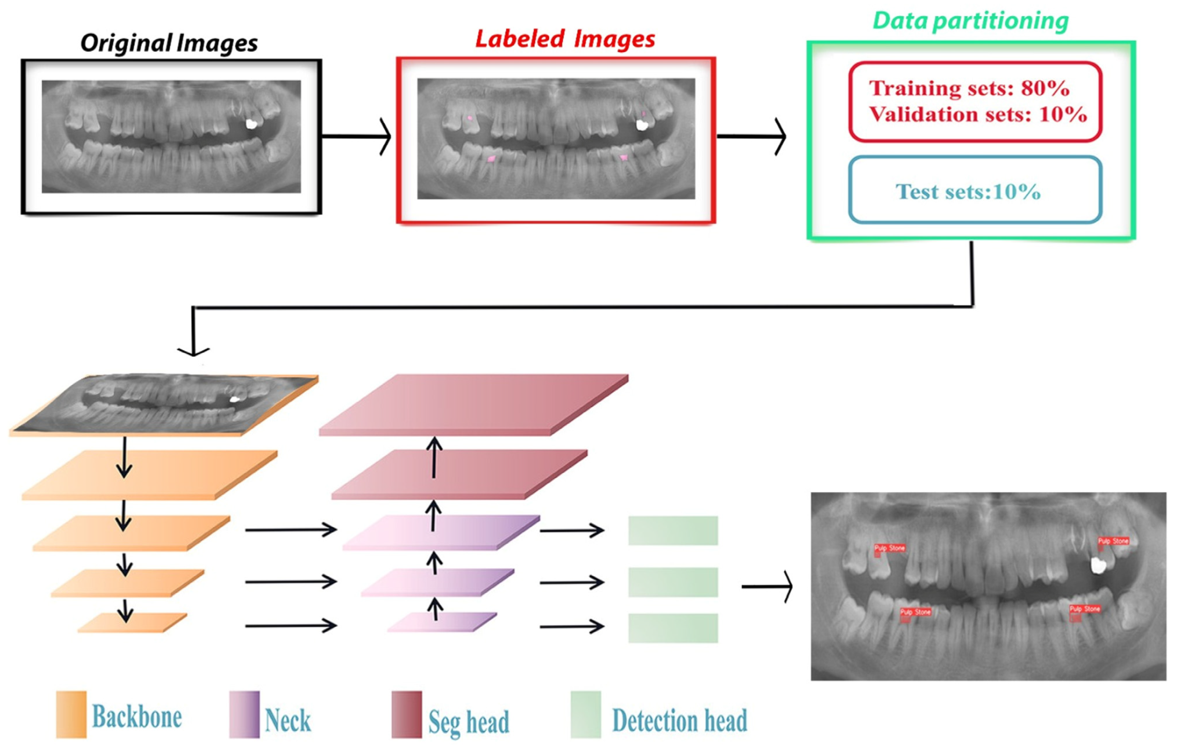
2.2.1. Labeling
2.2.2. Training
2.2.3. Success Evaluation
3. Results
4. Discussion
Limitations
5. Conclusions
Author Contributions
Funding
Institutional Review Board Statement
Informed Consent Statement
Data Availability Statement
Conflicts of Interest
References
- Şener, S.; Cobankara, F.K.; Akgünlü, F. Calcifications of the pulp chamber: Prevalence and implicated factors. Clin. Oral Investig. 2009, 13, 209–215. [Google Scholar] [CrossRef] [PubMed]
- Goga, R.; Chandler, N.P.; Oginni, A.O. Pulp stones: A review. Int. Endod. J. 2008, 41, 457–468. [Google Scholar] [CrossRef] [PubMed]
- Gulsahi, A.; Cebeci, A.; Özden, S. A radiographic assessment of the prevalence of pulp stones in a group of Turkish dental patients. Int. Endod. J. 2009, 42, 735–739. [Google Scholar] [CrossRef] [PubMed]
- Bauss, O.; Neter, D.; Rahman, A. Prevalence of pulp calcifications in patients with Marfan syndrome. Oral Surg. Oral Med. Oral Pathol. Oral Radiol. Endod. 2008, 106, e56–e61. [Google Scholar] [CrossRef] [PubMed]
- Abbott, P.; Yu, C. A clinical classification of the status of the pulp and the root canal system. Aust. Dent. J. 2007, 52, S17–S31. [Google Scholar] [CrossRef] [PubMed]
- Nayak, M.; Kumar, J.; Prasad, L.K. A radiographic correlation between systemic disorders and pulp stones. Ind. J. Dent. Res. 2010, 21, 369. [Google Scholar] [CrossRef]
- Ranjitkar, S.; Taylor, J.; Townsend, G. A radiographic assessment of the prevalence of pulp stones in Australians. Aust. Dent. J. 2002, 47, 36–40. [Google Scholar] [CrossRef]
- McCabe, P.; Dummer, P.M.H. Pulp canal obliteration: An endodontic diagnosis and treatment challenge. Int. Endod. J. 2012, 45, 177–197. [Google Scholar] [CrossRef]
- Pietrzycka, K.; Pawlicka, H.J.D.; Problems, M. Clinical aspects of pulp stones: A case report series. Dent. Med. Probl. 2020, 57, 213–220. [Google Scholar] [CrossRef]
- Shehab, M.; Abualigah, L.; Shambour, Q.; Abu-Hashem, M.A.; Shambour, M.K.Y.; Alsalibi, A.I.; Gandomi, A.H. Machine learning in medical applications: A review of state-of-the-art methods. Comput. Biol. Med. 2022, 145, 105458. [Google Scholar] [CrossRef]
- Singh, N.K.; Raza, K. Progress in deep learning-based dental and maxillofacial image analysis: A systematic review. Expert Syst. Appl. 2022, 199, 116968. [Google Scholar] [CrossRef]
- Arslan, E. Multiple Object Tracking with Data Association and Correlation Filter Based on Convolutional Neural Network Features; Bursa Uludag University: Bursa, Turkey, 2021. [Google Scholar]
- Bozkaya, F.; Yusefi, A.; Tiğlioğlu, Ş.; Kaya, A.K.; Kazanci, O.; Akmaz, M.Y.; Durdu, A.; Sungur, C. Improved Real Time Object Detection In Autonomous Systems Using Data Augmentation Methods. Eur. J. Sci. Technol. 2021, 2021, 83–87. [Google Scholar]
- Altındağ, A.; Sultan, U.; Bayrakdar, İ.Ş.; Çelik, Ö. Detecting Pulp Stones with Automatic Deep Learning in Bitewing Radiographs: A Pilot Study Of Artificial Intelligence. Eur. Ann. Dent. Sci. 2023, 50, 12–16. [Google Scholar] [CrossRef]
- Tamse, A.; Kaffe, I.; Littner, M.; Shani, R. Statistical evaluation of radiologic survey of pulp stones. J. Endod. 1982, 8, 455–458. [Google Scholar] [CrossRef] [PubMed]
- Orhan, K.; Bayrakdar, I.; Ezhov, M.; Kravtsov, A.; Özyürek, T. Evaluation of artificial intelligence for detecting periapical pathosis on cone-beam computed tomography scans. Int. Endod. J. 2020, 53, 680–689. [Google Scholar] [CrossRef]
- Hung, K.; Montalvao, C.; Tanaka, R.; Kawai, T.; Bornstein, M.M. The use and performance of artificial intelligence applications in dental and maxillofacial radiology: A systematic review. Dentomaxillofac. Radiol. 2020, 49, 20190107. [Google Scholar] [CrossRef]
- Musri, N.; Christie, B.; Ichwan, S.J.A.; Cahyanto, A. Deep learning convolutional neural network algorithms for the early detection and diagnosis of dental caries on periapical radiographs: A systematic review. Imaging Sci. Dent. 2021, 51, 237. [Google Scholar] [CrossRef]
- Sezgin, B.; Cakan, E.F.; Erdem, T.L. A Radiographic Assessment of the Prevalence and Distribution of Pulp Calcification. Eur. Oral Res. 2011, 45, 49. [Google Scholar]
- Altındağ, A.; Yüksel, İ.B. Pulpa Kalsifikasyonları Prevelansının Radyolojik Olarak Değerlendirilmesi. Necmettin Erbakan Üniversitesi Diş Hekim. Derg. 2021, 3, 102–107. [Google Scholar] [CrossRef]
- İlday, N.Ö.; Miloğlu, Ö.; Demirtaş, Ö.; Yildirim, E.; Seven, N.; Sağsöz, Ö. A Radiographic Assessment of the Prevalence of Pulp Stones in Patients Who Presented to Ataturk University Faculty of Dentistry Department of Oral Diagnosis and Radiology-Pulpa Taşı Prevalansının Atatürk Üniversitesi Diş Hekimliği Fakültesi Oral Diagnoz v. J. Istanb. Univ. Fac. Dent. 2014, 48, 9–16. [Google Scholar] [CrossRef]
- Hernandez-Solis, M.T.; Lara-Carrillo, E.; Toral-Rizo, V.H.; Bologna-Molina, R.E.; Garduño-Mejía, J.E.; Ibáñez-Mancera, N.G.; Hegazy-Hassan, W.; Santillán-Reyes, A.M. Frequency and Distribution of Pulpal Calcifications in Teeth Involved in Jaw Tumors. In Human Teeth—From Function to Esthetics; IntechOpen: Rijeka, Croatia, 2023. [Google Scholar]
- Hsieh, C.-Y.; Wu, Y.-C.; Su, C.-C.; Chung, M.-P.; Huang, R.-Y.; Ting, P.-Y.; Lai, C.-K.; Chang, K.S.; Tsai, Y.-W.C.; Shieh, Y.-S. The prevalence and distribution of radiopaque, calcified pulp stones: A cone-beam computed tomography study in a northern Taiwanese population. J. Dent. Sci. 2018, 13, 138–144. [Google Scholar] [CrossRef]
- Krastl, G.; Zehnder, M.S.; Connert, T.; Weiger, R.; Kühl, S. Guided endodontics: A novel treatment approach for teeth with pulp canal calcification and apical pathology. Dent. Traumatol. 2016, 32, 240–246. [Google Scholar] [CrossRef]
- Kumar, D.; Antony, S. Calcified canal and negotiation—A review. Res. J. Pharm. Technol. 2018, 11, 3727–3730. [Google Scholar] [CrossRef]
- Da Silva, P.Z.; Ribeiro, F.C.; Xavier, J.M.B.; Pratte-Santos, R.; Demuner, C. Radiographic evaluation of root canal treatment performed by undergraduate students, part I; iatrogenic errors. Iran. Endod. J. 2018, 13, 30. [Google Scholar]
- Schwendicke, F.; Rossi, J.; Göstemeyer, G.; Elhennawy, K.; Cantu, A.; Gaudin, R.; Chaurasia, A.; Gehrung, S.; Krois, J. Cost-effectiveness of artificial intelligence for proximal caries detection. J. Dent. Res. 2021, 100, 369–376. [Google Scholar] [CrossRef]
- Agrawal, P.; Nikhade, P.; Nikhade, P.P. Artificial intelligence in dentistry: Past, present, and future. Cureus 2022, 14, e27405. [Google Scholar] [CrossRef]
- Khanna, S.S.; Dhaimade, P.A. Artificial intelligence: Transforming dentistry today. Ind. J. Basic. Appl. Med. Res. 2017, 6, 161–167. [Google Scholar]
- Minaee, S.; Boykov, Y.; Porikli, F.; Plaza, A.; Kehtarnavaz, N.; Terzopoulos, D. Image segmentation using deep learning: A survey. IEEE Trans. Pattern Anal. Mach. Intell. 2021, 44, 3523–3542. [Google Scholar] [CrossRef]
- He, K.; Gkioxari, G.; Dollár, P.; Girshick, R. Mask r-cnn. In Proceedings of the IEEE International Conference on Computer Vision, Venice, Italy, 22–29 October 2017; pp. 2961–2969. [Google Scholar]
- Moutselos, K.; Berdouses, E.; Oulis, C.; Maglogiannis, I. Recognizing occlusal caries in dental intraoral images using deep learning. In Proceedings of the 2019 41st Annual International Conference of the IEEE Engineering in Medicine and Biology Society (EMBC), Berlin, Germany, 23–27 July 2019; pp. 1617–1620. [Google Scholar]
- Silva, G.; Oliveira, L.; Pithon, M. Automatic segmenting teeth in X-ray images: Trends, a novel data set, benchmarking and future perspectives. Expert Syst. Appl. 2018, 107, 15–31. [Google Scholar] [CrossRef]
- Selmi, A.; Syed, L.; Abdulkareem, B. Pulp stone detection using deep learning techniques. In Proceedings of the EAI International Conference on IoT Technologies for HealthCare, Virtual Event, 24–26 November 2021; pp. 113–124. [Google Scholar]
- Fariza, A.; Arifin, A.Z.; Astuti, E.R.; Kurita, T. Segmenting tooth components in dental x-ray images using Gaussian kernel-based conditional spatial Fuzzy C-Means clustering algorithm. Int. J. Intell. Eng. Syst. 2018, 12, 108–117. [Google Scholar] [CrossRef]
- Lee, S.; Oh, S.-I.; Jo, J.; Kang, S.; Shin, Y.; Park, J.-W. Deep learning for early dental caries detection in bitewing radiographs. Sci. Rep. 2021, 11, 16807. [Google Scholar] [CrossRef]
- Yang, Y.-M.; Guo, B.; Guo, L.-Y.; Yang, Y.; Hong, X.; Pan, H.-Y.; Zou, W.-L.; Hu, T. CBCT-aided microscopic and ultrasonic treatment for upper or middle thirds calcified root canals. BioMed Res. Int. 2016, 2016, 4793146. [Google Scholar] [CrossRef]
- Yuce, F.; Öziç, M.Ü.; Tassoker, M. Detection of pulpal calcifications on bite-wing radiographs using deep learning. Clin. Oral Investig. 2023, 27, 2679–2689. [Google Scholar] [CrossRef] [PubMed]
- Salahin, S.S.; Ullaa, M.S.; Ahmed, S.; Mohammed, N.; Farook, T.H.; Dudley, J. One-stage methods of computer vision object detection to classify carious lesions from smartphone imaging. Oral 2023, 3, 176–190. [Google Scholar] [CrossRef]
- Zhou, M.; Jie, W.; Tang, F.; Zhang, S.; Mao, Q.; Liu, C.; Hao, Y.J. Deep learning algorithms for classification and detection of recurrent aphthous ulcerations using oral clinical photographic images. J. Dent. Sci. 2024, 19, 254–260. [Google Scholar] [CrossRef] [PubMed]
- Ayan, E.; Bayraktar, Y.; Çelik, Ç.; Ayhan, B.J. Dental student application of artificial intelligence technology in detecting proximal caries lesions. J. Dent. Educ. 2024, 88, 490–500. [Google Scholar] [CrossRef]


| True Positive (TP) | False Positive (FP) | False Negative (FN) | Sensitivity | Precision | F1 Score |
|---|---|---|---|---|---|
| 614 | 177 | 151 | 0.8026 | 0.7762 | 0.7892 |
Disclaimer/Publisher’s Note: The statements, opinions and data contained in all publications are solely those of the individual author(s) and contributor(s) and not of MDPI and/or the editor(s). MDPI and/or the editor(s) disclaim responsibility for any injury to people or property resulting from any ideas, methods, instructions or products referred to in the content. |
© 2024 by the authors. Licensee MDPI, Basel, Switzerland. This article is an open access article distributed under the terms and conditions of the Creative Commons Attribution (CC BY) license (https://creativecommons.org/licenses/by/4.0/).
Share and Cite
Altındağ, A.; Bahrilli, S.; Çelik, Ö.; Bayrakdar, İ.Ş.; Orhan, K. The Detection of Pulp Stones with Automatic Deep Learning in Panoramic Radiographies: An AI Pilot Study. Diagnostics 2024, 14, 890. https://doi.org/10.3390/diagnostics14090890
Altındağ A, Bahrilli S, Çelik Ö, Bayrakdar İŞ, Orhan K. The Detection of Pulp Stones with Automatic Deep Learning in Panoramic Radiographies: An AI Pilot Study. Diagnostics. 2024; 14(9):890. https://doi.org/10.3390/diagnostics14090890
Chicago/Turabian StyleAltındağ, Ali, Serkan Bahrilli, Özer Çelik, İbrahim Şevki Bayrakdar, and Kaan Orhan. 2024. "The Detection of Pulp Stones with Automatic Deep Learning in Panoramic Radiographies: An AI Pilot Study" Diagnostics 14, no. 9: 890. https://doi.org/10.3390/diagnostics14090890