Review

Metallic Nanoparticles and Core-Shell Nanosystems in the Treatment, Diagnosis, and Prevention of Parasitic Diseases

1
Department of Microbiology and Immunology, Institute of Medical Science, Collegium Medicum, Jan Kochanowski University, IX Wieków Kielc 19A, 25-317 Kielce, Poland
2
Independent Laboratory of Nanomedicine, Medical University of Białystok, Mickiewicza 2B, 15-222 Białystok, Poland
3
Department of Medical Microbiology and Nanobiomedical Engineering, Medical University of Białystok, Mickiewicza 2C, 15-222 Białystok, Poland
4
Department of Public Health , Institute of Health Science, Collegium Medicum, Jan Kochanowski University, IX Wieków Kielc 19A, 25-317 Kielce, Poland
*
Author to whom correspondence should be addressed.
Pathogens 2023, 12(6), 838; https://doi.org/10.3390/pathogens12060838
Submission received: 25 April 2023 / Revised: 5 June 2023 / Accepted: 8 June 2023 / Published: 17 June 2023
(This article belongs to the Section Parasitic Pathogens)

Abstract

:
The usage of nanotechnology in the fight against parasitic diseases is in the early stages of development, but it brings hopes that this new field will provide a solution to target the early stages of parasitosis, compensate for the lack of vaccines for most parasitic diseases, and also provide new treatment options for diseases in which parasites show increased resistance to current drugs. The huge physicochemical diversity of nanomaterials developed so far, mainly for antibacterial and anti-cancer therapies, requires additional studies to determine their antiparasitic potential. When designing metallic nanoparticles (MeNPs) and specific nanosystems, such as complexes of MeNPs, with the shell of attached drugs, several physicochemical properties need to be considered. The most important are: size, shape, surface charge, type of surfactants that control their dispersion, and shell molecules that should assure specific molecular interaction with targeted molecules of parasites’ cells. Therefore, it can be expected that the development of antiparasitic drugs using strategies provided by nanotechnology and the use of nanomaterials for diagnostic purposes will soon provide new and effective methods of antiparasitic therapy and effective diagnostic tools that will improve the prevention and reduce the morbidity and mortality caused by these diseases.

1. Introduction

Despite continuous improvement of antiparasitic therapies, statistics show that parasites are responsible for about 14 million deaths per year and the total number of people affected by parasites is close to 3.5 billion [1]. Parasites have developed a wide range of potential transmission routes, from which it is difficult to determine which is of the greatest epidemiological importance. In the course of evolution, there have been many adaptations to specific forms of parasitism, including the ability to modulate the host’s immune response, high fecundity of parasites with long survival of invasive forms, as well as the use of parasitic vectors [2,3,4,5]. In humans, parasitic infections most often occur as a result of drinking contaminated water (e.g., Cryptosporidium parvum), consumption of raw or undercooked food (e.g., Giardia intestinalis, Diphyllobothrium latum, and Trichinella spiralis), inhalation of air with dust that can serve as a mean of transport for the parasites’ eggs (e.g., Enterobius vermicularis), contact with animals (e.g., Toxocara spp. or Diphylidium caninum), or contact with animal excrements (e.g., infection caused by Echinococcus multilocularis, and Toxoplasma gondii) [5,6,7,8,9,10,11,12,13,14]. An additional factor that contributes to parasite infections is travel to tropical areas, which often increases the chance of acquiring parasites, such as Schistosoma haematobium, Schistosoma mansoni, Plasmodium spp., and Trypanosoma spp. [15,16,17,18]. Sexual contact can also lead to infections caused by Trichomonas vaginalis or Pthirus pubis [19,20,21]. Nowadays parasitic diseases are a growing health problem in developing countries. Some of these diseases, such as leishmaniasis, African trypanosomiasis, or Chagas’ disease, belong to the group of so-called “neglected tropical diseases” (NTDs) which are prevalent in tropical and subtropical countries [22,23]. Therapeutic agents used to treat these diseases have limited efficacy and cause harmful or, sometimes, potentially fatal side-effects [24,25]. An additional problem of antiparasitic drugs (especially when administered against intracellular parasites) is their poor penetration through biological membranes of tissues and cells, which results in low bioavailability and, in consequence, lack of therapeutic effect [26,27,28]. These limitations might be greatly addressed by drugs in the form of nanosystems as they are usually characterized by increased tissue/cell penetration and high biocompatibility. The main ways of action of antiparasitic drugs are: blockade of parasite neuromuscular functions (causing muscle paralysis), induction of apoptosis, and inhibition of DNA and RNA synthesis. Antiprotozoal agents usually target the rapidly proliferating, cells. They also might target the synthesis of proteins or the specific metabolic pathways that distinguish host and parasite signal transmission pathways [27]. Standard treatment of parasitic diseases, apart from surgical removal of the parasite, is mainly based on the administration of one or a combination of few antiparasitic drugs (especially in the case of drug-resistance). The combination of several drugs is widely used. For example, in malaria a usual treatment is chloroquine combined with sulfadoxine–pyrimethamine [27] or, in the case of resistant strains, a combination of quinine and doxycycline or clindamycin. Such combinations often cause high toxicity, so new treatments are urgently needed. The drugs used in selected groups of parasites are listed in Table 1 [29,30,31].
One possible future solution to improve management of parasitic disease such as malaria may be the application of nanomaterials, such as metallic nanoparticles. Inorganic nanoparticles may be toxic. Factors affecting the cytotoxicity of metallic nanoparticles include, but are not limited to, size, shape, surface charge, nanoparticle synthesis, and metal ion release. This toxicity can be modulated by changing the shape and size of the particles and modifying their surface, thanks to which we can obtain nanoparticles with the desired properties, and controlled toxicity [27,32]. An increasing number of studies confirm this potential [27,33,34,35,36,37,38,39,40,41,42,43,44,45,46,47,48]. Currently, a significant number of nanosystems developed for medical use are composed of metallic nanoparticles with drug molecules attached to their surface, along with lipid particles or additional polymers. They can specifically affect tissues and cells by direct binding to specific receptors on the cell surface, which may be considered as their targets. They can also diffuse, accumulate within the tissue, and induce/provide some intrinsic and synergistic activity [24,49]. Their intrinsic properties, such as interaction with target cells, circulation time, renal clearance, protein absorption, and toxicity, can be governed by particle size, shape, diameter, and surface properties [35]. MeNPs can be synthetized using a wide spectrum of metals and metal oxides, such as gold, silver, iron oxide, copper oxide, zinc oxide, aluminium oxide, titanium dioxide, or gallium [50,51].

2. Different Strategies for Using MNPs and MNPs-Containing Nanosystems in Diagnostics, Prevention, and Therapy of Selected Parasitic Diseases

In the human body, parasites produce various chemical compounds and induce different physical effects, usually causing mechanical damage to tissues or organs. This process can be transient (reversible), but also permanent—such as liver or bladder damage occurring, respectively, during Echinoccocus multilocularis and Schisthosoma haemathobium infection. Some infections can directly cause the death of the host, such as neurocysticosis (Taenia solium), but, on the other hand, some parasites may not cause any symptoms, camouflaging themselves in the human body. Their presence might be revealed only in certain clinical conditions, such as immunosuppression or cancer (e.g., Toxoplasma gondii, and Cryptosporidium parvum) [12,52,53,54,55,56].
The use of nanotechnology can offer in the future many unique opportunities for better diagnostics of parasitic diseases. Diagnostic tools used nowadays for the detection of parasitic diseases include light and fluorescence microscopy, rapid diagnostic tests (RDT), such as immunochromatographic lateral flow tests, serology, quantitative Buffy-coat (QBC) techniques, and nucleic acid amplification techniques (mainly polymerase chain reaction—PCR) [44,57,58,59]. The diagnostics might be improved by the development of novel methods which use nanomaterials. Fluorescent, magnetic, and metal nanoparticles are currently being tested for their diagnostic potential. Among the latter, gold and silver nanoparticles are the most studied. They can emit intense absorption when excited by electromagnetic radiation [60]. Various molecules, such as antibodies, antigens, and enzymes, can be conjugated to nanoparticles as electrochemical markers, optical probes, and signal enhancers. An example is the use of magnetic nanoparticles with an iron oxide core and a silver coating for the early detection of malaria [61,62].
In the treatment of parasitosis, the key challenge is precise drug delivery to assure parasite elimination with low toxicity to host tissues and cells. The presence of specific homing ligands attached to the surface of the nanosystem might potentially allow selective binding to the parasite cell receptors, guaranteeing precise drug delivery [63,64,65]. Usage of MeNPs for drug delivery requires drugs attachment to nanoparticles surface that can be achieved by chemical adsorption, encapsulation, and conjugation. MeNPs and nanosystem administration can be conducted by oral, intragastric, intravenous, intraduodenal, pulmonary, and cutaneous methods [27,66,67,68,69]. Metal-based nanoparticles for biomedical applications need to meet several requirements such as stability, lack of aggregation, host biocompatibility, and cell/tissue selectivity [35,43,70,71]. There are experimental methods of usage of MeNPs to diagnose and treat cancers which display the theranostic potential of nanoparticles [72]. Parasitic diseases can interfere with, promote, or modulate carcinogenesis. Some parasites can modify the host’s immune response, thereby modifying the tumor microenvironment. For example, studies conducted in the case of active infection with Toxoplasma gondii in mice challenged with Lewis lung carcinoma cells showed increased survival. Similar studies resulted from the use of Plasmodium sp. Inducing strong innate and adaptive anti-cancer immune responses promoted survival and reduced the proliferation of Lewis lung cancer cells in an experimental model [73]. A separate group are parasites affecting the development of cancer, which include, among others: Schistosoma haematobium—associated with the development of bladder cancer; Schistosoma mansoni in people infected with hepatitis C virus (HCV)—increased risk of hepatocellular carcinoma; and Clonorchis synesis, the parasite that causes clonorchiasis, has been classified as highly carcinogenic, causing bile-duct carcinoma [73].
Magnetic hyperthermia using MNP comprises locally increasing the temperature above 42 °C, influencing the physiology of cancer cells by changing structural proteins, and inducing cell necrosis or apoptosis. In some parasitic diseases, there are intermediate forms in the form of cysts, e.g., Echinococcus granulosus, and Taenia multiceps. It can be assumed that the use of nanoparticles to induce hyperthermia, as in the case of cancer, can potentially be used in the fight against parasitic diseases in which one of the developmental forms is cysts. It might be supposed that this phenomenon, as in case of cancers, can potentially be used to fight parasitic diseases, especially those associated with organ tumor formation [50,51,74].
Recently, the development of new therapeutic strategies with the use of nanomaterials has been proposed for different parasitic diseases. A large number of studies were dedicated to leishmaniasis, African trypanosomiasis, American trypanosomiasis, malaria, toxoplasmosis, and schistosomiasis (see Figure 1, Table 2) [28,75,76].

2.1. Leishmaniasis

Leishmaniasis is a disease caused by a parasitic protozoan from the family of Trypanosomatidae. More than 20 species of leishmaniasis infect humans and the vector for it are female flies. The disease affects over 10 million people in about 90 tropical and subtropical countries. The WHO reports that the number of people at risk of leishmaniasis is ~350 million [98]. The most life-threatening form is visceral leishmaniasis (about 400,000 cases per year—95% mortality in untreated cases) [24,99]. The most common form is cutaneous leishmaniasis, which affects approximately 2 million people annually. It causes many clinical symptoms, including lumps and skin ulcers [99,100,101].
Many research groups have focused on development of novel approaches to treat and diagnose leishmaniasis. These efforts in the future can result in development of several potential drugs that are based on metallic nanoparticle. A considerable effectiveness was reported for silver nanoparticles (AgNPs) that cause damage to the structural integrity of Leishmania amazniensis amastigotes. AgNPs lead to the death of promastigotes and damage to amastigotes. The effect is a reduced number of infected macrophages, reduction of their metabolic activity, reduction of proliferation, and hastened deterioration [64,81,82,102,103]. Zinc oxide nanoparticles (ZnONPs) seem to be also effective. In in vitro studies, they display antiparasitic properties against Leishmania tropica [104]. Similarly, ZnONPs exert cytotoxicity against amastigote cells affecting the proliferation and metabolic activity of Leishmania donovanii. Interestingly, extracts of Verbena officinalis and Verbena tenuisecta might potentialize ZnONPs activity against the Leishmania parasite [85,102].
Gold nanoparticles conjugated with quercetin (QAuNPs) have also shown effectiveness and selectivity against Leishmania’s amastigote. Promising results were obtained when nanoparticles were used together with ultraviolet (UV) and infrared (IR) radiation—this enhances the killing effect of the nanoparticles, associated with increased generation of reactive oxygen species. While testing anti-leishmatic activity, UV and IR radiation effects were also assessed in the presence of silver nanoparticles (AgNPs), gold nanoparticles (AuNPs), titanium dioxide nanoparticles (TiO2NPs), zinc oxide nanoparticles (ZnONP), and magnesium oxide nanoparticles (MgONPs). All these experiments have shown promising anti-parasite effects, which was the most effective in case of silver and gold-based nanomaterials. However, when combined with UV and IR radiation, most of metallic nanoparticles displayed high toxicity to macrophages [80].
One possible way to address the high toxicity to macrophages might be the use of biogenic silver and gold nanoparticles. It was found that biogenic AgNPs are more efficient than those chemically synthesized—they have a similar effect on Leishmania at lower concentrations. They induce reactive oxygen species formation more strongly, resulting in the death of amastigotes [105,106]. It is worth underlining that biogenously synthesized AgNPs inhibit the growth of the parasite in a concentration-dependent manner. The effectiveness of biogenic silver nanoparticles (extract from Moringa oleifera, a horseradish tree) against leishmaniasis is worth pointing out due to their high efficiency. Indeed, the synthesis of AgNP using fungi, bacteria, or plants should be considered as an innovative approach in an attempt to reduce MNPs’ toxicity. Additionally, bioparticles conjugated with enzymes, proteins, polysaccharides, or amino acids that are present in plant extracts might have different, and usually better, therapeutic value when compared to those synthesized by stabilizing and reducing silver ions. Apart from microorganisms, biosynthesis of metallic nanomaterials can be achieved using plant species, such as Coriandrum sativum, Azadirachta indica, Nelumbo nucifera, Ocimum basilicum, and several others [107,108,109].

2.2. African Trypanosomiasis (African Coma)

Trypanosomes are a type of parasitic protozoan characterized by the presence of a single strand of dense DNA called a kinetoplast. They are responsible, inter alia, for trypanosomiasis (Trypanosoma brucei) and the American–Chagas’ disease (Trypanosoma cruzi). Infectious forms of Trypanosoma cruzi are trypomastigotes that can infect many different cell types through a process independent of the host cell’s actin polymerization. Parasite adhesion is driven by particles present on the surface of the parasite or secreted during the infection process; these molecules bind to specific receptors on the host cells [110]. The human African trypanosomiasis is caused by the protozoan Trypanosoma brucei gambiense (milder form) and Trypanosoma brucei rhodesiense (associated with faster disease progression) that involve development of neuronal symptoms and higher mortality [30,111]. The disease occurs only in Sub-Saharan Africa and is transmitted by vectors—the flies of genus Glossina [112,113,114]. When untreated, the disease often leads to coma and death due to multi-organ failure. Estimated data show that 65 million people living in the 36 Sub-Saharan African countries are at risk of contracting the disease [113]. Production of an effective vaccine is difficult due to the presence of a parasitic coat with variable surface glycoprotein (VSG) [24]. It allows trypanosomatic parasites to evade the host’s immune system through extensive antigenic variability [24,113]. Currently, drugs used to treat the infection cause serious adverse effects that were summarized in Table 1. The use of metal nanoparticles as a drug-delivery system for the above-mentioned compounds can, potentially, significantly reduce the occurrence of side-effects. Combinations of nanoparticles and drugs might have higher efficacy compared to anti-pathogenic agents alone, and can shorten the duration and doses of the drug administered. Functionalization of metallic nanoparticles with anti-pathogenic agents might also improve the pharmacokinetic properties of the drug by increasing its solubility, extending the half-life of the drug and the time of systemic circulation, which might result in lower doses and frequency of administration of the drug, thus reducing its cytotoxicity [27,115,116,117]. Metallic nanoparticles, such as silver and gold, display different potential against different species of Trypanosoma (Trypanosoma brucei, Trypanosoma congolense, and Trypanosoma evansi). Gold nanoparticles were less effective against Trypanosoma congolense and Trypanosoma evansi, with an average ≤50% inhibition of parasite growth compared to silver nanoparticles, which were ≥70%. Gold and silver nanoparticles strongly and selectively inhibit recombinant Trypanosoma brucei arginine kinase [77,111]. Arginine kinase is a phosphotransferase that is essential for the growth and survival of Trypanosoma brucei, particularly in the bloodstream of the infected host because it helps the parasite meet required energy demands to maintain a reductive environment [24,77]. The high selectivity of the nanoparticles for Trypanosoma spp.-specific kinase indicates a high potential for combining them with drugs.

2.3. Chagas’ Disease, American Trypanosomiasis
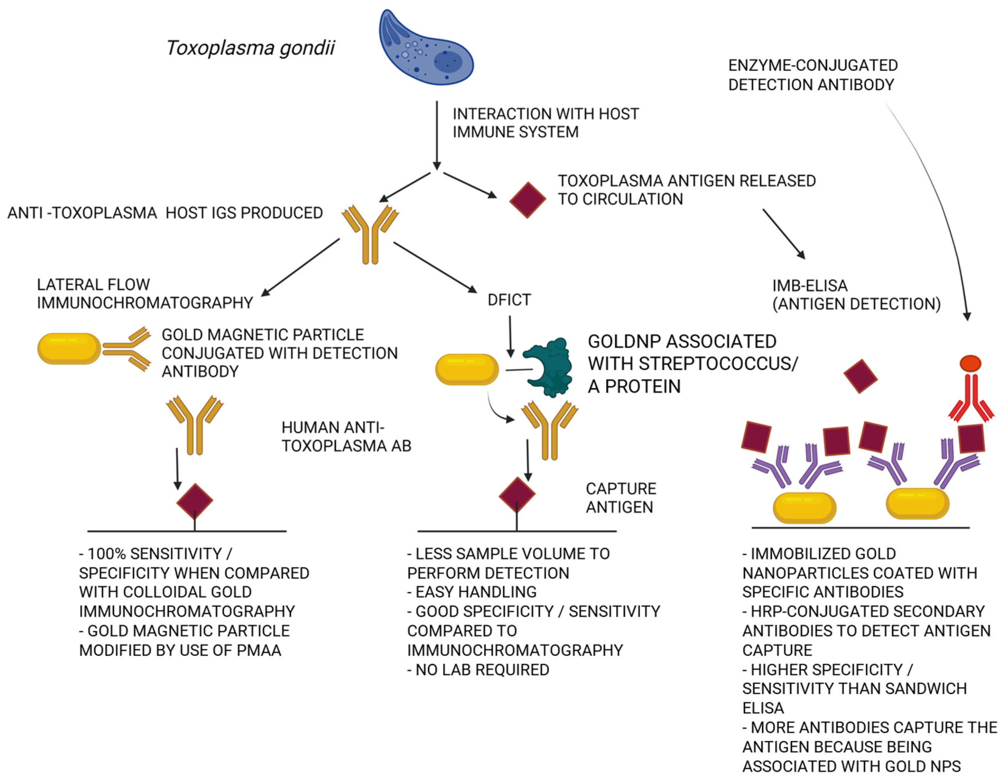
Chagas’ disease is caused by Trypanosoma cruzi. It is associated with a high level of morbidity and mortality, and its treatment is often not effective due to drug toxicity and low bioavailability [118]. The disease affects about 8 million people and causes about 50,000 deaths per year [22,119]. The disease can be transmitted perinatally; 15,000 newborns are born as infected each year [120,121]. The number of infected women of childbearing age is about 1 million and the risk of transmitting the parasite to the child is 3 to 5% [22]. Some recent Chagas’ disease diagnostics methods involved the use of nanomaterials—including metallic nanoparticles. More precisely, these diagnostic tools should be used in an integrated microflow system connected to a carbon-screen-printed electrode (SPCE) [122]. It is designed to quantify specific IgG antibodies in serum. It uses AuNPs functionalized with Trypanosoma cruzi proteins isolated from epimastigotes. The lower detection limit of the test was established to be 3.065 ng/mL and the coefficients of variation within and between tests were lower than 6.95% [122]. For early diagnosis of congenital Chagas’ disease, the Chunap test (Chagas’ urine nanoparticles test) is used. It is based on the Western blot detection of antigens that were secreted by trypomastigote forms [123]. The sensitivity and specificity of a urine-based test are, approximately, 90 and 95%, respectively, due to the use of nanoparticles that have been functionalized with polyunsaturated N-isopropylacrylamide and tryptan blue that can capture and concentrate T. cruzi [122]. Nifurtimox and benzimidazole are used in the treatment of Chagas’ disease. They are mainly associated with dermatological side-effects, fever, and lymphadenopathy [22,124]. Rarely, such conditions as gastrointestinal disorders, weight loss as well as more severe paresthesia, polyneuropathy, peripheral-nerve inflammation, bone-marrow dysfunction, and heart failure were reported [125,126,127,128,129]. The use of nanotechnology can have a significant impact on improving biopharmaceutical properties of the above-mentioned drugs. These features include increase in solubility, permeability, stability in the host’s bloodstream, bioavailability, and extend release time. Benzimidazole also has an anti-inflammatory effect. It is one of the main drugs used in the treatment of acute, early, and chronic Chagas’ disease, but it also causes systemic toxicity [130]. Nanosystems can help to transport the drug to the intracellular target and to reduce side-effects. This is achieved by increasing the effectiveness and bioavailability of the active compound against the pathogen, by extending the release and, thus, increasing the therapeutic index [123,131,132]. In the case of Trypanosoma, gold and silver nanoparticles have promising antiparasitic properties. This has been determined using both in vitro and in vivo settings [133]. However, in case of Trypanosoma congolense and Trypanosoma evansi, gold nanoparticles were less effective, with an average 50% reduction of parasite growth. Silver nanoparticles were found to reduce the growth of Trypanosoma congolense and Trypanosoma evansi by 70%, indicating that these nanoparticles might have more clearly defined targets in parasites cells [77]. Both AuNPs and AgNP displayed strong activity against Trypanosoma brucei, reducing parasite viability by more than 5%. These data suggest that the activity of nanoparticles against Trypanosoma is strongly selective for the parasite, which may also suggest strong potential for their use in infection with Trypanosoma cruzi [22].

2.4. Toxoplasmosis

Toxoplasmosis is caused by Toxoplasma gondii protozoan and in immunocompetent subjects in 80% of cases is asymptomatic. In the remaining, 20% is oligosymptomatic (enlargement of lymph nodes, flu-like symptoms, pneumonia very rarely, and myocarditis). Most often, the symptoms disappear and do not require treatment as the infection takes the latent form. The preventive measures and diagnostics of toxoplasmosis are of special importance in efforts to avoid development of its congenital type, which might cause birth defects and the death of fetuses. The course of toxoplasmosis depends, partly, on the particular Toxoplasma gondii strain and its intensity is determined mainly by the host’s immune response [134,135]. In most cases, the T. gondii infection does not produce any symptoms, but the protozoan is capable of producing tissue cysts that can be present for many years. Individuals particularly susceptible to symptomatic infections are those with cancer, AIDS, or ongoing immunosuppressive treatment, in whom the infection may cause neurological, ocular, and systemic complications [136,137,138,139,140,141].
In pregnant women, the consequences of Toxoplasma gondii infection depend on the stage of pregnancy. Miscarriages and nervous-system malformations of the fetus occur mainly in the first trimester of pregnancy [134,140]. In subsequent trimesters, the likelihood of damaging the fetus is lower, but it is more likely that the protozoan will overcome the mother–fetus barrier. The first prevention measure to undertake should be health education to limit the spread of toxoplasmosis in the human and animal population (especially, to reduce the exposure of pregnant women who will be more likely affected by primary infection). The second measure should be based on the mass screening of pregnant women and serological tests of neonatal blood to detect fetal infection. Diagnosis of pregnant women is essential in determining the type of infection (primary, or secondary), which translates to the probability of the parasite’s penetration through the placenta. The use of direct methods allows the detection of the presence of a parasite in the diagnostic specimens, such as amniotic fluid, placenta, umbilical cord blood, and fetal blood (identification of congenital infection) [142,143,144]. Indirect methods—biological tests involving the introduction of a parasite into laboratory animals or tissue cultures—are rare due to the time-consuming nature of the process and limited accessibility. MeNPs can be used in the diagnosis of toxoplasmosis, improving the effectiveness and sensitivity of tests. One example of such a test is a dynamic imaging test (DFICT) with sensitivity and specificity of 92% and 93.1%, respectively. This test involves use of gold nanoparticles [145,146,147,148]. Using nanoparticles modified with poly methacrylic acid (PMAA) coupled to the anti-human IgM provides a test with near-100% sensitivity and specificity, with the possibility of simultaneous detection of several pathogens [146,149]. A similar test is carried out using magnetic nanoparticles (IMB-ELISA) to detect Toxoplasma gondii surface antigens circulating in human-serum samples with a sensitivity of 98% and a specificity of 96.4% [150]. Nanotechnology has also found application in Toxoplasma gondii diagnostic tests involving the detection of genetic material using DNA probes with nanoparticles [151]. From a diagnostic platform, quantum dot probes synthesized with nickel nanoparticles (CdTE/Ni mQD) with magnetic and fluorescent properties, provide high sensitivity, specificity, and effectiveness in T. gondii DNA sensing [147].
In prevention methods against Toxoplasma gondii, the usage of nanoparticles for the development of an effective vaccine represents a vital alternative. The development of vaccines is particularly important from the perspective of immunocompromised subjects. Infection in people with a properly functioning immune system is usually asymptomatic, creating the dormant forms (tissues containing bradyzoites) in the brain and muscles [152]. As a result of decreased immunity, these dormant forms may reactivate and cause widespread tissue destruction. Such reactivation of latent infection is one of the main causes of mortality in AIDS-suffering patients. Different vaccination trials performed so far in animals with inactivated/attenuated vaccines, DNA vaccines, subunit vaccines, and genetically engineered vaccines indicate that development of such protective strategies for practical use in humans is possible [153]. The possible use of vaccines in humans against Toxoplasma gondii is still questionable, as some experiments that are effective in animal models still fail to provide protective immunity in humans. The Toxoplasma gondii life-cycle is very complex, resulting in the expression of different proteins with a high degree of antigenic polymorphism and variability, which depend on host and invasion pathways. So far, only a limited number of tested Toxoplasma gondii antigens were found to induce long-lasting immunity. Overall, the strategy to design effective anti-Toxoplasma vaccines should include optimal choice of the antigens and a proper administration method combined with optimized delivery systems that, taken together, will provide efficient induction of the immune response. In this context, MeNPs might serve as adjuvant for Toxoplasma gondii antigens to promote activation and migration of immunocompetent cells, which are involved in antigen presentation [33].
The vaccine against Toxoplasma gondii should provide high Th1-type immune response and prevent the development of the chronic form of the disease. Since there are many strains of Toxoplasma gondii and the parasite has innate mechanisms of avoiding immunity [154,155], this requirement is of particular importance. As mentioned above, the protein subunit vaccines or polyvalent subunit vaccines (“antigen cocktails”), as well as DNA vaccines, have been tested in animal models. They are an alternative to inactivated vaccines containing subcellular components of the parasite, such as SAG surface antigens. In Toxoplasma gondii tachyzoites, there are three types of characteristic surface antigens (SAG1, SAG2A, and SAG2B). The same situation is found in bradyzoites, although the antigens are different (SAG2C, SAG2D, and SAG4). The SAG3 antigen is a common future in both forms of Toxoplasma gondii. Vaccinations with combinations of the above-mentioned antigens in mice resulted in an increased survival rate after infection with a particularly lethal type 1 parasite. Better results were obtained after using poliglikolid (lactide-co-glycolide PLG) microparticles in combination with SAG1 and SAG2 protein antigens, which promote the sustained release of the antigens [155]. Studies were performed to develop a DNA-based vaccine against Toxoplasma gondii ribosomal P2 protein, which had its efficacy boosted by the usage of two nanomaterials—poly-lactic-co-glycolic acid (PLGA) and chitosan [156]. The DNA of the vaccine was trapped inside nanospheres, which were created from these nanomaterials. Study results show that both these vaccines elicited mixed Th1–Th2 immune response in mice model. The titers of both IgG1 antibodies (associated with Th2-related immunity) and IgG2a antibodies (associated with Th1-related immunity) were higher to the titers obtained when the DNA of the vaccine was administered without nanospheres. These results are promising from the point of view of the usage of nanomaterials in boosting the immune response against parasitic invasions.
Another research was conducted using self-replicating RNA amplicons, encapsulated in modified dendrimer nanoparticles (MDNP) [157]. The replicon used encoded GRA6, ROP2A, ROP18, SAG1, and SAG2A as well as apical membrane antigen-1. The nanosystem is capable of delivering this self-replicating RNA into the cytoplasm and to protect it against the action of cellular nucleases. The administration of this nanosystem-based RNA vaccine was able to protect mice against lethal infection by T. gondii. Additionally, the vaccine was effective enough to activate and stimulate the proliferation of CD8+ T lymphocytes, as well as to increase the expression IFN-γ and IL-2 [158]. Promising results in the treatment of toxoplasmosis have been achieved by the usage of chitosan nanoparticles (CSNPs), which display a high anti-parasitic activity against tachyzoites of T. gondii strain RH. The spiramycin-loaded chitosan nanoparticles (SLCNs) were demonstrated to increase the antiparasitic effect in both acute and chronic T. gondii infections [159,160]. A similar result was obtained with the use of calcium carbonate nanoparticles loaded with benznidazole (BZN@CaCO3) on the Trypanosoma cruzi species responsible for the Chagas’ disease. The use of BZN@CaCO3 decreased the viability of epimastigote and trypomastigote Trypanosoma cruzi. The antiparasitic effect was achieved with a lower-than-usual concentration of benznidazole; the effective usage of such a lower concentration of the drug may contribute to lessening the possibility of drug toxicity and, thus, adverse events [161].
The efficacy of similar nanoparticles can also be demonstrated in single-chamber echinococcosis caused by Echinococcus granulosus in which the colloimidic activity of biosynthesized nanoparticles (AgNPs) displayed up to 90% eradication ability of protoscolexes [162]. The efficacy of gold nanoparticles (AuNPs) was tested not only in the case of toxoplasmosis, but also to fight schistosomiasis, leishmaniasis, trypanosomiasis, cryptosporidiosis, and malaria [27,65,77,163,164,165,166,167,168]. The bioactivity of AuNPs was also tested in relation to selected species of insects of public-health importance, mainly including mosquito vectors, such as Aedes aegypti, Anopheles stephensi, Culex quinque fasciatus, Culex mites, and some tick species [169]. Very good results were observed with the use of silver and platinum nanoparticles, which caused >90% inhibition of T. gondii growth, with limited cytotoxicity to the host cells [170,171]. More effective action of gold and silver nanoparticles can also be observed using nanosystems AuNP-TRP and AgNP-TRP (both being of NPs conjugates with tryptophan) characterized by high activity against T. gondii. It happens so because Toxoplasma is auxotrophic for l-tryptophan (normally, it acquires tryptophan from the host in order to survive). The conjugation of NP with this amino acid promotes their uptake into parasite cells, thus, making them more sensitive to such nanosystems.
Atovaquone is used for the treatment of toxoplasmosis, especially in people with HIV/AIDS, malaria, or Pneumocystis jiroveci pneumonia. Unfortunately, this drug has low solubility and, thus, reduced bioavailability. Solubility may be improved by incorporation of atovaquone into conjugate nanosystems, for example, those with MeNPs [172,173,174]. The testing of polymeric polylactic nanocapsules (PLA) conjugated with atovaquone in the treatment of acute and chronic toxoplasmosis (animal model using mice) showed a positive therapeutic effect and improved drug activity. This therapy has shown a higher reduction of parasite load and an increased survival rate of experimental animals when compared to free atovaquone. A similar use of nanomaterials that improve bioavailability and activity of the drug has been demonstrated in experimental treatment with resveratrol. Resveratrol has a strong anti-inflammatory and antioxidant effect, as well as antibacterial, antifungal, antitumor, and antiparasitic properties. In addition, resveratrol regulates immunity by interfering with the regulation of immune cells, the synthesis of pro-inflammatory cytokines, and gene expression. Resveratrol participates in the activation of macrophages, T cells, and natural killer (NK) cells, and is involved in the suppressive regulatory functions of CD4 + CD25 + T cells. Pharmacokinetic analysis reveals that resveratrol is rapidly metabolized in the body and its oral bioavailability is very low, despite its absorption reaching 70%, which is associated with the use of high-dose concentrations that may cause immunosuppression [175]. To overcome these issues, resveratrol had been complexed with 2-hydroxypropyl-β-cyclodextrin (HPβCD). These complexes could also be additionally bound to sulphamethoxazol-trimethoprim particles. Treatment with such drug combinations reduced the number of brain cysts and inflammatory infiltrates in experimental animals—both in species infected with tachyzoites and bradyzoites of T. gondii. Such treatment had no effect on creatine kinase activity—an enzyme associated with energy metabolism of the brain—which proved better protection by resveratrol against inflammation within the CNS [138].
The promising use of nanotechnology in toxoplasmosis treatment might be expected with compounds such as ceragenins, which display a very broad antimicrobial spectrum of activity. The results of studies involving ceragenins CSA-13, CSA-44, CSA-131, and CSA-138 against the sensitive and metronidazole-resistant Trichomonas vaginalis protozoans allowed for an assumption, that these substances might be used against intracellular forms of Toxoplasma gondii. The application of metallic nanoparticles along with ceragenin as a core-shell nanosystem may result in mutual reinforcement of their biological action in relation to intracellular forms of Toxoplasma gondii [176]. The desired actions, i.e., the ability of drugs to cross biological barriers and to eliminate specific forms of this parasite (while minimizing side-effects) has yet to be achieved, but the likelihood of obtaining such therapeutic tools increases with the development and use of nanotechnology, as indicated in Table 3.

2.5. Malaria

Malaria is responsible for approximately 1.5 to 2.7 million deaths per year. Malaria is caused in humans by intra-erythrocyte protozoa of the Plasmodium species, which are transmitted by the Anopheles mosquito species. Of the Plasmodium species infectious in humans, Plasmodium falciparum and Plasmodium vivax are the most common malaria parasites in Africa and most of Asia. Plasmodium falciparum is responsible for cerebral malaria and the overwhelming majority of malaria deaths [181]. New methods for malaria diagnostics were developed using nanotechnology approach. A quantum-magnetic droplet test (MB—QD) allows for detection of Plasmodium falciparum protein 2 (HRP2—protein 2; rich in histidine) in serum or urine. Magnetic beads in combination with the anti-HRP2 antibody allow for the capture of target protein, which is then concentrated and detected using quantum dots [182]. For successful treatment of malaria, the main therapeutic option is artemisinin-based combination therapy (ACT)—currently one of the most effective antimalarial drugs, including the recommended mainstay in the treatment of Plasmodium falciparum infection. In addition, chloroquine and tafenoquine are used (please see Table 1), but the parasite shows resistance to these compounds in an increasing number of infections [183].
Gold and silver nanoparticles showed high activity against drug-resistant parasites [91]. The use of metallic nanoparticles in the treatment of malaria is supported by the fact that they can be synthesized according to the methods of “green chemistry”—using plant extracts such as those obtained from Andrographis paniculata or Calotropis gigantea. The plant extract might have its antiparasitic activity increased due to the presence of biological compounds, such as flavonoids, phenols, terpenes, steroids, and phenolic acids [184,185,186]. Some metal-oxide nanoparticles also have a strong anti-malaria effect. Oxide nanoparticles, such as Fe3O4, MgO, ZrO2, Al2O3, and CeO2, were coated with PDDS and their activity against Plasmodium falciparum was assessed. It was shown that PDDS-coated nanoparticles exhibit significantly better antimalarial activity than uncoated nanoparticles. Several researchers have demonstrated the anti-malarial effects of silver nanoparticles [76].
Studies have shown that photosynthetic nanoparticles have a lethal effect on different stages of malaria vectors. The best effect was observed in the use of gold and silver nanoparticles, especially in the larval and adult stages (mosquitoes egg are less susceptible to silver and gold nanoparticles) [184].

2.6. Schistosomiasis

Schistosomiasis is a very common parasitic disease. In humans, it is caused by the invasion of cercariae through the skin, usually during swimming in contaminated water. It is common in tropical and subtropical regions, mainly in places where safety and health-protection measures are limited. It is estimated that about 600–770 million people are at risk of being infected and that the disease currently affects over 200 million people worldwide. The estimated mortality in Sub-Saharan African countries is about 280,000 deaths per year [63].
The symptoms of schistosomiasis include abdominal pain, hematomas, anemia, and fibrosis of the bladder and liver. Currently, there is no vaccine against schistosomiasis, and treatment is mainly conducted with the use of the antihelmintic drug Praziquantel (PZQ), as indicated in Table 1. This drug is not effective against immature forms of the Schistosoma species and its misuse has led to the development of drug-resistant Schistosoma variants in many regions around the world [63].
To minimize the incidence and achieve the elimination of the pathogen, early diagnosis is the primary objective. Developing new ways of detection of Schistosoma, as well as observation of the progress of the disease, play a very important role.
Electrode biosensors are sensors that can capture trace amounts of analytes by detecting changes in electric current, electric potential, or conductivity caused by an immune response. They allow for significant differentiation in the shape of electrodes and material selection. The connection of nanomaterials to these sensors allowed for the increase in their sensitivity in detection of biomolecules at relatively lower thresholds. Biosensors based on nanotechnology (nanosensors) seem to be exceptionally sensitive, which is a promising solution in medical diagnostics. Modified biosensors are composed of various nanomaterials, i.e., carbon nanoparticles with gold and silver attached, magnetic nanoparticles, or carbon nanotubes. Nanomaterials can also be used to increase sensitivity and improve antigen- and antibody-based detection methods. So far, most studies have been based on the detection of the Schistosoma antigens. The example is an enzyme affinity test (magnetic affinity enzyme-linked immunoassay—MEIA). The MEIA test is based on the production of specific antibodies in response to the appearance of worm or larval antigen. In the MEIA test, magnetic beads have superparamagnetic nanoparticles and functional polymer materials. Additionally, magnetic beads were transformed by means of characteristic functional groups to be able to bond with charged molecules. The magnetic field was applied to the reactive system after setting molecular targets on magnetic beams. As a result, an immune complex was obtained by magnetic separation. In another diagnostic method, colloidal gold, a different variant of metallic nanoparticles was used. Thanks to the possibility of binding with biological macromolecules, i.e., immunoglobulins, such Ig-bonded colloidal gold is a practical device for key studies on clinical diagnostics. During the diagnostics process, schistosomal antigen produces an immune complex with IgG–colloidal gold [88].
As mentioned above, Praziquantel is active against all species of Schistosoma. It causes morphological changes in the parasite’s coating which increases antigen exposure to the action of the immune system. It also affects muscle contraction—probably through a rapid inflow of Ca2+ ions. In the past, some other drugs were used (metrifonate, oltipraz, niridazole, and oxamniquine). All of them were withdrawn due to high toxicity, carcinogenicity, and lack of desired efficacy against the parasite [63]. Facing so many flaws of current therapies, the use of nanotechnology in the precise delivery of the drug seems to be an appropriate approach. The main goal is the selection of appropriate nanoparticles for the delivery system in order to minimize side-effects and improve the effectiveness of the selected drug. This is particularly important in cases of co-infection with several strains of Schistosoma or in cerebral schistosomiasis. During the development of novel treatment regimens against schistosomiasis, the following nanosystems are used: liposomes, micelles, polymer nanoparticles, and metallic nanoparticles. Among the metallic nanoparticles, mostly silver and gold nanoparticles are used. It has been observed in Schistosoma mansoni infection (tested in mice model) treated with different doses of AuNPs, that nanoparticles have a beneficial effect on oxidative stress caused by the disease. AuNPs also alleviate inflammatory cell infiltration, reduce the diameter of granules, as well as reduce the extent of histological damage in the central nervous system [87].

3. Immune Response during Host—Parasite Interaction. Can MNPs and MNPs-Containing Nanosystems Be Used as Immunomodulators during the Course of Parasitic Diseases?

The host’s immune response to the parasitic infection depends mainly on the interaction of the parasite and the host’s immune system. The host defense mechanisms are usually not effective enough to eradicate the invading parasite. This allows the parasites to reach their target organ, therefore, gaining time for progression of the disease and development of the parasite itself [187].
The host’s immune response to the parasitic infection may be immediate characterized by significantly increased eosinophilia, and mast cell and basophil tissue infiltration. High levels of IgE antibodies were also observed (several times over the norm) as well as the adaptation process of T CD4+ lymphocytes [188,189,190]. These cells participate in the Th2 pathway of the immune system and can synthesize IL-4 cytokine that participates in starting physiological reactions to parasitic infections. The Th2-pathway-associated cytokines include interleukins production, including of IL-4, IL-13, IL-5, IL-9, IL-10, IL-25, and IL-31 [191]. During parasitic worm infections, the development of the Th2 response is observed, while during protozoan infections (e.g., in the Toxoplasma gondii invasion), the development of the Th1 response is reported. The main means of defense against intracellular parasites is the cellular response in individuals with properly functioning immune system. In the acute phase of the invasion, as well as in the chronic stage of the disease, CD4+ and CD8+ lymphocytes kill the parasite-infected cells (CD8+ also kill the extracellular forms), and they contribute to the development of immunity against Toxoplasma gondii through cytokine production [192]. Th1 lymphocytes producing IFN-γ, IL-2, TNF-β, and IL-12 are responsible for the defense against the intracellular forms, while the Th-2 response (characterized by the production of IL-4, IL 5, IL-6, IL-9, and IL-6) serves for strengthening the humoral reactions and destruction of the extracellular forms. So far, we do not have strong experimental data indicating the possibility of enhancing the antiparasitic immune response with metal nanoparticles. However, such a possibility may be suggested based on the physicochemical properties of metal nanoparticles coated with molecules derived from parasites, which stimulate the host’s immune response, allowing for their high local concentration based on an optimized surface-to-volume ratio within synthetized nanosystems.

4. Parasitic Invasion of Host Cells as Potential Targets for Nanosystem Action

In this article, the strategy based on the blocking of the parasitic invasion of host cells is illustrated with the examples of two parasites caused by Plasmodium and Toxoplasma gondii. Malaria in humans is caused by five Plasmodium species—Plasmodium falciparum, Plasmodium vivax, Plasmodium ovale, Plasmodium malariae, and Plasmodium knowlesi—and is a disease with acute symptoms, ranging from recurrent fever, headache, muscle and joint pain, vomiting, jaundice, and anemia, leading to serious complications, such as acidosis, respiratory failure, kidney failure, and cerebral malaria [193]. Parasites are located only in erythrocytes and, thus, theoretically, can reach all host tissues through circulation. The parasites (a form of merozoite) are released into the bloodstream from hepatocytes enveloped in merosomes and continue a repetitive cycle of erythrocyte invasion. The infection of erythrocytes causes their extensive remodeling and, thus, dysfunction, followed by cell lysis, which contributes to the development of symptoms such as severe anemia. The merozoite entry is a complex and dynamic process in which the apex end of the merosome must be directed towards the erythrocyte, where specialized secretory organelles (called the micronemes, rhoptries, and dense granules) are located. When the merosome is connected to the erythrocyte, its double layer is opened, and the parasite (merozoite) is absorbed. Once inside the erythrocyte, the parasites mature and, due to their protein production, remodelling of the cells can occur. The parasite proteins are released after erythrocyte destruction; these proteins interact with uninfected erythrocytes to change their antigen and vascular properties. As a result of this process, infected as well as non-infected erythrocytes are remodelled by the parasite which initiates their removal by the spleen. This process may exacerbate anemia [194]. Repeated cycles of erythrocyte invasion lead to the release of the parasite, deepening of anemia, toxic hem removal, activation of immune cells (thrombocyte modulation, neutrophilia, monocytes, macrophages, T cells, and B cells), and may cause a cytokine/chemokine storm to develop. The knowledge of repetitive cycles of erythrocyte invasion in malaria processes became a base for the development of a very sophisticated strategy of usage of nanomaterials to prevent Plasmodium falciparum entry into the host’s red blood cells [195]. The nanomaterials were named nanomimics, and two co-polymers were used to create it: polymersome-forming ABA block copolymer poly(2-methyl-2-oxazoline)-block-poly(dimethylsiloxane)-blockpoly(2-methyl-2-oxazoline) (PMOXA-b-PDMS-b-PMOXA) as well as the PDMS-heparin block copolymer. These nanomimics contained cellular receptor analogues for Plasmodium falciparum and were shown to efficiently block the entrance of the parasite into host red blood cells (as compared to soluble receptor proteins). The authors suggest that this blockade of entry can leave the parasite vulnerable to both the host’s immune response and the action of co-administered anti-plasmodial therapeutics.
In zoonosis caused by the protozoan Toxoplasma gondii, the parasite has the ability to actively penetrate and establish itself in most host cells, forming a parasitic vacuole. The protozoan affects the apoptosis process and induces changes in the host’s immune response. After penetration into the host, the parasite proliferates rapidly in the cells of the reticuloendothelial system (phagocytic system) and in the enterocytes that build the small intestinal mucous membrane, where it forms the pseudocysts containing many daughter cells (tachyzoites). Host enzymes lysozyme and hyaluronidase participate in the penetration of protozoan into these cells. Then, through blood or lymph (inside monocytes and neutrophils), the parasite is transported through the body. As a result of subsequent divisions, the destruction of host cells takes place and tachyzoites are released, which results in subsequent cell invasion and destruction, causing inflammatory and necrotic changes of tissues and organs. People with a properly functioning immune system, experience the acute phase to change into a chronic phase as a result of the immune response; in the chronic phase the tissue cysts with bradyzoites are formed. The cysts can develop in nearly all host cell types but most frequently they are formed in the nerve tissue (the central nervous system and retina neurons), striated muscle cells, and the cardiac muscle.
Overall, before these processes can begin, the protozoan must be positioned properly in relation to the affected cell (the so-called apical pole). In adhesion, the key component is the extracellular matrix lamina, which supports the Toxoplasma gondii cell. The presence of the lamina receptors on the parasite cell surface partly explains the possibility of infecting almost all the nucleated host cells. The lectins and the surface antigens from the SAG family (mainly SAG 1 protein) located on the surface of Toxoplasma gondii cells are also involved in the adhesion process. After reaching the proper location on the host cell and after recognition of surface receptors by SAG proteins, the concentration of Ca2+ ions in the parasite cell increases. This initiates the increase in the production of soluble proteins—micron (MIC). The parasite at the apical end of its cell has numerous secretory organelles (micronemes) from which the secretion of many MIC proteins takes place. The proteins mediate the invasion process; they are intended to stabilize the process of creating a parasitic vacuole, which is located in the apical end of the cell. The parasitic vacuole is a separated compartment within the host cell in which the parasite multiplies. The accumulation of proteins contributes to the formation of two adhesive complexes: MIC6-MIC1-MIC4 and MyoA complex (called A motor complex)—built of MIC2 and M2AP proteins that bind to aldolase, binding also the actin filaments. Then, as a result of the transformation, the cells of the parasite release the proteins with AMA1 and the proteins originating from the roptria neck (RON) create stable complexes that build the structure of the moving junction (MJ). ”Insertion” of the parasite cell depends on movement of this complex from the apical pole to the opposite pole with the participation of myosin. When the entire cell passes through the MJ structure, the outer membrane closes to form a parasitic vacuole, in which subsequent tachyzoites develop. The MIC6-MIC1-MIC4 is an adhesive complex containing the so-called “escorting protein” responsible for the correct access to the micron. MIC1 is the core of the complex—it is defined as a lactose-specific lectin which has the ability to bind glycoprotein receptors containing lactose, occurring on the surface of the attacked host cell. MIC 6 functions as an anchor of the adhesion complexes. MIC 2 companion proteins and M2AP bind to aldolase; this enzyme creates a bridge between them and the actin fibers of target cells. This complex, with the help of myosin, initiates the process of the parasite “slipping” into the host cell. The TgMyoA (myosin A motor complex) is comprised of micron proteins MIC 2 and M2AP, which are associated with aldolase binding to the actin pillars [196,197]. Different kinds of nanoparticles can affect the Toxoplasma ability to enter the host cells. AuNPs decrease the ability to invade host cells by >65%, AgNPs and PtNPs by >60% and >50%, respectively. At the same time, the nanoparticles reduce parasite replication in vacuoles by >67%. Probably, these anti-parasitic effects are caused by NPs’ stimulation of the host cells rather than by directly affecting the parasite itself. These anti-parasitic effects may be associated with the NPs’ ability to induce redox potential of the host cells (see Figure 2).

5. Challenges and Prospects for Future Use of Nanomaterials against Human Parasites

Metallic nanoparticles are considered to be effective as delivery systems for antiparasitic drugs. Currently, there is still a lack of such combined drugs on the market, but there are still many of them being subjected to studies, as well as preclinical and clinical trials. Although the use of nanoparticles offers great potential for the delivery of anti-parasitic drugs, there are still several limitations to overcome. Nanoparticles need to have increased absorption, prolonged release time, proper intracellular delivery systems, proper therapeutic effect, and should be affordable and safe to use. Unfortunately, artificially synthesized nanoparticles might be toxic, so it is important to continue research on new biocompatible and biodegradable nanomaterials. The combination of metallic nanoparticles with plant extracts has also resulted in obtaining better tools against parasitic worms. MeNPs have unique properties, such as electrical conductivity, high thermal and chemical stability, and the ability to transform into bioconjugates. Properly selected particles can effectively deliver drugs (which show high drug stability), reduce toxicity, have targeted affinity, controlled release, and safe degradation. Overall, research on the use of metallic nanoparticles in parasitic diseases is still relatively insignificant when compared to similar studies concerning other infectious diseases. There is clearly a need to create new nanoparticles and new nanosystems for safe and effective antiparasitic drug delivery. It is also crucial to study the toxicity and pharmacokinetics of metal-based compounds in the experimental setting related to pathophysiology of parasitic diseases.

Author Contributions

Conceptualization: G.K., M.M., E.P., B.D. and R.B.; validation: G.K. and P.P.; formal analysis: G.K., A.M. and K.F.; investigation: P.P., B.D. and G.K.; resources: G.K. and R.B.; data curation: P.P., M.K., A.L., A.S. and K.F.; writing—original draft preparation: G.K.; writing—review and editing: M.M., E.P., A.M., K.F., A.L., M.K., A.S., P.P., B.D. and R.B.; visualization: G.K., A.S. and M.K.; supervision: R.B. project administration: G.K., R.B. and B.D.; funding acquisition: G.K., R.B. and B.D. All authors have read and agreed to the published version of the manuscript.

Funding

This project was financed under the program of the Minister of Education and Science, called the Regional Initiative of Excellence, in the years 2019–2023 (project number 024/RID/2018/19). The amount of financing was 11 999 000,00 PLN. Project number SUPB.RN.23.032 (to B.D.). Scientific activity—Miniatura 5 (registration number 2021/05/X/NZ6/01485), to G.K., and the project Window to the World integrated program of the Jan Kochanowski University in Kielce for the development of the Świętokrzyskie region (contract number POWR.03.05.00-00-ZR24/18 of 4 June 2019), and concluded with the National Center for Research and Development (Intermediate Body). The project was cofinanced by the European Union under the European Social Fund.

Institutional Review Board Statement

Not applicable.

Informed Consent Statement

Not applicable.

Data Availability Statement

Not applicable.

Acknowledgments

Figures presented in manuscript were prepared using Biorender.com.

Conflicts of Interest

The authors declare no conflict of interest.

References

  1. Sitotaw, B.; Mekuriaw, H.; Damtie, D. Prevalence of intestinal parasitic infections and associated risk factors among Jawi primary school children, Jawi town, north-west Ethiopia. BMC Infect. Dis. 2019, 19, 341. [Google Scholar] [CrossRef] [Green Version]
  2. Tenter, A.M.; Heckeroth, A.R.; Weiss, L.M. Toxoplasma gondii: From animals to humans. Int. J. Parasitol. 2000, 30, 1217–1258. [Google Scholar] [CrossRef] [PubMed] [Green Version]
  3. Weedall, G.D.; Hall, N. Sexual reproduction and genetic exchange in parasitic protists. Parasitology 2015, 142 (Suppl. S1), S120–S127. [Google Scholar] [CrossRef] [PubMed] [Green Version]
  4. Aly, A.S.; Vaughan, A.M.; Kappe, S.H. Malaria parasite development in the mosquito and infection of the mammalian host. Annu. Rev. Microbiol. 2009, 63, 195–221. [Google Scholar] [CrossRef] [Green Version]
  5. Tyungu, D.L.; McCormick, D.; Lau, C.L.; Chang, M.; Murphy, J.R.; Hotez, P.J.; Mejia, R.; Pollack, H. Toxocara species environmental contamination of public spaces in New York City. PLoS Negl. Trop. Dis. 2020, 14, e0008249. [Google Scholar] [CrossRef] [PubMed]
  6. Koutsoumanis, K.; Allende, A.; Alvarez-Ordóñez, A.; Bolton, D.; Bover-Cid, S.; Chemaly, M.; Davies, R.; De Cesare, A.; Herman, L.; Hilbert, F.; et al. Public health risks associated with food-borne parasites. EFSA J. 2018, 16, e05495. [Google Scholar] [CrossRef]
  7. Pereira, A.; Martins, Â.; Brancal, H.; Vilhena, H.; Silva, P.; Pimenta, P.; Diz-Lopes, D.; Neves, N.; Coimbra, M.; Alves, A.C.; et al. Parasitic zoonoses associated with dogs and cats: A survey of Portuguese pet owners’ awareness and deworming practices. Parasit. Vectors 2016, 9, 245. [Google Scholar] [CrossRef] [Green Version]
  8. Lv, S.; Tian, L.G.; Liu, Q.; Qian, M.B.; Fu, Q.; Steinmann, P.; Chen, J.X.; Yang, G.J.; Yang, K.; Zhou, X.N. Water-related parasitic diseases in China. Int. J. Environ. Res. Public Health 2013, 10, 1977–2016. [Google Scholar] [CrossRef] [Green Version]
  9. De Niz, M.; Meehan, G.R.; Brancucci, N.M.B.; Marti, M.; Rotureau, B.; Figueiredo, L.M.; Frischknecht, F. Intravital imaging of host-parasite interactions in skin and adipose tissues. Cell Microbiol. 2019, 21, e13023. [Google Scholar] [CrossRef]
  10. Richard, R.L.; Ithoi, I.; Abd Majid, M.A.; Wan Sulaiman, W.Y.; Tan, T.C.; Nissapatorn, V.; Lim, Y.A. Monitoring of Waterborne Parasites in Two Drinking Water Treatment Plants: A Study in Sarawak, Malaysia. Int. J. Environ. Res. Public Health 2016, 13, 641. [Google Scholar] [CrossRef] [Green Version]
  11. Robertson, L.J.; Jore, S.; Lund, V.; Grahek-Ogden, D. Risk assessment of parasites in Norwegian drinking water: Opportunities and challenges. Food Waterborne Parasitol. 2021, 22, e00112. [Google Scholar] [CrossRef]
  12. Tarekegn, Z.S.; Dejene, H.; Addisu, A.; Dagnachew, S. Potential risk factors associated with seropositivity for Toxoplasma gondii among pregnant women and HIV infected individuals in Ethiopia: A systematic review and meta-analysis. PLoS Negl. Trop. Dis. 2020, 14, e0008944. [Google Scholar] [CrossRef] [PubMed]
  13. Virus, M.A.; Ehrhorn, E.G.; Lui, L.M.; Davis, P.H. Neurological and neurobehavioral disorders associated with Toxoplasma gondii infection in humans. J. Parasitol. Res. 2021, 2021, 6634807. [Google Scholar] [CrossRef] [PubMed]
  14. Macchiaroli, N.; Preza, M.; Pérez, M.G.; Kamenetzky, L.; Cucher, M.; Koziol, U.; Castillo, E.; Berriman, M.; Brehm, K.; Rosenzvit, M.C. Expression profiling of Echinococcus multilocularis miRNAs throughout metacestode development in vitro. PLoS Negl. Trop. Dis. 2021, 15, e0009297. [Google Scholar] [CrossRef] [PubMed]
  15. Utzinger, J.; Raso, G.; Brooker, S.; De Savigny, D.; Tanner, M.; Ornbjerg, N.; Singer, B.H.; N’goran, E.K. Schistosomiasis and neglected tropical diseases: Towards integrated and sustainable control and a word of caution. Parasitology 2009, 136, 1859–1874. [Google Scholar] [CrossRef] [PubMed] [Green Version]
  16. Qokoyi, N.K.; Masamba, P.; Kappo, A.P. Proteins as targets in anti-schistosomal drug discovery and vaccine development. Vaccines 2021, 9, 762. [Google Scholar] [CrossRef] [PubMed]
  17. Sato, S. Plasmodium-a brief introduction to the parasites causing human malaria and their basic biology. J. Physiol. Anthropol. 2021, 40, 1. [Google Scholar] [CrossRef]
  18. Magri, A.; Galuppi, R.; Fioravanti, M. Autochthonous. Pathogens 2021, 10, 334. [Google Scholar] [CrossRef]
  19. Bouchemal, K.; Bories, C.; Loiseau, P.M. Strategies for prevention and treatment of Trichomonas vaginalis infections. Clin. Microbiol. Rev. 2017, 30, 811–825. [Google Scholar] [CrossRef] [Green Version]
  20. Leitsch, D. Recent advances in the molecular biology of the protist parasite. Fac. Rev. 2021, 10, 26. [Google Scholar] [CrossRef]
  21. Amanzougaghene, N.; Mediannikov, O.; Ly, T.D.A.; Gautret, P.; Davoust, B.; Fenollar, F.; Izri, A. Molecular investigation and genetic diversity of Pediculus and Pthirus lice in France. Parasit. Vectors 2020, 13, 177. [Google Scholar] [CrossRef] [Green Version]
  22. Arrúa, E.C.; Seremeta, K.P.; Bedogni, G.R.; Okulik, N.B.; Salomon, C.J. Nanocarriers for effective delivery of benznidazole and nifurtimox in the treatment of chagas disease: A review. Acta Trop. 2019, 198, 105080. [Google Scholar] [CrossRef]
  23. Carrion, C.; Robles, N.; Sola-Morales, O.; Aymerich, M.; Ruiz Postigo, J.A. Mobile health strategies to tackle skin neglected tropical diseases with recommendations from innovative experiences: Systematic review. JMIR Mhealth Uhealth 2020, 8, e22478. [Google Scholar] [CrossRef]
  24. Volpedo, G.; Costa, L.; Ryan, N.; Halsey, G.; Satoskar, A.; Oghumu, S. Nanoparticulate drug delivery systems for the treatment of neglected tropical protozoan diseases. J. Venom. Anim. Toxins Incl. Trop. Dis. 2019, 25, e144118. [Google Scholar] [CrossRef] [Green Version]
  25. Al Jalali, V.; Zeitlinger, M. Systemic and target-site pharmacokinetics of antiparasitic agents. Clin. Pharmacokinet. 2020, 59, 827–847. [Google Scholar] [CrossRef] [Green Version]
  26. Sarangi, B.; Jana, U.; Sahoo, J.; Mohanta, G.P.; Manna, P.K. Systematic approach for the formulation and optimization of atorvastatin loaded solid lipid NANOAPARTICLES using response surface methodology. Biomed. Microdevices 2018, 20, 53. [Google Scholar] [CrossRef] [PubMed]
  27. Sun, Y.; Chen, D.; Pan, Y.; Qu, W.; Hao, H.; Wang, X.; Liu, Z.; Xie, S. Nanoparticles for antiparasitic drug delivery. Drug Deliv. 2019, 26, 1206–1221. [Google Scholar] [CrossRef] [PubMed]
  28. Deda, D.K.; Iglesias, B.A.; Alves, E.; Araki, K.; Garcia, C.R.S. Porphyrin derivative nanoformulations for therapy and antiparasitic agents. Molecules 2020, 25, 2080. [Google Scholar] [CrossRef] [PubMed]
  29. Liu, L.X.; Weller, P.F. Antiparasitic drugs. N. Engl. J. Med. 1996, 334, 1178–1184. [Google Scholar] [CrossRef]
  30. Franco, J.R.; Simarro, P.P.; Diarra, A.; Jannin, J.G. Epidemiology of human African trypanosomiasis. Clin. Epidemiol. 2014, 6, 257–275. [Google Scholar] [CrossRef]
  31. Altcheh, J.; Castro, L.; Dib, J.C.; Grossmann, U.; Huang, E.; Moscatelli, G.; Pinto Rocha, J.J.; Ramírez, T.E.; Group, C.S. Prospective, historically controlled study to evaluate the efficacy and safety of a new paediatric formulation of nifurtimox in children aged 0 to 17 years with Chagas disease one year after treatment (CHICO). PLoS Negl. Trop. Dis. 2021, 15, e0008912. [Google Scholar] [CrossRef] [PubMed]
  32. Skłodowski, K.; Chmielewska-Deptuła, S.J.; Piktel, E.; Wolak, P.; Wollny, T.; Bucki, R. Metallic nanosystems in the development of antimicrobial strategies with high antimicrobial activity and high biocompatibility. Int. J. Mol. Sci. 2023, 24, 2104. [Google Scholar] [CrossRef] [PubMed]
  33. Marques Neto, L.M.; Kipnis, A.; Junqueira-Kipnis, A.P. Role of metallic nanoparticles in vaccinology: Implications for infectious disease vaccine development. Front. Immunol. 2017, 8, 239. [Google Scholar] [CrossRef] [Green Version]
  34. Niemirowicz, K.; Piktel, E.; Wilczewska, A.Z.; Markiewicz, K.H.; Durnaś, B.; Wątek, M.; Puszkarz, I.; Wróblewska, M.; Niklińska, W.; Savage, P.B.; et al. Core-shell magnetic nanoparticles display synergistic antibacterial effects against. Int. J. Nanomed. 2016, 11, 5443–5455. [Google Scholar] [CrossRef] [PubMed] [Green Version]
  35. Piktel, E.; Niemirowicz, K.; Wątek, M.; Wollny, T.; Deptuła, P.; Bucki, R. Recent insights in nanotechnology-based drugs and formulations designed for effective anti-cancer therapy. J. Nanobiotechnol. 2016, 14, 39. [Google Scholar] [CrossRef] [Green Version]
  36. Khezerlou, A.; Alizadeh-Sani, M.; Azizi-Lalabadi, M.; Ehsani, A. Nanoparticles and their antimicrobial properties against pathogens including bacteria, fungi, parasites and viruses. Microb. Pathog. 2018, 123, 505–526. [Google Scholar] [CrossRef] [PubMed]
  37. Durnaś, B.; Wnorowska, U.; Pogoda, K.; Deptuła, P.; Wątek, M.; Piktel, E.; Głuszek, S.; Gu, X.; Savage, P.B.; Niemirowicz, K.; et al. Candidacidal activity of selected ceragenins and human cathelicidin LL-37 in experimental settings mimicking infection sites. PLoS ONE 2016, 11, e0157242. [Google Scholar] [CrossRef]
  38. Durnaś, B.; Piktel, E.; Wątek, M.; Wollny, T.; Góźdź, S.; Smok-Kalwat, J.; Niemirowicz, K.; Savage, P.B.; Bucki, R. Anaerobic bacteria growth in the presence of cathelicidin LL-37 and selected ceragenins delivered as magnetic nanoparticles cargo. BMC Microbiol. 2017, 17, 167. [Google Scholar] [CrossRef] [Green Version]
  39. Lim, S.S.; Othman, R.Y. Recent advances in Toxoplasma gondii immunotherapeutics. Korean J. Parasitol. 2014, 52, 581–593. [Google Scholar] [CrossRef]
  40. Adeyemi, O.S.; Murata, Y.; Sugi, T.; Han, Y.; Kato, K. Exploring amino acid-capped nanoparticles for selective anti-parasitic action and improved host biocompatibility. J. Biomed. Nanotechnol. 2018, 14, 847–867. [Google Scholar] [CrossRef]
  41. Adeyemi, O.S.; Murata, Y.; Sugi, T.; Han, Y.; Kato, K. Nanoparticles show potential to retard bradyzoites in vitro formation of Toxoplasma gondii. Folia Parasitol. 2019, 66, 1–6. [Google Scholar] [CrossRef]
  42. MubarakAli, D.; Thajuddin, N.; Jeganathan, K.; Gunasekaran, M. Plant extract mediated synthesis of silver and gold nanoparticles and its antibacterial activity against clinically isolated pathogens. Colloids Surf. B Biointerfaces 2011, 85, 360–365. [Google Scholar] [CrossRef]
  43. Huynh, K.H.; Pham, X.H.; Kim, J.; Lee, S.H.; Chang, H.; Rho, W.Y.; Jun, B.H. Synthesis, properties, and biological applications of metallic alloy nanoparticles. Int. J. Mol. Sci. 2020, 21, 5174. [Google Scholar] [CrossRef]
  44. Yetisgin, A.A.; Cetinel, S.; Zuvin, M.; Kosar, A.; Kutlu, O. Therapeutic nanoparticles and their targeted delivery applications. Molecules 2020, 25, 2193. [Google Scholar] [CrossRef]
  45. Torres-Sangiao, E.; Holban, A.M.; Gestal, M.C. Advanced nanobiomaterials: Vaccines, diagnosis and treatment of infectious diseases. Molecules 2016, 21, 867. [Google Scholar] [CrossRef] [PubMed] [Green Version]
  46. Akinsiku, A.A.; Dare, E.O.; Ajani, O.O.; Adekoya, J.A.; Adeyemi, A.O.; Ejilude, O.; Oyeyemi, K.D. Dataset on the evaluation of antimicrobial activity and optical properties of green synthesized silver and its allied bimetallic nanoparticles. Data Brief 2018, 21, 989–995. [Google Scholar] [CrossRef] [PubMed]
  47. Riezk, A.; Van Bocxlaer, K.; Yardley, V.; Murdan, S.; Croft, S.L. Activity of amphotericin B-loaded chitosan nanoparticles against experimental cutaneous leishmaniasis. Molecules 2020, 25, 4002. [Google Scholar] [CrossRef] [PubMed]
  48. Pimentel-Acosta, C.A.; Ramírez-Salcedo, J.; Morales-Serna, F.N.; Fajer-Ávila, E.J.; Chávez-Sánchez, C.; Lara, H.H.; García-Gasca, A. Molecular effects of silver nanoparticles on monogenean parasites: Lessons from Caenorhabditis elegans. Int. J. Mol. Sci. 2020, 21, 5889. [Google Scholar] [CrossRef] [PubMed]
  49. Petros, R.A.; DeSimone, J.M. Strategies in the design of nanoparticles for therapeutic applications. Nat. Rev. Drug Discov. 2010, 9, 615–627. [Google Scholar] [CrossRef]
  50. Kong, F.Y.; Zhang, J.W.; Li, R.F.; Wang, Z.X.; Wang, W.J.; Wang, W. Unique roles of gold nanoparticles in drug delivery, targeting and imaging applications. Molecules 2017, 22, 1445. [Google Scholar] [CrossRef] [Green Version]
  51. Dadfar, S.M.; Roemhild, K.; Drude, N.I.; von Stillfried, S.; Knüchel, R.; Kiessling, F.; Lammers, T. Iron oxide nanoparticles: Diagnostic, therapeutic and theranostic applications. Adv. Drug Deliv. Rev. 2019, 138, 302–325. [Google Scholar] [CrossRef] [PubMed]
  52. Benamrouz, S.; Conseil, V.; Creusy, C.; Calderon, E.; Dei-Cas, E.; Certad, G. Parasites and malignancies, a review, with emphasis on digestive cancer induced by Cryptosporidium parvum (Alveolata: Apicomplexa). Parasite 2012, 19, 101–115. [Google Scholar] [CrossRef] [PubMed] [Green Version]
  53. Vidal, J.E. HIV-related cerebral toxoplasmosis revisited: Current concepts and controversies of an old disease. J. Int. Assoc. Provid. AIDS Care 2019, 18, 2325958219867315. [Google Scholar] [CrossRef] [PubMed] [Green Version]
  54. Garcia, H.H.; Nash, T.E.; Del Brutto, O.H. Clinical symptoms, diagnosis, and treatment of neurocysticercosis. Lancet Neurol. 2014, 13, 1202–1215. [Google Scholar] [CrossRef] [PubMed]
  55. Bouzid, M.; Hunter, P.R.; Chalmers, R.M.; Tyler, K.M. Cryptosporidium pathogenicity and virulence. Clin. Microbiol. Rev. 2013, 26, 115–134. [Google Scholar] [CrossRef] [Green Version]
  56. Adamu, H.; Petros, B.; Zhang, G.; Kassa, H.; Amer, S.; Ye, J.; Feng, Y.; Xiao, L. Distribution and clinical manifestations of Cryptosporidium species and subtypes in HIV/AIDS patients in Ethiopia. PLoS Negl. Trop. Dis. 2014, 8, e2831. [Google Scholar] [CrossRef] [Green Version]
  57. Moyano, A.; Serrano-Pertierra, E.; Salvador, M.; Martínez-García, J.C.; Rivas, M.; Blanco-López, M.C. Magnetic lateral flow immunoassays. Diagnostics 2020, 10, 288. [Google Scholar] [CrossRef]
  58. Thakur, S.; Joshi, J.; Kaur, S. Leishmaniasis diagnosis: An update on the use of parasitological, immunological and molecular methods. J. Parasit. Dis. 2020, 44, 253–272. [Google Scholar] [CrossRef]
  59. Tangpukdee, N.; Duangdee, C.; Wilairatana, P.; Krudsood, S. Malaria diagnosis: A brief review. Korean J. Parasitol. 2009, 47, 93–102. [Google Scholar] [CrossRef] [Green Version]
  60. Tallury, P.; Malhotra, A.; Byrne, L.M.; Santra, S. Nanobioimaging and sensing of infectious diseases. Adv. Drug Deliv. Rev. 2010, 62, 424–437. [Google Scholar] [CrossRef]
  61. Baptista, V.; Peng, W.K.; Minas, G.; Veiga, M.I.; Catarino, S.O. Review of microdevices for hemozoin-based malaria detection. Biosensors 2022, 12, 110. [Google Scholar] [CrossRef]
  62. Nash, M.A.; Waitumbi, J.N.; Hoffman, A.S.; Yager, P.; Stayton, P.S. Multiplexed enrichment and detection of malarial biomarkers using a stimuli-responsive iron oxide and gold nanoparticle reagent system. ACS Nano 2012, 6, 6776–6785. [Google Scholar] [CrossRef] [Green Version]
  63. Adekiya, T.A.; Kondiah, P.P.D.; Choonara, Y.E.; Kumar, P.; Pillay, V. A review of nanotechnology for targeted anti-schistosomal therapy. Front. Bioeng. Biotechnol. 2020, 8, 32. [Google Scholar] [CrossRef]
  64. Bruni, N.; Stella, B.; Giraudo, L.; Della Pepa, C.; Gastaldi, D.; Dosio, F. Nanostructured delivery systems with improved leishmanicidal activity: A critical review. Int. J. Nanomed. 2017, 12, 5289–5311. [Google Scholar] [CrossRef] [Green Version]
  65. Varela-Aramburu, S.; Ghosh, C.; Goerdeler, F.; Priegue, P.; Moscovitz, O.; Seeberger, P.H. Targeting and Inhibiting. ACS Appl. Mater. Interfaces 2020, 12, 43380–43387. [Google Scholar] [CrossRef] [PubMed]
  66. Zhang, Z.H.; Zhang, Y.L.; Zhou, J.P.; Lv, H.X. Solid lipid nanoparticles modified with stearic acid-octaarginine for oral administration of insulin. Int. J. Nanomed. 2012, 7, 3333–3339. [Google Scholar] [CrossRef] [Green Version]
  67. Xu, Y.; Zhong, X.; Zhang, X.; Lv, W.; Yu, J.; Xiao, Y.; Liu, S.; Huang, J. Preparation of intravenous injection nanoformulation via co-assemble between cholesterylated gemcitabine and cholesterylated mPEG: Enhanced cellular uptake and intracellular drug controlled release. J. Microencapsul. 2017, 34, 185–194. [Google Scholar] [CrossRef] [PubMed]
  68. Mikušová, V.; Mikuš, P. Advances in chitosan-based nanoparticles for drug delivery. Int. J. Mol. Sci. 2021, 22, 9652. [Google Scholar] [CrossRef] [PubMed]
  69. Ghasemiyeh, P.; Mohammadi-Samani, S. Potential of Nanoparticles as permeation enhancers and targeted delivery options for skin: Advantages and disadvantages. Drug Des. Dev. Ther. 2020, 14, 3271–3289. [Google Scholar] [CrossRef]
  70. Luo, Y.H.; Chang, L.W.; Lin, P. Metal-based nanoparticles and the immune system: Activation, inflammation, and potential applications. Biomed. Res. Int. 2015, 2015, 143720. [Google Scholar] [CrossRef] [Green Version]
  71. Reddy, N.; Rapisarda, M. Properties and applications of nanoparticles from plant proteins. Materials 2021, 14, 3607. [Google Scholar] [CrossRef]
  72. Elgqvist, J. Nanoparticles as theranostic vehicles in experimental and clinical applications-focus on prostate and breast cancer. Int. J. Mol. Sci. 2017, 18, 1102. [Google Scholar] [CrossRef] [Green Version]
  73. Oliveira, F.M.S.; Cruz, R.E.; Pinheiro, G.R.G.; Caliari, M.V. Comorbidities involving parasitic diseases: A look at the benefits and complications. Exp. Biol. Med. 2022, 247, 1819–1826. [Google Scholar] [CrossRef]
  74. Amina, S.J.; Guo, B. A review on the synthesis and functionalization of gold nanoparticles as a drug delivery vehicle. Int. J. Nanomed. 2020, 15, 9823–9857. [Google Scholar] [CrossRef]
  75. Genetu Bayih, A.; Debnath, A.; Mitre, E.; Huston, C.D.; Laleu, B.; Leroy, D.; Blasco, B.; Campo, B.; Wells, T.N.C.; Willis, P.A.; et al. Susceptibility testing of medically important parasites. Clin. Microbiol. Rev. 2017, 30, 647–669. [Google Scholar] [CrossRef] [PubMed] [Green Version]
  76. Aderibigbe, B.A. Metal-based nanoparticles for the treatment of infectious diseases. Molecules 2017, 22, 1370. [Google Scholar] [CrossRef] [PubMed] [Green Version]
  77. Adeyemi, O.S.; Molefe, N.I.; Awakan, O.J.; Nwonuma, C.O.; Alejolowo, O.O.; Olaolu, T.; Maimako, R.F.; Suganuma, K.; Han, Y.; Kato, K. Metal nanoparticles restrict the growth of protozoan parasites. Artif. Cells Nanomed. Biotechnol. 2018, 46, S86–S94. [Google Scholar] [CrossRef] [Green Version]
  78. Nadhman, A.; Khan, M.I.; Nazir, S.; Khan, M.; Shahnaz, G.; Raza, A.; Shams, D.F.; Yasinzai, M. Annihilation of Leishmania by daylight responsive ZnO nanoparticles: A temporal relationship of reactive oxygen species-induced lipid and protein oxidation. Int. J. Nanomed. 2016, 11, 2451–2461. [Google Scholar] [CrossRef] [Green Version]
  79. Delavari, M.; Dalimi, A.; Ghaffarifar, F.; Sadraei, J. In vitro study on cytotoxic effects of ZnO nanoparticles on promastigote and amastigote forms of Leishmania major (MRHO/IR/75/ER). Iran. J. Parasitol. 2014, 9, 6–13. [Google Scholar]
  80. Jebali, A.; Kazemi, B. Nano-based antileishmanial agents: A toxicological study on nanoparticles for future treatment of cutaneous leishmaniasis. Toxicol. Vitr. 2013, 27, 1896–1904. [Google Scholar] [CrossRef] [PubMed]
  81. Fanti, J.R.; Tomiotto-Pellissier, F.; Miranda-Sapla, M.M.; Cataneo, A.H.D.; Andrade, C.G.T.J.; Panis, C.; Rodrigues, J.H.D.S.; Wowk, P.F.; Kuczera, D.; Costa, I.N.; et al. Biogenic silver nanoparticles inducing Leishmania amazonensis promastigote and amastigote death in vitro. Acta Trop. 2018, 178, 46–54. [Google Scholar] [CrossRef] [PubMed]
  82. Rahul, S.; Chandrashekhar, P.; Hemant, B.; Bipinchandra, S.; Mouray, E.; Grellier, P.; Satish, P. In vitro antiparasitic activity of microbial pigments and their combination with phytosynthesized metal nanoparticles. Parasitol. Int. 2015, 64, 353–356. [Google Scholar] [CrossRef] [PubMed]
  83. Perinoto, Â.; Maki, R.M.; Colhone, M.C.; Santos, F.R.; Migliaccio, V.; Daghastanli, K.R.; Stabeli, R.G.; Ciancaglini, P.; Paulovich, F.V.; de Oliveira, M.C.F.; et al. Biosensors for efficient diagnosis of leishmaniasis: Innovations in bioanalytics for a neglected disease. Anal. Chem. 2010, 82, 9763–9768. [Google Scholar] [CrossRef] [PubMed]
  84. Alti, D.; Veeramohan Rao, M.; Rao, D.N.; Maurya, R.; Kalangi, S.K. Gold-Silver bimetallic nanoparticles reduced with herbal leaf extracts induce ROS-mediated death in both promastigote and amastigote stages of Leishmania donovani. ACS Omega 2020, 5, 16238–16245. [Google Scholar] [CrossRef]
  85. Sumaira; Salman Hashmi, S.; Ali, G.S.; Zia, M.; Haider Abbasi, B. Comparative antileishmanial efficacy of the biosynthesised ZnO NPs from genus Verbena. IET Nanobiotechnol. 2018, 12, 1067–1073. [Google Scholar] [CrossRef]
  86. Dkhil, M.A.; Khalil, M.F.; Bauomy, A.A.; Diab, M.S.; Al-Quraishy, S. Efficacy of gold nanoparticles against nephrotoxicity induced by Schistosoma mansoni infection in mice. Biomed. Environ. Sci. 2016, 29, 773–781. [Google Scholar] [CrossRef]
  87. Dkhil, M.A.; Khalil, M.F.; Diab, M.S.M.; Bauomy, A.A.; Al-Quraishy, S. Effect of gold nanoparticles on mice splenomegaly induced by schistosomiasis mansoni. Saudi J. Biol. Sci. 2017, 24, 1418–1423. [Google Scholar] [CrossRef]
  88. Wu, Y.; Liu, J.; Lin, Y.; Weng, R.; Chen, R.; Li, J.; Lv, Z. Diagnosis, monitoring, and control of schistosomiasis-an update. J. Biomed. Nanotechnol. 2018, 14, 430–455. [Google Scholar] [CrossRef]
  89. Dkhil, M.A.; Bauomy, A.A.; Diab, M.S.; Wahab, R.; Delic, D.; Al-Quraishy, S. Impact of gold nanoparticles on brain of mice infected with Schistosoma mansoni. Parasitol. Res. 2015, 114, 3711–3719. [Google Scholar] [CrossRef]
  90. Hendiger, E.B.; Padzik, M.; Sifaoui, I.; Reyes-Batlle, M.; López-Arencibia, A.; Rizo-Liendo, A.; Bethencourt-Estrella, C.J.; Nicolás-Hernández, D.S.; Chiboub, O.; Rodríguez-Expósito, R.L.; et al. Silver Nanoparticles as a novel potential preventive agent against Acanthamoeba keratitis. Pathogens 2020, 9, 350. [Google Scholar] [CrossRef]
  91. Barabadi, H.; Alizadeh, Z.; Rahimi, M.T.; Barac, A.; Maraolo, A.E.; Robertson, L.J.; Masjedi, A.; Shahrivar, F.; Ahmadpour, E. Nanobiotechnology as an emerging approach to combat malaria: A systematic review. Nanomedicine 2019, 18, 221–233. [Google Scholar] [CrossRef] [PubMed]
  92. Murugan, K.; Panneerselvam, C.; Subramaniam, J.; Madhiyazhagan, P.; Hwang, J.S.; Wang, L.; Dinesh, D.; Suresh, U.; Roni, M.; Higuchi, A.; et al. Eco-friendly drugs from the marine environment: Spongeweed-synthesized silver nanoparticles are highly effective on Plasmodium falciparum and its vector Anopheles stephensi, with little non-target effects on predatory copepods. Environ. Sci. Pollut. Res. Int. 2016, 23, 16671–16685. [Google Scholar] [CrossRef] [PubMed]
  93. Murugan, K.; Panneerselvam, C.; Samidoss, C.M.; Madhiyazhagan, P.; Suresh, U.; Roni, M.; Chandramohan, B.; Subramaniam, J.; Dinesh, D.; Rajaganesh, R.; et al. In vivo and in vitro effectiveness of Azadirachta indica-synthesized silver nanocrystals against Plasmodium berghei and Plasmodium falciparum, and their potential against malaria mosquitoes. Res. Vet. Sci. 2016, 106, 14–22. [Google Scholar] [CrossRef] [PubMed]
  94. Jaganathan, A.; Murugan, K.; Panneerselvam, C.; Madhiyazhagan, P.; Dinesh, D.; Vadivalagan, C.; Aziz, A.T.; Chandramohan, B.; Suresh, U.; Rajaganesh, R.; et al. Earthworm-mediated synthesis of silver nanoparticles: A potent tool against hepatocellular carcinoma, Plasmodium falciparum parasites and malaria mosquitoes. Parasitol. Int. 2016, 65, 276–284. [Google Scholar] [CrossRef]
  95. Mishra, A.; Kaushik, N.K.; Sardar, M.; Sahal, D. Evaluation of antiplasmodial activity of green synthesized silver nanoparticles. Colloids Surf. B Biointerfaces 2013, 111, 713–718. [Google Scholar] [CrossRef]
  96. Antczak, M.; Dzitko, K.; Długońska, H. Human toxoplasmosis—Searching for novel chemotherapeutics. Biomed. Pharmacother. 2016, 82, 677–684. [Google Scholar] [CrossRef]
  97. Zhou, Q.; Wang, Q.; Shen, H.; Zhang, Y.; Zhang, S.; Li, X.; Acharya, G. Seroepidemiological map of Toxoplasma gondii infection and associated risk factors in preconception period in China: A nationwide cross-sectional study. J. Obstet. Gynaecol. Res. 2018, 44, 1134–1139. [Google Scholar] [CrossRef]
  98. Abdellahi, L.; Iraji, F.; Mahmoudabadi, A.; Hejazi, S.H. Vaccination in Leishmaniasis: A review article. Iran. Biomed. J. 2022, 26, 1–35. [Google Scholar] [CrossRef]
  99. Pace, D.; Williams, T.N.; Grochowska, A.; Betts, A.; Attard-Montalto, S.; Boffa, M.J.; Vella, C. Manifestations of paediatric Leishmania infantum infections in Malta. Travel. Med. Infect. Dis. 2011, 9, 37–46. [Google Scholar] [CrossRef] [Green Version]
  100. Scorza, B.M.; Carvalho, E.M.; Wilson, M.E. Cutaneous manifestations of human and murine Leishmaniasis. Int. J. Mol. Sci. 2017, 18, 1296. [Google Scholar] [CrossRef] [Green Version]
  101. Saldanha, M.G.; Pagliari, C.; Queiroz, A.; Machado, P.R.L.; Carvalho, L.; Scott, P.; Carvalho, E.M.; Arruda, S. Tissue damage in human cutaneous Leishmaniasis: Correlations between inflammatory cells and molecule expression. Front. Cell. Infect. Microbiol. 2020, 10, 355. [Google Scholar] [CrossRef]
  102. Saleem, K.; Khursheed, Z.; Hano, C.; Anjum, I.; Anjum, S. Applications of nanomaterials in Leishmaniasis: A focus on recent advances and challenges. Nanomaterials 2019, 9, 1749. [Google Scholar] [CrossRef] [Green Version]
  103. Nafari, A.; Cheraghipour, K.; Sepahvand, M.; Shahrokhi, G.; Gabal, E.; Mahmoudvand, H. Nanoparticles: New agents toward treatment of leishmaniasis. Parasite Epidemiol. Control 2020, 10, e00156. [Google Scholar] [CrossRef]
  104. Ali, A.; Ambreen, S.; Javed, R.; Tabassum, S.; Ul Haq, I.; Zia, M. ZnO nanostructure fabrication in different solvents transforms physio-chemical, biological and photodegradable properties. Mater. Sci. Eng. C Mater. Biol. Appl. 2017, 74, 137–145. [Google Scholar] [CrossRef] [PubMed]
  105. Allahverdiyev, A.M.; Abamor, E.S.; Bagirova, M.; Ustundag, C.B.; Kaya, C.; Kaya, F.; Rafailovich, M. Antileishmanial effect of silver nanoparticles and their enhanced antiparasitic activity under ultraviolet light. Int. J. Nanomed. 2011, 6, 2705–2714. [Google Scholar] [CrossRef] [PubMed] [Green Version]
  106. Mohammadpour, R.; Dobrovolskaia, M.A.; Cheney, D.L.; Greish, K.F.; Ghandehari, H. Subchronic and chronic toxicity evaluation of inorganic nanoparticles for delivery applications. Adv. Drug Deliv. Rev. 2019, 144, 112–132. [Google Scholar] [CrossRef]
  107. El-Khadragy, M.; Alolayan, E.M.; Metwally, D.M.; El-Din, M.F.S.; Alobud, S.S.; Alsultan, N.I.; Alsaif, S.S.; Awad, M.A.; Abdel Moneim, A.E. Clinical efficacy associated with enhanced antioxidant enzyme activities of silver nanoparticles biosynthesized using Moringa oleifera leaf extract, against cutaneous Leishmaniasis in a murine model of Leishmania major. Int. J. Environ. Res. Public Health 2018, 15, 1037. [Google Scholar] [CrossRef] [Green Version]
  108. Malapermal, V.; Botha, I.; Krishna, S.B.N.; Mbatha, J.N. Enhancing antidiabetic and antimicrobial performance of. Saudi J. Biol. Sci. 2017, 24, 1294–1305. [Google Scholar] [CrossRef] [PubMed]
  109. Pandey, V.; Swami, R.K.; Narula, A. Harnessing the potential of roots of traditional power plant: Ocimum. Front. Plant Sci. 2021, 12, 765024. [Google Scholar] [CrossRef]
  110. Salassa, B.N.; Romano, P.S. Autophagy: A necessary process during the. Virulence 2019, 10, 460–469. [Google Scholar] [CrossRef] [Green Version]
  111. Adeyemi, O.S.; Arowolo, A.T.; Hetta, H.F.; Al-Rejaie, S.; Rotimi, D.; Batiha, G.E. Apoferritin and apoferritin-capped metal nanoparticles inhibit Arginine kinase of of Trypanosoma brucei. Molecules 2020, 25, 3432. [Google Scholar] [CrossRef]
  112. Mulenga, P.; Chenge, F.; Boelaert, M.; Mukalay, A.; Lutumba, P.; Lumbala, C.; Luboya, O.; Coppieters, Y. Integration of human african trypanosomiasis control activities into primary healthcare services: A scoping review. Am. J. Trop. Med. Hyg. 2019, 101, 1114–1125. [Google Scholar] [CrossRef] [PubMed]
  113. Onyilagha, C.; Uzonna, J.E. Host immune responses and immune evasion strategies in African trypanosomiasis. Front. Immunol. 2019, 10, 2738. [Google Scholar] [CrossRef] [PubMed] [Green Version]
  114. Kimani, N.M.; Backhaus, S.; Matasyoh, J.C.; Kaiser, M.; Herrmann, F.C.; Schmidt, T.J.; Langer, K. Preparation of sesquiterpene lactone-loaded PLA nanoparticles and evaluation of their Antitrypanosomal activity. Molecules 2019, 24, 2110. [Google Scholar] [CrossRef] [PubMed] [Green Version]
  115. Rodgers, J.; Steiner, I.; Kennedy, P.G.E. Generation of neuroinflammation in human African trypanosomiasis. Neurol. Neuroimmunol. Neuroinflamm. 2019, 6, e610. [Google Scholar] [CrossRef] [PubMed] [Green Version]
  116. Baptista, P.V.; McCusker, M.P.; Carvalho, A.; Ferreira, D.A.; Mohan, N.M.; Martins, M.; Fernandes, A.R. Nano-strategies to fight multidrug resistant bacteria—“A Battle of the Titans”. Front. Microbiol. 2018, 9, 1441. [Google Scholar] [CrossRef] [PubMed] [Green Version]
  117. Albalawi, A.E.; Alanazi, A.D.; Baharvand, P.; Sepahvand, M.; Mahmoudvand, H. High Potency of organic and inorganic nanoparticles to treat cystic echinococcosis: An evidence-based review. Nanomaterials 2020, 10, 2538. [Google Scholar] [CrossRef]
  118. Echeverría, L.E.; Marcus, R.; Novick, G.; Sosa-Estani, S.; Ralston, K.; Zaidel, E.J.; Forsyth, C.; RIbeiro, A.L.P.; Mendoza, I.; Falconi, M.L.; et al. WHF IASC roadmap on chagas disease. Glob. Heart 2020, 15, 26. [Google Scholar] [CrossRef] [Green Version]
  119. Ribeiro, V.; Dias, N.; Paiva, T.; Hagström-Bex, L.; Nitz, N.; Pratesi, R.; Hecht, M. Current trends in the pharmacological management of Chagas disease. Int. J. Parasitol. Drugs Drug Resist. 2020, 12, 7–17. [Google Scholar] [CrossRef]
  120. Dumonteil, E.; Herrera, C.; Buekens, P. A therapeutic preconceptional vaccine against Chagas disease: A novel indication that could reduce congenital transmission and accelerate vaccine development. PLoS Negl. Trop. Dis. 2019, 13, e0006985. [Google Scholar] [CrossRef] [Green Version]
  121. Costales, J.A.; Sánchez-Gómez, A.; Silva-Aycaguer, L.C.; Cevallos, W.; Tamayo, S.; Yumiseva, C.A.; Jacobson, J.O.; Martini, L.; Carrera, C.A.; Grijalva, M.J. A national survey to determine prevalence of Trypanosoma cruzi infection among pregnant women in Ecuador. Am. J. Trop. Med. Hyg. 2015, 92, 807–810. [Google Scholar] [CrossRef] [PubMed]
  122. Quijia Quezada, C.; Azevedo, C.S.; Charneau, S.; Santana, J.M.; Chorilli, M.; Carneiro, M.B.; Bastos, I.M.D. Advances in nanocarriers as drug delivery systems in Chagas disease. Int. J. Nanomed. 2019, 14, 6407–6424. [Google Scholar] [CrossRef] [PubMed]
  123. Choudhury, S.D. Nano-medicines a hope for Chagas disease! Front. Mol. Biosci. 2021, 8, 655435. [Google Scholar] [CrossRef] [PubMed]
  124. Mazzeti, A.L.; Capelari-Oliveira, P.; Bahia, M.T.; Mosqueira, V.C.F. Review on experimental treatment strategies against Trypanosoma cruzi. J. Exp. Pharmacol. 2021, 13, 409–432. [Google Scholar] [CrossRef] [PubMed]
  125. Olivera, M.J.; Cucunubá, Z.M.; Valencia-Hernández, C.A.; Herazo, R.; Agreda-Rudenko, D.; Flórez, C.; Duque, S.; Nicholls, R.S. Risk factors for treatment interruption and severe adverse effects to benznidazole in adult patients with Chagas disease. PLoS ONE 2017, 12, e0185033. [Google Scholar] [CrossRef] [PubMed]
  126. Tanowitz, H.B.; Machado, F.S.; Spray, D.C.; Friedman, J.M.; Weiss, O.S.; Lora, J.N.; Nagajyothi, J.; Moraes, D.N.; Garg, N.J.; Nunes, M.C.; et al. Developments in the management of Chagas cardiomyopathy. Expert. Rev. Cardiovasc. Ther. 2015, 13, 1393–1409. [Google Scholar] [CrossRef] [Green Version]
  127. Bern, C. Antitrypanosomal therapy for chronic Chagas’ disease. N. Engl. J. Med. 2011, 364, 2527–2534. [Google Scholar] [CrossRef]
  128. Jackson, Y.; Alirol, E.; Getaz, L.; Wolff, H.; Combescure, C.; Chappuis, F. Tolerance and safety of nifurtimox in patients with chronic chagas disease. Clin. Infect. Dis. 2010, 51, e69–e75. [Google Scholar] [CrossRef] [Green Version]
  129. Maldonado, E.; Rojas, D.A.; Urbina, F.; Solari, A. The use of antioxidants as potential co-adjuvants to treat chronic Chagas disease. Antioxidants 2021, 10, 1022. [Google Scholar] [CrossRef]
  130. Cevey, Á.; Penas, F.N.; Alba Soto, C.D.; Mirkin, G.A.; Goren, N.B. IL-10/STAT3/SOCS3 axis is involved in the anti-inflammatory effect of benznidazole. Front. Immunol. 2019, 10, 1267. [Google Scholar] [CrossRef]
  131. Al-Kassas, R.; Bansal, M.; Shaw, J. Nanosizing techniques for improving bioavailability of drugs. J. Control Release 2017, 260, 202–212. [Google Scholar] [CrossRef] [PubMed]
  132. Hasslocher-Moreno, A.M.; Saraiva, R.M.; Sangenis, L.H.C.; Xavier, S.S.; de Sousa, A.S.; Costa, A.R.; de Holanda, M.T.; Veloso, H.H.; Mendes, F.S.N.S.; Costa, F.A.C.; et al. Benznidazole decreases the risk of chronic Chagas disease progression and cardiovascular events: A long-term follow up study. EClinicalMedicine 2021, 31, 100694. [Google Scholar] [CrossRef]
  133. Brito, T.K.; Viana, R.L.S.; Moreno, C.J.G.; Barbosa, J.D.S.; Júnior, F.L.D.S.; de Medeiros, M.J.C.; Melo-Silveira, R.F.; Almeida-Lima, J.; Pontes, D.D.L.; Silva, M.S.; et al. Synthesis of silver nanoparticle employing corn cob xylan as a reducing agent with anti-Trypanosoma cruzi activity. Int. J. Nanomed. 2020, 15, 965–979. [Google Scholar] [CrossRef] [PubMed] [Green Version]
  134. Attias, M.; Teixeira, D.E.; Benchimol, M.; Vommaro, R.C.; Crepaldi, P.H.; De Souza, W. The life-cycle of Toxoplasma gondii reviewed using animations. Parasit. Vectors 2020, 13, 588. [Google Scholar] [CrossRef] [PubMed]
  135. Konstantinovic, N.; Guegan, H.; Stäjner, T.; Belaz, S.; Robert-Gangneux, F. Treatment of toxoplasmosis: Current options and future perspectives. Food Waterborne Parasitol. 2019, 15, e00036. [Google Scholar] [CrossRef]
  136. Machala, L.; Kodym, P.; Malý, M.; Geleneky, M.; Beran, O.; Jilich, D. Toxoplasmosis in immunocompromised patients. Epidemiol. Mikrobiol. Imunol. 2015, 64, 59–65. [Google Scholar]
  137. Abou-El-Naga, I.F.; El Kerdany, E.D.; Mady, R.F.; Shalaby, T.I.; Zaytoun, E.M. The effect of lopinavir/ritonavir and lopinavir/ritonavir loaded PLGA nanoparticles on experimental toxoplasmosis. Parasitol. Int. 2017, 66, 735–747. [Google Scholar] [CrossRef]
  138. Miller, C.M.; Boulter, N.R.; Ikin, R.J.; Smith, N.C. The immunobiology of the innate response to Toxoplasma gondii. Int. J. Parasitol. 2009, 39, 23–39. [Google Scholar] [CrossRef]
  139. Li, Y.; Zeng, Y.M.; Liu, M.; Lu, Y.Q.; Liu, X.Y.; Zhang, Y.L.; Jiang, Z.S.; Yang, T.T.; Sun, Y.; Lan, K.; et al. Development of a risk scoring system for prognostication in HIV-related toxoplasma encephalitis. BMC Infect. Dis. 2020, 20, 923. [Google Scholar] [CrossRef]
  140. da Cruz, A.B.; Maia, M.M.; Pereira, I.S.; Taniwaki, N.N.; Namiyama, G.M.; Telles, J.P.M.; Vidal, J.E.; Spegiorin, L.C.J.F.; Brandão de Mattos, C.C.; Mattos, L.C.; et al. Human extracellular vesicles and correlation with two clinical forms of toxoplasmosis. PLoS ONE 2020, 15, e0229602. [Google Scholar] [CrossRef] [Green Version]
  141. Daher, D.; Shaghlil, A.; Sobh, E.; Hamie, M.; Hassan, M.E.; Moumneh, M.B.; Itani, S.; El Hajj, R.; Tawk, L.; El Sabban, M.; et al. Comprehensive overview of Toxoplasma gondii—Induced and associated diseases. Pathogens 2021, 10, 1351. [Google Scholar] [CrossRef]
  142. Zhang, K.; Lin, G.; Han, Y.; Li, J. Serological diagnosis of toxoplasmosis and standardization. Clin. Chim. Acta 2016, 461, 83–89. [Google Scholar] [CrossRef]
  143. Ybañez, R.H.D.; Ybañez, A.P.; Nishikawa, Y. Review on the current trends of toxoplasmosis serodiagnosis in humans. Front. Cell. Infect. Microbiol. 2020, 10, 204. [Google Scholar] [CrossRef]
  144. Smit, G.S.A.; Vu, B.T.L.; Do, D.T.; Do, Q.H.; Pham, H.Q.; Speybroeck, N.; Devleesschauwer, B.; Padalko, E.; Roets, E.; Dorny, P. Sero-epidemiological status and risk factors of toxoplasmosis in pregnant women in Northern Vietnam. BMC Infect. Dis. 2019, 19, 329. [Google Scholar] [CrossRef] [PubMed]
  145. Hegazy, S.; Farid, A.; Rabae, I.; El-Amir, A. Novel IMB-ELISA assay for rapid diagnosis of human toxoplasmosis using SAG1 antigen. Jpn. J. Infect. Dis. 2015, 68, 474–480. [Google Scholar] [CrossRef] [PubMed] [Green Version]
  146. Li, X.; Zhang, Q.; Hou, P.; Chen, M.; Hui, W.; Vermorken, A.; Luo, Z.; Li, H.; Li, Q.; Cui, Y. Gold magnetic nanoparticle conjugate-based lateral flow assay for the detection of IgM class antibodies related to TORCH infections. Int. J. Mol. Med. 2015, 36, 1319–1326. [Google Scholar] [CrossRef] [PubMed] [Green Version]
  147. Assolini, J.P.; Concato, V.M.; Goncalves, M.D.; Carloto, A.C.M.; Conchon-Costa, I.; Pavanelli, W.R.; Melanda, F.N.; Costa, I.N. Nanomedicine advances in toxoplasmosis: Diagnostic, treatment, and vaccine applications. Parasitol. Res. 2017, 116, 1603–1615. [Google Scholar] [CrossRef] [PubMed]
  148. Jiang, W.; Liu, Y.; Chen, Y.; Yang, Q.; Chun, P.; Yao, K.; Han, X.; Wang, S.; Yu, S.; Wang, Q. A novel dynamic flow immunochromatographic test (DFICT) using gold nanoparticles for the serological detection of Toxoplasma gondii infection in dogs and cats. Biosens. Bioelectron. 2015, 72, 133–139. [Google Scholar] [CrossRef]
  149. Medawar-Aguilar, V.; Jofre, C.F.; Fernández-Baldo, M.A.; Alonso, A.; Angel, S.; Raba, J.; Pereira, S.V.; Messina, G.A. Serological diagnosis of Toxoplasmosis disease using a fluorescent immunosensor with chitosan-ZnO-nanoparticles. Anal. Biochem. 2019, 564–565, 116–122. [Google Scholar] [CrossRef]
  150. Aly, I.; Taher, E.E.; El Nain, G.; El Sayed, H.; Mohammed, F.A.; Hamad, R.S.; Bayoumy, E.M. Advantages of bioconjugated silica-coated nanoparticles as an innovative diagnosis for human toxoplasmosis. Acta Trop. 2018, 177, 19–24. [Google Scholar] [CrossRef]
  151. Sousa, S.; Castro, A.; Correia da Costa, J.M.; Pereira, E. Biosensor based immunoassay: A new approach for serotyping of Toxoplasma gondii. Nanomaterials 2021, 11, 2065. [Google Scholar] [CrossRef]
  152. Mammari, N.; Halabi, M.A.; Yaacoub, S.; Chlala, H.; Dardé, M.L.; Courtioux, B. Modulates the host cell responses: An overview of apoptosis pathways. Biomed. Res. Int. 2019, 2019, 6152489. [Google Scholar] [CrossRef] [Green Version]
  153. Liu, Q.; Singla, L.D.; Zhou, H. Vaccines against Toxoplasma gondii: Status, challenges and future directions. Hum. Vaccin. Immunother. 2012, 8, 1305–1308. [Google Scholar] [CrossRef] [PubMed] [Green Version]
  154. Jongert, E.; Roberts, C.W.; Gargano, N.; Förster-Waldl, E.; Förster-Wald, E.; Petersen, E. Vaccines against Toxoplasma gondii: Challenges and opportunities. Mem. Inst. Oswaldo Cruz 2009, 104, 252–266. [Google Scholar] [CrossRef] [Green Version]
  155. Chu, K.B.; Quan, F.S. Advances in Toxoplasma gondii vaccines: Current strategies and challenges for vaccine development. Vaccines 2021, 9, 413. [Google Scholar] [CrossRef]
  156. Yu, Z.; He, K.; Cao, W.; Aleem, M.T.; Yan, R.; Xu, L.; Song, X.; Li, X. Nano vaccines for T. gondii ribosomal P2 protein with nanomaterials as a promising DNA vaccine against toxoplasmosis. Front. Immunol. 2022, 13, 839489. [Google Scholar] [CrossRef]
  157. Chahal, J.S.; Khan, O.F.; Cooper, C.L.; McPartlan, J.S.; Tsosie, J.K.; Tilley, L.D.; Sidik, S.M.; Lourido, S.; Langer, R.; Bavari, S.; et al. Dendrimer-RNA nanoparticles generate protective immunity against lethal Ebola, H1N1 influenza, and Toxoplasma gondii challenges with a single dose. Proc. Natl. Acad. Sci. USA 2016, 113, E4133–E4142. [Google Scholar] [CrossRef] [PubMed] [Green Version]
  158. Adeyemi, O.S.; Murata, Y.; Sugi, T.; Kato, K. Inorganic nanoparticles kill Toxoplasma gondii via changes in redox status and mitochondrial membrane potential. Int. J. Nanomed. 2017, 12, 1647–1661. [Google Scholar] [CrossRef] [PubMed] [Green Version]
  159. Teimouri, A.; Azami, S.J.; Keshavarz, H.; Esmaeili, F.; Alimi, R.; Mavi, S.A.; Shojaee, S. Anti-Toxoplasma activity of various molecular weights and concentrations of chitosan nanoparticles on tachyzoites of RH strain. Int. J. Nanomed. 2018, 13, 1341–1351. [Google Scholar] [CrossRef] [PubMed] [Green Version]
  160. Etewa, S.E.; El-Maaty, D.A.A.; Hamza, R.S.; Metwaly, A.S.; Sarhan, M.H.; Abdel-Rahman, S.A.; Fathy, G.M.; El-Shafey, M.A. Assessment of spiramycin-loaded chitosan nanoparticles treatment on acute and chronic toxoplasmosis in mice. J. Parasit. Dis. 2018, 42, 102–113. [Google Scholar] [CrossRef] [PubMed]
  161. Tessarolo, L.D.; de Menezes, R.R.P.P.; Mello, C.P.; Lima, D.B.; Magalhães, E.P.; Bezerra, E.M.; Sales, F.A.M.; Barroso Neto, I.L.; Oliveira, M.F.; Dos Santos, R.P.; et al. Nanoencapsulation of benznidazole in calcium carbonate increases its selectivity to Trypanosoma cruzi. Parasitology 2018, 145, 1191–1198. [Google Scholar] [CrossRef]
  162. Rahimi, M.T.; Ahmadpour, E.; Rahimi Esboei, B.; Spotin, A.; Kohansal Koshki, M.H.; Alizadeh, A.; Honary, S.; Barabadi, H.; Ali Mohammadi, M. Scolicidal activity of biosynthesized silver nanoparticles against Echinococcus granulosus protoscolices. Int. J. Surg. 2015, 19, 128–133. [Google Scholar] [CrossRef]
  163. Benelli, G. Gold nanoparticles—Against parasites and insect vectors. Acta Trop. 2018, 178, 73–80. [Google Scholar] [CrossRef]
  164. Adeyemi, O.S.; Whiteley, C.G. Interaction of metal nanoparticles with recombinant arginine kinase from Trypanosoma brucei: Thermodynamic and spectrofluorimetric evaluation. Biochim. Biophys. Acta 2014, 1840, 701–706. [Google Scholar] [CrossRef] [Green Version]
  165. Tomiotto-Pellissier, F.; Miranda-Sapla, M.M.; Machado, L.F.; Bortoleti, B.T.D.S.; Sahd, C.S.; Chagas, A.F.; Assolini, J.P.; Oliveira, F.J.A.; Pavanelli, W.R.; Conchon-Costa, I.; et al. Nanotechnology as a potential therapeutic alternative for schistosomiasis. Acta Trop. 2017, 174, 64–71. [Google Scholar] [CrossRef] [PubMed]
  166. Shah, A.; Gupta, S.S. Anti-leishmanial nanotherapeutics: A current perspective. Curr. Drug Metab. 2019, 20, 473–482. [Google Scholar] [CrossRef] [PubMed]
  167. Diouani, M.F.; Ouerghi, O.; Belgacem, K.; Sayhi, M.; Ionescu, R.; Laouini, D. Casein-conjugated gold nanoparticles for amperometric detection of Leishmania infantum. Biosensors 2019, 9, 68. [Google Scholar] [CrossRef] [PubMed] [Green Version]
  168. Dykman, L.A. Gold nanoparticles for preparation of antibodies and vaccines against infectious diseases. Expert Rev. Vaccines 2020, 19, 465–477. [Google Scholar] [CrossRef] [Green Version]
  169. Sutthanont, N.; Attrapadung, S.; Nuchprayoon, S. Larvicidal activity of synthesized silver nanoparticles from Curcuma zedoaria essential oil against Culex quinquefasciatus. Insects 2019, 10, 27. [Google Scholar] [CrossRef] [Green Version]
  170. Adeyemi, O.S.; Murata, Y.; Sugi, T.; Han, Y.; Kato, K. Modulation of host HIF-1α activity and the tryptophan pathway contributes to the anti-Toxoplasma gondii potential of nanoparticles. Biochem. Biophys. Rep. 2017, 11, 84–92. [Google Scholar] [CrossRef]
  171. Gaafar, M.R.; Mady, R.F.; Diab, R.G.; Shalaby, T.I. Chitosan and silver nanoparticles: Promising anti-toxoplasma agents. Exp. Parasitol. 2014, 143, 30–38. [Google Scholar] [CrossRef]
  172. Dunay, I.R.; Heimesaat, M.M.; Bushrab, F.N.; Müller, R.H.; Stocker, H.; Arasteh, K.; Kurowski, M.; Fitzner, R.; Borner, K.; Liesenfeld, O. Atovaquone maintenance therapy prevents reactivation of toxoplasmic encephalitis in a murine model of reactivated toxoplasmosis. Antimicrob. Agents Chemother. 2004, 48, 4848–4854. [Google Scholar] [CrossRef] [PubMed] [Green Version]
  173. Shubar, H.M.; Lachenmaier, S.; Heimesaat, M.M.; Lohman, U.; Mauludin, R.; Mueller, R.H.; Fitzner, R.; Borner, K.; Liesenfeld, O. SDS-coated atovaquone nanosuspensions show improved therapeutic efficacy against experimental acquired and reactivated toxoplasmosis by improving passage of gastrointestinal and blood-brain barriers. J. Drug Target. 2011, 19, 114–124. [Google Scholar] [CrossRef]
  174. Sordet, F.; Aumjaud, Y.; Fessi, H.; Derouin, F. Assessment of the activity of atovaquone-loaded nanocapsules in the treatment of acute and chronic murine toxoplasmosis. Parasite 1998, 5, 223–229. [Google Scholar] [CrossRef] [PubMed] [Green Version]
  175. Malaguarnera, L. Influence of resveratrol on the immune response. Nutrients 2019, 11, 946. [Google Scholar] [CrossRef] [Green Version]
  176. Polat, Z.A.; Cetin, A.; Savage, P.B. Evaluation of the in vitro activity of ceragenins against Trichomonas vaginalis. Acta Parasitol. 2016, 61, 376–381. [Google Scholar] [CrossRef]
  177. Azami, S.J.; Teimouri, A.; Keshavarz, H.; Amani, A.; Esmaeili, F.; Hasanpour, H.; Elikaee, S.; Salehiniya, H.; Shojaee, S. Curcumin nanoemulsion as a novel chemical for the treatment of acute and chronic toxoplasmosis in mice. Int. J. Nanomed. 2018, 13, 7363–7374. [Google Scholar] [CrossRef] [Green Version]
  178. Wang, H.; Lei, C.; Li, J.; Wu, Z.; Shen, G.; Yu, R. A piezoelectric immunoagglutination assay for Toxoplasma gondii antibodies using gold nanoparticles. Biosens. Bioelectron. 2004, 19, 701–709. [Google Scholar] [CrossRef]
  179. Wang, H.; Li, Q.; Reyes, S.; Zhang, J.; Zeng, Q.; Zhang, P.; Xie, L.; Lee, P.J.; Roncal, N.; Melendez, V.; et al. Nanoparticle formulations of decoquinate increase antimalarial efficacy against liver stage Plasmodium infections in mice. Nanomedicine 2014, 10, 57–65. [Google Scholar] [CrossRef] [PubMed]
  180. Pissinate, K.; dos Santos Martins-Duarte, É.; Schaffazick, S.R.; de Oliveira, C.P.; Vommaro, R.C.; Guterres, S.S.; Pohlmann, A.R.; de Souza, W. Pyrimethamine-loaded lipid-core nanocapsules to improve drug efficacy for the treatment of toxoplasmosis. Parasitol. Res. 2014, 113, 555–564. [Google Scholar] [CrossRef]
  181. Tran, T.M.; Crompton, P.D. Decoding the complexities of human malaria through systems immunology. Immunol. Rev. 2020, 293, 144–162. [Google Scholar] [CrossRef] [PubMed]
  182. Castro-Sesquen, Y.E.; Kim, C.; Gilman, R.H.; Sullivan, D.J.; Searson, P.C. Nanoparticle-based histidine-rich protein-2 assay for the detection of the malaria parasite Plasmodium falciparum. Am. J. Trop. Med. Hyg. 2016, 95, 354–357. [Google Scholar] [CrossRef] [PubMed]
  183. Cohen, A. Role of the European pharmacist in the implementation of the latest WHO guidelines for malaria. Pathogens 2023, 12, 729. [Google Scholar] [CrossRef]
  184. Rahman, K.; Khan, S.U.; Fahad, S.; Chang, M.X.; Abbas, A.; Khan, W.U.; Rahman, L.; Haq, Z.U.; Nabi, G.; Khan, D. Nano-biotechnology: A new approach to treat and prevent malaria. Int. J. Nanomed. 2019, 14, 1401–1410. [Google Scholar] [CrossRef] [Green Version]
  185. Ponarulselvam, S.; Panneerselvam, C.; Murugan, K.; Aarthi, N.; Kalimuthu, K.; Thangamani, S. Synthesis of silver nanoparticles using leaves of Catharanthus roseus Linn. G. Don and their antiplasmodial activities. Asian Pac. J. Trop. Biomed. 2012, 2, 574–580. [Google Scholar] [CrossRef] [Green Version]
  186. Kojom Foko, L.P.; Eya’ane Meva, F.; Eboumbou Moukoko, C.E.; Ntoumba, A.A.; Ngaha Njila, M.I.; Belle Ebanda Kedi, P.; Ayong, L.; Lehman, L.G. A systematic review on anti-malarial drug discovery and antiplasmodial potential of green synthesis mediated metal nanoparticles: Overview, challenges and future perspectives. Malar. J. 2019, 18, 337. [Google Scholar] [CrossRef] [Green Version]
  187. Maizels, R.M.; McSorley, H.J. Regulation of the host immune system by helminth parasites. J. Allergy Clin. Immunol. 2016, 138, 666–675. [Google Scholar] [CrossRef] [Green Version]
  188. Mukai, K.; Tsai, M.; Starkl, P.; Marichal, T.; Galli, S.J. IgE and mast cells in host defense against parasites and venoms. Semin. Immunopathol. 2016, 38, 581–603. [Google Scholar] [CrossRef] [Green Version]
  189. Tedla, M.G.; Every, A.L.; Scheerlinck, J.Y. Investigating immune responses to parasites using transgenesis. Parasit. Vectors 2019, 12, 303. [Google Scholar] [CrossRef] [Green Version]
  190. Obata-Ninomiya, K.; Domeier, P.P.; Ziegler, S.F. Basophils and eosinophils in nematode infections. Front. Immunol. 2020, 11, 583824. [Google Scholar] [CrossRef] [PubMed]
  191. Allen, J.E.; Sutherland, T.E. Host protective roles of type 2 immunity: Parasite killing and tissue repair, flip sides of the same coin. Semin. Immunol. 2014, 26, 329–340. [Google Scholar] [CrossRef] [PubMed]
  192. Miller, C.M.; Smith, N.C.; Ikin, R.J.; Boulter, N.R.; Dalton, J.P.; Donnelly, S. Immunological interactions between 2 common pathogens, Th1-inducing protozoan Toxoplasma gondii and the Th2-inducing helminth Fasciola hepatica. PLoS ONE 2009, 4, e5692. [Google Scholar] [CrossRef] [PubMed] [Green Version]
  193. Coban, C.; Lee, M.S.J.; Ishii, K.J. Tissue-specific immunopathology during malaria infection. Nat. Rev. Immunol. 2018, 18, 266–278. [Google Scholar] [CrossRef] [PubMed]
  194. Haldar, K.; Mohandas, N. Malaria, erythrocytic infection, and anemia. Hematol. Am. Soc. Hematol. Educ. Program 2009, 2009, 87–93. [Google Scholar] [CrossRef] [Green Version]
  195. Najer, A.; Wu, D.; Bieri, A.; Brand, F.; Palivan, C.G.; Beck, H.P.; Meier, W. Nanomimics of host cell membranes block invasion and expose invasive malaria parasites. ACS Nano 2014, 8, 12560–12571. [Google Scholar] [CrossRef]
  196. Jewett, T.J.; Sibley, L.D. The toxoplasma proteins MIC2 and M2AP form a hexameric complex necessary for intracellular survival. J. Biol. Chem. 2004, 279, 9362–9369. [Google Scholar] [CrossRef] [Green Version]
  197. Harper, J.M.; Huynh, M.H.; Coppens, I.; Parussini, F.; Moreno, S.; Carruthers, V.B. A cleavable propeptide influences Toxoplasma infection by facilitating the trafficking and secretion of the TgMIC2-M2AP invasion complex. Mol. Biol. Cell 2006, 17, 4551–4563. [Google Scholar] [CrossRef] [Green Version]
Figure 1. Possible usage of nanosystems in antiparasitic treatment.
Pathogens 12 00838 g001
Figure 2. Possible enhancements of current immunological assays by the introduction of nanoparticles.
Pathogens 12 00838 g002
Table 1. Selected drugs used against parasites.
Group of Antiparasitic DrugsAntiparasitic Drugs
Anti-malarialsartemisinin chloroquine, amodiaquine,
atovaquone, primaquine, mefloquine,
atovaquone-proguanil, doxycycline
Anti-babesial agentsatovaquone, azithromycin, clindamycin, quinine
Anti-amoebic agentsiodoquinol, paromomycin sulfate, diloxanide furoate, metronidazole, tinidazole, emetine, chloroquine
Anti-giardial agentsmetronidazol, tinidazole, furazolidone, albendazole
Trypanocidal agentsnifurtimox, benznidazole, pentamidine,
eflornithine, suramin, melarsoprol
Anti-leishmanial agentsmeglumine antimoniate, pentamidine, amphotericin B
Anti-toxoplasma agentssulfadiazine, pyrimethamine,
sulfamethazine, sulfamerazine
Anti-cestodal drugspraziquantel, niclosamide, albendazole
Anti-nematodal drugspraziquantel, metrifonate, oxamniquine Albendazole,
diethylcarbamazine, ivermectin
Table 2. Selected studies on metal-based nanoparticles used in eradication of parasites.
Species of the ParasiteNanoparticlesTherapeutic ResultBibliography
Trypanosoma
brucei
AgNP, AuNP, PtNPstrong
anti-parasitic effect
[77]
Leishmania
tropica
ZnONPeffect on the cell
membrane of
the parasite
[78]
Leishmania
major
ZnONPinduction of apoptosis, toxic effect on the
promastigote
of L. major
[79]
AgNP, AuNP, TiO2NP, ZnONPiMgONPhigh
antiparasitic
activity
[80,81,82,83,84]
Leishmania
donovanii
ZnONPcytotoxic effect
on the amastigote
[85]
Schistosma
mansoni
AuNPantihelmintic
action
[63,86,87]
carbon nanoparticles with gold and silver,
magnetic
nanoparticles,
carbon nanotubes,
colloidal gold
high
anti-parasitic
activity
[86,88,89]
Acanthamoeba
keratitis
AgNPanti-amoebic effect
on Acanthamoeba
trophozoites
[90,91,92,93,94,95]
Plasmodium
falciparum
metal oxide
nanoparticles Fe3O4, MgO, ZrO2, Al2O3, CeO2,
AuNP
demonstrated
anti-plasmoid
activity
[65,91]
Toxoplasma
gondii
AuNP
AgNP
high anti-parasitic
activity
[96,97]
Table 3. Examples of nanoparticles and nanosystems used in diagnostics and therapy of Toxoplasma gondii infection.
Nanoparticles and Nanosystems Used in Management of Toxoplasmosis
in Diagnostics:in Therapy:
Nanoparticles of zinc oxide
(ZnO-NP)
covered with chitosan [149]
Chitosan and silver
nanoparticles [171]
Silicon coated nanoparticles
(SiO2NP) [150]
Curcumin
nanoemulsion [177]
Gold nanoparticles
[178,179]
Gold and iron nanoparticles [33]
Lipid-core nanocapsules with pyrimethamine [180]
Disclaimer/Publisher’s Note: The statements, opinions and data contained in all publications are solely those of the individual author(s) and contributor(s) and not of MDPI and/or the editor(s). MDPI and/or the editor(s) disclaim responsibility for any injury to people or property resulting from any ideas, methods, instructions or products referred to in the content.

