Article

Isospora and Lankesterella Parasites (Eimeriidae, Apicomplexa) of Passeriform Birds in Europe: Infection Rates, Phylogeny, and Pathogenicity

1
Institute of Pathology, Department of Biological Sciences and Pathobiology, University of Veterinary Medicine Vienna, 1210 Vienna, Austria
2
Institute of Zoology, Slovak Academy of Sciences, 845 06 Bratislava, Slovakia
3
Nature Research Centre, 084 12 Vilnius, Lithuania
4
Clinical Institutes of the MedUni Vienna, Department of Pathology, Medical University of Vienna, 1090 Vienna, Austria
*
Authors to whom correspondence should be addressed.
These authors contributed equally to this work.
Pathogens 2024, 13(4), 337; https://doi.org/10.3390/pathogens13040337
Submission received: 20 February 2024 / Revised: 9 April 2024 / Accepted: 15 April 2024 / Published: 18 April 2024
(This article belongs to the Collection Pathology and Parasitic Diseases of Animals)

Abstract

:
Wild birds are common hosts to numerous intracellular parasites such as single-celled eukaryotes of the family Eimeriidae (order Eucoccidiorida, phylum Apicomplexa). We investigated the infection rates, phylogeny, and pathogenicity of Isospora and Lankesterella parasites in wild and captive passerine birds. Blood and tissue samples of 815 wild and 15 deceased captive birds from Europe were tested using polymerase chain reaction and partial sequencing of the mitochondrial cytochrome b and cytochrome c oxidase I and the nuclear 18S rRNA gene. The infection rate for Lankesterella in wild birds was 10.7% compared to 5.8% for Isospora. Chromogenic in situ hybridization with probes targeting the parasites’ 18S rRNA was employed to identify the parasites’ presence in multiple organs, and hematoxylin–eosin staining was performed to visualize the parasite stages and assess associated lesions. Isospora parasites were mainly identified in the intestine, spleen, and liver. Extraintestinal tissue stages of Isospora were accompanied by predominantly lymphohistiocytic inflammation of varying severity. Lankesterella was most frequently detected in the spleen, lung, and brain; however, infected birds presented only a low parasite burden without associated pathological changes. These findings contribute to our understanding of Isospora and Lankesterella parasites in wild birds.

Graphical Abstract

1. Introduction

Apicomplexa is a diverse group of single-celled eukaryotic protozoans, many of which are obligate endoparasites of animals. The most common and studied apicomplexan parasites of birds belong to the orders Haemosporida (class Aconoidasida) and Eucoccidiorida (class Conoidasida). Parasites of the genera Isospora and Lankesterella (family Eimeriidae, order Eucoccidiorida) have received less attention, and the effects of their infections in wild birds are less understood. Additionally, these two parasite genera were often confused because their life cycles remain insufficiently investigated to this day, and their life stages in host tissue closely resemble each other [1,2,3,4,5]. Several of the latter studies have described the life stages of Lankesterella in bird tissues, but based on the currently available information, these observations likely referred to Isospora [6]. It is also necessary to mention that several parasites described as Hepatozoon species probably belong to Lankesterella, which has been pointed out previously [7,8,9].
The genus Isospora Schneider, 1881, is currently assumed to comprise a group of monoxenous coccidian parasites of passeriform birds [10], including parasites previously assigned to the genus Atoxoplasma Garnham, 1950, which is a synonym of Isospora according to Barta et al. [11]. Transmission of Isospora between vertebrate hosts occurs via the shedding of sporulated oocysts through feces and oral intake by other individuals [12] (Figure 1). The parasites infect the intestines and can spread to other organs via extraintestinal merozoites in blood-circulating mononuclear cells. Commonly affected organs are the spleen, liver, heart, and lung [10,12]. Isospora species in passerine birds encompass organisms with short intestinal life cycles (enteric isosporosis) and others with extended life cycles, including extraintestinal merogony (systemic isosporosis) [12,13]. Historically, there were suggestions that intestinal and extraintestinal forms represent distinct parasites [14]; however, molecular studies of these parasites from various tissues revealed that these forms belong to the same parasite species [12,15,16].
Isosporosis can be subclinical or lead to severe disease with a fatal outcome [13,17]. While enteric isosporosis affects birds from diverse orders, systemic isosporosis has evolved only in birds of the order Passeriformes according to Flach et al. [13]. Clinical signs of infection are non-specific and may include apathy, loss of appetite, diarrhea, dehydration, progressive weight loss, reduction in pectoral musculature, dyspnea, tachypnea, ruffled feathering, abdominal distension, retraced neck, and loss of balance [18,19,20,21,22]. Because these symptoms often go unnoticed, detection of disease ante mortem is very challenging, especially in wild birds. High mortality associated with virulent systemic infection was predominantly observed in fledglings but may also occur in adults [18,23]. Traditionally, Isospora parasites are identified from feces rather than blood, and currently known species are distinguished based on their oocyst morphology. Only a few studies have investigated Isospora parasites from birds using molecular techniques [13,24,25,26,27].
The genus Lankesterella Labbé, 1899, was first described from amphibians [28]. The parasites are assumed to follow a heteroxenous life cycle with merogony, gametogony, and development of sporulated oocysts of Lankesterella taking place in cells of the mononuclear phagocyte system throughout the anuran body, followed by excystation to release sporozoites which invade erythrocytes [29,30,31,32,33,34]. Blood-sucking invertebrate vectors such as leeches subsequently ingest the blood-circulating sporozoites and transmit them to another host during their next blood meal [29,30,31]. An experimental infection of Bufo marinus has also been reported in which transmission occurred through the forced oral ingestion of liver tissue and blood from infected toads [32]. In recent years, studies have also been published on Lankesterella species in reptiles [35,36,37,38]. Transmission occurs in saurian hosts by predation on the infected invertebrate vectors such as dipterans and acarines [37,39], and the parasites are considered to be highly host-specific [38]. As in anurans, merogony, gametogony, and sporogony of Lankesterella presumably take place in the saurian host, with invasive sporozoites developing as blood stages that infect erythrocytes as well as leucocytes [36]. During the vector’s blood meal, sporozoites are ingested to complete the life cycle, whereby no further development takes place in the vector serving as a paratenic host [35]. Research on avian Lankesterella parasites is a relatively young field [6,13,40]. It is likely that the Lankesterella life cycle is similar to the one described for amphibians, with merogony, gametogony, and sporogony of Lankesterella parasites taking place entirely within the vertebrate host, leading to the development of sporozoites, which infect thrombocytes and leucocytes of birds. After their uptake by the vectors, the parasites’ sporozoites can persist within them for a long time and were seen up to 42 days post-infection in Aedes aegypti [6] (Figure 2). Attempts to experimentally infect canaries with Lankesterella strains isolated from other bird species did not succeed in the past [1,6], probably because of the high host specificity of Lankesterella parasites [6].
This study on Isospora and Lankesterella parasites aimed to (1) determine the infection rates of these parasites in wild passerine birds through molecular analyses, (2) investigate the phylogenetic relationships of these parasite lineages, (3) develop molecular probes for the specific detection of Isospora and Lankesterella parasites in formalin-fixed and paraffin-wax-embedded (FFPE) tissue samples using chromogenic in situ hybridization (CISH), and (4) analyze pathomorphological changes in FFPE tissue sections of infected birds to investigate the potential pathogenicity of these parasites.

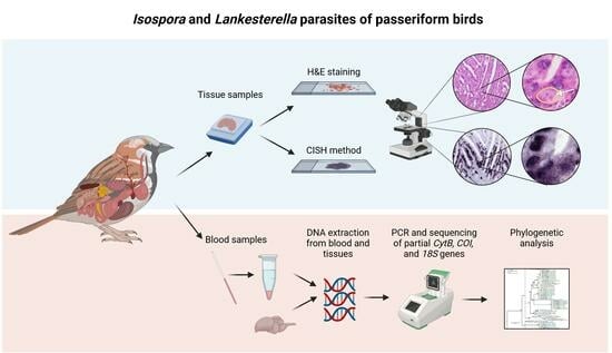
2. Materials and Methods

2.1. Sample Collection

The present study comprised samples of 815 wild passeriform birds from Slovakia (302 individuals), Hungary (218), Austria (216), Lithuania (42), Switzerland (23), Bosnia and Herzegovina (7), and Russia (7). It included birds of 64 species from 24 families, most of which belonged to the Paridae (161 individuals), Sylviidae (145), Muscicapidae (135), Passeridae (57), Acrocephalidae (63), Hirundinidae (48), Phylloscopidae (42), Turdidae (40), and Fringillidae (38). The selection particularly included small passeriform birds, which were assumed to be more prone to infections with Lankesterella parasites due to their insectivorous feeding habits.
In addition to the samples from wild birds (n = 815), FFPE tissue samples from 15 deceased captive birds were included in this study. These birds were sent to the University of Veterinary Medicine Vienna (Vetmeduni) for pathological examination with the suspicion of being infected with ‘Atoxoplasma’ (=Isospora) parasites and comprised the following species: Carduelis carduelis (1), Padda oryzivora (1), Passerina ciris (1), Serinus canaria (3), Spinus cucullatus (3), Spinus xanthogastrus (3), and Spinus yarrellii (3).
The material investigated originated from a range of different sources. The 302 Slovakian birds were captured using mist nets from 2017 to 2019 and in 2022 at the Drienovec Bird Ringing Station in the south-east part of the country near the Drienovec village as described previously [41]. Blood samples were taken by puncturing the brachial vein and stored in 70% ethanol at 4 °C until DNA extraction. Similarly, blood samples were taken from 128 birds captured at the Biological Station Illmitz in eastern Austria and used to prepare both blood spots on high-grade filter papers Whatman™ 903 for DNA extraction and blood smears on microscope slides. The same procedure was followed when taking blood samples from 15 bird patients housed at the Clinical Unit of Internal Medicine Small Animals of the Vetmeduni. Blood samples were also collected from seven birds caught with mist nets at the ‘Rybachy’ Biological Station on the Curonian spit in Kaliningrad oblast in 2014 and used to prepare blood spots and smears. All Hungarian (218) and Swiss (23) samples, and 41 of the Austrian ones, originated from dead birds collected for an Usutu virus monitoring study conducted by the Institutes of Pathology and Virology (Vetmeduni) from 2003 to 2007. These birds were dissected, and tissue samples were collected from the heart, lung, liver, spleen, kidney, brain, skeletal muscle, proventriculus, gizzard, and intestine, fixed in formalin, and embedded in paraffin wax. Brain tissue was stored at −80 °C for total RNA/DNA extraction [42]. FFPE tissue samples were also prepared from 32 dead birds collected during a Citizen Science project in eastern Austria in the summer of 2020. Liver and lung samples of the latter birds were frozen at −80° until DNA extraction [43]. Samples from Bosnia and Herzegovina were collected from seven dead birds submitted for routine pathological examinations at the Department of Pathology at the Sarajevo Faculty of Veterinary Medicine (University of Sarajevo) between 2017 and 2018, and DNA was extracted from frozen liver tissue. The 42 Lithuanian samples originated from birds caught with mist nets at the Ventės Ragas Ornithological station in Ventė Cape (western Lithuania) between 2018 and 2020. Blood spots and smears were prepared from fresh blood, and smears were fixed in absolute methanol and stained with 10% Giemsa solution [44]. FFPE tissue samples were prepared from various organs following euthanization by decapitation and dissection. In total, FFPE tissues from 367 birds were used for CISH-based parasite screenings. Most of the FFPE tissue blocks contained brain, heart, lung, liver, crop, proventriculus and gizzard, intestine, pancreas, kidney, ovary, testicle, skeletal muscle, bone marrow, spleen, and bursa of Fabricius tissues. However, some bird cadavers were not well preserved, and the FFPE blocks did not include the complete set of the above-listed organs.

2.2. DNA Extraction

DNA extraction of the Slovakian samples was performed at the Institute of Zoology of the Slovak Academy of Sciences in Bratislava. DNA was extracted using the QIAamp DNA Blood Mini Kit (QIAGEN GmbH, Hilden, Germany) following the manufacturer’s recommendation, resolved to a final concentration of ca. 100 ng/µL, and stored at −20 °C [41]. The DNA extractions of all other samples were performed at the Institute of Pathology, Vetmeduni Vienna. The total RNA/DNA of the samples collected for the Usutu virus project was extracted from 140 µL homogenates of brain tissue using the QIAamp Viral RNA Mini Kit (QIAGEN) [45]. In the case of the samples from the 15 deceased captive birds sent for pathological investigation to the Institute of Pathology, DNA was extracted from FFPE tissue blocks. In short, every 10 sections of 10 µm thickness were cut from the FFPE blocks, deparaffinized with limonene and ethanol, and then extracted with the QIAamp DNA Micro-Kit (QIAGEN) according to the manufacturer’s protocol. The DNA of the other samples was either extracted from blood spots on filter paper (ca. 1 cm2) or frozen liver and lung tissue (ca. 4 mm3) using the DNeasy Blood & Tissue Kit (QIAGEN GmbH) following the standard protocol. All DNA samples were stored at −20 °C either at the Institute of Zoology, Slovak Academy of Sciences, Bratislava, or the Institute of Pathology, Vetmeduni Vienna.

2.3. Primer Design

A nested PCR assay was designed to sequence an 827 base pair (bp) section of the mitochondrial (mt) cytochrome b gene (CytB). The primers were designed based on an alignment comprising all published mitochondrial genomes of Eimeriidae. The primers were placed in conserved sequence regions and should allow amplification of this CytB fragment in most members of the family Eimeriidae. The first primer set, LankCytB_F1 (5′-GATCTCATTTACAATCATATCCATGTC-3′) and LankCytB_R1 (5′-TAGATTACGGTAAAGAATCTACCA-3′), amplifies a 1033 bp fragment (982 bp excl. primers). The second (nested) primer set, LankCytB_F2 (5′-GTGAAATTTCACATGCCTTTGC-3′) and LankCytB_R2 (5′-CTTCCTGAGGTAGTTGAGCTC-3′), amplifies an 870 bp fragment (827 bp excl. primers).
Since the DNA in FFPE samples is usually degraded to fragments of less than 400 bp, a second PCR assay was designed for amplifying three overlapping CytB fragments, which can be combined to an 823 bp CytB fragment covering roughly the same sequence region as the nested PCR assay. The primers I_CytB_F1 (5′-TGAAATGTCACATGCCTTTGC-3′) and I_CytB_R1 (5′-AGGACTATCTACATAAAAACCTCC-3′) [alternative for Lankesterella: L_CytB_R1 (5′-GTAGGATGATCTACATAGAATCCTC-3′)], I_CytB_F2 (5′-GGGTCAAATGAGCTTCTGG-3′) and I_CytB_R2 (5′-TATCTGGATGTGATAGTTCCAT-3′), and I_CytB_F3 (5′-TCCTATTAGCTCAATCATTCTTTG-3′) and I_CytB_R3 (5′-TGAGGTAATTGAGCTCCAAC-3′) amplify sections of 340 bp (295 bp excl. primers), 344 bp (303 bp), and 334 bp (290 bp), respectively. While the outer primers (I_CytB_F1 and I_CytB_R3) should also target the CytB of most Eimeriidae, the inner primers were designed based on the Isospora CytB sequences obtained in the present study only.
Another nested PCR assay was designed to sequence a 906 bp section of the mt cytochrome c oxidase I gene (COI) from a selection of approximately 70 samples containing 17 Isospora and 20 Lankesterella lineages obtained with the nested CytB PCR assay. The COI primers were also placed in sequence regions conserved within most members of the family Eimeriidae. The first primer set, LankCOI_F1 (5′-CTGCTGCAAACCATAAAGAATTAGG-3′) and LankCOI_R1 (5′-CAGGAATTCTACGTGGCATAACAT-3′), amplifies a 1335 bp fragment (1286 bp excl. primers). The second primer set, LankCOI_F2 (5′-TGGTTCAGGTATTGGTTGGA-3′) and LankCOI_R2 (5′-GACCATACTCTTAAGAATGGAGAATC-3′), amplifies a 906 bp fragment (860 bp excl. primers).
The nested PCR assay by Chagas et al. [6] was used to sequence approx. 1000 bp fragments of the 18S ribosomal RNA gene (18S) in a subset of samples containing a variety of Isospora and Lankesterella lineages. The first primer set, Cocc18S_n1F (5′-CAGCTTTCGACGGTATGGTATTGG-3′) and Cocc18S_n1R (5′-CAGACCTGTTATTGCCTCAAACTTCCT-3′), amplifies a fragment of 1135 bp. The nested primer set, Cocc18S_n2F (5′-GTATTGGCTTACCGTGGCAGTGAC-3′) and Cocc18S_n2R (5′-GCCTCAAACTTCCTTGCGTTAGACA-3′), amplifies a fragment of 1105 bp. The 18S sequences were primarily used as a basis for designing the in situ hybridization probes to specifically target and differentiate parasites of the two genera.

2.4. PCR and Sequencing

All 830 individuals were screened for the presence of Isospora and Lankesterella parasites with the nested PCR approach targeting CytB. Birds for which only FFPE samples were available (n = 15) were additionally screened with the PCR assay using the three sets of overlapping primers. The COI and 18S PCRs were performed on 48 samples.
All PCRs were performed with the KAPA2G Fast HotStart PCR-Kit (Sigma-Aldrich, St. Louis, MO, USA) in 25 µL volumes containing 12.5 µL KAPA2G Fast HotStart polymerase mix, 8.5 µL AD, 1 µL MgCl2 (1.5 mM), each 1 µL primer (10 mM), and 1 µL template. The nested PCRs started with an initial denaturation for 2 min at 94 °C, followed by 20 (first PCR) or 35 (nested PCR) cycles with 15 s at 94 °C, 15 s at defined annealing temperatures for each primer set (see below), 30 s at 72 °C, and a final extension for 10 min at 72 °C. Each 1 µL PCR product of the first PCRs was used as a template for the nested PCRs. The annealing temperature for the two nested PCR assays (CytB and COI) was 53 °C, while the one for the PCRs targeting the short, overlapping CytB fragments was 48 °C. The PCRs targeting the 18S rDNA were performed at 56 °C annealing temperature.
The PCR products (3 µL) were visualized on 1% agarose gels stained with ROTI®GelStain (Carl Roth GmbH & Co, Karlsruhe, Germany) and imaged with a BioSens SC-Series 710 gel documentation system (GenXpress, Wiener Neudorf, Austria). Positive PCR products were sent to Microsynth Austria (Vienna, Austria) for purification and sequencing in both directions using the PCR primers. Raw forward and reverse sequences were aligned using MAFFT v.7 [46] applying the default settings, and the electropherograms were visually inspected with Bioedit v.7.0.8.0 [47]. Mixed infections were present in some samples, and positions with double peaks in the electropherograms were first coded with the corresponding ambiguity codes. Then, all CytB sequences were aligned and sorted by sequence similarity. Unphasing of sequences was straightforward because the individuals mostly contained one prevalent lineage present in other birds of the same species, or the lineages differed only by one or a few bp while one lineage was dominating.
New haplotypes identified in the present study were assigned lineage names consisting of “i” for Isospora or “k” for Lankesterella, followed by the first three letters of the bird genera and bird species, and ascending numbers starting with “01”. The naming scheme was chosen following the system established for avian haemosporidian parasites in the MalAvi database (http://130.235.244.92/Malavi/; accessed on 1 December 2023 [48]), which summarizes DNA barcode sequences (476 bp CytB fragments) of parasite lineages and provides information on bird hosts, vectors, geographic regions, and literature.
All sequences were uploaded to NCBI GenBank and can be retrieved under accession numbers PP666884–PP667037 (CytB), PP667038–PP667082 for (COI), and PP660150–PP660193 for (18S).

2.5. Phylogenetic Analysis

Phylogenetic trees were calculated with the CytB sequences of Isospora spp. and Lankesterella spp. and the COI sequences of Isospora spp. The only mt DNA sequences published for Lankesterella are the genomes of two Lankesterella minima lineages isolated from Lithobates clamitans frogs; therefore, no phylogenetic tree was calculated with the Lankesterella COI sequences.
The alignments for the CytB trees of Isospora and Lankesterella contained 74 and 93 sequences, respectively (including those from mt genomes available on GenBank) and covered 827 bp. Sequences of Lankesterella sp. kSYLATR01 (AH0893) and Isospora sp. iPASDOM02 (AH1562) were used as outgroups. To visualize the distribution in host species, the sequences of these two alignments were not collapsed to haplotypes.
COI sequences of Isospora parasites have been retrieved from various species of wild and captive birds. We included all Isospora COI sequences from GenBank, which covered the selected 648 bp COI fragment and contained less than two ambiguity characters. The initial alignment contained 217 sequences from GenBank and 24 obtained in the present study. The sequences were collapsed to 194 unique haplotypes with DAMBE v.7.0.5.1 [49], and a sequence of Lankesterella sp. kERIRUB01 (AH0896) was included as an outgroup.
Phylogenetic trees were also calculated with partitioned datasets containing the concatenated CytB (827 bp) and COI (860 bp) sequences of Isospora spp. and Lankesterella spp. The alignment for Isospora spp. contained the sequences of 18 different lineages and the outgroup Lankesterella sp. kSYLATR01 (SK162/19). The alignment of Lankesterella spp. contained sequences of 22 different lineages and the outgroup Isospora sp. iPASDOM02 (AH1553). The trees contain fewer haplotypes than the CytB trees because we were not able to sequence the COI sequences for all CytB lineages.
The best-fit substitution models according to the corrected Akaike Information Criterion were evaluated using IQ-TREE [50], resulting in the model GTR+G+I for all three alignments. Maximum likelihood (ML) majority-rule consensus trees were calculated with W-IQ-TREE by performing 10,000 replicates. Bayesian Inference (BI) trees were calculated with MrBayes v.3.2 [51], running 5 million generations (two runs each with four chains, one of which was heated) and sampling every thousandth tree. The first 25% of trees were discarded as burn-in, and a 50% majority rule consensus tree was calculated from the remaining 3750 trees each.
All alignments were visually inspected using Bioedit v.7.0.8.0 [47]. The phylogenetic trees were graphically prepared using FigTree v.1.4.4 (http://tree.bio.ed.ac.uk/software/figtree/; accessed on 14 March 2023; Andrew Rambaut) and finalized with Adobe Illustrator CC v.2015 (Adobe Inc., San José, CA, USA).

2.6. Genetic Distances between Isospora and Lankesterella CytB Lineages

Uncorrected genetic distances between the Isospora and Lankesterella CytB (823 bp) lineages detected in the present study were calculated using MEGA v.10.2.2 [52]. The distances were calculated separately based on the alignments containing 31 Isospora and 27 Lankesterella lineages. Sequences of Lankesterella sp. kPASDOM02 and Isospora sp. iSYLATR01 were included for comparison. The alignments were trimmed to 823 bp, the length of the 4 bp shorter Isospora sequences obtained from the FFPE samples.

2.7. Chromogenic In Situ Hybridization

To detect Isospora and Lankesterella parasite stages by CISH, two probes were designed based on an alignment containing all available 18S sequences of Isospora and Lankesterella (including 28 sequences generated in the present study), most other Eimeriidae, and several bird species. The probes were commercially synthesized by Microsynth Austria (Vienna, Austria) including digoxigenin labels at the 3′ ends. The probes are complementary to the DNA sequence of the 18S rRNA gene and directly target the 18S rRNA. The probe Iso18S_ISH (5′-GGAAGAAAGCCGAGGGCAAACCAGGTGC-3′) binds the 18S rRNA of all avian Isospora lineages with a maximum of one or two mismatches. Moreover, it binds the 18S rRNA of numerous closely related Eimeria species isolated from Muridae, Sciuridae, Bovidae, Leporidae, and galliform birds. It was not possible to design a probe specific to avian Isospora parasites because their 18S sequences differed only in a few bp from those of many other Eimeriidae. The probe Lan18S_ISH (5′-GGAAATATGCCACAATCCATCCTC-3′) binds the 18S rRNA of all published avian Lankesterella lineages with a maximum of one or two mismatches. The probe is specific to this parasite group and does not match the 18S rRNA of other Eimeriidae, including Lankesterella parasites found in American lizard species by Megía-Palma et al. [37].
The specificity of the probes was checked in silico. AmplifX v.2.0.7 (Nicolas Jullien, Aix-Marseille Univ., CNRS, INP, Marseille, France; https://inp.univ-amu.fr/en/amplifx-manage-test-and-design-your-primers-for-pcr; accessed on 3 September 2021) was used to check the quality of the newly designed probes. All probes were blasted against genomes of apicomplexan parasites and birds in NCBI GenBank to exclude unintentional binding. To confirm the specificity of the probes, cross-checking was also performed by CISH to rule out the chance that the probe designed for Isospora binds to Lankesterella in the tissue and vice versa, potentially causing false-positive results (Supplementary File Figure S1).
CISH was performed on all Eimeriidae PCR-positive birds of which FFPE samples were available (n = 53). The procedure followed a previously established CISH protocol [53] with some modifications. Paraffin-wax-embedded tissue samples were sectioned (2–3 µm) and placed on Superfrost Plus slides (Menzel-Gläser, Braunschweig, Germany). After drying, sections were dewaxed in orange terpene (SAV Liquid Production GmbH, Flintsbach am Inn, Germany) and rehydrated in a series of graded alcohols (100%, 96% and 70%) and distilled water. Proteolytic treatment was performed with Proteinase K 3.3 µg/mL (Roche Diagnostics, Vienna, Austria) in Tris-buffered saline (pH 7.4) at 37 °C for 40 min. After proteolysis, the slides were rinsed with distilled water and dehydrated in 96% and 100% ethanol, followed by air-drying. The hybridization mixture consisted of distilled water, 20× standard saline citrate buffer (SSC), formamide, 50× Denhardt’s solution, herring sperm DNA, dextran sulfate, and the appropriate oligonucleotide probe (10 ng/100 µL). Slides were covered with the hybridization mixture and incubated at 95 °C for 6 min and then immediately cooled down on crushed ice and hybridized overnight in a humid chamber at 40 °C. On the second day, three stringency washing procedures (2 × SSC, 1 × SSC, and 0.1 × SSC, 10 min each at room temperature) were carried out to remove unspecific bindings. For immunological detection of hybrids, slides were covered with an equilibration buffer containing distilled water, buffer I (2×) (1M Tris-HCl 20%, 5M NaCl 6%, and DEPC-treated water), normal goat serum (VECS-1000, Szabo-Scandic, Vienna, Austria), and Triton X-100 10% (Merck, Darmstadt, Germany) for 30 min at room temperature. Afterward, slides were incubated with anti-digoxigenin-AP Fab fragments (#11093274910, Merck KgA, Darmstadt, Germany) at a concentration of 1:200 for 1 h at room temperature followed by two 15 min washing steps in buffer I (1×) and briefly in buffer III (100 mM Tris-HCl, 100 mM NaCl, 50 mM MgCl2 in DEPC-treated water, final pH 9.5). To visualize the probe–parasite hybrids, the chromogenic substrates 4.5 µL/mL NBT (4-nitro blue tetrazolium) (Roche Diagnostics) and 3.5 µL/mL BCIP (5-bromo-4-chloro-3-indolyl phosphate) (Roche Diagnostics) were mixed with 2.5 µL/mL Levamisol (Merck) in buffer III and applied to the slides. After incubation in a dark humid chamber for at least 60 min at room temperature, the chromogenic reaction was stopped by placing the slides in TE buffer (pH 8.0) for 10 min. As a final step, the slides were counterstained with hematoxylin, Gill 3 (Merck, Darmstadt, Germany), and mounted with Aquatex (Merck, Darmstadt, Germany).

2.8. Microscopic Examination

Hematoxylin–eosin (HE)-stained and CISH-treated tissue sections were evaluated by bright field microscopy using 100×, 200×, 400×, and 1000× magnifications. Microphotographs were taken using an Olympus UC90 camera attached to an Olympus BX51 microscope (Olympus Europa, Hamburg, Germany).
All tissue samples were examined to determine the degree of parasite burden following the semiquantitative scoring system developed as part of this study: negative (−; no signal in sections detected), low-grade (+), moderate (++), and high-grade (+++).
The received score was determined by using a reward point system in three steps. First, points were assigned regarding the signal’s distribution. Tissues with an oligofocal distribution received 1 point. Tissues that exhibited multifocal signals received 2 points, and 3 points corresponded to a diffuse distribution. In the next step, the number of signals per foci was recorded. Single signals or a cluster of up to 20 signals resulted in 1 point. Two points were given if a cluster showed between 20 and 40 signals, and 3 points if more than 40 but less than 60 signals were seen. If there were more than 60 signals per cluster, 4 points were given. Finally, the points awarded were added, resulting in a final score, distinguishing between + (1–3 points; low-grade parasite burden), ++ (4–5 points; moderate parasite burden), and +++ (≥6 points; high-grade parasite burden).
Corresponding HE-stained tissue sections were examined to confirm the presence of parasites based on references [10,18,54]. In addition, pathological changes in all tissue samples were recorded to evaluate potential pathogenicity. It should be noted that many organs showed autolytic changes of varying severity, which led to limited histological evaluation in some cases.

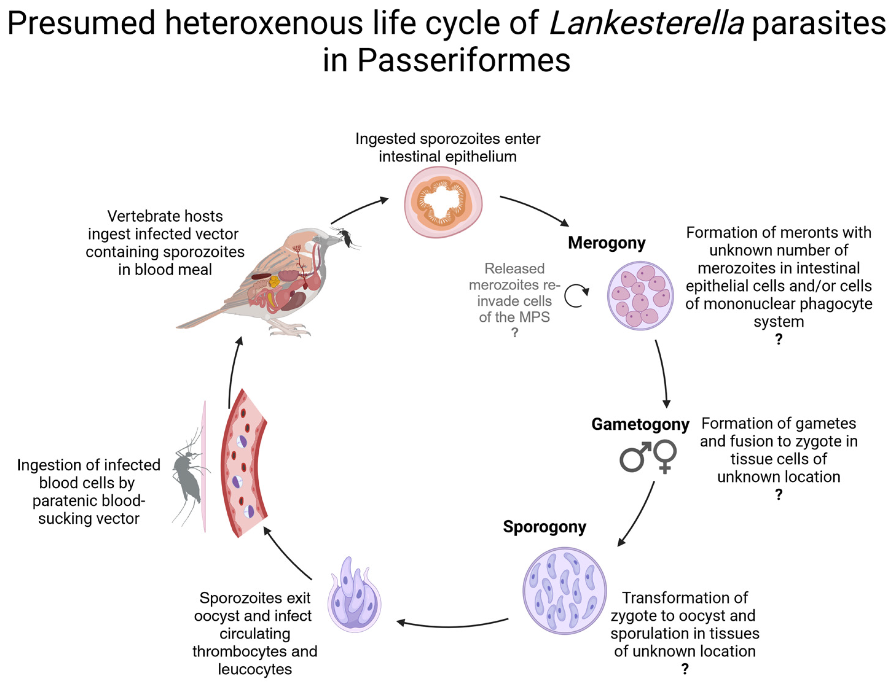
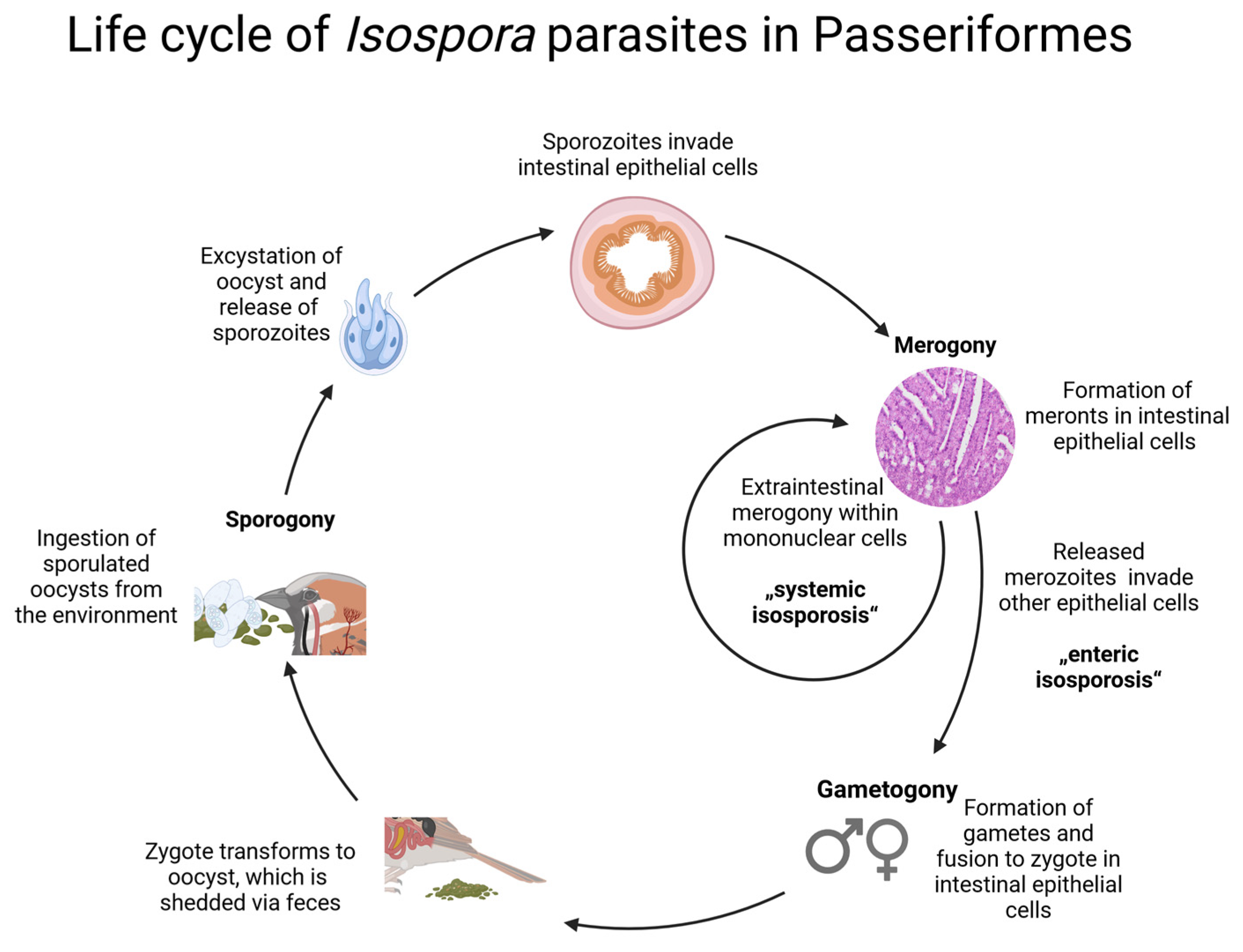
2.9. Graphical Representation of the Parasites’ Life Cycles

The illustration software BioRender (https://www.biorender.com) was used for the graphical creation of the presumed parasites’ life cycles based on descriptions in [6,18].

3. Results

3.1. Infection Rates
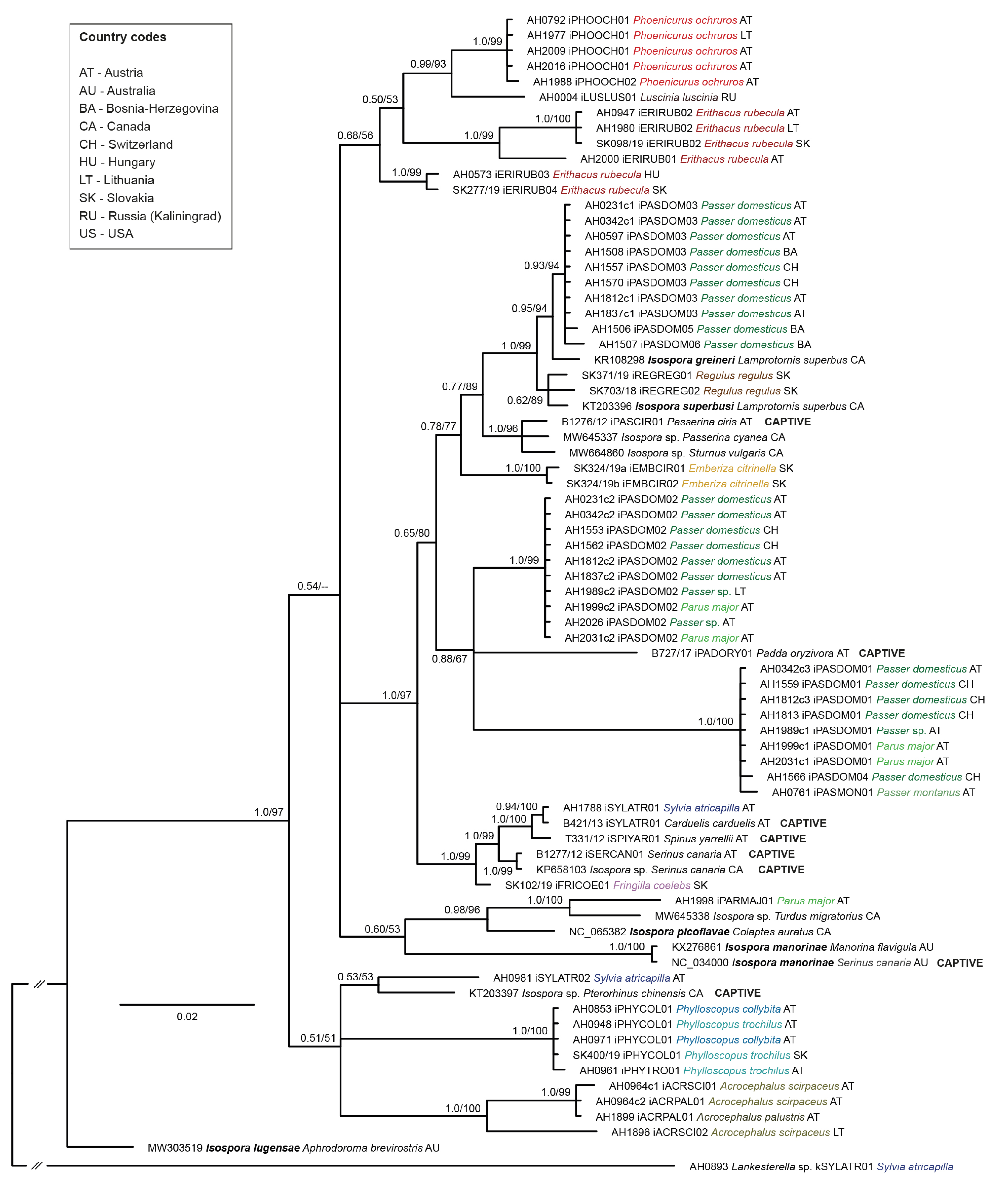
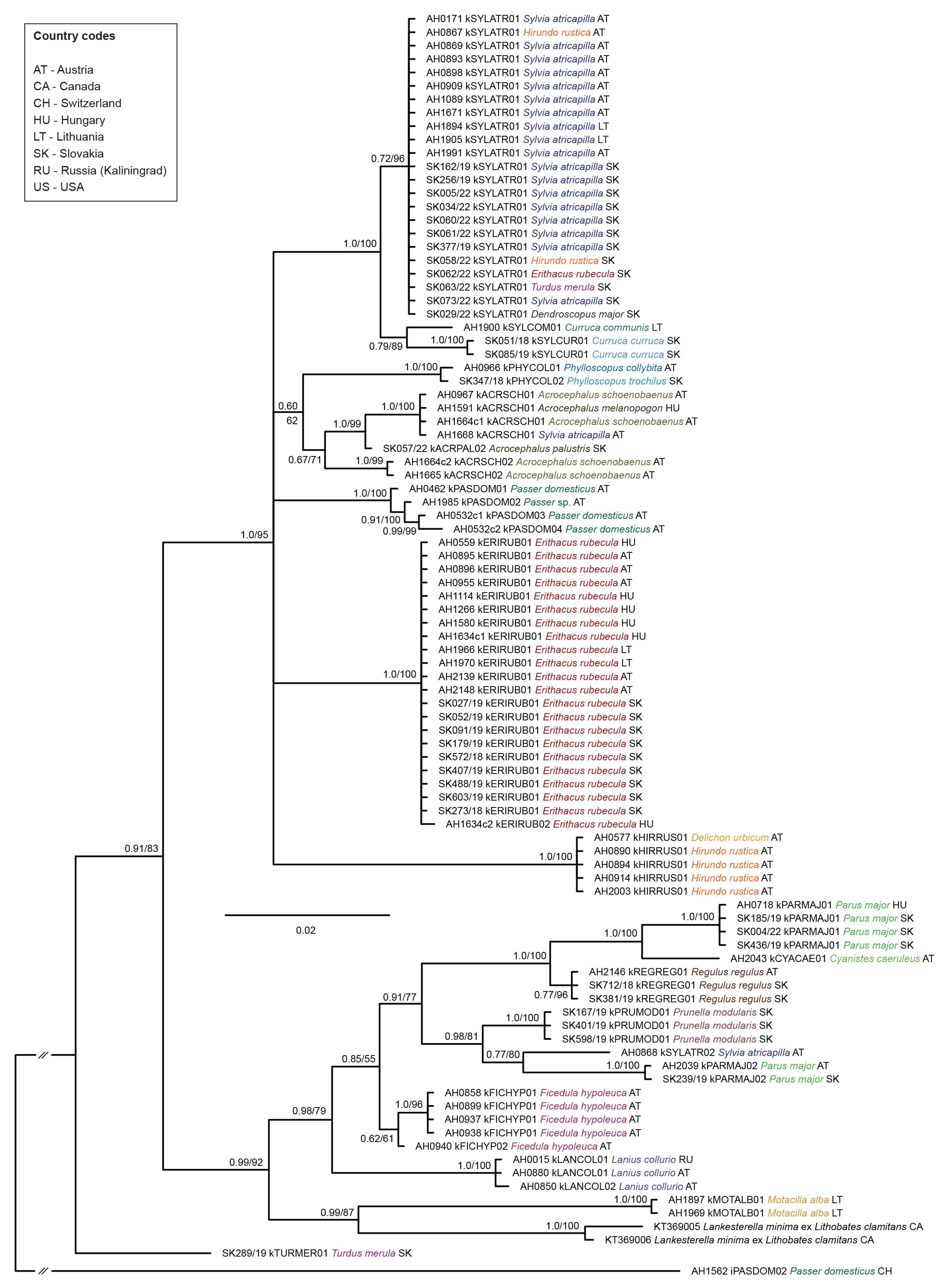
Based on PCRs and CytB sequencing results, the overall infection rate for Isospora in the wild birds was 5.8% (47/815). Double and triple infections with different Isospora lineages were detected in seven and two individuals, respectively. In total, 26 Isospora CytB lineages were recorded, most of which were specific to one or two host species. The Isospora infection rates differed strongly between the most frequently sampled bird species, with 50% in Passer domesticus (n = 46), 5.8% in Erithacus rubecula (n = 104), 3.5% in Parus major (n = 86), 2.5% in Sylvia atricapilla (n = 118), and no records in Cyanistes caeruleus (n = 65) and Hirundo rustica (n = 43). High infection rates were also recorded in Luscinia luscinia (100%), Phoenicurus ochruros (62.5%), and Regulus regulus (27.3%); however, bird sample sizes of these species were small, with one to eleven individuals (Table 1).
Moreover, 5 of the 15 captive birds of which only FFPE samples were available were confirmed positive for Isospora spp.: P. ciris (1/1), P. oryzivora (1/1), C. carduelis (1), S. canaria (1/3), and S. yarrellii (1/3). They featured five lineages, four of which were not found in the wild bird samples.
The overall infection rate for Lankesterella was 10.7% (87/815), and only two individuals featured double infections. In total, 25 Lankesterella CytB lineages were recorded, which were mostly restricted to single host species as well. The infection rates also differed strongly with 21.2% in E. rubecula, 17.8% in S. atricapilla, 14.0% in H. rustica, 7.0% in P. major, 6.5% in P. domesticus, and 1.5% in C. caeruleus. The highest infection rates were recorded in Motacilla alba (50%), Ficedula hypoleuca (45.5%), Lanius collurio (42.9%), and Acrocephalus schoenobaenus (37.5%) (Table 1).
In addition to CytB sequences, COI sequences were obtained from 20 of the 26 Isospora lineages (from wild birds) and from 23 of the 25 Lankesterella lineages. The 18S sequences were obtained from 18 Isospora and 10 Lankesterella lineages. In both genera, CytB sequences of some lineages differed by one to a few bp, but the corresponding COI sequences were identical. This was the case for the Isospora lineage pairs iREGREG01 and iREGREG02, iPASDOM03 and iPASDOM06, and iEMBCIR01 and iEMBCIR02, which differed by 5 bp, 2 bp, and 1 bp, respectively. Similarly, the Lankesterella lineages kPASDOM02 and kPASDOM03 and kPHYCOL01 and kPHYCOL02 both differed by 1 bp in CytB, but the COI sequences were identical.

3.2. Phylogeny

The Isospora CytB tree contained 43 unique lineages (including sequences from ten mt genomes published on GenBank) (Figure 3). The highest p-distance measured between the Isospora lineages was 8.0%, and the mean distance was 5.0%. A lineage of Isospora lugensae (GenBank accession MW303519) formed the sister group to a major clade containing all other Isospora lineages, which clustered into several subclades. The subclades mainly contained lineages retrieved from bird species of the same family, suggesting the Isospora lineages had high host specificity. The birds kept in captivity (FFPE samples) featured five Isospora lineages, two of which were found in other birds. The lineage iSYLATR01 from a captive C. carduelis (B421/13) was also detected in S. atricapilla (AH1788) from Austria and differed by 0.4% from lineage iSPIYAR01 from S. yarrellii (T331/12), and one S. canaria (B1277/12) was infected with the lineage iSERCAN01, which previously was isolated from S. canaria (KP658103) in Canada [55]. All the latter lineages clustered into a highly supported clade together with lineage iFRICOE01 from Fringilla coelebs in Slovakia, which also belongs to the Fringillidae. The lineage iPASCIR01 from P. ciris (B1276/12) differed by 6 bp and 7 bp from Isospora lineages found in Passerina cyanea (MW645337) and Sturnus vulgaris (MW664860) from Canada, respectively (published on GenBank only). The subclade containing the three latter lineages fell in a clade with other lineages mostly isolated from Passeridae and Sturnidae. The lineage iPADORY01 from a captive P. oryzivora formed an independent branch and clustered with two subclades containing parasite lineages from Passeridae and Paridae. Genetic p-distances between the Isospora CytB lineages (823 bp) detected in the present study are provided in Supplementary File Table S1.
The Lankesterella CytB tree contained 29 unique lineages (including sequences of two mt genomes of L. minima isolated from L. clamitans (Figure 4). The highest p-distance between different Lankesterella lineages was 8.6%, and the mean distance was 5.0%. Most lineages were restricted to single bird species; however, lineage kSYLATR01 was detected in four bird species: S. atricapilla, H. rustica, E. rubecula, and Turdus merula. The lineage kTURMER01 from T. merula in Slovakia was the sister group to all other Lankesterella lineages, which were grouped into two well-supported clades, each containing several subclades. Interestingly, the two L. minima lineages from the green frog L. clamitans clustered in a clade with lineage kMOTALB01 from M. alba in Lithuania. Genetic p-distances between the Lankesterella CytB lineages (823 bp) detected in the present study are provided in Supplementary File Table S2.
A phylogenetic tree was also calculated with the COI sequences of Isospora spp. from the present study and sequences published from other wild bird species on GenBank (Supplementary File Figure S2). None of the Isospora COI lineages was detected in previous studies. The tree contained a total of 193 different Isospora lineages, most of which (>140) were published by Kubiski et al. [24], who screened samples from captive and wild birds collected by the San Diego Zoo Wildlife Alliance.
The phylogenetic trees calculated with the concatenated COI and CytB sequences contained 18 Isospora and 22 Lankesterella lineages. The topologies resembled those in the CytB trees, but the support values were also low at the deeper nodes (Supplementary File Figure S3). Future phylogenetic studies would benefit from analyzing the complete mt genomes and the more variable nuclear coding genes.

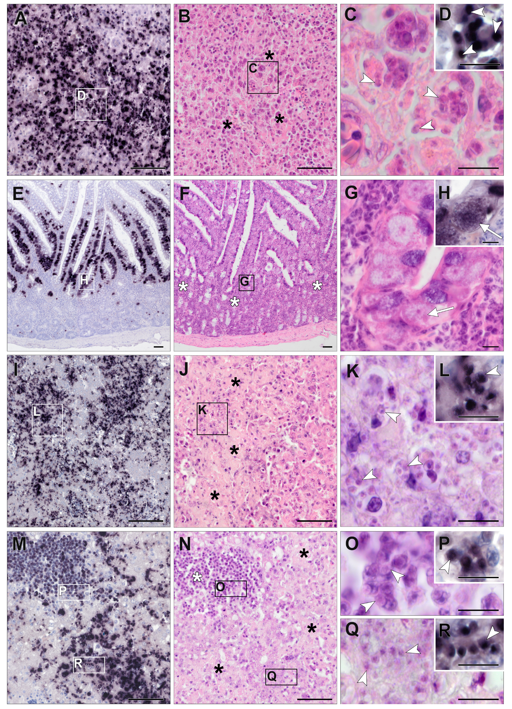
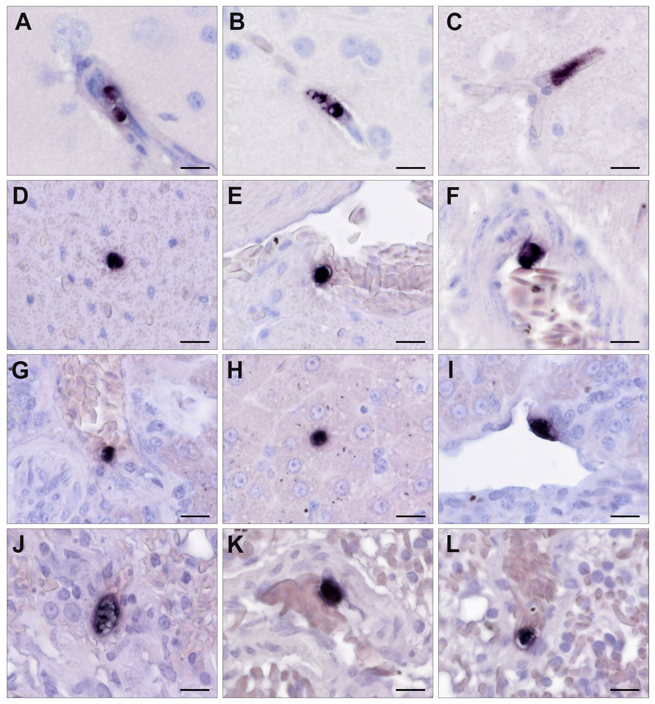
3.3. Chromogenic In Situ Hybridization

Among the birds that were PCR-positive for Isospora (n = 53), FFPE tissue samples were available for subsequent CISH in 26 cases. In the Lankesterella-infected birds (n = 87), FFPE tissue samples were available for CISH from 20 individuals. However, FFPE tissue samples did not always contain the same set of organs for every individual. In both parasite genera, the brain was most frequently part of the organ set and was only missing in one case each. Tissue samples from the heart, liver or kidney were present for at least 80% of these individuals. Lung, stomach (proventriculus and/or ventriculus), skeletal muscle, and spleen samples were available for CISH in at least half of the Isospora- and Lankesterella-infected birds. In 54% of Isospora-infected birds, the intestine was also part of the tissue samples. In many birds, the esophagus, pancreas, gonads, trachea, and bursa of Fabricius were not available. Only in one bird infected with Isospora, tissue samples also included crop and bone marrow. In 34 of 46 samples (74%), Isospora or Lankesterella were successfully identified by the dark-purple-to-black color reaction using the respective genus-specific probes. In both Isospora- and Lankesterella-infected birds, signals appeared in various organs, including the heart, lung, liver, spleen, kidney, brain, skeletal muscle, proventriculus and gizzard, intestine, esophagus, testicle, ovary, and pancreas. The distribution of detected signals varied between the individual hosts, ranging from single infected organs to a generalized infection and also between the two parasite genera, which is described in more detail below.

3.4. Detection of Isospora by CISH

In 22 out of 26 (85%) examined Isospora-infected birds, CISH signals were found, with the distribution and abundance of signals in the different organs varying between individuals (Table 2). In most cases, systemic infections were observed, which appeared mild in most (P. major, P. ochruros) and severe in some cases (P. ciris, P. oryzivora, S. yarrellii, P. domesticus). A low-grade parasite burden in at least one organ was found in twelve birds (46%). In only two cases (8%), moderate numbers of stained parasites in at least one organ were detected. Eight birds (31%) showed a high-grade parasite burden.
Isospora parasites frequently appeared in the spleen (87%), intestine (79%), and liver (71%) of CISH-examined birds (Table 2), followed by the kidney (45%), gizzard and/or proventriculus (41%), and lung (40%). Signals were less commonly observed in the skeletal muscle (33%) and heart (29%). In three cases, signals were present in the brain (12%). Parasites were also found in the bone marrow, crop, pancreas, ovary, and esophagus. However, the sample size of individuals from which these organs were available for CISH was small (Table 2), ranging from one to seven. No signals were detected in the testicle, trachea, and bursa of Fabricius.
High parasite burdens, determined using the CISH score, were mostly found in the liver and spleen (Figure 5) of captive P. ciris, P. oryzivora, C. carduelis, S. yarrellii, and three wild P. domesticus. Detected signals were mostly roundish to oval, varied in size, and were assumed to be located in mononuclear cells, corresponding to extraintestinal merogony stages (=merozoites) (Figure 5, Supplementary File Figure S4). The bone marrow of the aforementioned P. ciris (B1276/12)—the only individual including bone marrow in the organ set—also revealed a high parasite burden. Furthermore, a high parasite burden was detected in the intestines of each P. oryzivora and E. rubecula, with much larger, roundish to oval or polygonal signals of varying intensity located in epithelial cells, corresponding to micro- and macrogamonts (Figure 5). A moderate parasite burden of presumably merozoites was found in the lung, kidney, gizzard and proventriculus (Supplementary File Figure S4), ovary, and crop of various bird species. The heart, brain, skeletal muscles, esophagus, and pancreas of infected birds were the least affected and showed low parasite burden (Supplementary File Figure S4). This was particularly evident in Passeridae.
Relatively small, roundish to oval signals appeared in the lumina of larger vessels or capillaries of various organs and were therefore evaluated as parasitic blood stages. Signals were more frequently found in endothelial cells or closely associated with the endothelia of differently sized blood vessels; however, their cellular location (intra- or extracellular, intra- or extraluminal) could not be determined with certainty.
In the case of Isospora-infected birds, signals could often be distinguished either as tissue stages or blood stages. However, for some signals, it could not be determined with absolute confidence whether they were representing tissue or blood stages due to the low intensity of the signal or poor preservation status of the tissue.

3.5. Detection of Lankesterella by CISH

In 12 of 20 (60%) Lankesterella-infected birds, CISH signals were detected (Table 3). In those birds, a low-grade parasite burden in at least one organ was observed.
Lankesterella parasites frequently appeared in the spleen (44%), lung (36%), and brain (32%) of CISH-examined birds (Table 3). The heart, liver, and kidney were affected in 19% of examined birds. Signals were also observed in the gizzard, including the proventriculus (20%), and skeletal muscle (14%). In addition, signals were observed in testicles (40%) and intestines (14%), but again, the number of birds in which these two organs were part of the examined set of organs was rather low, being five and seven individuals, respectively (Table 3). Signals were absent in the trachea, esophagus, ovary, bursa of Fabricius, and pancreas. FFPE tissue samples tested for Lankesterella parasites did not contain bone marrow or crop tissue. The detected signals varied in terms of shape (roundish to elongated oval) and size (Figure 6). Similar to the CISH results for Isospora-infected birds, many signals appeared to be closely associated with the endothelia of differently sized blood vessels, which was observed in various organs of several birds. Some relatively small signals appeared to be located in the lumina of blood vessels indicating blood stages. However, the exact cellular location of the signals could often not be determined, partly due to low signal intensity or autolytic changes.

3.6. Histopathology of Isospora Infections

Histological evaluation of HE-stained tissue samples was often difficult due to autolytic changes. Therefore, reliable histologic identification of Isospora stages was not always possible. Histological lesions were mainly observed in the liver, spleen, intestine, proventriculus, and lung, with lymphocytic to lymphohistiocytic or histiocytic inflammation predominantly in a focal to multifocal distribution. In various organs of good preservation status, large numbers of Isospora parasites (presumably merozoites) were recognizable intralesionally in mononuclear cells like lymphocytes and macrophages (Figure 5, Supplementary File Figure S4). The number of parasites often appeared to correlate with the severity of lesions. In addition, mild to severe, oligo- to multifocal acute necrosis associated with recognizable parasites was observed in the liver and spleen (Figure 5). In some cases, intestinal epithelia contained large numbers of Isospora parasites (presumably micro- and macrogamonts) (Figure 5). In the intestine, lymphocytic inflammatory cells were seen mainly in the lamina propria (Figure 5). Low-grade to moderate proventriculitis, ventriculitis, or both were seen in several cases, sometimes accompanied by parasitic stages (presumably merozoites, Supplementary File Figure S4). Focal low-grade to moderate serositis of the proventriculus, gizzard, and kidney with concomitant parasites (presumably merozoites) was noted in single cases (Supplementary File Figure S4). The kidney and ovary showed low- to moderate-grade inflammation with associated parasites in individual cases. Inside the wall of a blood vessel, parasites (presumably merozoites) were detected in the brain of one bird (Supplementary File Figure S4). In another case, many parasites were identified in bone marrow. Low-grade, lymphocytic to granulomatous myocarditis was observed in a few birds, but concomitant parasites were absent.

3.7. Histopathology of Lankesterella Infections

Several birds showed pathological lesions in various HE-stained tissues; however, these lesions were not accompanied by CISH signals for Lankesterella. Furthermore, parasitic stages of Lankesterella matching the CISH signals’ locations were not identified in subsequent HE-stained sections, probably due to their small size.
The intestines of three birds showed focal to multifocal lymphocytic enteritis, with moderate to severe inflammation in two cases and only mild enteritis in the third bird. In the intestinal epithelium of these three individuals, some roundish, protozoan-like objects that were not stained with the Lankesterella-specific CISH probe were found. Cross-testing of one of these birds using the Isospora-specific probe revealed positive staining of these structures, indicating co-infection (Supplementary File Figure S5).

4. Discussion

This study presents the first systematic exploration of Isospora and Lankesterella parasites in wild songbirds, based on a sample including 815 individuals. The overall infection rates for Isospora and Lankesterella were 5.8% and 10.7%, respectively. The analysis of partial CytB sequences revealed 26 Isospora and 25 Lankesterella lineages. Most of the parasite lineages were detected exclusively in one host species, supporting high host specificity. In addition to the partial CytB sequences, partial COI and 18S sequences were obtained from a subset of samples covering most parasite lineages. Specific probes were designed for the detection of Isospora and Lankesterella parasites in tissues using CISH. Our findings revealed that Isospora most commonly infects the spleen, intestine, and liver, while Lankesterella is predominantly located in the spleen, lung, and brain.
Passer domesticus was the host species showing the highest infection rate of Isospora, whereas E. rubecula and S. atricapilla had the highest infection rates of Lankesterella parasites. This difference in host preference is likely attributed to the behavioral aspects of the hosts and the life cycles of the parasites. Since Isospora is transmitted through feces [12], which are most commonly found on the ground, it is conceivable that key hosts for this parasite would be species primarily moving and feeding on the ground. Hence, we observed high infection rates in ground-dwelling species such as P. domesticus and lower infection rates in species like E. rubecula or S. atricapilla. A completely different transmission route is observed for Lankesterella, which is most likely not transmitted through feces but by eating the infected vector [6].
Reports of avian Isospora infections either originate from captive birds [19,20,22,56,57] or single individuals of deceased wild birds [26,27], and one study investigated both captive and wild birds [24]. Only a few studies exclusively analyzed infection rates of Isospora in larger groups of free-ranging wild birds [25,54,58,59]. Gill and Paperna [54] examined fecal samples of wild P. domesticus and found Isospora oocysts by floatation in the feces of birds trapped in the Jordan valley (90/124), the coastal plain south of Tel Aviv (27/32), and in the desert region in southern Israel (17/22), revealing infection rates of over 70% in each region. In our study, P. domesticus was also frequently infected with an infection rate of 50%, which could be related to its habits as a ground-dwelling species. In Western Australia, fecal samples from 34 Corvus coronoides were examined by flotation and sequence analysis, resulting in an Isospora infection rate of 44.1% [25]. Svobodová [59] analyzed fecal samples of 571 individuals belonging to 46 species of passeriform birds caught in different areas of the Czech Republic and detected a considerably higher overall infection rate for Isospora, 36.4%, by direct light microscopy of sporulated oocysts as compared to our study (6%). Hippolais icterina of the family Acrocephalidae had the highest infection rate in the study presented in [59], namely 79%. We also found Acrocephalidae species to be infected, namely Acrocephalus scirpaceus (9.1%) and Acrocephalus palustris (5.3%), while the only H. icterina individual examined was uninfected.
The infection rate of Lankesterella in the present study was only slightly higher, at 10.7%, than the one reported by Chagas et al. [6], namely 8.6%. In our study, birds belonging to the family Muscicapidae and Sylviidae presented a higher infection rate of Lankesterella; however, Chagas et al. [6] analyzed a small number of Sylviidae and no Muscicapidae. Saravana Bhavan Venkatachalam et al. [40] analyzed several bird species but did not report the rates of infection. Acrocephalus schoenobaenus was sampled in different studies at different localities. The Lankesterella infection rate was 47% in Poland [7], 17.2% in Lithuania [6], and 37.5% in the present study. Merino et al. [9] investigated C. caeruleus in Spain for Lankesterella and reported an infection rate of 31.2%, which is much higher than the one reported in our study (1.5%), and no infections were found in the three individuals investigated by Chagas et al. [6]. The closely related P. major presented a higher infection rate in our study (7%) than that found by Chagas et al. [6]. Such differences between the rates of infection are still not completely understood and should be better investigated. The sampling season might partially explain these discrepancies since juveniles might be less frequently infected than adults [6]. Additionally, the availability of vectors is also a determinant for the rate of infections, and the complete life cycle of Lankesterella is still unknown.
The phylogenetic analyses demonstrated that avian Isospora parasites are a reciprocally monophyletic group based on their mt COI and CytB genes. This study revealed 43 distinct Isospora lineages, with a high degree of host specificity. The distribution of lineages seems to be rather correlated with host species than geographic regions. Our results are generally in line with those of Kubiski et al. [24], who emphasized the host specificity of Isospora lineages, highlighting co-evolutionary aspects of these interactions. In the present study, Isospora sequences generally were specific to one host species or closely related species of the same genus. However, for several lineages, the phylogenetic analysis revealed a broader host range including species from different families; e.g., the lineages iPASDOM02 and iPASDOM01 both infected P. domesticus and P. major. The presence of the Isospora lineage iSYLATR01 in S. atricapilla (family Sylviidae) and C. carduelis also illustrates the parasite’s ability to infect hosts across different bird families. This suggests a broader host range than previously understood, potentially indicating opportunistic host switching or more flexible parasite–host interactions. Insectivorous bird species were found to be more likely infected with Lankesterella parasites. However, most of these bird species also contained Isospora parasites (Figure 3 and Figure 4).
Another explanation for the detection of the same lineage in different bird hosts could be pseudoparasitism. Trefancová et al. [60] highlighted the phenomenon of pseudoparasitism in Isospora; they found oocysts of avian Isospora parasites in the feces of bank voles and yellow-necked mice. Additional data are needed to verify our observations.
The phylogenetic analysis of Lankesterella CytB sequences revealed 29 distinct lineages differing by a maximum of 8.6%, highlighting a significant genetic diversity within the genus. This study found that most lineages were specific to one host, except for the lineage kSYLATR01, which was found in multiple bird species from different families (S. atricapilla, H. rustica, E. rubecula, and T. merula), indicating complex host–parasite dynamics. This pattern aligns with the findings of Chagas et al. [6] and Saravana Bhavan Venkatachalam et al. [40]. The phylogenetic analysis also revealed that some bird species featured Lankesterella lineages from different clades. Notably, the lineage kPARMAJ01 exhibits a sister lineage relationship with kCYACAE01 in C. caeruleus, a pattern not observed by Saravana Bhavan Venkatachalam et al. [40]. However, Saravana Bhavan Venkatachalam et al. [40] analyzed partial 18S sequences, while our study mainly focused on CytB. The distinct positioning of kTURMER01 as a sister group to all other lineages indicates a potential early divergence within the genus. This branching pattern resonates with the evolutionary insights provided by Megía-Palma et al. [61] for the family Eimeriidae, which includes both Isospora and Lankesterella. Furthermore, the finding that the frog parasite L. minima and the lineage kMOTALB01 from M. alba clustered together highlights the potential for cross-species transmission and evolutionary adaptability within Lankesterella. This observation is in line with the broader understanding of parasite–host co-evolution as discussed by Lindsay et al. [62], particularly in the context of the life cycles of coccidian parasites and host interactions.
While pathological lesions associated with protozoal infection of the Eimeriidae family like lymphohistiocytic inflammation or necrosis can be detected in HE-stained sections of infected birds, histologic identification of parasite stages can be very difficult, or they may be overlooked because of their small size [22,57]. In addition, tissue samples of sufficient quality are not always available for light microscopic examination due to advanced autolysis [56]. In such cases, particularly low-grade infections may remain undetected. Conversely, there is a risk of misidentifying cell alterations like karyorrhexis, cellular inclusions, or pigments as merozoites in HE-staining [57]. Therefore, to clearly identify the protozoa in tissue sections and recognize their associated pathogenicity, a CISH procedure for Isospora and Lankesterella was developed. Furthermore, a semiquantitative scoring system was applied to record parasite burden in the affected organ, and HE-staining was performed to confirm the presence or absence of parasites and to detect associated pathological changes.
To our knowledge, Isospora parasites in birds have not yet been investigated using CISH. Systemic isosporosis with intraleukocytic merozoites in various organs was observed in previously published studies by using HE-staining or tissue impression smears [10,20,63]. In our study, based on visible CISH signals, we were able to detect small parasite stages (presumably merozoites) within mononuclear cells in various organs, and larger gamonts in enterocytes, with the highest parasite burden found in the liver, spleen, and intestine. Especially in organs with a high CISH score, we confirmed the presence of extraintestinal tissue stages (presumably merozoites) in correlating HE-stained sections. This was particularly the case in the liver and spleen. This is consistent with the results of Schrenzel et al. [10], who highlighted the spleen, liver, heart, and lung as frequently affected organs in systemic infections. In numerous other studies, merozoites were detected in the liver or spleen as well [13,20,63,64].
In tissue sections that showed only single signals or small clusters by CISH, parasites could only rarely be identified in corresponding HE-stained sections. One explanation for this result would be that due to the small size of protozoa, the parasite was no longer present in the following HE-stained sections. In addition, autolysis sometimes limited histological evaluation. These results indicate that CISH is a useful tool for identifying Isospora stages in tissue sections, even in mild infections or in case the infected tissue is poorly preserved.
For this study, the same set of organs was not always available for examination in each case. For example, bone marrow tissue was only available in a captive P. ciris (B1276/12), in which numerous Isospora parasites (presumably merozoites) were detected using CISH. This individual suffered from systemic isosporosis and revealed CISH signals in multiple organs, including the liver and spleen. It therefore remains unclear whether this was an isolated case or whether bone marrow is affected more frequently in the case of a systemic infection. Only few studies examined bone marrow for parasites in addition to typically infected organs such as the liver, spleen, and intestine [10,20,54,64]. Although Schrenzel et al. [10] described merozoites in histiocytic or lymphohistiocytic infiltrates of the bone marrow in examined birds, there was no precise information on other affected organs and which bird species was infected. In the past, HE-stained tissue sections from a captive Spinus tristis [64] and a Saltator similis [20] as well as Giemsa-stained impression smears from wild P. domesticus [54] also revealed intraleukocytic parasites (presumably merozoites) in the bone marrow.
Besides bone marrow, the trachea, esophagus, gonads, crop, and bursa of Fabricius were only available for examination in a few cases, and the occurrence of infected organs based on CISH varied greatly between the cases that included these organs. Some of these organs were also examined for Isospora parasites in previous studies [10,22]. Like in the present study, Schrenzel et al. [10] observed histiocytic or lymphohistiocytic infiltrates in the ovaries of passerine birds, in some cases associated with intracellular merozoites. In the work of Sánchez-Cordón et al. [22], the trachea and esophagus were also among the examined organs of three S. canaria, all suffering from systemic isosporosis. While parasites were absent in these two organs, one bird showed necrotic foci containing mycotic structures (presumably Candida spp.) in its esophagus accompanied by mononuclear cells. Further studies on individuals with a complete set of organs are needed to clarify this question. As far as the authors are aware, this is the first study that examined tissue of the crop and bursa of Fabricius for Isospora parasites.
Scattered signals were visible in blood vessels of various organs, indicating the presence of extraintestinal merozoites, which are transported via circulating leucocytes. These stages can cause a significant mononuclear inflammatory response and necrosis [12,64,65,66,67].
It is noteworthy that in four out of five captive birds in which CISH signals were found, at least one organ had a high CISH score, indicating a higher susceptibility of captive birds to severe infection compared to wild birds. This could be explained by the fact that captive animals are generally exposed to higher infection pressure due to the restricted habitat or because they were moved outside their natural range. All captive birds examined in this study appeared to suffer from a systemic isosporosis with lymphocytic to lymphohistiocytic inflammation of multiple organs associated with intralesional parasites, accompanied by severe liver necrosis in two cases. In three of these captive birds, the involved lineages were only found in these species. However, in the fourth case (C. carduelis, B421/13), the same lineage occurred in a wild S. atricapilla.
Isospora-associated tissue lesions were consistent with the results of previous studies [19,20,21,22,63]. In many cases of our study, a lymphocytic to lymphohistiocytic inflammation of varying severity was noted in various organs, sometimes accompanied by necrosis in the liver and spleen. The severity of histologic lesions often appeared to correlate with the CISH score. Unfortunately, it was difficult to describe the detailed morphological appearance of Isospora stages in HE-stained tissue sections. Intracytoplasmic merozoites were often only faintly stained and could sometimes hardly be distinguished from cell structures of the host tissue cell due to autolytic changes. Partington et al. [57] also described the identification of parasites in HE-stained tissue samples as challenging, except for severe cases. In the past, merozoites of Isospora have been detected and morphologically described in mononuclear cells using impression smears of the liver, spleen, lung, kidney, and intestine [15,20]. Impression smears could therefore serve as an additional method for morphological description. However, this method has been described as unreliable for low-grade, subclinical infections [19].
Previous knowledge about the life cycle of Isospora (Figure 1) is consistent with our findings of intracytoplasmic merozoites in mononuclear cells such as lymphocytes and macrophages in various organs and gamonts in the intestinal epithelium. It is noteworthy that every bird with detectable CISH signals in the intestine also showed signals in at least one other organ (in most cases the liver). This indicates that all CISH-positive birds suffered from a systemic infection and raises the question of whether there are really Isospora infections with only an intestinal life cycle. Further investigations are required to answer this question.
These observations justify the rationale of the present study to use the data of blood or tissue investigations as a basis for judging whether a case is Isospora-infected or not. The number of Isospora-positive cases might have been higher by consistently examining intestinal samples or feces (as suggested by the detection of single Isospora-cases in PCR-negative samples), but fecal samples were not available in the current study. Kubiski et al. [24] followed a different approach by a priori selecting samples from birds with diagnosed protozoal infections (Isospora, Coccidia), which were mostly collected by the San Diego Zoo Wildlife Alliance. They generated sequences from 136 individual birds, whereby intestines or feces were only available for half of the individuals. They also detected Isospora DNA in liver, lung, and spleen samples originating from the other half of bird specimens, supporting the presence of parasite stages in the blood at least. Illera et al. [58] screened fresh fecal samples of 248 Curruca conspicillata from Macaronesia, Morocco, and Spain and detected Isospora spp. at all 14 sites studied, with infection rates ranging from 7.7% to 52.0%. The infection rates for Isospora spp. determined from fecal samples were thus much higher than those in blood samples of Sylviidae screened for the present study with only 2.5% for S. atricapilla. Thus, the analysis of blood and non-intestinal tissue samples likely led to much lower infection rates detected than actually present in these birds.
To the best of our knowledge, the present study is the first to detect the presence of Lankesterella in tissue sections of passerine birds using CISH. The percentage of samples showing CISH signals to confirm the presence of parasites was lower in Lankesterella-PCR-positive birds (60%) than in Isospora-PCR-positive birds (85%). CISH signals of Lankesterella parasite stages were found more frequently in the spleen, lung, and brain, followed by the liver, kidney, and heart. Unfortunately, as with Isospora, the same set of organ samples was not available for each case. One explanation for the lower presence in CISH compared to Isospora could be that the spleen, which appeared most frequently affected, was missing in some CISH-negative birds. All Lankesterella-infected organs showed only a low parasite burden with predominantly scattered signals. HE-staining was subsequently performed to visualize parasite stages identified by CISH in tissue sections. However, parasitic stages could not be identified with certainty in corresponding HE-stained sections in any case. Interestingly, signals were frequently found closely associated with the endothelia of different-sized blood vessels in various organs such as the lungs, heart, liver, and brain. This correlates with the results of previously published studies on Lankesterella species in anurans, where developmental stages such as meronts and/or oocysts were detected in capillary endothelial cells and in macrophages of the liver, spleen, lung, or kidney [30,33]. Furthermore, zygotes and oocysts of Lankesterella have been found in endothelial cells of blood vessels in the muscle, intestinal wall, and ovary and in the envelopes of the brain of B. marinus [32]. Desser et al. [30] proposed vascular endothelial cells as the primary site for oocyst formation, which, when sporulated, can contain 32 or more sporozoites lacking a sporocyst [31]. In anurans, sporozoites released from oocysts were observed in hepatocytes and macrophages of the liver parenchyma before being established in circulating erythrocytes [31,33,34].
In case the life cycle of Lankesterella in birds is similar to that of amphibians, observed CISH signals closely associated with the endothelia could therefore represent meronts or oocytes. This demonstrates the diagnostic value of CISH as a method for detecting Lankesterella stages in tissue sections, providing further information on the life cycle of Lankesterella in passerines. In addition, molecular techniques such as CISH are an important method for distinguishing between parasites of the order Eucoccidiorida such as Lankesterella, Isospora, and Hepatozoon, which were often confused in the past due to morphologically similar life stages [3,9].
Although studies that have investigated Lankesterella species in amphibians provide insights into the developmental stages of the parasites [29,30,32,33,34,68], the pathogenicity of Lankesterella in anurans has hardly been investigated so far. And if so, it remained unclear to what extent the protozoan infection contributed to the host’s death [31].
Corresponding pathological changes associated with CISH signals for Lankesterella were absent in the examined birds. Although some birds showed predominantly mild inflammation in various organs, these were not associated with CISH signals. Therefore, Lankesterella parasites were not considered to be responsible for the observed lesions, and the actual etiology of the detected pathological changes remained unclear. These results indicate subclinical infection and imply that the individuals may have served merely as reservoir hosts.

5. Conclusions

In the present study, infection with Isospora and Lankesterella was detected in 64 species of wild passerine birds using PCR and sequencing. The overall infection rate of Lankesterella was 10.7% and therefore slightly higher than the infection rate of Isospora which was 5.8%. Partial CytB sequence analysis revealed 26 Isospora and 25 Lankesterella lineages, with the majority of lineages detected only in one bird species, indicating high host specificity. Using the designed specific probes, parasite stages of Isospora and Lankesterella could be detected and differentiated in tissue sections of infected birds by CISH. Systemic isosporosis, formerly described as atoxoplasmosis, resulted in some cases in significant pathological lesions in the hosts’ organs. We therefore postulate that wild birds are not only subclinically infected with Isospora but can also become severely ill. By contrast, low detection of CISH signals for Lankesterella in tissue sections and the absence of corresponding pathological changes in HE-stained sections indicate more frequent subclinical infections in wild passerines.

Supplementary Materials

The following supporting information can be downloaded at https://www.mdpi.com/article/10.3390/pathogens13040337/s1: Figure S1: Histological sections of the performed probe cross-checking using chromogenic in situ hybridization (A–D). Intestine of a wild E. rubecula (AH1980), which was tested positive for Isospora by PCR and sequencing, showed abundant Isospora-specific signals within the enterocytes (A), whereas no Lankesterella-specific signals were present (B). A Lankesterella-specific signal (D) was detected in the heart of an infected M. alba (AH1969, Lankesterella infection was confirmed by PCR and sequencing), but Isospora-specific signals were absent (C). This confirms the specificity of the probes and rules out cross-reactivity. Scale bars: 50 µm; Figure S2: Bayesian Inference tree of partial cytochrome c oxidase I sequences (648 bp) of avian Isospora parasites from GenBank and the present study. Posterior probabilities and maximum likelihood bootstrap values are indicated at most nodes. The scale bar indicates the expected mean number of substitutions per site according to the model of sequence evolution applied. A sequence of Lankesterella sp. kERIRUB01 from E. rubecula was included as outgroup; Figure S3: Bayesian Inference trees of partial concatenated COI (860 bp) and CytB (827 bp) sequences of Isospora (A) and Lankesterella (B) lineages. Posterior probabilities and maximum likelihood bootstrap values are indicated at all nodes. The scale bars indicate the expected mean number of substitutions per site according to the model of sequence evolution applied. Sequence of Lankesterella sp. kSYLATR01 and Isospora sp. iPASDOM02 were included as outgroups; Figure S4: Histological sections of the proventriculus (A–D) and brain (I–L) of a captive P. ciris (B1276/12) and serosa of the proventriculus (E–H) of a wild P. domesticus (AH0597). The images in the right-hand column show higher magnifications of the regions marked by rectangles in the left columns. In the HE-stained sections, the proventriculus showed lymphocytic to lymphohistiocytic infiltrates with intracytoplasmic Isospora parasites (C,G, white arrowheads, presumably merozoites). Note the Isospora parasites (white arrowhead, presumably merozoites) inside the wall of a blood vessel in the brain (K). Due to their small size, the parasites were not always readily identifiable in HE-stained sections but could easily be detected using CISH (B,D,F,H,J,L). Scale bars: (A,B,E,F,I,J) 50 µm; (C,D,G,H,K,L) 10 µm; Figure S5: Histological sections of the intestine (A–C) of a wild E. rubecula (AH1966), which was confirmed to be infected with Lankesterella by PCR and sequencing and showed Lankesterella-specific chromogenic in situ hybridization signals in liver and lung. Parasite stages (white arrow) were also found in the intestinal epithelium (A), but Lankesterella-specific CISH signals were not seen in the tissue (B). Cross-testing with the Isospora-specific probe revealed a positive staining, indicating a co-infection (C). Scale bars: 50 µm; Table S1: Genetic p-distances between the Isospora cytochrome b lineages (823 bp) detected in the present study. A sequence of Lankesterella sp. kPASDOM02 was added for comparison; Table S2: Genetic p-distances between the Lankesterella cytochrome b lineages (823 bp) detected in the present study. A sequence of Isospora sp. iSYLATR01 was added for comparison.

Author Contributions

Conceptualization, C.K., A.Š., J.H. and H.W.; Methodology, A.Š. and J.H.; Validation, C.K., A.Š. and J.H.; Formal analysis, C.K., A.Š. and J.H.; Investigation, C.K., A.Š., J.M., N.N. and J.H.; Resources, H.W.; Data Curation, C.K., A.Š. and J.H.; Writing—Original Draft Preparation, C.K., A.Š. and J.H.; Writing—Review and Editing, C.K., A.Š., T.H., J.M., N.N., C.R.F.C., J.H. and H.W.; Visualization, C.K., A.Š., T.H. and J.H.; Supervision, H.W.; Project Administration, J.H. and H.W. All authors have read and agreed to the published version of the manuscript.

Funding

This research received no external funding.

Institutional Review Board Statement

The collection of blood samples from birds in Austria was approved by the institutional ethics and animal welfare committee and the Federal Ministry of Education, Science and Research according to §§ 26ff. of Animal Experiments Acts, Tierversuchsgesetz 2012—TVG 2012, Austria (BMWFW-68.205/0036-WF/V/3b/2017; 14 February 2017). Material collection in Lithuania was performed by licensed researchers and approved by the Environmental Protection Agency, Lithuania (permits 5 May 2016, No. 23; 26 April 2017, No. 23; 5 June 2017, No. 33; 13 April 2018, No. 24; 19 April 2019, No. 23; 7 April 2020, No. 21; 5 May 2021, No. 26-SR-96). The Slovakian birds were captured and sampled under the permits of the Ministry of the Environment of the Slovak Republic No. 269/132/05-5.1_p (13 May 2005) and 9830/2017-6.3 (18 December 2017). The sampling of birds from Russia was approved by the International Research Cooperation Agreement between the Biological Station Rybachy of the Zoological Institute of the Russian Academy of Sciences and the Institute of Ecology of Nature Research Centre (25 May 2010).

Informed Consent Statement

Not applicable.

Data Availability Statement

Data are contained within the article. The generated sequence data were deposited in the NCBI GenBank database.

Acknowledgments

We would like to thank Amer Alić (Department of Pathology, Sarajevo Faculty of Veterinary Medicine), Hanspeter Steinmetz (Clinic for Zoo Animals, Exotic Pets and Wildlife, University of Zurich), and Tamás Bakonyi (Department of Microbiology and Infectious Disease, University of Veterinary Medicine Budapest) for providing samples, as well as Karin Bittermann from the Molecular Pathology Laboratory, Vetmeduni Vienna, for her excellent technical support. Open Access Funding by the University of Veterinary Medicine Vienna.

Conflicts of Interest

The authors declare no conflicts of interest.

References

  1. Baker, J.R.; Lainson, R.; Killick-Kendrick, R. Lankesterella corvi n. sp., a blood parasite of the English rook, Corvus f. frugilegus L. J. Protozool. 1959, 6, 233–238. [Google Scholar] [CrossRef]
  2. Box, E.D. Influence of Isospora infections on patency of avian Lankesterella (Atoxoplasma, Garnham, 1950). J. Parasitol. 1967, 53, 1140–1147. [Google Scholar] [CrossRef]
  3. Khan, R.A.; Desser, S.S. Avian Lankesterella infections in Algonquin Park, Ontario. Can. J. Zool. 1971, 49, 1105–1110. [Google Scholar] [CrossRef] [PubMed]
  4. Lainson, R. Atoxoplasma Garnham, 1950, as a synonym for Lankesterella Labbé, 1899. Its life cycle in the English sparrow (Passer domesticus domesticus, Linn.). J. Protozool. 1959, 6, 360–371. [Google Scholar] [CrossRef]
  5. Lainson, R. The transmission of Lankesterella (= Atoxoplasma) in birds by the mite Dermanyssus gallinae. J. Protozool. 1960, 7, 321–322. [Google Scholar] [CrossRef]
  6. Chagas, C.R.F.; Harl, J.; Preikša, V.; Bukauskaitė, D.; Ilgūnas, M.; Weissenböck, H.; Valkiūnas, G. Lankesterella (Apicomplexa, Lankesterellidae) blood parasites of passeriform birds: Prevalence, molecular and morphological characterization, with notes on sporozoite persistence in vivo and development in vitro. Animals 2021, 11, 1451. [Google Scholar] [CrossRef]
  7. Biedrzycka, A.; Kloch, A.; Migalska, M.; Bielański, W. Molecular characterization of putative Hepatozoon sp. from the sedge warbler (Acrocephalus schoenobaenus). Parasitology 2013, 140, 695–698. [Google Scholar] [CrossRef]
  8. Martínez, J.; Merino, S.; Badás, E.P.; Almazán, L.; Moksnes, A.; Barbosa, A. Hemoparasites and immunological parameters in snow bunting (Plectrophenax nivalis) nestlings. Polar Biol. 2018, 41, 1855–1866. [Google Scholar] [CrossRef]
  9. Merino, S.; Martínez, J.; Martínez-de la Puente, J.; Criado-Fornelio, Á.; Tomás, G.; Morales, J.; Lobato, E.; García-Fraile, S. Molecular characterization of the 18S rDNA gene of an avian Hepatozoon reveals that it is closely related to Lankesterella. J. Parasitol. 2006, 92, 1330–1335. [Google Scholar] [CrossRef]
  10. Schrenzel, M.D.; Maalouf, G.A.; Gaffney, P.M.; Tokarz, D.; Keener, L.L.; McClure, D.; Griffey, S.; McAloose, D.; Rideout, B.A. Molecular characterization of isosporoid Coccidia (Isospora and Atoxoplasma spp.) in passerine birds. J. Parasitol. 2005, 91, 635–647. [Google Scholar] [CrossRef]
  11. Barta, J.R.; Schrenzel, M.D.; Carreno, R.; Rideout, B.A. The genus Atoxoplasma (Garnham 1950) as a junior objective synonym of the genus Isospora (Schneider 1881) species infecting birds and resurrection of Cystoisospora (Frenkel 1977) as the correct genus for Isospora species infecting mammals. J. Parasitol. 2005, 91, 726–727. [Google Scholar] [CrossRef] [PubMed]
  12. Box, E.D. Life cycles of two Isospora species in the canary, Serinus canarius Linnaeus. J. Protozool. 1977, 24, 57–67. [Google Scholar] [CrossRef] [PubMed]
  13. Flach, E.J.; Dodhia, H.S.; Guthrie, A.; Blake, D.P. Systemic isosporiasis (atoxoplasmosis) in passerine birds at the Zoological Society of London, London Zoo. J. Zoo Wildl. Med. 2022, 53, 70–82. [Google Scholar] [CrossRef] [PubMed]
  14. Labbé, A. Sur les coccidies des oiseaux. Comptes Rendus l’Académie Sci. 1893, 116, 1300–1303. [Google Scholar]
  15. Ball, S.J.; Brown, M.A.; Daszak, P.; Pittilo, R.M. Atoxoplasma (Apicomplexa: Eimeriorina: Atoxoplasmatidae) in the greenfinch (Carduelis chloris). J. Parasitol. 1998, 84, 813–817. [Google Scholar] [CrossRef]
  16. Boulard, Y.; Landau, I.; Grulet, O.; Baccam, D. Ultrastructure of chronic reticuloendothelial forms of Isospora of sparrows. Ann. Parasitol. Hum. Comp. 1987, 62, 181–184. [Google Scholar] [PubMed]
  17. Greiner, E.C. Isospora, Atoxoplasma, and Sarcocystis. In Parasitic Diseases of Wild Birds; Wiley: Hoboken, NJ, USA, 2008; pp. 108–119. [Google Scholar]
  18. Terio, K.A.; Adkesson, M.J. Systemic isosporosis in passerine birds. In Fowler’s Zoo and Wild Animal Medicine Current Therapy; Elsevier: Amsterdam, The Netherlands, 2019; Volume 9, pp. 454–458. ISBN 978-0-323-55228-8. [Google Scholar]
  19. Adkesson, M.J.; Zdziarski, J.M.; Little, S.E. Atoxoplasmosis in tanagers. J. Zoo Wildl. Med. 2005, 36, 265–272. [Google Scholar] [CrossRef] [PubMed]
  20. de Oliveira, A.R.; de Souza, T.D.; Mol, J.P.S.; Flecher, M.C.; Hiura, E.; Santos, R.L. Pathological and molecular characterization of systemic isosporosis (atoxoplasmosis) in captive green-winged saltator (Saltator similis). Vet. Parasitol. 2018, 255, 98–101. [Google Scholar] [CrossRef]
  21. Maslin, W.R.; Latimer, K.S. Atoxoplasmosis in canary fledglings: Severe lymphocytic enteritis with preferential parasitism of B lymphocytes. Avian Dis. 2009, 53, 473–476. [Google Scholar] [CrossRef]
  22. Sánchez-Cordón, P.J.; Gómez-Villamandos, J.C.; Gutiérrez, J.; Sierra, M.A.; Pedrera, M.; Bautista, M.J. Atoxoplasma spp. infection in captive canaries (Serinus canaria). J. Vet. Med. Ser. A 2007, 54, 23–26. [Google Scholar] [CrossRef]
  23. Trupkiewicz, J.; Garner, M.M.; Juan-Sallés, C. Passeriformes, Caprimulgiformes, Coraciiformes, Piciformes, Bucerotiformes, and Apodiformes. In Pathology of Wildlife and Zoo Animals; Elsevier: Amsterdam, The Netherlands, 2018; pp. 799–823. ISBN 978-0-12-805306-5. [Google Scholar]
  24. Kubiski, S.V.; Witte, C.; Burchell, J.A.; Conradson, D.; Zmuda, A.; Barbon, A.R.; Vilches-Moure, J.G.; Felt, S.A.; Rideout, B.A. Mitochondrial gene diversity and host specificity of Isospora in passerine birds. Front. Vet. Sci. 2022, 9, 847030. [Google Scholar] [CrossRef] [PubMed]
  25. Liu, D.; Brice, B.; Elliot, A.; Ryan, U.; Yang, R. Isospora coronoideae n. sp. (Apicomplexa: Eimeriidae) from the Australian raven (Corvus coronoides) (Passeriformes: Corvidae) (Linnaeus, 1758) in Western Australia. Parasitol. Res. 2019, 118, 2399–2408. [Google Scholar] [CrossRef] [PubMed]
  26. Rejman, E.E.; Hák-Kovács, K.; Barta, J.R. The first Isospora species (Apicomplexa: Eimeriidae) described from the northern yellow-shafted flicker (Colaptes auratus luteus) in Ontario, Canada. Acta Parasitol. 2022, 67, 1162–1171. [Google Scholar] [CrossRef] [PubMed]
  27. Trefancová, A.; Kvičerová, J. Isospora svecica sp. n. (Apicomplexa: Eimeriidae), a new species of coccidium from the white-spotted bluethroat Luscinia svecica cyanecula (Aves: Passeriformes: Muscicapidae). Parasitol. Res. 2019, 118, 3043–3051. [Google Scholar] [CrossRef] [PubMed]
  28. Labbé, A. Sporozoa. In Das Tierreich. Eine Zusammenstellung und Kennzeichnung der Rezenten Tierformen; Schulze, F.E., Butschli, O., Eds.; Walter de Gruyter: Berlin, Germany, 1899; p. 180. [Google Scholar]
  29. Barta, J.R.; Martin, D.S.; Carreno, R.A.; Siddall, M.E.; Profous-Juchelka, H.; Hozza, M.; Ann Powles, M.; Sundermann, C. Molecular phylogeny of the other tissue Coccidia: Lankesterella and Caryospora. J. Parasitol. 2001, 87, 121–127. [Google Scholar] [CrossRef] [PubMed]
  30. Desser, S.S.; Siddall, M.E.; Barta, J.R. Ultrastructural observations on the developmental stages of Lankesterella minima (Apicomplexa) in experimentally infected Rana catesbeiana tadpoles. J. Parasitol. 1990, 76, 97–103. [Google Scholar] [CrossRef] [PubMed]
  31. Gericota, B.; Garner, M.M.; Barr, B.; Nordhausen, R.; Larsen, R.S.; Lowenstine, L.J.; Murphy, B.G. Morphologic, immunohistochemical, and molecular characterization of a novel Lankesterella protozoan in two white’s tree frogs (Litoria caerulea). J. Zoo Wildl. Med. 2010, 41, 242–248. [Google Scholar] [CrossRef] [PubMed]
  32. Lainson, R.; Paperna, I. Light and electron microscope study of a Lankesterella petiti n. sp., (Apicomplexa: Lankesterellidae) infecting Bufo marinus (Amphibia: Anura) in Pará, North Brazil. Parasite 1995, 2, 307–313. [Google Scholar] [CrossRef]
  33. Paperna, I.; Bastien, P.; Chavatte, J.-M.; Landau, I. Lankesterella poeppigii n. sp. (Apicomplexa, Lankesterellidae) de Bufo poeppigii (Tschudi, 1845) del Perú. Rev. Peru. Biol. 2009, 16, 165–168. [Google Scholar] [CrossRef]
  34. Paperna, I.; Martin, C. The Development and fine structure of Lankesterella cf. dicroglossi (Apicomplexa: Lankesterellidae) infecting frogs in Niger, West Africa. Folia Parasitol. 2001, 48, 178–186. [Google Scholar] [CrossRef]
  35. Chang, Y.-C.; Lin, T.-S.; Huang, W.-W.; Lee, H.-Y.; Shih, C.-H.; Wu, Y.-C.; Huang, C.-C.; Chen, T.-H. Reevaluation of hemoparasites in the black spiny-tailed iguana (Ctenosaura similis) with the first pathological and molecular characterizations of Lankesterella desseri n. sp. and redescription of Hepatozoon gamezi. Microorganisms 2023, 11, 2374. [Google Scholar] [CrossRef] [PubMed]
  36. Hajiyan, R.; Javanbakht, H. Molecular characterization of haemoparasites, genus Lankestrella (Apicomplexa: Coccidia) in two lizard species from Iran. J. Wildl. Biodivers. 2024, 8, 192–202. [Google Scholar] [CrossRef]
  37. Megía-Palma, R.; Martínez, J.; Paranjpe, D.; D’Amico, V.; Aguilar, R.; Palacios, M.G.; Cooper, R.; Ferri-Yáñez, F.; Sinervo, B.; Merino, S. Phylogenetic analyses reveal that Schellackia parasites (Apicomplexa) detected in American lizards are closely related to the genus Lankesterella: Is the range of Schellackia restricted to the Old World? Parasites Vectors 2017, 10, 470. [Google Scholar] [CrossRef] [PubMed]
  38. Megía-Palma, R.; Martínez, J.; Cuervo, J.J.; Belliure, J.; Jiménez-Robles, O.; Gomes, V.; Cabido, C.; Pausas, J.G.; Fitze, P.S.; Martín, J.; et al. Molecular evidence for host–parasite co-speciation between lizards and Schellackia Parasites. Int. J. Parasitol. 2018, 48, 709–718. [Google Scholar] [CrossRef] [PubMed]
  39. Telford, S.R. The Hemoparasites of the Reptilia; CRC: Boca Raton, FL, USA, 2009. [Google Scholar]
  40. Saravana Bhavan Venkatachalam, A.K.; Čepička, I.; Hrazdilová, K.; Svobodová, M. Host specificity of passerine Lankesterella (Apicomplexa: Coccidia). Eur. J. Protistol. 2023, 90, 126007. [Google Scholar] [CrossRef] [PubMed]
  41. Šujanová, A.; Špitalská, E.; Václav, R. Seasonal dynamics and diversity of haemosporidians in a natural woodland bird community in Slovakia. Diversity 2021, 13, 439. [Google Scholar] [CrossRef]
  42. Chvala, S.; Bakonyi, T.; Bukovsky, C.; Meister, T.; Brugger, K.; Rubel, F.; Nowotny, N.; Weissenböck, H. Monitoring of Usutu virus activity and spread by using dead bird surveillance in Austria, 2003–2005. Vet. Microbiol. 2007, 122, 237–245. [Google Scholar] [CrossRef] [PubMed]
  43. Himmel, T.; Harl, J.; Matt, J.; Weissenböck, H. A Citizen science-based survey of avian mortality focusing on haemosporidian infections in wild passerine birds. Malar. J. 2021, 20, 417. [Google Scholar] [CrossRef] [PubMed]
  44. Valkiūnas, G.; Iezhova, T.A.; Križanauskienė, A.; Palinauskas, V.; Sehgal, R.N.M.; Bensch, S. A Comparative analysis of microscopy and PCR-based detection methods for blood parasites. J. Parasitol. 2008, 94, 1395–1401. [Google Scholar] [CrossRef] [PubMed]
  45. Weissenböck, H.; Kolodziejek, J.; Url, A.; Lussy, H.; Rebel-Bauder, B.; Nowotny, N. Emergence of Usutu virus, an African mosquito-borne flavivirus of the Japanese encephalitis virus group, Central Europe. Emerg. Infect. Dis. 2002, 8, 652–655. [Google Scholar] [CrossRef]
  46. Katoh, K.; Standley, D.M. MAFFT Multiple Sequence Alignment Software Version 7: Improvements in performance and usability. Mol. Biol. Evol. 2013, 30, 772–780. [Google Scholar] [CrossRef] [PubMed]
  47. Hall, T.A. BioEdit: A user-friendly biological sequences alignment editor and analysis program for Windows 95/98/NT. Nucleic Acids Symp. Ser. 1999, 41, 95–98. [Google Scholar]
  48. Bensch, S.; Hellgren, O.; Pérez-Tris, J. MalAvi: A public database of malaria parasites and related haemosporidians in avian hosts based on mitochondrial cytochrome b lineages. Mol. Ecol. Resour. 2009, 9, 1353–1358. [Google Scholar] [CrossRef] [PubMed]
  49. Xia, X.; Xie, Z. DAMBE: Data analysis in molecular biology and evolution. J. Hered. 2001, 92, 371–373. [Google Scholar] [CrossRef] [PubMed]
  50. Nguyen, L.-T.; Schmidt, H.A.; von Haeseler, A.; Minh, B.Q. IQ-TREE: A fast and effective stochastic algorithm for estimating maximum-likelihood phylogenies. Mol. Biol. Evol. 2015, 32, 268–274. [Google Scholar] [CrossRef] [PubMed]
  51. Ronquist, F.; Teslenko, M.; Van Der Mark, P.; Ayres, D.L.; Darling, A.; Höhna, S.; Larget, B.; Liu, L.; Suchard, M.A.; Huelsenbeck, J.P. MrBayes 3.2: Efficient Bayesian phylogenetic inference and model choice across a large model space. Syst. Biol. 2012, 61, 539–542. [Google Scholar] [CrossRef] [PubMed]
  52. Kumar, S.; Stecher, G.; Li, M.; Knyaz, C.; Tamura, K. MEGA X: Molecular evolutionary genetics analysis across computing platforms. Mol. Biol. Evol. 2018, 35, 1547–1549. [Google Scholar] [CrossRef] [PubMed]
  53. Dinhopl, N.; Mostegl, M.M.; Richter, B.; Nedorost, N.; Maderner, A.; Fragner, K.; Weissenböck, H. In situ hybridisation for the detection of Leishmania species in paraffin wax-embedded canine tissues using a digoxigenin-labelled oligonucleotide probe. Vet. Rec. 2011, 169, 525. [Google Scholar] [CrossRef] [PubMed]
  54. Gill, H.; Paperna, I. Proliferative visceral Isospora (atoxoplasmosis) with morbid impact on the Israeli sparrow Passer domesticus biblicus Hartert, 1904. Parasitol Res. 2008, 103, 493–499. [Google Scholar] [CrossRef]
  55. Ogedengbe, M.E.; Brash, M.; Barta, J.R. The complete mitochondrial genome sequence of an Isospora sp. (Eimeriidae, Eucoccidiorida, Coccidiasina, Apicomplexa) causing systemic coccidiosis in domestic canaries (Serinus canaria Linn.). Mitochondrial DNA Part A 2016, 27, 3315–3317. [Google Scholar] [CrossRef]
  56. Mohr, F.; Betson, M.; Quintard, B. Investigation of the presence of Atoxoplasma spp. in blue-crowned laughingthrush (Dryonastes courtoisi) adults and neonates. J. Zoo Wildl. Med. 2017, 48, 1–6. [Google Scholar] [CrossRef] [PubMed]
  57. Partington, C.J.; Gardiner, C.H.; Fritz, D.; Phillips, L.G.; Montali, R.J. Atoxoplasmosis in Bali mynahs (Leucopsar rothschildi). J. Zoo Wildl. Med. 1989, 20, 328–335. [Google Scholar]
  58. Illera, J.C.; Fernández-Álvarez, Á.; Hernández-Flores, C.N.; Foronda, P. Unforeseen biogeographical patterns in a multiple parasite system in Macaronesia. J. Biogeogr. 2015, 42, 1858–1870. [Google Scholar] [CrossRef]
  59. Svobodová, M. Isospora, Caryospora and Eimeria (Apicomplexa: Passeriform birds from Czech Republic. Acta Protozool. 1994, 33, 101–108. [Google Scholar]
  60. Trefancová, A.; Mácová, A.; Kvičerová, J. Isosporan oocysts in the faeces of bank voles (Myodes glareolus; Arvicolinae, Rodentia): Real parasites, or pseudoparasites? Protist 2019, 170, 104–120. [Google Scholar] [CrossRef] [PubMed]
  61. Megía-Palma, R.; Martínez, J.; Nasri, I.; Cuervo, J.J.; Martín, J.; Acevedo, I.; Belliure, J.; Ortega, J.; García-Roa, R.; Selmi, S.; et al. Phylogenetic relationships of Isospora, Lankesterella, and Caryospora species (Apicomplexa: Eimeriidae) infecting lizards. Org. Divers. Evol. 2016, 16, 275–288. [Google Scholar] [CrossRef]
  62. Lindsay, D.S.; Dubey, J.P.; Santín-Durán, M. Coccidia and other protozoa. In Diseases of Swine; Zimmerman, J.J., Karriker, L.A., Ramirez, A., Schwartz, K.J., Stevenson, G.W., Zhang, J., Eds.; Wiley: Hoboken, NJ, USA, 2019; pp. 1015–1027. ISBN 978-1-119-35085-9. [Google Scholar]
  63. Barbón, A.R.; López, J.; Jamriška, J.; Thomasson, A.; Braun, J.; Stidworthy, M.F. Clinical and pathological aspects of systemic Isospora infection in blue-crowned laughing thrushes (Garrulax courtoisi) at Jersey Zoo. J. Avian Med. Surg. 2019, 33, 265–277. [Google Scholar] [CrossRef] [PubMed]
  64. Cushing, T.L.; Schat, K.A.; States, S.L.; Grodio, J.L.; O’Connell, P.H.; Buckles, E.L. Characterization of the host response in systemic isosporosis (atoxoplasmosis) in a colony of captive American goldfinches (Spinus tristis) and house sparrows (Passer domesticus). Vet. Pathol. 2011, 48, 985–992. [Google Scholar] [CrossRef] [PubMed]
  65. Laird, M. Atoxoplasma paddae (Aragão) from several South Pacific silvereyes (Zosteropidae) and a New Zealand rail. J. Parasitol. 1959, 45, 47–52. [Google Scholar] [CrossRef]
  66. Middleton, A.L.A.; Julian, R.J. Lymphoproliferative disease in the american goldfinch, Carduelis tristis. J. Wildl. Dis. 1983, 19, 280–285. [Google Scholar] [CrossRef]
  67. Quiroga, M.I.; Aleman, N.; Vazquez, S.; Nieto, J.M. Diagnosis of atoxoplasmosis in a canary (Serinus canarius) by histopathologic and ultrastructural examination. Avian Dis. 2000, 44, 465–469. [Google Scholar] [CrossRef] [PubMed]
  68. Stenberg, P.L.; Bowerman, W.J. Hemoparasites in Oregon spotted frogs (Rana pretiosa) from Central Oregon, USA. J. Wildl. Dis. 2008, 44, 464–468. [Google Scholar] [CrossRef] [PubMed]
Figure 1. Schematic illustration of the life cycle of Isospora parasites in Passeriformes based on descriptions in [13,17,18]. The life cycle of Isospora in avian hosts consists of an endogenous and exogenous part whereby transmission occurs by the ingestion of infectious sporulated oocysts (tetrasporozoic diplosporocystic) from the environment via the fecal–oral route. Excystation takes place in the bird’s intestine, releasing sporozoites that invade epithelial cells of the mucosa. Subsequently, merogony occurs, producing merozoites which invade other epithelial cells and undergo replication. Gametogony is initiated by the merozoites from the last merogony generation. Micro- and macrogamonts fuse during fertilization to form a zygote (enteric isosporosis). Unsporulated oocysts are excreted with the feces to sporulate in the environment within a few days to become infectious for the next avian host. In systemic isosporosis, formerly known as atoxoplasmosis, merozoites produced in intestinal epithelial cells invade mononuclear cells and leave the intestine, leading to extraintestinal merogony.
Pathogens 13 00337 g001
Figure 2. Schematic illustration of the proposed heteroxenous life cycle of Lankesterella parasites in Passeriformes based on descriptions in published studies on birds [6] and amphibians [31]. Transmission of Lankesterella parasites in birds presumably occurs by predation of infected invertebrate vectors, followed by merogony, gametogony, and sporogony, likely in intestinal epithelial cells and/or cells of the mononuclear phagocytic system, producing sporozoites which invade leucocytes and thrombocytes. Infected circulating blood cells are then ingested by hematophagous vectors without further development. After their uptake by the vectors, the parasites’ sporozoites can persist within them for a long time and were seen up to 42 days post-infection in Aedes aegypti. In this illustration, a mosquito is shown as a potential vector, but it is still not known which vectors are actually capable of transmitting Lankesterella.
Pathogens 13 00337 g002
Figure 3. Bayesian Inference tree of partial cytochrome b sequences (827 bp) of avian Isospora parasites. Posterior probabilities and maximum likelihood bootstrap values are indicated at all nodes. The scale bar indicates the expected mean number of substitutions per site according to the model of sequence evolution applied. A sequence of Lankesterella sp. kSYLATR01 from Sylvia atricapilla was included as an outgroup. Different colors are provided for the host species investigated in the present study to enhance comparability.
Pathogens 13 00337 g003
Figure 4. Bayesian Inference tree of partial cytochrome b sequences (827 bp) of avian and amphibian Lankesterella parasites. Posterior probabilities and maximum likelihood bootstrap values are indicated at all nodes. The scale bar indicates the expected mean number of substitutions per site according to the model of sequence evolution applied. A sequence of Isospora sp. iPASDOM02 from Passer domesticus was included as an outgroup. Different colors are provided for the host species investigated in the present study to enhance comparability.
Pathogens 13 00337 g004
Figure 5. Histological sections of the spleen (AD) of a captive Passerina ciris (B1276/12), intestine (EH) of a wild E. rubecula (AH1980), and liver (IR) of a captive Padda oryzivora (B727/17). The images in the right-hand column show higher magnifications of the regions marked by rectangles in the left columns. Note that the CISH images in the right column show a smaller field of view than contained in the rectangles. CISH using an Isospora-specific probe showed distinct purple signals, confirming the parasites’ presence (A,D,E,H,I,L,M,P,R). Multiple Isospora parasites (white arrowheads, presumably merozoites) were seen in the spleen (C) and liver (K,O,Q) in corresponding HE-stained sections. Note the Isospora parasites (white arrowheads) within the cytoplasm of mononuclear cells indenting the nuclei (K,O). Multifocal necrosis (black asterisks) in the spleen (B) and liver (J,N) was associated with Isospora parasites. With CISH, Isospora parasites were easily detected by their purple staining within enterocytes (H, white arrow). The corresponding HE-stained section showed Isospora parasites of different shapes and sizes (G, white arrow, presumably macrogamont). Inflammatory response (F, white asterisks) was observed in the lamina propria mucosae of the intestine. Scale bars: (A,B,E,F,I,J,M,N) 50 µm; (C,D,G,H,K,L,O,R) 10 µm.
Pathogens 13 00337 g005
Figure 6. Parasite stages of Lankesterella spp. in infected wild birds, identified by CISH in brain (AC), heart (DF), liver (GI), and lung (JL) sections. The tissue sections originate from a Motacilla alba (AH1897) with the exception of (C,F). (C) belongs to the brain of a Hirundo rustica (AH2003) and (F) to the heart of another M. alba (AH1969). (A,B) Relatively small parasites within capillaries were discernible by their purple staining. (C,EG,I,K,L) Roundish to oval signals appeared to be closely associated with endothelia of different-sized blood vessels. (D,H,J) Single signals of varying size and color intensity, in which the exact cellular localization of the parasite could not be precisely determined in the respective organ. Scale bars: 10 µm.
Pathogens 13 00337 g006
Table 1. Bird species investigated for Isospora and Lankesterella parasites. For each host species, infection rates (IRs) of Isospora and Lankesterella parasites and detected cytochrome b (CytB) lineages are indicated with numbers of records in brackets. Note that some individuals had mixed infections with two or three lineages of the same parasite genus.
Host FamilyHost SpeciesNIR (%)Isospora LineageIR (%)Lankesterella Lineage
AcrocephalidaeAcrocephalus arundinaceus3- -
Acrocephalus melanopogon9- 11.1kACRSCH01 (1)
Acrocephalus palustris195.3iACRPAL01 (1)10.5kACRPAL02 (2)
Acrocephalus schoenobaenus8- 37.5kACRSCH01 (2), kACRSCH02 (2)
Acrocephalus scirpaceus229.1iACRSCI01 (1), iACRSCI02 (1), iACRPAL01 (1)-
Acrocephalus sp.1- -
Hippolais icterina1- -
AegithalidaeAegithalos caudatus1- -
AlaudidaeCalandrella brachydactyla1- -
CerthiidaeCerthia brachydactyla1- -
CorvidaeGarrulus glandarius5- -
EmberizidaeEmberiza citrinella1020.0iEMBCIR01 (1), iEMBCIR02 (1)-
Emberiza schoeniclus7- -
FringillidaeCarduelis carduelisF3- -
Coccothraustes coccothraustes19- -
Fringilla coelebs616.6iFRICOE01 (1)-
Pyrrhula pyrrhula7- -
HirundinidaeDelichon urbicum3- 33.3kHIRRUS01 (1)
Hirundinidae sp.1- -
Hirundo rustica43- 14.0kHIRRUS01 (4), kSYLATR01 (2)
Hirundo sp.1- -
LaniidaeLanius collurio7- 42.9kLANCOL01 (2), kLANCOL02 (1)
LocustellidaeLocustella luscinioides9- -
Locustella naevia1- -
MotacillidaeMotacilla alba4- 50.0kMOTALB01 (2)
Motacilla flava1- -
MuscicapidaeErithacus rubecula1045.8iERIRUB01 (1), iERIRUB02 (3), iERIRUB03 (1), iERIRUB04 (1)21.2kERIRUB01 (21), kERIRUB02 (1), kSYLATR01 (1)
Ficedula hypoleuca11- 45.5kFICHYP01 (4), kFICHYP02 (1)
Luscinia luscinia1100.0iLUSLUS01 (1)-
Luscinia megarynchos5- -
Muscicapa striata1- -
Phoenicurus ochruros862.5iPHOOCH01 (4), iPHOOCH02 (1)-
Phoenicurus phoenicurus3- -
Phoenicurus sp.1- -
Saxicola rubicola1- -
PanuridaePanurus biarmicus4- -
ParidaeCyanistes caeruleus65- 1.5kCYACAE01 (1)
Parus major863.5iPARMAJ01 (1), iPASDOM01 (1), iPASDOM02 (1)7.0kPARMAJ01 (4), kPARMAJ02 (2)
Parus sp.1- -
Periparus ater1- -
Poecile palustris8- -
PasseridaePasser domesticus4650.0iPASDOM01 (5), iPASDOM02 (7), iPASDOM03 (8), iPASDOM04 (1), iPASDOM05 (1), iPASDOM06 (1)6.5kPASDOM01 (1), kPASDOM03 (1), kPASDOM04 (1)
Passer montanus812.5iPASMON01 (1)-
Passer sp.3100.0iPASDOM01 (1), iPASDOM02 (2)33.3kPASDOM02 (1)
PhylloscopidaePhylloscopus collybita1811.1iPHYCOL01 (2)5.6kPHYCOL01 (1)
Phylloscopus sibilatrix4- -
Phylloscopus trochiloides2- -
Phylloscopus trochilus1816.7iPHYCOL01 (2), iPHYTRO01 (1)5.6kPHYCOL02 (1)
PrunellidaePrunella modularis14- 21.4kPRUMOD01 (3)
RegulidaeRegulus ignicapilla3 -
Regulus regulus1127.3iREGREG01 (2), iREGREG02 (1)9.1kREGREG01 (1)
SylviidaeSylvia atricapilla1182.5iSYLATR01 (1), iSYLATR02 (2)17.8kSYLATR01 (18), kSYLATR02 (1), kACRSCH01 (1), kPARMAJ01 (1)
Sylvia borin7- -
Curruca communis9- 11.1kSYLCOM01 (1)
Curruca curruca10- 20.0kSYLCUR01 (2)
Sylvia sp.1- -
TroglodytidaeTroglodytes troglodytes5- -
TurdidaeTurdus merula28- 7.1kTURMER01 (1), kSYLATR01 (1)
Turdus philomelos12- -
Captive birds
CardinalidaePasserina ciris1100.0iPASCIR01 (1)-
EstrildidaePadda oryzivora1100.0iPADORY01 (1)-
FringillidaeCarduelis carduelis1100.0iSYLATR01 (1)-
Spinus cucullatus3- -
Serinus canaria333.3iSERCAN01 (1)-
Spinus xanthogastrus3- -
Spinus yarrellii333.3iSPIYAR01 (1)-
Table 2. Results of chromogenic in situ hybridization (CISH) targeting Isospora parasites in bird tissue.
Host FamilyHost SpeciesHost-IDCISHHELULISPKIBRMUGIINTRESTEOVBUPCCRBM
AcrocephalidaeAcrocephalus palustrisAH1899pos+n/a+n/a+n/an/an/an/an/an/an/an/an/an/a
Acrocephalus scirpaceusAH1896pos++++n/an/an/an/an/an/an/an/an/an/a
CardinalidaePasserina ciris (c)B1276/12pos++++++++++++++++n/an/an/a++n/an/a+++++
Estrildidae Padda oryzivora (c)B727/17pos+++++ +++ ++++++++n/an/an/an/an/a+n/an/a
FringillidaeCarduelis carduelisB421/13posn/an/a+++n/a+n/an/an/an/an/an/an/an/an/an/an/an/a
Serinus canaria (c)B1277/12negn/an/an/an/an/an/an/an/an/an/an/an/an/an/a
Spinus yarrellii (c)T331/12pos++++++n/a++n/an/an/an/an/a+n/an/a
MuscicapidaeErithacus rubeculaAH1980pos+++++n/an/an/an/an/an/a
Erithacus rubeculaAH2000negn/an/an/an/an/an/an/an/a
Phoenicurus ochrurosAH0792pos++n/an/an/an/an/an/an/a
Phoenicurus ochrurosAH1977posn/a+n/a+n/an/an/an/an/an/an/a
Phoenicurus ochrurosAH1988posn/a++n/an/an/an/an/an/an/a
Phoenicurus ochrurosAH2009pos+n/a+n/an/an/an/an/a
ParidaeParus majorAH1998negn/an/an/an/an/an/an/a
Parus majorAH1999pos+n/a++n/an/an/an/an/an/an/a
PasseridaePasser domesticusAH0597posn/a+++n/a++n/an/an/an/an/an/an/an/an/a
Passer domesticusAH1553negn/an/an/an/an/an/an/an/an/an/a
Passer domesticusAH1557posn/a+n/an/an/an/an/an/an/an/an/an/an/an/a
Passer domesticusAH1559pos++++n/an/an/an/an/an/an/an/an/an/an/a
Passer domesticusAH1562pos+++++++n/a++n/an/a+n/an/an/an/an/an/a
Passer domesticusAH1566pos+++++++n/an/an/an/an/an/a+n/an/an/an/a
Passer domesticusAH1570posn/a++n/a+n/an/an/an/an/an/an/an/an/an/an/a
Passer domesticusAH2031pos+n/a+++n/an/an/an/an/a
Passer montanusAH0761pos+++n/a+n/an/an/an/an/an/an/an/an/an/an/a
Passer sp.AH1989pos+++++n/an/an/a+n/an/a
Passer sp.AH2026pos+++n/a++n/an/an/an/an/a
Semiquantitative CISH results for Isospora of PCR-positive wild and captive (c) birds scored as absence of signals (−), low-grade (+), moderate (++), or high-grade (+++) in context with the location of signals within the heart (HE), lung (LU), liver (LI), spleen (SP), kidney (KI), brain (BR), skeletal muscle (MU), gizzard and/or proventriculus (GI), intestine (IN), trachea (TR), esophagus (ES), testicle (TE), ovary (OV), bursa of Fabricius (BU), pancreas (PC), crop (CR), and bone marrow (BM). n/a: organ not available; pos: positive (bird showed CISH signals in at least one organ); neg: negative (no signal in organs detected).
Table 3. Results of CISH targeting Lankesterella parasites in bird tissue.
Host FamilyHost SpeciesHost-IDCISHHELULISPKIBRMUGIINTRESTEOVBUPCCRBM
AcrocephalidaeAcrocephalus melanopogonAH1591posn/an/an/an/an/a+n/an/an/an/an/an/an/an/an/an/an/a
EmberizidaeEmberiza citrinellaAH1888negn/an/an/an/an/an/an/an/an/an/a
HirundinidaeHirundo rusticaAH2003posn/a+n/an/an/an/an/an/a
MotacillidaeMotacilla albaAH1897pos+++++++n/an/an/an/a+n/an/an/an/a
Motacilla albaAH1969pos+n/a++++++n/an/a+n/an/an/an/a
MuscicapidaeErithacus rubeculaAH1580negn/an/an/an/an/an/an/an/an/an/an/an/an/an/an/an/a
Erithacus rubeculaAH1634posn/an/an/an/an/a+n/an/an/an/an/an/an/an/an/an/an/a
Erithacus rubeculaAH1966pos++n/an/an/an/an/an/a
Erithacus rubeculaAH1970negn/an/an/an/an/an/an/an/a
Erithacus rubeculaAH2139negn/an/an/an/an/an/an/a
Erithacus rubeculaAH2148posn/an/an/an/an/a+n/an/an/an/an/an/an/an/an/a
ParidaeCyanistes caeruleusAH2043negn/an/an/an/an/an/an/an/an/an/an/an/a
Parus majorAH2039negn/an/an/an/an/an/an/a
PasseridaePasser domesticusAH1569negn/an/an/an/an/an/an/an/an/an/an/a
Passer domesticusAH0462posn/an/a+n/an/an/an/an/an/an/an/a
RegulidaeRegulus regulusAH2146negn/an/an/an/an/an/an/a
SylviidaeSylvia atricapillaAH1894pos+n/an/an/an/an/an/an/an/an/an/a
Sylvia atricapillaAH1905pos+n/an/an/an/an/an/an/an/an/a
Sylvia atricapillaAH1991pos++++n/an/an/an/an/an/an/a
Curruca communisAH1900pos++n/an/an/an/an/an/an/an/an/a
Semiquantitative CISH results for Lankesterella of PCR-positive wild birds scored as absence of signals (−), low-grade (+), moderate (++), or high-grade (+++) in context with the location of signals within the heart (HE), lung (LU), liver (LI), spleen (SP), kidney (KI), brain (BR), skeletal muscle (MU), gizzard and/or proventriculus (GI), intestine (IN), trachea (TR), esophagus (ES), testicle (TE), ovary (OV), bursa of Fabricius (BU), pancreas (PC), crop (CR), and bone marrow (BM). n/a: organ not available; pos: positive (bird showed CISH signals in at least one organ); neg: negative (no signal in organs detected).
Disclaimer/Publisher’s Note: The statements, opinions and data contained in all publications are solely those of the individual author(s) and contributor(s) and not of MDPI and/or the editor(s). MDPI and/or the editor(s) disclaim responsibility for any injury to people or property resulting from any ideas, methods, instructions or products referred to in the content.

