Article

Lactobacillus acidophilus LA-5 Ameliorates Inflammation and Alveolar Bone Loss Promoted by A. actinomycetemcomitans and S. gordonii in Mice and Impacts Oral and Gut Microbiomes

by
Manuela R. Bueno
1,2,*,†,
Fernando H. Martins
1,3,†,
Catarina M. Rocha
1,3,
Dione Kawamoto
3,
Karin H. Ishikawa
3,
Ellen S. Ando-Suguimoto
3,
Aline R. Carlucci
1,3,
Leticia S. Arroteia
4,
Renato V. Casarin
4 and
Marcia P. A. Mayer
1,3
1
Department of Microbiology, Institute of Biomedical Sciences, University of São Paulo, São Paulo 05508-000, SP, Brazil
2
Department of Periodontology, Faculdade São Leopoldo Mandic, Campinas 13045-755, SP, Brazil
3
Department of Stomatology, School of Dentistry, University of São Paulo, São Paulo 05508-220, SP, Brazil
4
Department of Prosthesis and Periodontology, School of Dentistry, University of Campinas, Campinas 13083-875, SP, Brazil
*
Author to whom correspondence should be addressed.
These authors contributed equally to this work.
Microorganisms 2024, 12(4), 836; https://doi.org/10.3390/microorganisms12040836
Submission received: 1 March 2024 / Revised: 26 March 2024 / Accepted: 29 March 2024 / Published: 22 April 2024
(This article belongs to the Special Issue Probiotics, Prebiotics, and Gut Microbes)

Abstract

:
The benefits of probiotics on dysbiotic microbiomes and inflammation are dependent on the tested strain, host factors, and the resident microbiome. There is limited knowledge on the effects of probiotics in A. actinomycetemcomitans-associated periodontitis. Thus, Lactobacillus acidophilus LA5 (LA5) was orally inoculated for 30 days in C57Bl/6 mice infected with A. actinomycetemcomitans JP2 (Aa) and S. gordonii (Sg). Alveolar bone loss, gingival gene expression, and oral and gut microbiomes were determined. LA5 controlled bone loss in Aa+Sg-infected mice, downregulated the expression of Il-1β and upregulated Il-10 in gingival tissues, and altered the oral and gut microbiomes. LA5 increased the diversity of the oral microbiome of Aa+Sg infected mice, and Aa+Sg and Aa+Sg+LA5 oral or gut microbiomes clustered apart. LA5 induced shifts in Aa+Sg infected mice by increasing the abundance of Muribaculaceae and decreasing Bifidobacteriaceae in the oral cavity and increasing the abundance of Verrucomicrobiae and Eggerthellales in the gut. In conclusion, LA5 oral administration controls experimental Aa-associated periodontitis by altering inflammatory gene expression and the oral and gut microbiomes.

1. Introduction

Periodontal diseases (PD) are inflammatory conditions of the tooth-supporting structures induced by dysbiotic subgingival biofilms. The Gram-negative facultative bacterium Aggregatibacter actinomycetemcomitans (Aa) is involved in the polymicrobial community of periodontitis, especially of rapidly progressing disease with molar-incisor pattern (MIP) affecting young subjects (previously known as localized aggressive periodontitis) [1,2]. A. actinomycetemcomitans is 50-times more abundant in MIP sites than in subgingival sites of age-/race-matched healthy controls, and dysbiosis is seem not only in the oral but also in the gut microbiome of these diseased patients [2]. A. actinomycetemcomitans-associated periodontitis usually requires systemic antibiotic for successful treatment [3] and life-long supportive periodontal therapy [4].
Animal and clinical studies indicated that interventions with beneficial bacteria such as probiotics would be able to control the dysbiotic biofilm and modulate host response in periodontitis [5]. Recent meta-analyses data revealed that the oral administration of probiotics results in an improvement in clinical parameters and immunological biomarkers in gingivitis patients and periodontally healthy subjects [6]. These data also showed that the administration of probiotics as adjuvant treatment in combination with scaling and root planning can improve clinical parameters, reduce proinflammatory markers, and change the microbial profile in chronic periodontitis patients [6,7]. However, these randomized clinical trials of probiotics may not be comparable, since variables such as criteria for patient selection, probiotic strain, dose, frequency, and period of probiotic treatment can potentially affect the experimental outcomes.
The benefits of probiotics for each disease are specific to each isolate and differ even between isolates of the same species [8]. Since probiotic health benefits should be predictable based on the strain’s properties and their underlying mechanisms [9], we selected the commercially available probiotic strain Lactobacillus acidophilus LA5 for this study due to its antimicrobial and immunomodulatory properties. The data of in vitro analyses revealed that L. acidophilus LA5 can impair the establishment of pathogens in a subgingival multispecies biofilm model [10], reduce the apoptosis of infected gingival epithelial cells [11], regulate the transcription of bacteria virulence factors [12,13], and reduce the dysbiosis of the diabetic gut microbiota [14]. L. acidophilus LA5 modulatory properties comprise the attenuation of epithelial cells response to Porphyromonas gingivalis [13] and to A. actinomycetemcomitans [11] and of dendritic cells to LPS [15], whereas this strain induced a high response of otherwise non stimulated CD14 + monocytes [16]. We have also recently shown that LA5 was able to control bone destruction induced by a pathogenic consortium formed by P. gingivalis, Prevotella intermedia, Fusobacterium nucleatum, and S. gordonii in a murine periodontitis model [17]. Thus, to evaluate whether L. acidophilus LA5 would also impact periodontitis associated with A. actinomycetemcomitans, such as MIP periodontitis, we tested its effect in an A. actinomycetemcomitans and S. gordonii periodontitis experimental model by evaluating the alveolar bone loss, expression of inflammatory mediators and pathogens’ recognition patterns in the gingiva, as well as the oral and gut microbiomes.

2. Materials and Methods

2.1. Animals and Group Allocation

Thirty-two 4-week-old old C57Bl/6 male mice, bred under specific pathogen-free conditions, were acquired from the Central Facility of School of Medicine, USP, and kept in the mouse breeding facility of the Department of Microbiology and Parasitology, Institute of Biomedical Sciences, USP. Animals were kept in microisolators, with an artificial light–dark cycle of 12 h and room temperature of 22 °C, with water and food available ad libidum and randomly allocated in four groups (n = 8): non-infected negative control (SHAM), positive control (Aa+Sg), probiotic control (LA5) and test group (Aa+Sg+LA5). The animals were monitored for weight gain, loss of mobility, and skin appearance throughout the experimental period. Procedures were performed following National Institutes of Health Guidelines for Experimental Animal Welfare and approved by the Institutional Animal Care and Use Committee (ICB/USP numbers: 3104200220 and 4828281020).

2.2. Blinding

Each animal was assigned a temporary random number within the group. Based on their position on the rack, cages were given a numerical designation. For each group, a cage was selected randomly from the pool of all cages. Blinding was carried out during the allocation, evaluation of the results, and data analysis. Blindness was unfeasible during the experiment since the bacterial suspensions differed in color from the vehicle.

2.3. Exclusion Criteria

Animals presenting alteration in growth, weight and/or physical defects at baseline were excluded.

2.4. Sample Size

Alveolar bone loss was the primary outcome and therefore used for sample size calculation. A pilot study was conducted taking into consideration a difference in the bone volume of 4719 cubic pixels at a standard area, and a sample size of 7.84 animals was adequate to obtain a Type I error rate of 5% and power greater than 80% [18,19]. Thus, each group was formed by 8 animals.

2.5. Bacteria Strains and Culture Conditions

Lactobacillus acidophilus LA-5™ (CHR Hansen Holding A/S, Hørsholm, Denmark) was used as the probiotic strain. The microbial consortium consisted of A. actinomycetemcomitans strain JP2 [20] and S. gordonii DL1 [21].
LA5 was cultured in MRS Lactobacilli agar and broth, A. actinomycetemcomitans in tryptone soy agar with 0.5% yeast extract or brain heart infusion (BHI) broth, and S. gordonii in BHI agar or broth, incubated at 37 °C, 5% CO2.
Standard broth cultures were obtained, and cells were harvested and resuspended in 500 μL lyophilization solution (10% skin milk with 5% L-Glutamic acid monosodium salt hydrate, and 5% dithiothreitol) (Sigma-Aldrich, Darmstadt, Germany). Aliquots were lyophilized (Freezone Triad Freezer Dryers, Labconco, Kansas City, MI, USA) and maintained at −80 °C. Lyophilized bacteria of the microbial consortium were inoculated in BHI broth, incubated for 6 h under 37 °C/5% CO2 to recover to physiological state prior being inoculated. Viability was estimated for each lot.

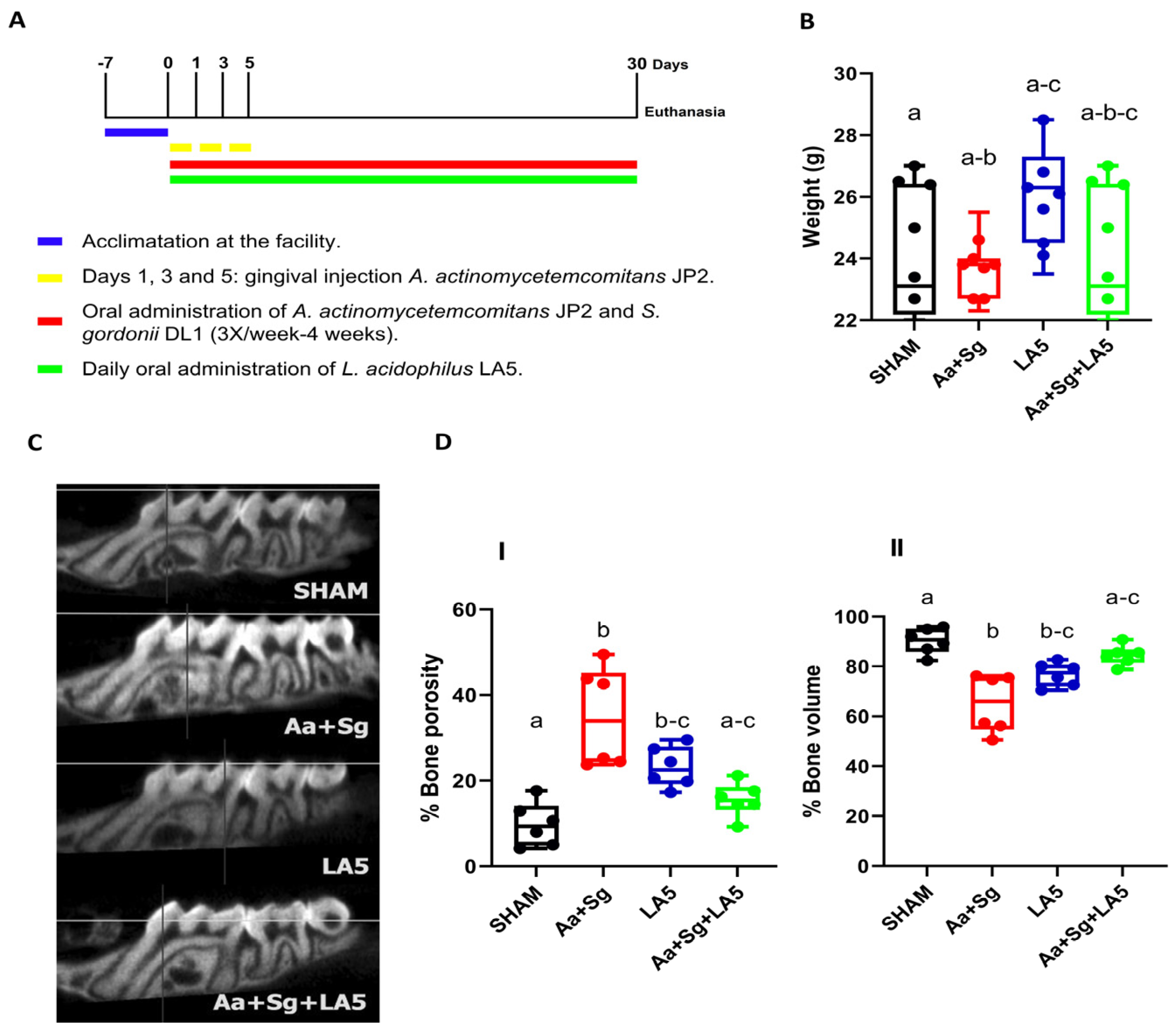
2.6. Experimental Treatments

Groups Aa+Sg and Aa+Sg+LA5 received 50 µL aliquots containing 1 × 10⁹ CFU A. actinomycetemcomitans and 1 × 10⁸ CFU S. gordonii in PBS/1.5% carboxymethylcellulose, into the oral cavity with the aid of a gavage needle, three times a week for four weeks [22]. These groups also received, under anesthesia, a palatal injection of 10 µL containing 1 × 10⁷ CFU A. actinomycetemcomitans in PBS in the interproximal gingiva between the first and second molars on the left hemimaxilla [23] at days 01, 03, and 05 of the experimental period. The non-infected group (SHAM) and the probiotic control group (LA5) were orally inoculated with PBS/1.5% carboxymethylcellulose and received a palatal injection with PBS at the same days and volumes used in the Aa+Sg infected groups.
Groups LA5 and Aa+Sg+LA5 received the probiotic daily in the oral cavity for 30 days, in 50 µL aliquots containing 1 × 10⁸ CFU of LA5 in PBS/1.5% carboxymethylcellulose. SHAM and Aa+Sg groups received the vehicle (Figure 1A).

2.7. Euthanasia and Samples Collection

After 30 days, the animals were anesthetized (ketamine (100 mg/kg IP) + xylazine (10 mg/kg IP)) and sacrificed by exsanguination.
Oral biofilm was obtained with a microbrush, the content of the jejunum was obtained with a spatula and both samples were kept in TRIS-EDTA, pH 7.4, for microbiome analysis. The gingival tissue around the buccal and palatal surfaces of the left molars was removed and stored in RNAlaterTM Stabilization Solution (Invitrogen Life Technologies, Carlsbad, CA, USA) for gene expression analyses. The left hemimaxilla was kept in 4% formaldehyde solution for 24 h, transferred to PBS, and stored for alveolar bone analysis.

2.8. Alveolar Bone Loss Analysis

Alveolar bone resorption was determined by micro tomography (SkyScan 1174 version 1.1, Kontich, Belgium) at 45 kV voltage, 550 uA current, 8.71 μm pixel size, 0.2 mm aluminum filter. The left hemimaxillae were scanned, and a blinded examiner selected a standard area of 60 × 30 pixel (Roi) at the interproximal region between the first and second molar from the second molar cementoenamel junction in 15 coronal sections. The images were analyzed by calculating percentages of bone volume and porosity using CT Analyzer software Version 1.15.4.0, SkyScan.

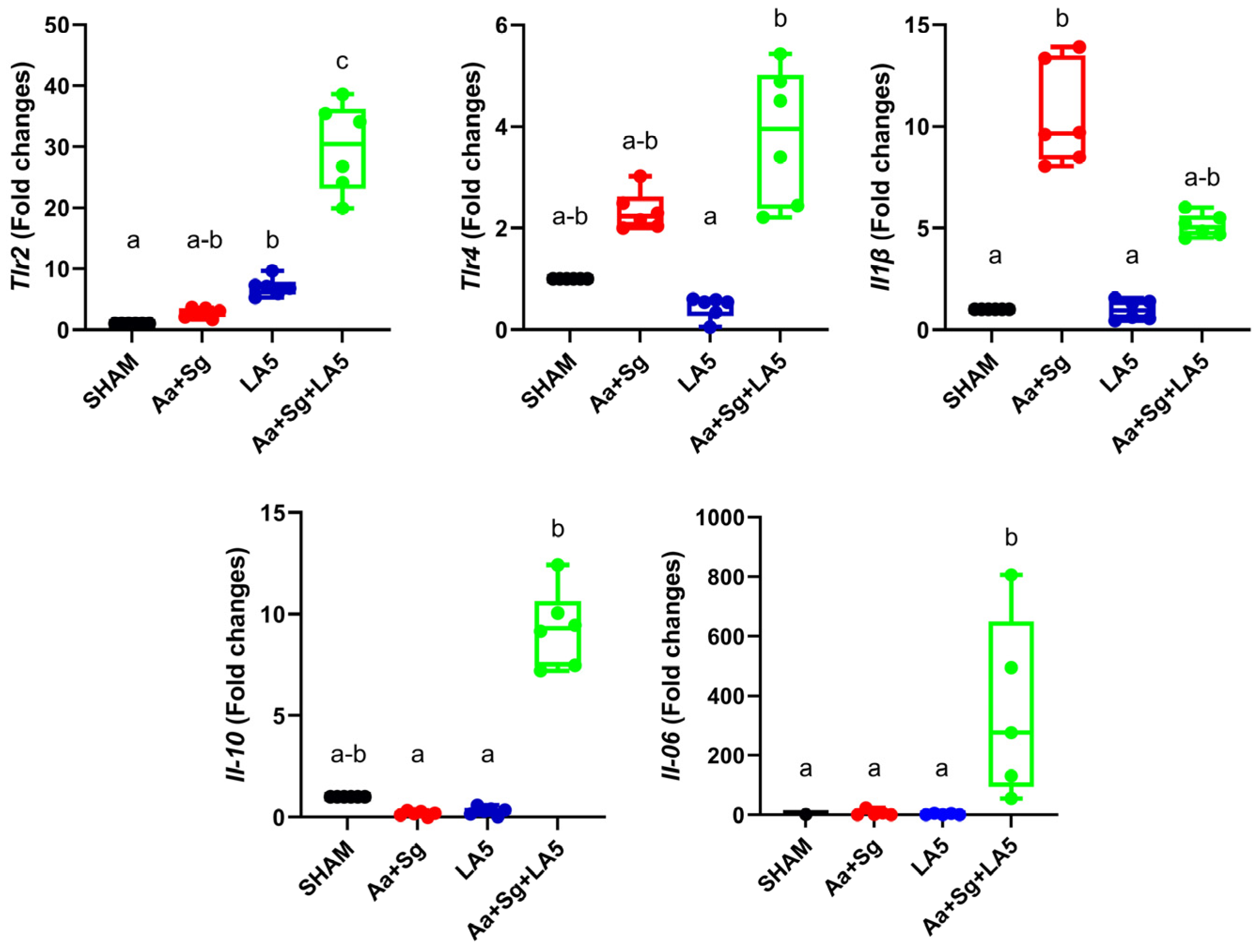
2.9. Gene Expression in Gingiva

RNA was extracted using Trizol LS Reagent (Invitrogen Life Technologies, Carlsbad, CA, USA) in a cell disrupter (BioSpec 3110BX Mini-BeadBeater-1 High Energy Cell Disrupter, Campinas, SP, Brazil) for 20 s, twice. After deoxyribonuclease (Ambion™ DNase I, Invitrogen Life Technologies) treatment, cDNA was obtained using the SuperScriptTM ViloTM Synthesis Kit for RT-PCR (Invitrogen Life Technologies). Quantitative PCR was performed in StepOne Plus System thermocycler (Applied Biosystems, Foster City, CA, USA) with 100 ng cDNA using TaqMan™ Gene Expression Assay (Invitrogen by Thermo Fisher Scientific, Vilnius, Lithuania). Commercial Taqman primers and probes (Invitrogen Life Technologies, Carlsbad, CA, USA) comprised Tlr-2 (Mm01213946_g1), Tlr-4 (Mm00445273_m1), Il-1β (Mm00434228_m1), Il-10 (Mm01288386_m1), Tnf (Mm00443258_m1), β-actin (Mm00607939_s1), and Gapdh (Mm99999915_g1). Relative expression of target genes was calculated by the ΔΔCT method, using β-actin and Gapdh as endogenous controls [24], and expressed as fold changes in relation to control group (SHAM).

2.10. Oral and Gut Microbiomes

DNA from oral biofilm and gut samples of six animals per group was extracted using the Master pureTM DNA Purification Kit (Epicentre® Illumina Company, Madison, WI, USA). A barcoded primer set Bakt_341F CCTACGGGNGGCWGCAG and Bakt_805R GACTACHVGGGTATCTAATCC [25] was used to amplify the hypervariable V3–V4 region of 16SrRNA. DNA was sequenced by ByMyCell (Ribeirão Preto, São Paulo, Brazil) using the Illumina MiSeq 2 × 250 platform. Data were submitted to Sequence Read Archive (SRA) under BioProject identification #PRJNA994574.

2.11. Statistical Analysis

Data normality was checked using the Kolmogorov–Smirnov statistical test with Lilliefors correlation, and homogeneity of variances was assessed using the F test. Parametric data were analyzed by Kruskal–Wallis, followed by Dunn’s post-hoc test. Statistical significance was set at p < 0.05. The GraphPad Prisma® Version 9.0.0 program was used (GraphPad Software, La Jolla, CA, USA).
Microbiome data were analyzed using Qiime 2 2022.8 [26]. Demultiplexed sequences and reads were filtered using Dada 2, and quality score threshold = 25. Trimmed sequences were clustered into amplicon sequence variants (ASVs), and taxonomy was assigned using Silva138 database [27,28]. Alpha diversity indices (Faith, Pielou and Shannon), and Beta diversity by Weighted and Unweighted UniFrac distances were calculated. Clustering was visualized by principal Coordinates Analysis (PCA) [26,29] and differences among groups determined by Permanova (999 permutations). Differences in mean relative abundance of taxa were determined by ANCOM (analysis of composition of microbiomes) [30], using 75% as the empirical cut-off value. The expanded HOMD (eHOMD) database was assessed to search for the inoculated species.

3. Results

3.1. Animals Changes

Animals did not exhibit any changes in fur, skin, and mobility throughout the experiment. There were no differences in animals’ weight at baseline. There was a trend to increased final weight in the groups that received LA5 (LA5 and Aa+Sg+LA5) when compared to SHAM and Aa+Sg, respectively (Figure 1B).

3.2. Alveolar Bone Loss

The inoculation of Aa+Sg induced alveolar bone loss. Administration of LA5 in otherwise non-infected animals (probiotic control group-LA5) also showed some degree of destruction compared to SHAM. However, administration of LA5 to Aa+Sg-infected animals (Aa+Sg+LA5) prevented bone destruction induced by Aa+Sg infection (Figure 1C,D).
Figure 1. C57Bl/6 mice were allocated into four groups (n = 8) and submitted to different treatments for 30 days: negative control (SHAM); positive control: A. actinomycetemcomitans and S. gordonii (Aa+Sg); probiotic control: L. acidophilus (LA5), and test group: A. actinomycetemcomitans, S. gordonii and L. acidophilus LA5 (Aa+Sg+LA5). (A) Study design. Control groups received the vehicles. (B) Animals weight in grams at the end of the experimental period. (C) Alveolar bone analysis determined by microtomography in the interproximal region of first and second molar at the left maxilla. Representative image of the alveolar bone of different groups. (D) Percentage of alveolar bone porosity (I) and percentage of alveolar bone volume (II). Different letters indicate statistical difference among groups. Kruskal–Wallis, post-hoc Dunn (p < 0.05).
Microorganisms 12 00836 g001

3.3. Transcription Analysis in Gingival Tissue

Infection with Aa+Sg increased mRNA levels of Il-1β. Administration of LA5 to otherwise non-infected animals led to the upregulation of Tlr2 but did not alter the transcription of other studied genes. There was no difference in Tnf expression levels among groups. However, administration of LA5 to Aa+Sg-infected animals led to the upregulation of Tlr2 and Tlr4, downregulation of Il-1β, and upregulation of Il-10 and Il-6 (Figure 2).

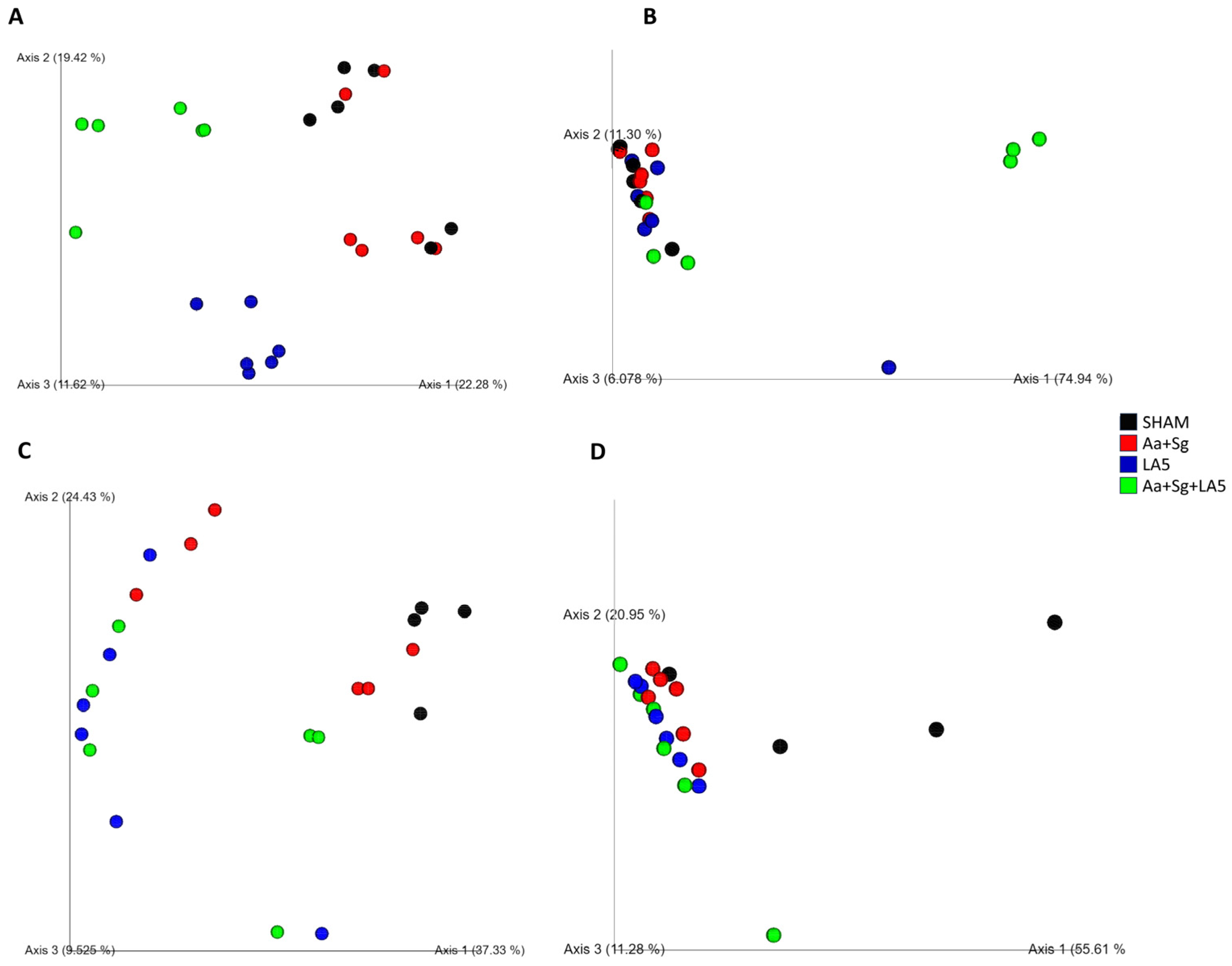
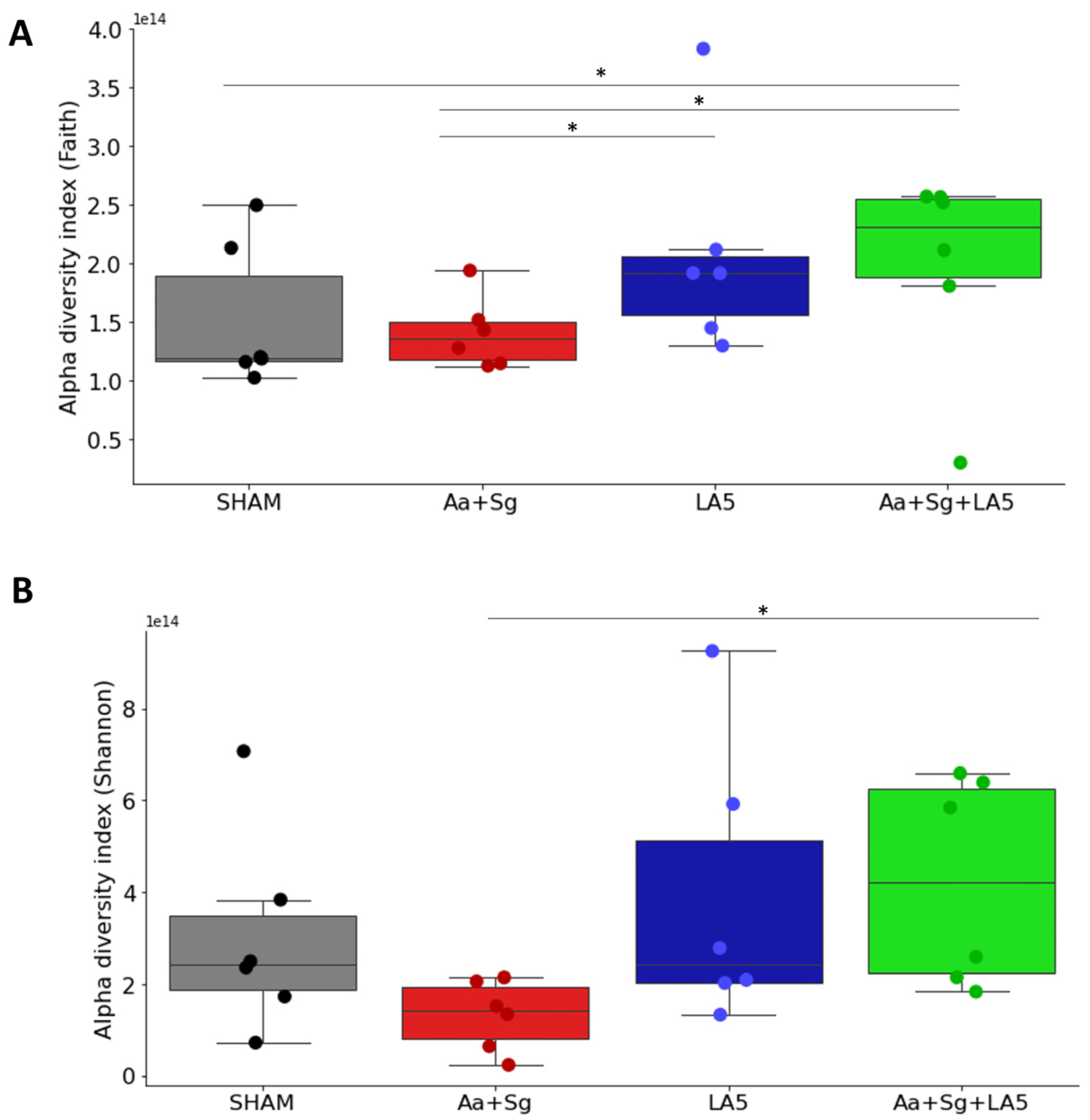
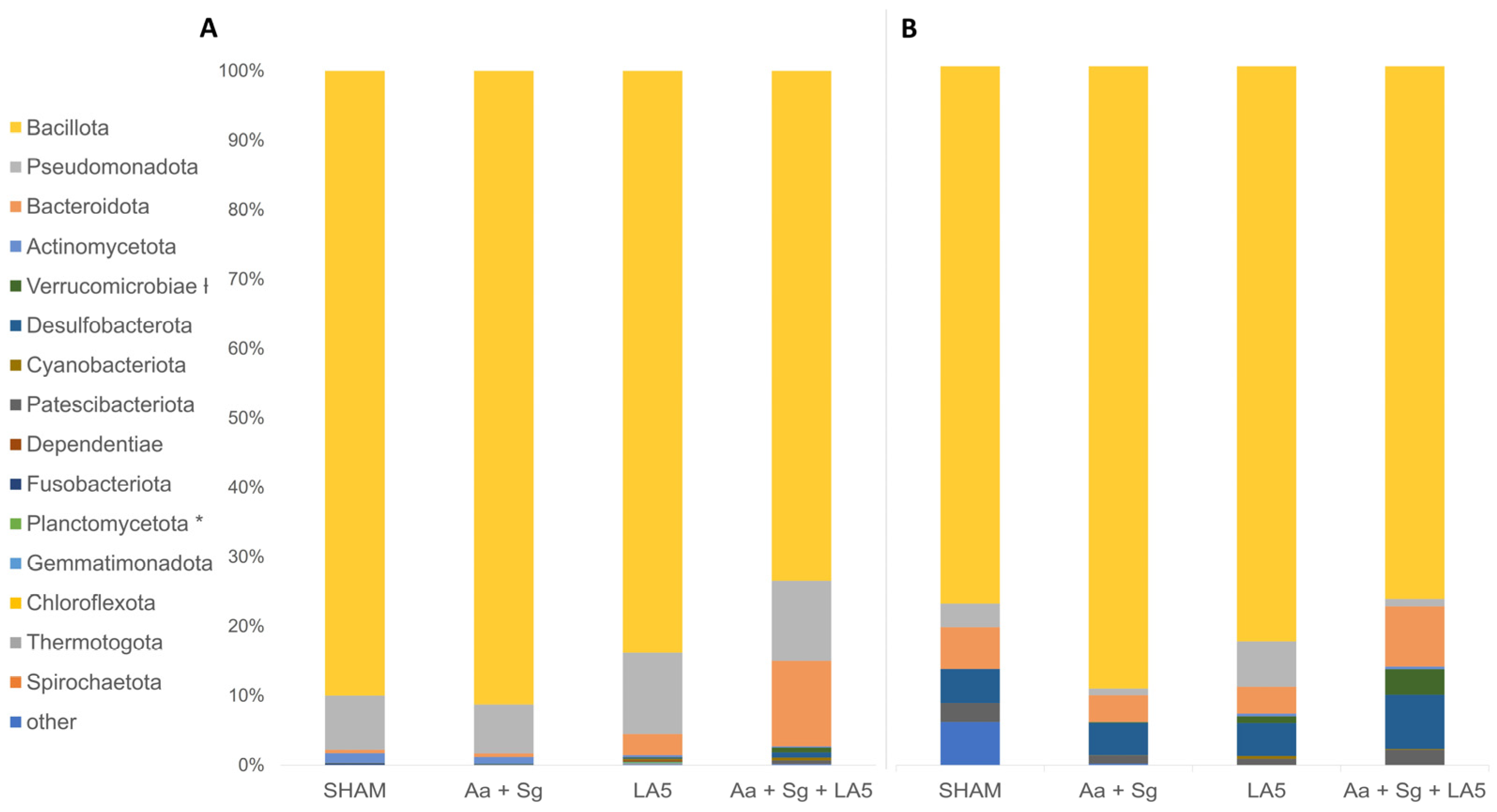
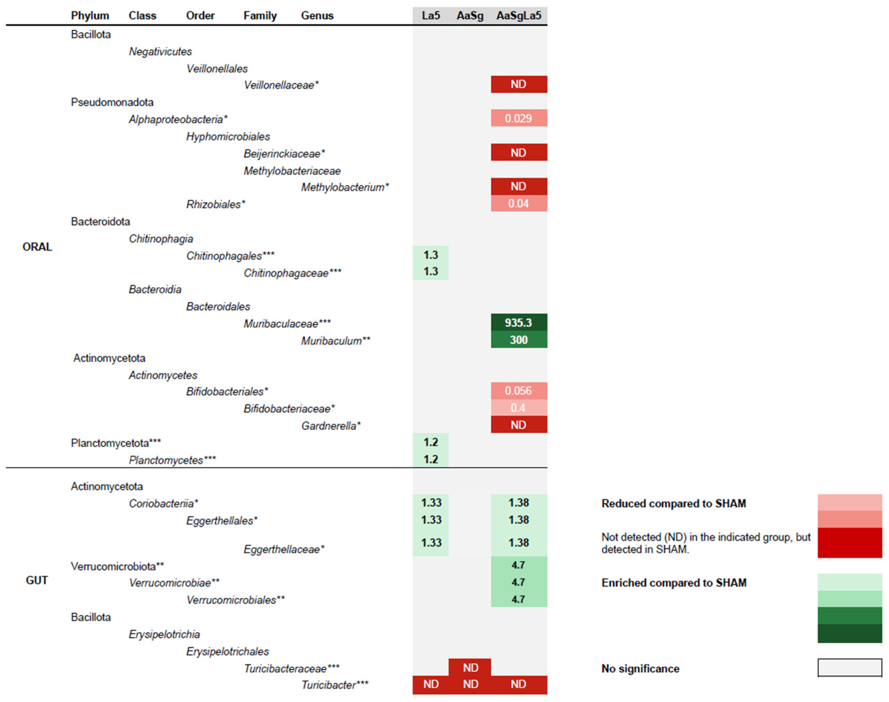
3.4. Oral and Gut Microbiomes

Oral biofilm and gut samples from six animals/group were evaluated for the microbiome analyses. However, the data were obtained only from four animals of the SHAM group due to the poor recovery of DNA from oral biofilm samples. The total number of sequences obtained after the amplification of 16SrRNA of oral and gut samples was 7893.351 (max 386,428 and min 92,229). After filtering and removal of chimeras, the total number was 257,7399 for oral (max 147,037 and min 82,876) and 1,609,759 for gut samples (max 229,627 and min 4859).
Inoculation of Aa+Sg tended to decrease alpha diversity indices (not significant) of oral biofilms. However, administration of LA5 induced a more diverse phylogenetic community (Faith) and increased the richness (Shannon) of the oral microbiome of Aa+Sg-infected mice (Figure 3A,B). There were no differences in Alpha diversity indices (Faith, Pielou, and Shannon) of the gut microbiome among groups.
Inoculation of the microbial consortium Aa+Sg did not alter the population structure of the oral microbiome, since the SHAM and Aa+Sg microbial communities did not differ based on Unweighted and Weighted Unifrac distances (Figure 4). However, infection with Aa+Sg led to a shift in the gut microbiome compared to SHAM-infected animals based on Unweighted and Weighted Unifrac distances measurements (Figure 5).
Inoculation of LA5 either alone (probiotic control group) or in mice infected with Aa+Sg altered the population structure of the oral microbiome, based on the Unweighted and Weighted Unifrac distances, as shown in the PCoA plots (Figure 4A,B).
The probiotic LA5 also interfered with the structure of the gut microbiome. Beta diversity analysis based on Unweighted Unifrac distances demonstrated that all groups clustered apart, except for groups LA5 and Aa+Sg+LA5 (Figure 4C). Analyses based on Weighted Unifrac distances revealed that the microbial communities in the gut of groups SHAM, Aa+Sg and Aa+Sg+LA5 differed from each other, and LA5 differed from SHAM, as visualized in PCoA plots and determined by PERMANOVA (Figure 4D).
Bacillota was the most abundant phylum in all oral and gut communities independently on the treatment (Figure 5A,B). ANCOM revealed differences in RA of Planctomycetota among oral samples (Figure 5A) and Verrucomicrobiae in gut samples (Figure 5B). Furthermore, differences in RA of several taxa in oral and gut samples at lower taxonomic levels were demonstrated (Figure 6).
The eHOMD database analyses indicated that Aa was detected at a low abundance (~0.02%) in three out of six oral samples of the Aa+Sg group but in no other oral or gut samples. Nevertheless, S. gordonii and L. acidophilus were not detected in any of the studied oral and gut samples.

4. Discussion

We have tested the effect of the probiotic strain L. acidophilus LA5 in an experimental model of periodontitis induced by the pathogen A. actinomycetemcomitans associated with S. gordonii due to their synergistic effect [31,32]. Although most species of oral streptococci of sanguinis-mitis groups, which includes S. gordonii, are commensals, S. gordonii can be considered an “accessory pathogen” [31]. Cooperation between S. gordonii and A. actinomycetemcomitans increased the survival and persistence of A. actinomycetemcomitans and promoted its virulence in a murine abscess model [33]. This cooperation involves the production of L-lactate and H2O2 by S. gordonii, providing nutrients for A. actinomycetemcomitans [34] but favoring its dispersal throughout the oral cavity [35].
Aggregatibacter actinomycetemcomitans was detected in the oral biofilm of three out of six mice of the Aa+Sg-infected group, suggesting successful colonization in accordance with data of in vitro and in vivo abscess experimental model [32,33]. Furthermore, Aa+Sg-infected mice exhibited alveolar bone loss and the upregulation of Il-1β in gingival tissues. Moreover, oral administration of L. acidophilus LA5 was able to control alveolar bone loss induced by Aa+Sg infection, reduced Aa to undetectable levels in the oral biofilm of all studied animals, and altered the transcriptional profile of gingival tissues.
Administration of LA5 to otherwise non-infected animals resulted in a slight increase in Tlr2 mRNA levels, but no other evident result was observed in the mice of the LA5 group, except for a shift in the oral and gut microbiomes. Thus, the increase in Tlr2 transcription promoted by LA5 in the gingival tissues of mice may increase surveillance in the oral mucosa, maintaining a healthy associated microbiota in balance with the host.
On the other hand, the administration of LA5 to Aa+Sg-infected animals led to the upregulation of Tlr2 and Tlr4, suggesting further increased surveillance in the oral mucosa, but was able to reduce inflammation, as observed by the altered transcription profile of inflammatory mediators. The capacity to differentially modulate Toll-like receptors (TLRs) is considered an important characteristic of immunobiotic strains [36]. Although the mechanisms underlying the modulation of inflammation induced by L. acidophilus LA5 are not fully understood, our previous data in LPS-mature dendritic cells indicate that L. acidophilus LA5 is able to alter the transcription of several genes involved in TLRs signaling and in the regulation of NF-kappa B activation [15]. Tlr2 upregulation is a common trait observed after probiotics treatment in the gut [37], and LA5 was shown to induce Tlr2 expression in gingival epithelial cells [11,13]. This mechanism is relevant to the anti-inflammatory effects of lactobacilli probiotics, leading to an increased recognition of cell surface lipoproteins and teichoic acid, as well as the production of negative regulators of the NF-κB signaling pathway in a TLR2-dependent manner [38,39]. In contrast, the upregulation of Tlr4 promoted by the probiotic in Aa+Sg-infected mice was unexpected since L. acidophilus LA5 decreased Tlr4 expression in gingival epithelial cells and monocytes in vitro [11,40], and the present data indicate that the probiotic altered cytokines transcription profile toward an anti-inflammatory effect.
The downregulation of Il-1β and upregulation of Il-10 promoted by LA5 in Aa+Sg infected mice are attractive effects to modulate bone resorption and are expected in successful periodontal treatment in humans [41,42,43]. On the other hand, the effect of the increased Il-6 transcription levels induced by the probiotic is less clear. Il-6 is a proinflammatory cytokine, and treatment with monoclonal antibodies against Il-6 receptor resulted in decreased periodontal inflammation and improved periodontal status [43]. However, IL-6 increased response is a common feature after probiotic treatment and is associated with an increased production of IgAS against pathogens in Peyer Patches in the gut [44] in a TLR2-dependent mechanism [45]. The overall attenuation of inflammation and reduced bone resorption promoted by LA5 in vivo after Aa+Sg challenge could not be predicted by in vitro studies since LA5 induced Il-1β and Il-6 transcription and Il-1β release by DCs and monocytes [16,40] but reduced their expression by epithelial cells [11,13].
Infection with Aa+Sg promoted changes in the oral microbiome. However, administration of L. acidophilus LA5 induced more shifts in the oral microbiome than infection with the pathobionts. LA5 administration increased diversity of Aa+Sg-infected mice and impacted their oral microbiomes, although there were no shifts in the abundance of the main phyla Bacillota (former Firmicutes), Pseudomonadota (former Proteobacteria), or Bacteroidota (former Bacteroidetes) [46]. Dysbiosis of the oral microbiome is characterized by increased abundance of opportunistic/inflammophilic organisms and decreased abundance of commensals species. However, the administration of LA5 to Aa+Sg-infected animals resulted in an decreased abundance of some Alpha Pseudomonadota, Veillonellaceae, and Bifidobacteriaceae with no known pathogenic potential in the oral cavity, as well as an increased abundance of Muribaculaceae. Muribaculaceae is considered beneficial to the gut due to production of short-chain fatty acids (SCFA), especially propionate [47], and its abundance was increased by other probiotic lactobacilli [48]. The abundance of Muribaculaceae is reduced in inflammatory diseases [49,50,51], including in periodontitis [52], but its role in the oral cavity remains unknown.
Animal experimental studies have suggested that swallowing of oral bacteria results in dysbiosis in the gut and systemic inflammation [53]. These data are reinforced by human studies indicating an altered microbiome in the gut of periodontitis patients [2,54,55]. Our data indicated that infection with Aa+Sg altered the gut microbiome by increasing the abundance of Eggerthelaceae and decreasing the abundance of Turicibacteraceae.
The oral administration of LA5 was also able to alter the gut microbiome. Although LA5 viability may decrease throughout the passage in the hazardous environment of the stomach, its beneficial effects of LA5 toward a healthier gut microbiome were indicated by simulating the digestive system in vitro [14]. Our data indicated that the administration of LA5 increased the abundance of Verrucomicrobiae and Eggerthelaceae in the gut, especially of Aa+Sg-infected mice. The role of Eggerthelaceae in the gut is not fully understood, but its high abundance was associated to the beneficial effects of curcumin in a mouse model [50]. More importantly, members of the phylum Verrucomicrobiae improve the integrity of the intestinal barrier and regulate host metabolism and immunity [56]. Thus, our data corroborate the existing evidence that LA5 administration can enrich the gut microbiome with beneficial organisms related to health [44,45,57,58].
Overall, the probiotic treatment showed antimicrobial as well as anti-inflammatory effects, leading to the control of periodontitis induced by A. actinomycetemcomitans, as previously expected from in vitro studies [11,12]. However, these data should be interpreted under the limitations of the animal model. A. actinomycetemcomitans’ main virulence factor, the leukotoxin, does not affect murine cells [59], whereas adhesion to epithelial cells mediated by A. actinomycetemcomitans’ adhesins Aae and OMP100 is also impaired in mice [58,60]. Furthermore, interaction of A. actinomycetemcomitans with S. gordonii can alter the expression of key virulence factors of A. actinomycetemcomitans [33]. Another drawback from this model was observed when the administration of the probiotic alone also induced a slight alveolar bone resorption (Figure 1D), contrasting with our previous data, where LA5 induced no significant alveolar bole destruction in mice with a reduced microbiome [17].
Other uses of L. acidophilus LA5 include the control of diabetes [14], gestational diabetes [61], and Clostridium difficile infection [62]. Regarding periodontitis, this strain could control alveolar bone loss induced by experimental inoculation of P. gingivalis and other anaerobic organisms, as well as by A. actinomycetemcomitans, suggesting its beneficial effect against different forms of periodontitis in humans.
The ideal probiotic treatment for any infectious-inflammatory disease should be based not only on the nature of the disease but also on the initial resident microbiome prior to probiotic treatment, and other factors, such as genetic susceptibility and disease progression, should be considered. Future research should not only evaluate the efficacy of different probiotic regimens in randomized clinical trials but should also provide the principles to select the better probiotic regimen on an individual basis, focusing on the mechanisms underlying probiotics effects and on the factors involved in the success and failures of probiotic administration for each disease. Moreover, side effects should be observed as probably not all patients can be safely treated due to their systemic health [63].

5. Conclusions

Under the limitations of previous in vitro studies and of this in vivo experimental murine model, we can conclude that L acidophilus LA5 is a potential candidate to control periodontitis in humans due to its immunomodulatory properties, its antimicrobial effects against bacteria implicated in the disease, and its ability to alter the oral and gut microbiomes in experimental periodontitis. These results should be taken with reservation as future clinical trials should be performed to assess the therapeutic effects of L. acidophilus LA5 in the control of periodontal disease progression.

Author Contributions

M.P.A.M. drew the project, planned the assays, and wrote the manuscript. M.R.B. and F.H.M. planned and ran the assays, contributed with the statistical analysis, and helped writing the manuscript. C.M.R., D.K., K.H.I., E.S.A.-S. and A.R.C. helped with the sample analyses. R.V.C. and L.S.A. helped with microtomography assays. All authors have read and agreed to the published version of the manuscript.

Funding

This research was supported by the São Paulo Research Foundation (FAPESP) grant #2015/18273-9. MRB, DK, EAS, KHI were supported by scholarships from FAPESP 2017/16377-7, 2016/13159-6, 2016/14687-6, and 2016/13156-7. EAS was also supported by CAPES.

Data Availability Statement

Data are contained within the article.

Acknowledgments

We thank the São Paulo Research Foundation (FAPESP) for supporting this study and for the scholarships of MRB, DK, EAS, and KHI. We also thank Coordination for the improvement of higher Education Personnel (CAPES) for the scholarship of EAS.

Conflicts of Interest

The authors declare no conflicts of interest.

References

  1. Haubek, D.; Johansson, A. Pathogenicity of the highly leukotoxic JP2 clone of Aggregatibacter actinomycetemcomitans and its geographic dissemination and role in aggressive periodontitis. J. Oral Microbiol. 2014, 6, 23980. [Google Scholar] [CrossRef] [PubMed]
  2. Amado, P.P.P.; Kawamoto, D.; Albuquerque-Souza, E.; Franco, D.C.; Saraiva, L.; Casarin, R.C.V.; Horliana, A.; Mayer, M.P.A. Oral and Fecal Microbiome in Molar-Incisor Pattern Periodontitis. Front. Cell Infect. Microbiol. 2020, 10, 583761. [Google Scholar] [CrossRef] [PubMed]
  3. Feres, M.; Figueiredo, L.C.; Soares, G.M.; Faveri, M. Systemic antibiotics in the treatment of periodontitis. Periodontol. 2000 2015, 67, 131–186. [Google Scholar] [CrossRef] [PubMed]
  4. Sanz, M.; Herrera, D.; Kebschull, M.; Chapple, I.; Jepsen, S.; Beglundh, T.; Sculean, A.; Tonetti, M.S.; Participants, E.F.P.W.; Methodological, C. Treatment of stage I-III periodontitis-The EFP S3 level clinical practice guideline. J. Clin. Periodontol. 2020, 47 (Suppl. S22), 4–60. [Google Scholar] [CrossRef] [PubMed]
  5. Myneni, S.R.; Brocavich, K.; Wang, H.H. Biological strategies for the prevention of periodontal disease: Probiotics and vaccines. Periodontol. 2000 2020, 84, 161–175. [Google Scholar] [CrossRef] [PubMed]
  6. Gheisary, Z.; Mahmood, R.; Harri Shivanantham, A.; Liu, J.; Lieffers, J.R.L.; Papagerakis, P.; Papagerakis, S. The Clinical, Microbiological, and Immunological Effects of Probiotic Supplementation on Prevention and Treatment of Periodontal Diseases: A Systematic Review and Meta-Analysis. Nutrients 2022, 14, 1036. [Google Scholar] [CrossRef] [PubMed]
  7. Li, J.; Zhao, G.; Zhang, H.M.; Zhu, F.F. Probiotic adjuvant treatment in combination with scaling and root planing in chronic periodontitis: A systematic review and meta-analysis. Benef. Microbes 2023, 14, 95–107. [Google Scholar] [CrossRef] [PubMed]
  8. Vincenzi, A.; Goettert, M.I.; Volken de Souza, C.F. An evaluation of the effects of probiotics on tumoral necrosis factor (TNF-α) signaling and gene expression. Cytokine Growth Factor. Rev. 2021, 57, 27–38. [Google Scholar] [CrossRef]
  9. Kleerebezem, M.; Binda, S.; Bron, P.A.; Gross, G.; Hill, C.; van Hylckama Vlieg, J.E.; Lebeer, S.; Satokari, R.; Ouwehand, A.C. Understanding mode of action can drive the translational pipeline towards more reliable health benefits for probiotics. Curr. Opin. Biotechnol. 2019, 56, 55–60. [Google Scholar] [CrossRef]
  10. Bueno, M.R.; Dudu-Silva, G.; Macedo, T.T.; Gomes, A.P.d.A.P.; Rodrigues Oliveira Braga, A.; Aguiar Silva, L.D.; Bueno-Silva, B. Lactobacillus acidophilus impairs the establishment of pathogens in a subgingival multispecies biofilm. Front. Dent. Med. 2023, 4, 1212773. [Google Scholar] [CrossRef]
  11. Bueno, M.R.; Ishikawa, K.H.; Almeida-Santos, G.; Ando-Suguimoto, E.S.; Shimabukuro, N.; Kawamoto, D.; Mayer, M.P.A. Lactobacilli Attenuate the Effect of Aggregatibacter actinomycetemcomitans Infection in Gingival Epithelial Cells. Front. Microbiol. 2022, 13, 846192. [Google Scholar] [CrossRef] [PubMed]
  12. Ishikawa, K.H.; Bueno, M.R.; Kawamoto, D.; Simionato, M.R.L.; Mayer, M.P.A. Lactobacilli postbiotics reduce biofilm formation and alter transcription of virulence genes of Aggregatibacter actinomycetemcomitans. Mol. Oral Microbiol. 2021, 36, 92–102. [Google Scholar] [CrossRef] [PubMed]
  13. Albuquerque-Souza, E.; Balzarini, D.; Ando-Suguimoto, E.S.; Ishikawa, K.H.; Simionato, M.R.L.; Holzhausen, M.; Mayer, M.P.A. Probiotics alter the immune response of gingival epithelial cells challenged by Porphyromonas gingivalis. J. Periodontal Res. 2019, 54, 115–127. [Google Scholar] [CrossRef] [PubMed]
  14. Salgaço, M.K.; de Oliveira, F.L.; Sartoratto, A.; Mesa, V.; Mayer, M.P.A.; Sivieri, K. Impact of Lactobacillus acidophilus—La5 on Composition and Metabolism of the Intestinal Microbiota of Type 2 Diabetics (T2D) and Healthy Individuals Using a Microbiome Model. Fermentation 2023, 9, 740. [Google Scholar] [CrossRef]
  15. Vale, G.C.; Mota, B.I.S.; Ando-Suguimoto, E.S.; Mayer, M.P.A. Lactobacilli Probiotics Modulate Antibacterial Response Gene Transcription of Dendritic Cells Challenged with LPS. Probiotics Antimicrob. Proteins 2024, 16, 293–307. [Google Scholar] [CrossRef] [PubMed]
  16. Vale, G.C.; Mota, B.I.S.; Ando-Suguimoto, E.S.; Mayer, M.P.A. Effect of Probiotics Lactobacillus acidophilus and Lacticaseibacillus rhamnosus on Antibacterial Response Gene Transcription of Human Peripheral Monocytes. Probiotics Antimicrob. Proteins 2023, 15, 264–274. [Google Scholar] [CrossRef] [PubMed]
  17. Catarucci, A.C.S.K.D.; Shimabukuro, N.; Ishikawa, K.H.; Ando-Suguimoto, E.S.; Ribeiro, R.A.; Nicastro, G.G.; Albuquerque-Souza, E.; Robson Souza, R.F.; Mayer, M.P.A. Oral Administration of Lactobacillus Acidophilus LA5 Prevents Alveolar Bone Loss and Alters Oral and Gut Microbiomes in a Murine Periodontitis Experimental Model; University of São Paulo: São Paulo, Brazil, 2024; to be submitted. [Google Scholar]
  18. Charan, J.; Kantharia, N.D. How to calculate sample size in animal studies? J. Pharmacol. Pharmacother. 2013, 4, 303–306. [Google Scholar] [CrossRef] [PubMed]
  19. Shimabukuro, N.; Cataruci, A.C.S.; Ishikawa, K.H.; de Oliveira, B.E.; Kawamoto, D.; Ando-Suguimoto, E.S.; Albuquerque-Souza, E.; Nicoli, J.R.; Ferreira, C.M.; de Lima, J.; et al. Bifidobacterium Strains Present Distinct Effects on the Control of Alveolar Bone Loss in a Periodontitis Experimental Model. Front. Pharmacol. 2021, 12, 713595. [Google Scholar] [CrossRef] [PubMed]
  20. Tsai, C.C.; Shenker, B.J.; DiRienzo, J.M.; Malamud, D.; Taichman, N.S. Extraction and isolation of a leukotoxin from Actinobacillus actinomycetemcomitans with polymyxin B. Infect. Immun. 1984, 43, 700–705. [Google Scholar] [CrossRef]
  21. Hsu, S.D.; Cisar, J.O.; Sandberg, A.L.; Kilian, M. Adhesive Properties of Viridans Streptoccocal Species. Microb. Ecol. Health Dis. 2009, 7, 125–137. [Google Scholar]
  22. Repeke, C.E.; Ferreira, S.B., Jr.; Claudino, M.; Silveira, E.M.; de Assis, G.F.; Avila-Campos, M.J.; Silva, J.S.; Garlet, G.P. Evidences of the cooperative role of the chemokines CCL3, CCL4 and CCL5 and its receptors CCR1+ and CCR5+ in RANKL+ cell migration throughout experimental periodontitis in mice. Bone 2010, 46, 1122–1130. [Google Scholar] [CrossRef] [PubMed]
  23. Madeira, M.F.; Queiroz-Junior, C.M.; Costa, G.M.; Werneck, S.M.; Cisalpino, D.; Garlet, G.P.; Teixeira, M.M.; Silva, T.A.; Souza, D.G. Platelet-activating factor receptor blockade ameliorates Aggregatibacter actinomycetemcomitans-induced periodontal disease in mice. Infect. Immun. 2013, 81, 4244–4251. [Google Scholar] [CrossRef] [PubMed]
  24. Pfaffl, M.W. A new mathematical model for relative quantification in real-time RT-PCR. Nucleic Acids Res. 2001, 29, e45. [Google Scholar] [CrossRef]
  25. Herlemann, D.P.; Labrenz, M.; Jurgens, K.; Bertilsson, S.; Waniek, J.J.; Andersson, A.F. Transitions in bacterial communities along the 2000 km salinity gradient of the Baltic Sea. ISME J. 2011, 5, 1571–1579. [Google Scholar] [CrossRef] [PubMed]
  26. Bolyen, E.; Rideout, J.R.; Dillon, M.R.; Bokulich, N.A.; Abnet, C.C.; Al-Ghalith, G.A.; Alexander, H.; Alm, E.J.; Arumugam, M.; Asnicar, F.; et al. Reproducible, interactive, scalable and extensible microbiome data science using QIIME 2. Nat. Biotechnol. 2019, 37, 852–857. [Google Scholar] [CrossRef] [PubMed]
  27. Quast, C.; Pruesse, E.; Yilmaz, P.; Gerken, J.; Schweer, T.; Yarza, P.; Peplies, J.; Glockner, F.O. The SILVA ribosomal RNA gene database project: Improved data processing and web-based tools. Nucleic Acids Res. 2013, 41, D590–D596. [Google Scholar] [CrossRef] [PubMed]
  28. Yilmaz, P.; Parfrey, L.W.; Yarza, P.; Gerken, J.; Pruesse, E.; Quast, C.; Schweer, T.; Peplies, J.; Ludwig, W.; Glockner, F.O. The SILVA and “All-species Living Tree Project (LTP)” taxonomic frameworks. Nucleic Acids Res. 2014, 42, D643–D648. [Google Scholar] [CrossRef] [PubMed]
  29. Lozupone, C.A.; Stombaugh, J.I.; Gordon, J.I.; Jansson, J.K.; Knight, R. Diversity, stability and resilience of the human gut microbiota. Nature 2012, 489, 220–230. [Google Scholar] [CrossRef] [PubMed]
  30. Lin, H.; Peddada, S.D. Analysis of compositions of microbiomes with bias correction. Nat. Commun. 2020, 11, 3514. [Google Scholar] [CrossRef]
  31. Nobbs, A.; Kreth, J. Genetics of sanguinis-Group Streptococci in Health and Disease. Microbiol. Spectr. 2019, 7. [Google Scholar] [CrossRef]
  32. Stacy, A.; Everett, J.; Jorth, P.; Trivedi, U.; Rumbaugh, K.P.; Whiteley, M. Bacterial fight-and-flight responses enhance virulence in a polymicrobial infection. Proc. Natl. Acad. Sci. USA 2014, 111, 7819–7824. [Google Scholar] [CrossRef] [PubMed]
  33. Stacy, A.; Fleming, D.; Lamont, R.J.; Rumbaugh, K.P.; Whiteley, M. A Commensal Bacterium Promotes Virulence of an Opportunistic Pathogen via Cross-Respiration. mBio 2016, 7, e00782-16. [Google Scholar] [CrossRef] [PubMed]
  34. Brown, S.A.; Whiteley, M. A novel exclusion mechanism for carbon resource partitioning in Aggregatibacter actinomycetemcomitans. J. Bacteriol. 2007, 189, 6407–6414. [Google Scholar] [CrossRef] [PubMed]
  35. Ramsey, M.M.; Rumbaugh, K.P.; Whiteley, M. Metabolite cross-feeding enhances virulence in a model polymicrobial infection. PLoS Pathog. 2011, 7, e1002012. [Google Scholar] [CrossRef] [PubMed]
  36. Kanmani, P.; Kim, H. Functional capabilities of probiotic strains on attenuation of intestinal epithelial cell inflammatory response induced by TLR4 stimuli. Biofactors 2019, 45, 223–235. [Google Scholar] [CrossRef] [PubMed]
  37. Vizoso Pinto, M.G.; Rodriguez Gomez, M.; Seifert, S.; Watzl, B.; Holzapfel, W.H.; Franz, C.M. Lactobacilli stimulate the innate immune response and modulate the TLR expression of HT29 intestinal epithelial cells in vitro. Int. J. Food Microbiol. 2009, 133, 86–93. [Google Scholar] [CrossRef] [PubMed]
  38. Sengupta, R.; Altermann, E.; Anderson, R.C.; McNabb, W.C.; Moughan, P.J.; Roy, N.C. The role of cell surface architecture of lactobacilli in host-microbe interactions in the gastrointestinal tract. Mediators Inflamm. 2013, 2013, 237921. [Google Scholar] [CrossRef]
  39. Sun, K.Y.; Xu, D.H.; Xie, C.; Plummer, S.; Tang, J.; Yang, X.F.; Ji, X.H. Lactobacillus paracasei modulates LPS-induced inflammatory cytokine release by monocyte-macrophages via the up-regulation of negative regulators of NF-kappaB signaling in a TLR2-dependent manner. Cytokine 2017, 92, 1–11. [Google Scholar] [CrossRef] [PubMed]
  40. Vale, G.C.; Mayer, M.P.A. Effect of probiotic Lactobacillus rhamnosus by-products on gingival epithelial cells challenged with Porphyromonas gingivalis. Arch. Oral Biol. 2021, 128, 105174. [Google Scholar] [CrossRef]
  41. Taiete, T.; Monteiro, M.F.; Casati, M.Z.; do Vale, H.F.; Ambosano, G.M.B.; Nociti, F.H.; Sallum, E.A.; Casarin, R.C.V. Local IL-10 level as a predictive factor in generalized aggressive periodontitis treatment response. Scand. J. Immunol. 2019, 90, e12816. [Google Scholar] [CrossRef]
  42. Rabelo, M.S.; Gomes, G.H.; Foz, A.M.; Stadler, A.F.; Cutler, C.W.; Susin, C.; Romito, G.A. Short-term effect of non-surgical periodontal treatment on local and systemic cytokine levels: Role of hyperglycemia. Cytokine 2021, 138, 155360. [Google Scholar] [CrossRef] [PubMed]
  43. Balta, M.G.; Papathanasiou, E.; Blix, I.J.; Van Dyke, T.E. Host Modulation and Treatment of Periodontal Disease. J. Dent. Res. 2021, 100, 798–809. [Google Scholar] [CrossRef]
  44. Jain, S.; Yadav, H.; Sinha, P.R.; Naito, Y.; Marotta, F. Dahi containing probiotic Lactobacillus acidophilus and Lactobacillus casei has a protective effect against Salmonella enteritidis infection in mice. Int. J. Immunopathol. Pharmacol. 2008, 21, 1021–1029. [Google Scholar] [CrossRef]
  45. Yamasaki-Yashiki, S.; Miyoshi, Y.; Nakayama, T.; Kunisawa, J.; Katakura, Y. IgA-enhancing effects of membrane vesicles derived from Lactobacillus sakei subsp. sakei NBRC15893. Biosci. Microbiota Food Health 2019, 38, 23–29. [Google Scholar] [CrossRef]
  46. Verma, D.; Garg, P.K.; Dubey, A.K. Insights into the human oral microbiome. Arch. Microbiol. 2018, 200, 525–540. [Google Scholar] [CrossRef] [PubMed]
  47. Smith, B.J.; Miller, R.A.; Ericsson, A.C.; Harrison, D.C.; Strong, R.; Schmidt, T.M. Changes in the gut microbiome and fermentation products concurrent with enhanced longevity in acarbose-treated mice. BMC Microbiol. 2019, 19, 130. [Google Scholar] [CrossRef]
  48. Ma, F.; Sun, M.; Song, Y.; Wang, A.; Jiang, S.; Qian, F.; Mu, G.; Tuo, Y. Lactiplantibacillus plantarum-12 Alleviates Inflammation and Colon Cancer Symptoms in AOM/DSS-Treated Mice through Modulating the Intestinal Microbiome and Metabolome. Nutrients 2022, 14, 1916. [Google Scholar] [CrossRef] [PubMed]
  49. Rooks, M.G.; Veiga, P.; Wardwell-Scott, L.H.; Tickle, T.; Segata, N.; Michaud, M.; Gallini, C.A.; Beal, C.; van Hylckama-Vlieg, J.E.; Ballal, S.A.; et al. Gut microbiome composition and function in experimental colitis during active disease and treatment-induced remission. ISME J. 2014, 8, 1403–1417. [Google Scholar] [CrossRef] [PubMed]
  50. Cui, C.; Han, Y.; Li, H.; Yu, H.; Zhang, B.; Li, G. Curcumin-driven reprogramming of the gut microbiota and metabolome ameliorates motor deficits and neuroinflammation in a mouse model of Parkinson’s disease. Front. Cell Infect. Microbiol. 2022, 12, 887407. [Google Scholar] [CrossRef]
  51. Krych, Ł.; Nielsen, D.S.; Hansen, A.K.; Hansen, C.H. Gut microbial markers are associated with diabetes onset, regulatory imbalance, and IFN-γ level in NOD mice. Gut Microbes 2015, 6, 101–109. [Google Scholar] [CrossRef]
  52. Xie, Z.; Li, M.; Qian, M.; Yang, Z.; Han, X. Co-Cultures of Lactobacillus acidophilus and Bacillus subtilis Enhance Mucosal Barrier by Modulating Gut Microbiota-Derived Short-Chain Fatty Acids. Nutrients 2022, 14, 4475. [Google Scholar] [CrossRef] [PubMed]
  53. Nakajima, M.; Arimatsu, K.; Kato, T.; Matsuda, Y.; Minagawa, T.; Takahashi, N.; Ohno, H.; Yamazaki, K. Oral Administration of P. gingivalis Induces Dysbiosis of Gut Microbiota and Impaired Barrier Function Leading to Dissemination of Enterobacteria to the Liver. PLoS ONE 2015, 10, e0134234. [Google Scholar] [CrossRef] [PubMed]
  54. Kawamoto, D.; Borges, R.; Ribeiro, R.A.; de Souza, R.F.; Amado, P.P.P.; Saraiva, L.; Horliana, A.; Faveri, M.; Mayer, M.P.A. Oral Dysbiosis in Severe Forms of Periodontitis Is Associated With Gut Dysbiosis and Correlated with Salivary Inflammatory Mediators: A Preliminary Study. Front. Oral Health 2021, 2, 722495. [Google Scholar] [CrossRef] [PubMed]
  55. Lourenço, T.G.B.; de Oliveira, A.M.; Tsute Chen, G.; Colombo, A.P.V. Oral-gut bacterial profiles discriminate between periodontal health and diseases. J. Periodontal Res. 2022, 57, 1227–1237. [Google Scholar] [CrossRef] [PubMed]
  56. Zhao, Q.; Yu, J.; Hao, Y.; Zhou, H.; Hu, Y.; Zhang, C.; Zheng, H.; Wang, X.; Zeng, F.; Hu, J.; et al. Akkermansia muciniphila plays critical roles in host health. Crit. Rev. Microbiol. 2023, 49, 82–100. [Google Scholar] [CrossRef]
  57. Ondee, T.; Pongpirul, K.; Visitchanakun, P.; Saisorn, W.; Kanacharoen, S.; Wongsaroj, L.; Kullapanich, C.; Ngamwongsatit, N.; Settachaimongkon, S.; Somboonna, N.; et al. Lactobacillus acidophilus LA5 improves saturated fat-induced obesity mouse model through the enhanced intestinal Akkermansia muciniphila. Sci. Rep. 2021, 11, 6367. [Google Scholar] [CrossRef] [PubMed]
  58. Fine, D.H.; Velliyagounder, K.; Furgang, D.; Kaplan, J.B. The Actinobacillus actinomycetemcomitans autotransporter adhesin Aae exhibits specificity for buccal epithelial cells from humans and old world primates. Infect. Immun. 2005, 73, 1947–1953. [Google Scholar] [CrossRef]
  59. Tsai, C.C.; Ho, Y.P.; Chou, Y.S.; Ho, K.Y.; Wu, Y.M.; Lin, Y.C. Aggregatibacter (Actinobacillus) actimycetemcomitans leukotoxin and human periodontitis—A historic review with emphasis on JP2. Kaohsiung J. Med. Sci. 2018, 34, 186–193. [Google Scholar] [CrossRef]
  60. Yue, G.; Kaplan, J.B.; Furgang, D.; Mansfield, K.G.; Fine, D.H. A second Aggregatibacter actinomycetemcomitans autotransporter adhesin exhibits specificity for buccal epithelial cells in humans and Old World primates. Infect. Immun. 2007, 75, 4440–4448. [Google Scholar] [CrossRef]
  61. Hajifaraji, M.; Jahanjou, F.; Abbasalizadeh, F.; Aghamohammadzadeh, N.; Abbasi, M.M.; Dolatkhah, N. Effect of probiotic supplements in women with gestational diabetes mellitus on inflammation and oxidative stress biomarkers: A randomized clinical trial. Asia Pac. J. Clin. Nutr. 2018, 27, 581–591. [Google Scholar]
  62. Goldstein, E.J.C.; Johnson, S.J.; Maziade, P.J.; Evans, C.T.; Sniffen, J.C.; Millette, M.; McFarland, L.V. Probiotics and prevention of Clostridium difficile infection. Anaerobe 2017, 45, 114–119. [Google Scholar] [CrossRef] [PubMed]
  63. Suez, J.; Zmora, N.; Segal, E.; Elinav, E. The pros, cons, and many unknowns of probiotics. Nat. Med. 2019, 25, 716–729. [Google Scholar] [CrossRef] [PubMed]
Figure 2. Relative transcription of genes encoding receptors Tlr-2 and Tlr-4 and cytokines Il-1β, Il-10, and Il-6 in gingival tissue of C57Bl/6 mice submitted to different treatments: Groups (n = 6): negative control (SHAM); positive control: A. actinomycetemcomitans and S. gordonii (Aa+Sg); probiotic control: L. acidophilus (LA5), and test group: A. actinomycetemcomitans, S. gordonii and L. acidophilus LA5 (Aa+Sg+LA5). Data on target genes were normalized to mRNA levels of Gapdh and/or β-actin reference genes (internal controls). Different letters indicate statistical difference among groups. Kruskal–Wallis, post-hoc Dunn (p < 0.05).
Microorganisms 12 00836 g002
Figure 3. Alpha diversity analyses of the oral biofilm microbiome of C57Bl/6 mice of the following groups (n = 6): negative control (SHAM); positive control: A. actinomycetemcomitans and S. gordonii (Aa+Sg); probiotic control: L. acidophilus (LA5), and test: A. actinomycetemcomitans, S. gordonii and L. acidophilus LA5 (Aa+Sg+LA5). (A) Richness (Faith’s PD) index and (B) Richness Shannon index analyses * Kruskal–Wallis (p ≤ 0.05).
Microorganisms 12 00836 g003
Figure 4. Treatment with LA5 induced alterations in the oral and gut microbiome of infected animals. Principal coordinate analysis (PCoA), based on unweighted (A) and weighted (B) UniFrac distance metrics performed on oral biofilm microbial communities and unweighted (C) and weighted (D) UniFrac distance metrics on gut microbial communities of C57Bl/6 mice from different groups (n = 6): negative control (SHAM), positive control (A. actinomycetemcomitans and S. gordonii-Aa+Sg), probiotic control (L. acidophilus-LA5), and test group (Aa+Sg+LA5). The PCoA revealed significant changes in the oral and gut composition of C57Bl/6 mice. Treatments LA5 and Aa+Sg+LA5 exhibited similar effects, with Aa+Sg+LA5 causing the most significant shift. These findings underscore treatment-specific impacts on oral microbial communities (PERMANOVA, 999 permutations, p < 0.01).
Microorganisms 12 00836 g004
Figure 5. Microbial abundance of bacteria at the phylum level in the oral (A) and gut (B) microbiomes of C57Bl/6 mice. The following groups were examined: negative control (SHAM), positive control (A. actinomycetemcomitans and S. gordonii-Aa+Sg), probiotic control (L. acidophilus-LA5), and test group (Aa+Sg+LA5). ANCOM (*) indicates statistical differences in the oral microbiome for Planctomycetota. (ƚ) denotes statistical differences in the gut microbiome for Verrucomicrobiota. The Kruskal–Wallis test was performed, with utilized p-values: p < 0.05, p < 0.01, and p < 0.001.
Microorganisms 12 00836 g005
Figure 6. Disparities in the relative abundance (expressed as fold changes relative to SHAM) of bacterial taxa in the oral and gut microbiomes of C57Bl-6 mice across distinct groups: negative control (SHAM), positive control (A. actinomycetemcomitans and S. gordonii-Aa+Sg), probiotic control (L. acidophilus-LA5), and test group (Aa+Sg+LA5). Data were subjected to analysis via ANCOM, with the 75th percentile of the W distribution serving as the empirical cut-off value. Statistically significant differences among groups were validated using Kruskal–Wallis (* p < 0.05, ** p < 0.01, *** p < 0.001).
Microorganisms 12 00836 g006
Disclaimer/Publisher’s Note: The statements, opinions and data contained in all publications are solely those of the individual author(s) and contributor(s) and not of MDPI and/or the editor(s). MDPI and/or the editor(s) disclaim responsibility for any injury to people or property resulting from any ideas, methods, instructions or products referred to in the content.

