Article

Comparison of the Operator and Surrounding Dose When Using Portable Intraoral X-ray Devices

by
Mehrdad Abdinian
1,
Maedeh Aminian
2,
Forouzan Keymasi
3,
Parisa Soltani
1,4,*,
Mariangela Cernera
4,
Niccolo Giuseppe Armogida
4 and
Gianrico Spagnuolo
4,5,*
1
Department of Oral and Maxillofacial Radiology, Dental Implants Research Center, Dental Research Institute, School of Dentistry, Isfahan University of Medical Sciences, Isfahan 8174673461, Iran
2
Department of Oral and Maxillofacial Radiology, School of Dentistry, Ardabil University of Medical Sciences, Ardabil 5618953141, Iran
3
Student Research Committee, School of Dentistry, Isfahan University of Medical Sciences, Isfahan 8174673461, Iran
4
Department of Neurosciences, Reproductive and Odontostomatological Sciences, University of Naples “Federico II”, 80138 Naples, Italy
5
Therapeutic Dentistry Department, Institute for Dentistry, Sechenov University, 119991 Moscow, Russia
*
Authors to whom correspondence should be addressed.
Appl. Sci. 2024, 14(8), 3515; https://doi.org/10.3390/app14083515
Submission received: 20 March 2024 / Revised: 16 April 2024 / Accepted: 17 April 2024 / Published: 22 April 2024
(This article belongs to the Section Applied Dentistry and Oral Sciences)

Abstract

:

Featured Application

It is highly unlikely that the operator would reach occupational dose limits when using iRay D3 and EZRay Air. The Epix radiographic device allows for fewer daily radiographs.

Abstract

This study aimed to investigate the scattered radiation dose using three portable dental radiographic units: iRay D3, EZRay Air, and Epix. The absorbed dose was measured at 0.5 and 1 m distances, every 15° in the horizontal plane, using an ionization chamber. The maximum number of radiographs per day using the portable units was calculated considering a dose limit of 50 mSv/year and 20 mSv/year. The doses were higher in the Epix unit compared to the other two devices. Anterior exposure was generally higher than the sides or posterior exposure. With a dose limit of 50 mSv/year, considering a distance of 0.5 m between the operator and the X-ray unit, a maximum of 961, 565, and 38 radiographs are permitted daily using iRay D3, EZRay Air, and Epix, respectively. Considering a dose limit of 20 mSv/year, with a distance of 0.5 m between the operator and the radiographic device, a maximum of 384, 226, and 15 radiographs are permitted daily using iRay D3, EZRay Air, and Epix portable units, respectively. It is highly unlikely that an operator would reach occupational dose limits when using iRay D3 and EZRay Air. The Epix radiographic device allows for fewer daily radiographs and should be avoided for daily use.

1. Introduction

Ionizing radiation is widely used in dentistry for several dental purposes such as diagnosis, prognosis, and treatment. Several radiographic devices are used for dental radiography. Guidelines recommend a minimum distance of 2 m from the dental X-ray source when use of barriers is not possible [1]. Portable handheld dental X-ray devices are currently used for forensic purposes and instances in which access to dental facilities is difficult or not possible, such as distant and deprived locations, nursing homes, and patients under general anesthesia. In addition, portable X-ray units can be used in mass disasters and for post-mortem analysis and identification [2,3,4,5,6]. These handheld dental radiographic devices are equipped with a protective shield attached to the device [7]. The proper method of use for these devices is exposure with the device being held with stretched arms far from the body parallel to the floor. Nevertheless, concerns exist regarding the operator’s dose during exposure with handheld dental X-ray devices [8]. In addition, these portable radiographic units can also be equipped with a remote exposure button, allowing the operator to place the portable device on a stationary tripod and stand farther away from the X-ray source. However, the principal difference between portable and conventional wall-mounted units is the fact that the operator often touches the X-ray generator during the action of taking radiographs [9].
Several studies have evaluated radiation safety and dosage of portable X-ray devices [7,10]. Danforth et al. conducted a study to measure the radiation dose received by operators using the Nomad portable device and reported that the whole-body annual dose amounted to 0.047 mSv, which corresponds to only 0.09% of the annual dose limit of 50 mSv [11]. In another investigation, Leadbeatter et al. utilized the Rextar X handheld dental radiography unit. They reported absorbed doses resulting from a single adult maxillary molar radiography: 0.69 µGy at the left hand, 0.78 µGy at the right hand, and 0.47 µGy at the operator’s eyes [12]. Additionally, Makdissi et al. studied operator doses while using a portable radiographic device in various positions. Notably, the received dose was higher when the operator’s arm was bent [8]. Lastly, Ghafari et al. employed a Geiger Muller counter and found that the radiation dose near the Port-XII portable device remained below the annual dose limit [13].
However, there are some concerns regarding various aspects of radiation safety, especially in newly developed radiographic units. Several organizations in different parts of the world have set public and occupational dose limits per annum. The International Commission on Radiation Protection (ICRP) has suggested an annual occupational dose limit of 20 mSv (averaged over 5 consecutive years). In the United States, however, the National Council on Radiation Protection and Measurements (NCRP) enforces an annual dose limit of 50 mSv for radiology personnel, a limit higher than ICRP recommendations [13,14]. These occupational dose limits are established to ensure that operators do not encounter any deterministic effects, while also minimizing the risk of stochastic effects. Therefore, the exposure specifications claimed by manufacturers of different radiographic devices must be tested in experimental settings to ensure their safety. This study aimed to measure the absorbed radiation dose using three different portable dental radiographic units. In addition, the maximum permitted radiographs per day were calculated considering the operator dose limits.

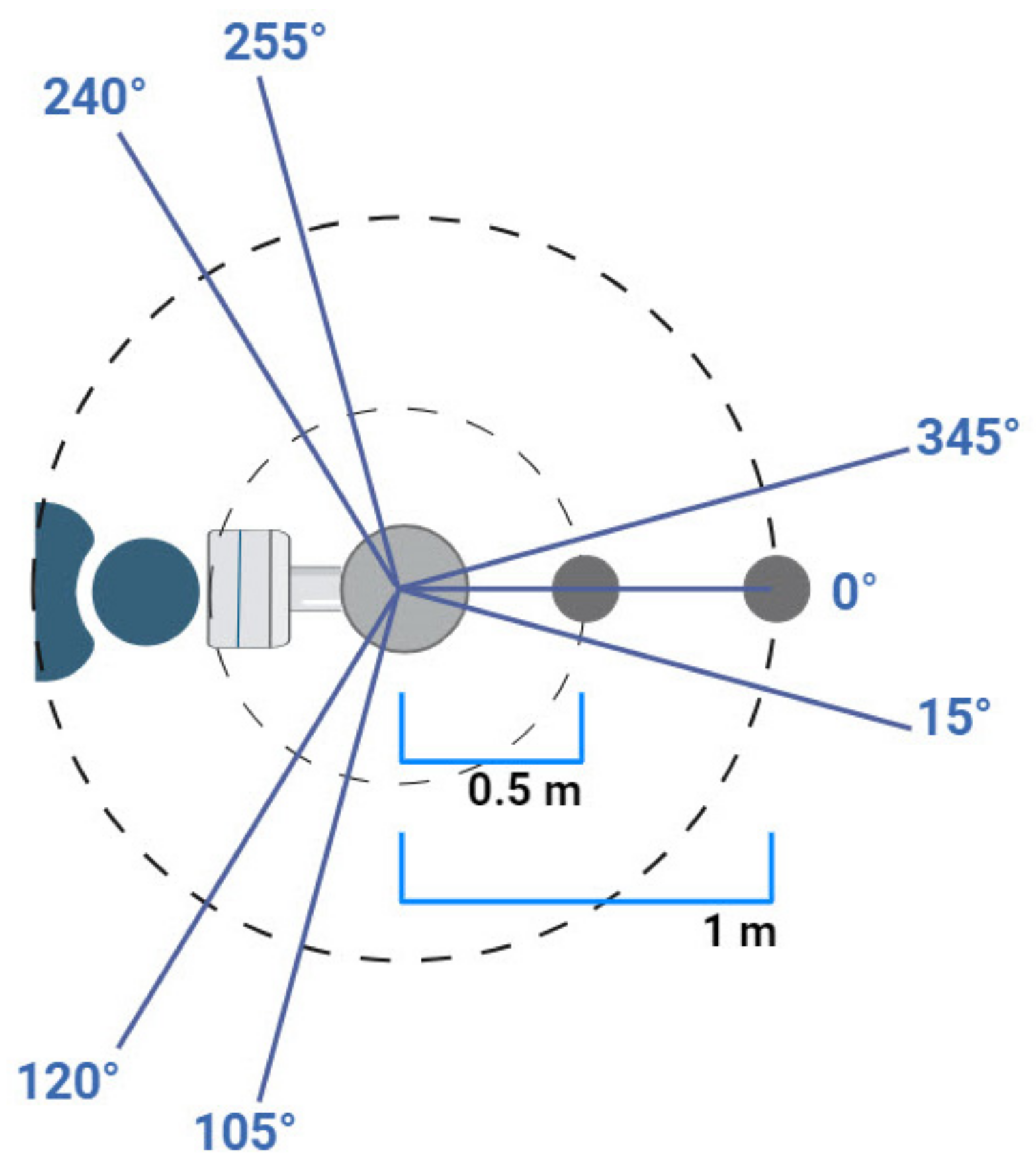
2. Materials and Methods

2.1. Devices and Equipment

The following equipment was used in the present study.
Three types of handheld dental radiographic units were investigated: (1) iRay D3 (Dexcowin Co., Ltd., Seoul, Republic of Korea); (2) EZRay Air (Vatech Co., Ltd., Gyeonggi-do, Hwaseong-si, Republic of Korea); and (3) Epix (EPPCO, Isfahan, Iran). The characteristics of the portable dental radiographic devices are mentioned in Table 1.
A CBCT image quality test phantom (DVTap, Quart, Zorneding, Germany) with a diameter of 16 cm and height of 15 cm, with standard test objects of PMMA, Air, and PVC, was used as the subject.
An ionization chamber (Piranha 657, RTI Electronics, Mölndal, Sweden) with a dose range of 1.3–650 Gy was used for dosimetry.

2.2. Measurement of the X-ray Output

Due to the potential errors in the measurement of scattered radiation and different dose measurements at the cone of the dental radiographic units, the output of each X-ray unit was first determined, so that further measurements could be standardized by dividing the measured scattered dose by the output of the radiographic unit. The measurement of the X-ray output was performed by placing the dosimeter directly at the opening of the cone of each radiographic dental unit and operating the device using the following parameters: 60 kVp, 2 mA (for iRay D3 and Epix) and 2.5 mA (for EZRay Air), and 1 s exposure time.

2.3. Measurement of the Scattered Dose

The opening of the cone of each portable dental radiographic device was placed in direct contact with the side of the CBCT image quality test phantom in order for the central X-ray emitted from the unit to pass through the center of the phantom. The exposure parameters were set at 60 kVp, 2 mA (for iRay D3 and Epix) and 2.5 mA (for EZRay Air), and 1 s exposure time.
On the horizontal and vertical planes, the direction of the central X-ray was set at 0°. The dose was measured at 0.5 and 1 m distances, every 15° clockwise in the horizontal plane. Based on a study by Iwawaki et al. [15], the following angles on the horizontal plane were considered: 0° (the most anterior) was set as exposure to the surrounding personnel; 15–105° and 255–345° were designated as sides, and 120–240° posterior to the unit was considered to cause exposure to the operator (Figure 1).
The measurements of the ionization chamber expressed in mGy were regarded as the absorbed dose. The measured exposure in each angle and distance was then divided by the output exposure, to standardize the measurements and obtain the air dose (µGy/Gy). Next, the angular values in each distance (0.5 or 1 m) were averaged to obtain the air dose for the anterior, sides, and posterior regions according to the values mentioned earlier, as suggested by Iwawaki et al.

2.4. Measurement Based on the Operator Dose Limits

The equivalent dose was calculated considering a radiation weighting factor (Wt) of 1 for X-rays and expressed as mSv. According to the NCRP, occupational exposure must not exceed 50 mSv annually. Therefore, a dose limit of 50 mSv/year must be considered for radiology personnel [13]. On the other hand, the ICRP set an occupational dose limit of 20 mSv per year [14]. One year comprises 52 weeks and one week comprises 5 working days. Thus, one year comprises 260 working days. To prevent exceeding the dose limits set by the NCRP and ICRP, exposure has to be 192.3 μSv or less and 76.9 μSv or less per day, respectively. The limit of the number of radiographs acquired per day using the portable units was calculated by dividing this daily dose (192.3 μSv for the NCRP dose limit and 76.9 μSv for the ICRP dose limit) by the operator dose (posterior region) for each portable radiographic unit.

3. Results

3.1. Output Measurements

X-ray output exposures detected by the ionization chamber were 1.808 mGy, 3.597 mGy, and 8.931 mGy for the iRay D3, EZRay Air, and Epix portable radiographic units, respectively.

3.2. Scattered Dose Measurements

The absorbed doses were generally higher in the Epix portable X-ray unit compared to the other two radiographic devices. In all of the portable radiographic units, anterior exposure was higher than side exposure or posterior exposure. Additionally, as predicted, the exposure decreased at 1 m distance compared to 0.5 m distance (Table 2). Table 3 demonstrates the air doses in different regions using different portable X-ray devices.

3.3. Dose Limit Considerations

According to NCRP guidelines, considering a distance of 0.5 m between the operator and the X-ray unit, a maximum of 961, 565, and 38 radiographs per day are permitted using iRay D3, EZRay Air, and Epix portable radiographic units, respectively (Table 4).
Considering ICRP guidelines, with a distance of 0.5 m between the operator and the radiographic device, a maximum of 384, 226, and 15 radiographs per day are permitted using iRay D3, EZRay Air, and Epix portable radiographic units, respectively (Table 5).

4. Discussion

Based on the findings of this study, the doses were generally higher in the Epix portable dental radiographic unit compared to the other two dental radiographic devices. In all of the portable radiographic units, anterior exposure was higher than side or posterior exposure. Considering a distance of 0.5 m between the operator and the X-ray unit, a maximum of 961, 565, and 38 radiographs per day are permitted using iRay D3, EZRay Air, and Epix portable dental devices, respectively, to reach the NCRP dose limit of 50 mSv. To reach the dose limit of 20 mSv set by ICRP, with a distance of 0.5 m between the operator and the radiographic device, a maximum of 384, 226, and 15 radiographs per day are permitted using iRay D3, EZRay Air, and Epix portable units, respectively. It must be pointed out that due to the settings and specifications of the three portable dental radiographic units, the EZRay Air radiographic device was operated at 2.5 mA, while the iRay D3 and EZRay Air radiographic devices used 2 mA tube currents.
Danforth et al. measured the dose received by the operator while using a Nomad portable device. According to their findings, whole-body annual dose was 0.047 mSv, 0.09% of the annual dose limit of 50 mSv [11]. In another study, Leadbeatter et al. used the Rextar X camera style handheld dental radiography unit. They reported that absorbed doses from a single adult maxillary molar radiography were 0.69, 0.78, and 0.47 µGy at the left hand, right hand, and eyes of the operator, respectively [12]. Makdissi et al. reported their findings on operator dose while using portable radiographic devices in three different positions. The received dose of the operator’s thyroid, eyes, and feet were higher in the bent arm position [8]. In another study, Ghafari et al. used a Geiger Muller counter and stated that the radiation dose in proximity to the Port-XII portable radiographic device was less than the annual dose limit [13]. In a study conducted by Otaka et al., it was observed that when acquiring more than 100 images per day using portable X-ray units, the use of 0.25 mm lead protective clothing or screens was necessary to avoid reaching the 20 mSv dose limit set by ICRP. These protective measures have been reported to reduce scattered rays from 60 kV X-rays by 99% [16]. In another study, Altındağ et al. compared radiation doses of Nomad Pro 2, Rextar X, and Diox 602 handheld dental radiographic units using calibrated thermoluminescent dosimeters. Their results showed that, for most organs, the Nomad dental X-ray unit led to higher equivalent doses, while for the right hand and left hand, the Rextar X and Diox portable radiographic units had the highest equivalent dose, respectively. Peak measurements without protection were as follows: gonad, 24.4 μGy; thyroid, 30.5 μGy; right eye, 31.9 μGy; left eye, 27.9 μGy; right hand, 111.6 μGy; left hand, 71.7 μGy [17]. While setting up a controlled area is not imperative for imaging a small number of subjects during visiting care, protective actions—such as wearing appropriate clothing and maintaining a 2 m distance—can mitigate the risk of exposure exceeding the dose limit. However, in scenarios involving the imaging of mass disasters which may entail multiple radiographic exposures by a single operator, both radiation protection and a controlled area become crucial safety considerations. Additionally, it is worth mentioning that patients often require several radiographs, and the number of permitted daily radiographs must not be mistaken for the number of patients. Therefore, the Epix portable dental radiographic system is not suitable for obtaining radiographs on a daily basis, particularly with shorter distances between the operator and the portable dental radiographic unit.
Portable dental radiographic units are an indispensable tool in dentistry and forensics, allowing for access to radiographic evaluation of the dentition and surrounding structures in underprivileged zones, nursing homes, hospitals, and disaster scenes. Studies have shown that radiographic images obtained by these units have comparable quality with that of wall-mounted ones. Kim et al. have shown that the percentage average error value of the tube voltage is lower than the standard. Therefore, they concluded that images of appropriate quality can be taken with a portable unit, offering acceptable diagnostic value [18]. In another study, Hoogeveen et al. examined the subjective quality of bitewing radiographs obtained by handheld and stationary dental radiographic units. They reported that subjectively assessed diagnostic image quality in bitewing radiographs acquired by handheld and stationary X-ray devices did not differ significantly [19]. Another study by Amani et al. revealed that while no significant difference existed between the wall-mounted and portable dental radiographic devices in sharpness, contrast, and error in vertical angulation, there is a significant difference between the two groups in cone-cutting and error in horizontal angulation of intraoral radiographs. The images taken with portable devices showed better image quality, as judged by five observers, compared to the stationary device [20]. Concerns over the safety of portable dental radiographic units, as well as the difficulty to ensure the correct geometry of the source, intraoral detector, and patient can be considered as the main pitfalls of these handheld dental X-ray units. Therefore, a general suggestion is that handheld intraoral dental radiographic units should supplement but not replace conventional radiographic equipment for daily practice [9].
As mentioned, there are currently two sets of occupational dose limits. The NCRP enforces a dose limit of 50 mSv per year, while the ICRP recommends a more conservative annual dose limit of 20 mSv averaged over 5 consecutive years. The ICRP was established in 1928 following the initiative of the first International Congress of Radiology held in 1925. This organization has made general recommendations on limits of exposure for radiation workers and public. The NCRP was established in the United States in 1929 as the national counterpart to the ICRP. Initially conceived as an advisory committee for the United States members of these international bodies, the NCRP quickly gained independence and began making recommendations for use within the United States, starting in 1931. Over time, it evolved into a formal Council with a charter from the United States Congress in 1964 [21]. The NCRP also offers dose limit recommendations which closely resemble those set by the ICRP. However, there are occasional variations. The key differences between recent limits lie in the greater flexibility that the NCRP allows when formulating recommendations. The dose limits recommended by both organizations have generally decreased in each recommendation, since their establishment [21,22]. The shift in radiation protection guidelines has been driven by the recognition that deterministic effects—those directly causing tissue damage and cell death—are not the primary limiting factor. Exposure can be effectively controlled using recent advances in radiologic equipment to stay below the thresholds for these effects. Instead, the critical concerns lie in stochastic effects, such as cancer and genetic impacts. These effects, occurring later, have required considerable time to fully understand, quantify, and contextualize. Even now, there is great uncertainty about the biological effects of low-dose radiation (less than 100 mGy). Currently, the radiological protection systems and recommendations operate under the assumption that the risk of radiation-related stochastic effects is directly proportional to the dose received. Importantly, this model does not incorporate a dose threshold below which there is no risk. Thus, it is commonly known as the linear no-threshold model. The linear no-threshold model has been a topic of extensive scientific debate and controversy. Some researchers propose that it may overestimate the actual risk and suggest the existence of a dose threshold below which no risk exists or even the possibility of health benefits of lower doses of radiation (hormesis effects). Other critics argue that the linear no-threshold model underestimates the true risk at low doses of ionizing radiation. They propose that the relationship between the received dose and undesirable effects is supra-linear, meaning the risk increases more significantly than predicted by the linear no-threshold model [23]. In the absence of definitive supportive evidence, the regulatory organizations, such as the ICRP and the NCRP, still use the linear no-threshold model as the basis for recommending public and occupational dose limits.
Mobile and portable X-ray devices, as well as X-ray devices used during surgery, must be operated at a distance of 2 m or more from the X-ray tube [8,15]. It has to be considered that in several settings where the use of portable dental radiographic devices is indicated, setting up the equipment in a radiation-controlled area becomes challenging, and operators and surrounding workers may unavoidably acquire images within the 2 m range from the X-ray tube. Therefore, it is important to study the exposure doses in distances less than 2 m from the radiation source. In the present study, the 0.5 and 1 m distances were applied for measurements of scattered dose of different portable dental radiographic units.
NCRP and ICRP guidelines provide whole-body occupational dose limits of 50 mSv and 20 mSv (averaged over 5 consecutive years), respectively [21]. The presumption for this study was that one operator is in charge of all of these exposures. In real situations, more than one individual is usually in charge of exposures, thus the number of permitted daily radiographs is even higher than the calculations in this study. On the other hand, some patients may require several radiographs and the number of permitted daily radiographs must not be mistaken for the number of patients. However, the NCRP reports that the annual dose for dental radiology operators does not seem to exceed 1 mSv in each year.
Protective measures devised in portable dental radiographic devices include a plastic shield providing a safe zone for the operator behind the device when the device is held horizontally. Also, internal shielding material reduces the leakage of radiation from the radiographic system [24]. Cho et al. presented approaches for radiation protection while using portable devices such as protective tube shields, protective gloves, and longer collimators [24,25]. The present study has some limitations. In vivo experiment was not possible due to ethical concerns. However, efforts were made in order to replicate the clinical situation for radiographic exposures. Additionally, although all the portable dental units were operated at the same voltages and exposure times, EZRay Air used 2.5 mA (the lowest mA setting in the device) while iRay D3 and Epix used 2 mA tube currents. Further studies can use different portable devices and measure specific effective doses of different organs, thus providing more detailed knowledge considering the organ sensitivity factors. To the authors’ knowledge, no previous study has been performed comparing the portable dental radiographic units employed in this study. Additionally, determination of the maximum permitted number of radiographs using these three devices provides useful and practical information for radiation technicians in healthcare.

5. Conclusions

It is highly unlikely that the operator would reach occupational dose limits set by organizations when using iRay D3 and EZRay Air portable dental radiographic units. The Epix radiographic device allows for fewer daily radiographs, and should be avoided for daily use. Operator dose limits have to be adhered to for obtaining radiographs using portable dental radiographic devices.

Author Contributions

Conceptualization, M.A. (Mehrdad Abdinian) and M.A. (Maedeh Aminian); methodology, F.K. and P.S.; validation, G.S.; formal analysis, M.C. and N.G.A.; resources, M.A. (Mehrdad Abdinian); data curation, F.K. and P.S.; writing—original draft preparation, M.A. (Mehrdad Abdinian), M.A. (Maedeh Aminian) and F.K.; writing—review and editing, P.S., M.C., N.G.A. and G.S.; supervision, M.A. (Mehrdad Abdinian); funding acquisition, M.A. (Mehrdad Abdinian). All authors have read and agreed to the published version of the manuscript.

Funding

This research was funded by Isfahan University of Medical Sciences, grant number 397197.

Institutional Review Board Statement

This study was approved by the Research Ethics Committee at Isfahan University of Medical Sciences (#IR.MUI.REC.1397.3.197).

Informed Consent Statement

Not applicable.

Data Availability Statement

The raw data supporting the conclusions of this article will be made available by the authors on request.

Conflicts of Interest

The authors declare no conflicts of interest.

References

  1. White, S.C.; Pharoah, M.J. White and Pharoah’s Oral Radiology E-Book: Principles and Interpretation; Elsevier Health Sciences: Amsterdam, The Netherlands, 2018. [Google Scholar]
  2. Charlton, D.G. Portable dental equipment: Dental units and X-ray equipment. Gen. Dent. 2009, 57, 336–341. [Google Scholar]
  3. Coy, J. Hand-held dental X-ray (HDX) with medical collimator: Use in casualty radiology. Mil. Med. 1996, 161, 428–431. [Google Scholar] [CrossRef]
  4. Coy, J.; Vandre, R.H.; Davidson, W.R. Use of the hand-held dental X-ray machine during joint operation, NATO exercise Display Determination-92. Mil. Med. 1997, 162, 575–577. [Google Scholar] [CrossRef]
  5. Van Dis, M.L.; Miles, D.A.; Parks, E.T.; Razmus, T.F. Information yield from a hand-held dental X-ray unit. Oral. Surg. Oral. Med. Oral. Pathol. 1993, 76, 381–385. [Google Scholar] [CrossRef]
  6. Varghese, S.; Kimmel, A.; Radmer, T.; Bradley, T.G.; Bahcall, J. In vitro evaluation of the XR-15 portable X-ray unit for forensic odontology. J. Forensic Odontostomatol. 2004, 22, 5–8. [Google Scholar]
  7. McGiff, T.J.; Danforth, R.A.; Herschaft, E.E. Maintaining radiation exposures as low as reasonably achievable (ALARA) for dental personnel operating portable hand-held X-ray equipment. Health Phys. 2012, 103, S179–S185. [Google Scholar] [CrossRef] [PubMed]
  8. Makdissi, J.; Pawar, R.R.; Johnson, B.; Chong, B.S. The effects of device position on the operator’s radiation dose when using a handheld portable X-ray device. Dentomaxillofac. Radiol. 2016, 45, 20150245. [Google Scholar] [CrossRef] [PubMed]
  9. Geist, J.R. Handheld intraoral dental X-ray devices should supplement but not replace conventional radiographic equipment. Oral Surg. Oral Med. Oral Pathol. Oral Radiol. 2021, 132, 257–259. [Google Scholar] [CrossRef] [PubMed]
  10. Hosseini Pooya, S.; Hafezi, L.; Manafi, F.; Talaeipour, A. Assessment of the radiological safety of a Genoray portable dental X-ray unit. Dentomaxillofac. Radiol. 2015, 44, 20140255. [Google Scholar] [CrossRef] [PubMed]
  11. Danforth, R.A.; Herschaft, E.E.; Leonowich, J.A. Operator Exposure to Scatter Radiation from a Portable Hand-held Dental Radiation Emitting Device (Aribex™ NOMAD™) While Making 915 Intraoral Dental Radiographs. J. Forensic Sci. 2009, 54, 415–421. [Google Scholar] [CrossRef] [PubMed]
  12. Leadbeatter, J.; Diffey, J. Evaluation of radiation exposure to operators of portable hand-held dental X-ray units. Phys. Eng. Sci. Med. 2021, 44, 377–385. [Google Scholar] [CrossRef] [PubMed]
  13. Ghafari, R.; Khabiri, M.; Rafieian, M.; Khorrami, E.; Nikzad, S. Determination of scattered radiation around PORT-XII portable dental radiography unit. J. Isfahan Dent. Sch. 2013, 9, 329–336. [Google Scholar]
  14. ICRP. The 2007 Recommendations of the International Commission on Radiological Protection; ICRP: Ottawa, ON, Canada, 2007; pp. 2–4. [Google Scholar]
  15. Iwawaki, A.; Otaka, Y.; Asami, R.; Ozawa, T.; Izawa, M.; Saka, H. The study of protection of operators and surrounding workers at the time of using portable intraoral X-ray unit. Leg. Med. 2018, 33, 66–71. [Google Scholar] [CrossRef] [PubMed]
  16. Otaka, Y.; Harata, Y.; Izawa, M.; Iwawaki, A.; Asami, R.; Saka, H.; Okumura, Y. On the safe use of portable intraoral X-ray units in large-scale disasters. Jpn. J. Oral. Diag./Oral Med. 2017, 30, 311–326. [Google Scholar] [CrossRef]
  17. Altındağ, A.; Eren, H.; Orhan, K.; Görgün, S. Evaluation of Operator and Patient Doses after Irradiation with Handheld X-ray Devices. Appl. Sci. 2023, 13, 10414. [Google Scholar] [CrossRef]
  18. Kim, E.; Park, H.; Choi, H.; Kim, J. An assessment of the usefulness of handheld X-ray devices in general radiography based on a performance evaluation experiment. Int. J. Radiat. Res. 2023, 21, 545–551. [Google Scholar] [CrossRef]
  19. Hoogeveen, R.C.; Ouchene, S.; Berkhout, W. Diagnostic image quality of hand-held and wall-mounted X-ray devices in bitewing radiography: A non-inferiority clinical trial. Dentomaxillofac. Radiol. 2021, 50, 20200471. [Google Scholar] [CrossRef] [PubMed]
  20. Amani, T.; Surenthar, M.; Umamaheswari, T.; Prethipa, R.; Kumar, L. Image Quality Assessment of Digital Radiographs Captured by Hand-Held Devices versus Wall-Mounted Devices: A Retrospective Comparative Study. Cureus 2024, 16, e52900. [Google Scholar] [CrossRef] [PubMed]
  21. Sinclair, W.K. Radiation protection recommendations on dose limits: The role on the NCRP and the ICRP and future developments. Int. J. Radiat. Oncol. Biol. Phys. 1995, 31, 387–392. [Google Scholar] [CrossRef]
  22. Wei, Y.; Dewji, S. A comprehensive review of dose limits, triage systems and measurement tools for consequence management of nuclear and radiological emergencies. Radiat. Phys. Chem. 2024, 217, 111533. [Google Scholar] [CrossRef]
  23. Laurier, D.; Billarand, Y.; Klokov, D.; Leuraud, K. The scientific basis for the use of the linear no-threshold (LNT) model at low doses and dose rates in radiological protection. J. Radiol. Prot. 2023, 43, 024003. [Google Scholar] [CrossRef] [PubMed]
  24. Turner, D.C.; Kloos, D.K.; Morton, R. Radiation safety characteristics of the NOMAD™ portable X-ray system. Qual. Regul. Serv. 2005, 95648, 57. [Google Scholar]
  25. Iwawaki, A.; Otaka, Y.; Asami, R.; Ishii, T.; Kito, S.; Tamatsu, Y.; Aboshi, H.; Saka, H. Comparison of air dose and operator exposure from portable X-ray units. Leg. Med. 2020, 47, 101787. [Google Scholar] [CrossRef] [PubMed]
Figure 1. Schematic demonstration of the experimental settings (scales of objects are not realistic).
Applsci 14 03515 g001
Table 1. Characteristics of the portable dental radiographic devices.
ModelTube VoltageTube CurrentExposure TimeFocal SpotTotal Filtration
iRay D360 kV (fixed)2 mA (fixed)0.05–1.35 s0.8 mm2.3 mmAl
EZRay Air60–65 kV2.5–3 mA0.05–1 s0.4 mm1.5 mmAl (min)
Epix60 kV (fixed)2 mA (fixed)1 s0.8 mm1.6 mmAl
Table 2. Absorbed doses of portable dental radiographic devices (µGy).
Radiographic Unit Region 0.5 m 1 m
iRay D3Anterior 1.430.09
Sides0.160.00
Posterior0.200.00
EZRay AirAnterior 0.820.51
Sides0.480.15
Posterior0.340.24
EpixAnterior 5.510.80
Sides4.221.30
Posterior5.041.01
Table 3. Air doses of portable dental radiographic devices (µGy/Gy).
Radiographic UnitRegion 0.5 m 1 m
iRay D3Anterior 789.2747.22
Sides89.510.00
Posterior93.430.00
EZRay AirAnterior 228.77140.64
Sides134.1042.35
Posterior95.3367.33
EpixAnterior 616.8489.34
Sides472.22145.31
Posterior564.66112.85
Table 4. Maximum number of radiographs per day using portable dental radiographic devices considering the National Council on Radiation Protection and Measurements (NCRP) dose limits (50 mSv).
Radiographic Unit0.5 m1 m
iRay D3961-
EZRay Air565801
Epix38190
Table 5. Maximum number of radiographs per day using portable dental radiographic devices considering the International Commission on Radiation Protection (ICRP) dose limits (20 mSv).
Radiographic Unit0.5 m1 m
iRay D3384-
EZRay Air226320
Epix1576
Disclaimer/Publisher’s Note: The statements, opinions and data contained in all publications are solely those of the individual author(s) and contributor(s) and not of MDPI and/or the editor(s). MDPI and/or the editor(s) disclaim responsibility for any injury to people or property resulting from any ideas, methods, instructions or products referred to in the content.

