Article

Examination of the Bactericidal and Fungicidal Activity of Bacillus amyloliquefaciens M Isolated from Spring Waters in Bulgaria

1
Department of Microbiology, University of Food Technologies, 26 Maritza Boulevard, 4002 Plovdiv, Bulgaria
2
Department of Biochemistry and Molecular Biology, University of Food Technologies, 26 Maritza Boulevard, 4002 Plovdiv, Bulgaria
3
Department of Wine and Beer Technology, University of Food Technologies, 26 Maritza Boulevard, 4002 Plovdiv, Bulgaria
*
Author to whom correspondence should be addressed.
Appl. Sci. 2024, 14(9), 3612; https://doi.org/10.3390/app14093612
Submission received: 22 March 2024 / Revised: 16 April 2024 / Accepted: 17 April 2024 / Published: 24 April 2024
(This article belongs to the Special Issue Natural Products and Bioactive Compounds)

Abstract

:
In order for a strain to be considered a probiotic or suitable plant bioprotective agent, it must have proven antimicrobial activity against pathogenic bacteria and phytopathogenic fungi. Bacillus amyloliquefaciens M exhibited significantly high antifungal activity against pathogenic fungi of the genera Aspergillus, Penicillium, Fusarium, and Sclerotinia; yeasts of the genera Candida and Saccharomyces; as well as high antibacterial activity against pathogens of the genera Escherichia, Salmonella, Staphylococcus, Listeria, Pseudomonas, and Bacillus. The manifested antimicrobial activity was influenced by the composition of the growth medium. The antifungal activity of the strain was investigated at growth temperatures of 30 °C and 37 °C, and at different pH values in aerobic and anaerobic cultivation, under static and dynamic culturing conditions. High antifungal activity was observed at the 24th h on both growth media (LBG broth and MPB broth) at pH = 6 and pH = 7 in aerobic and anaerobic cultivation. Bacillus amyloliquefaciens M produced antibiotic substances at pH > 5.0, and the antibiotic substances were either secreted into the medium or associated with the cell surface. Four compounds with different antifungal activity and different Rf values were registered through thin-layer chromatography (Rf1 = 0.47; Rf2 = 0.55; Rf3 = 0.67; and Rf4 = 0.75), two of the compounds were ninhydrin-positive. Bacillus amyloliquefaciens M was cultured in a bioreactor with stirring, and the parameters of the growth kinetics and the sporulation kinetics have been modeled. A spore concentrate of Bacillus amyloliquefaciens M has been obtained. In further research, the efficiency of the concentrate as a plant bioactive agent will be tested.

1. Introduction

Antibiotics are administered to control the growth and spread of pathogenic microorganisms in the gastrointestinal tract, as well as for good digestion and absorption and metabolism of nutrients [1,2]. This is of particular importance for animal husbandry, since antibiotics applied to accelerate growth have a positive effect. Regardless of the positive effect of the applied antibiotics, their widespread use has led to new problems for humans, plants, and animals because of the occurrence of cross- and multi-antibiotic resistance and dysbacteriosis [3,4]. This necessitated the use of other natural products as an alternative to antibiotics. Probiotics, plant extracts, essential oils, antimicrobial peptides, acidifiers, and enzymes fall within this definition [5]. These agents can be applied either alone or, more often, in combinations [6]. According to FAO/WHO 2002 [7], probiotics are defined as follows: “Non-pathogenic bacteria for humans, possessing antagonistic activity towards pathogenic and opportunistic bacteria and ensuring the restoration of normal microflora”. Taken in adequate amounts (viable cell concentration of 106–109 cfu/g), they have a beneficial effect on the functioning of the digestive system and related organs and systems [8,9,10]. Gram-positive lactic acid bacteria of the genus Lactobacillus, the genus Bifidobacterium, yeast of the species Saccharomyces boulardii, fungi of the genus Aspergillus, spore-forming bacteria of the genus Bacillus are most frequently included in the composition of probiotic preparations. Bacillus species and strains are of significant interest for the development of probiotic preparations. Bacillus species are characterized by rapid growth, they exhibit competitive exclusion of pathogenic microorganisms, and their spores are resistant to the conditions of the gastrointestinal tract (low pH values and the presence of pepsin, pH = 8, and the presence of pancreatin, high concentrations of bile salts), protecting the host’s immune system [11,12]. Bacillus sp. produce substances with antimicrobial activity of different chemical nature. Antimicrobial substances include peptides, amino sugars, phospholipids, etc. According to Montesinos, 2007 and Bravo et al., 2017 [13,14], antimicrobial compounds produced by Bacillus strains exhibit antifungal activity against phytopathogenic fungi. Bacillus sp. also produce a wide range of peptides with antimicrobial activity against pathogenic bacteria and fungi [15]. In recent years, the issue of aflatoxins in food and protecting the population from their carcinogenic effect has been raised. The development of preparations with high antifungal activity against Aspergillus flavus would allow both soil and plant individuals to be treated. This in turn would enable the production of safe foods. Sclerotinia sp. (Sclerotinia sclerotiorum) affects the root system of plants and manifests itself as a wet light brown rotting spot at the base of the stem, which is later covered with a white cotton-like coating. Regardless of the infection phase, plant species die [16,17]. Bacillus sp. peptides are classified as ribosomally synthesized bacteriocins and polyketides and lipopeptides produced by non-ribosomal enzymes. Bacteriocins are able to act on the cytoplasmic membrane, increasing its semi-permeability and causing depolarization of the cell membrane of pathogenic bacteria. Some bacteriocins suppress microbial cell viability and cell division processes [18]. Polyketides are secondary metabolites composed of fatty acids with high antifungal activity. They act on pathogenic microorganisms by repressing genes related to cell wall synthesis and DNA replication [19], and some block the synthesis of a number of proteins [20]. The best-studied bacteriocins are lipopeptides, which are synthesized using peptide synthetase enzymes [21], and consist of a hydrophilic peptide part (consisting of 7 to 10 amino acids) connected to a hydrophobic fatty tail [22]. Depending on their structure, they form three groups: biosurfactants, iturins, and fengycins. Biosurfactants act on the lipid bilayer of various biological membranes [22]. Iturins possess high polymorphism with diverse biological, physiological, and biochemical properties [22]. Fengicides exhibit specific antifungal activity [22]. Bacillus amyloliquefaciens is a species of the genus Bacillus that secretes extracellular enzymes, such as β-amylases, cellulase, metalloproteases, and proteases, through which it improves digestion, nutrient absorption, and intestinal resistance to infection [23]. Bacillus amyloliquefaciens LFB112 secreted bacteriocins with good thermostability and acid/alkaline tolerance, active against a wide range of pathogenic bacteria—Gram-positive and Gram-negative bacteria, causing various host diseases.
Microelements, which play an important role in the enzymatic reactions occurring in the cell and in the sporulation process, are necessary for the normal growth and development of bacilli. The bacilli cell walls are cation-selective. Bacilli cells take up Cu2+ during the period of exponential growth and sporulation. The intensity of the sporulation process depends on the presence of mineral salts containing ions of Ca2+, Mg2+, K+, Mn2+, Zn2+, Cu2+, etc. in the medium. In the absence of some of them, the process of sporulation does not take place [24].
The role of Mn2+ in the sporulation process in Bacillus strains was established. Mn2+ ions are required by the cell as a cofactor for phosphate glycerosynthetase, which is a strictly Mn2+-dependent enzyme required for sporulation in normal cultural media. Metal ions are necessary for the normal functioning of a number of enzymes, which act as stabilizers or cofactors of enzymes and also as regulators of the sporulation process. Therefore, salts of Cu2+, Mn2+, and Mg2+ have to be added to the regular composition of MPB [24].
Species of the genus Bacillus with proven antifungal and antibacterial properties are part of biopreparations for agriculture (such as plant protection agents, for example, phytosporin, and probiotics for animal husbandry, for example, biosporin). The activity of these forms depends on the concentration of the resulting spores. In order for a preparation to be used in one direction or another, it is necessary to contain a high concentration of spores of the relevant strain (not less than 108–109 cfu/g). A mandatory condition for achieving a high concentration of spores is the research and optimization of the cultivation process [25,26].
The aim of the present study was to determine the antibacterial and antifungal activity of Bacillus amyloliquefaciens M and to examine the production of antibiotic substances.

2. Materials and Methods

2.1. Microorganisms

The studies in the present research were conducted with Bacillus amyloliquefaciens strain M that had been isolated from spring waters in the village of Yabalkovo, Haskovo region, Bulgaria, and had been identified to species level by sequencing the 16S rRNA gene (unpublished data).
The following test-pathogenic microorganisms were used in the examination of the antibacterial activity of Bacillus amyloliquefaciens M: Escherichia coli ATCC 25922, Salmonella enterica ssp. enterica serovar Enteritidis ATCC 25928, Salmonella abony NTCC 6017, Staphylococcus aureus ATCC 6538P, Listeria monocytogenes ATCC 19111, Pseudomonas aeruginosa NBIMCC 1390, and Bacillus cereus ATCC 14579.
The following test-saprophytic microorganisms were used in the examination of the antifungal activity of Bacillus amyloliquefaciens M: yeast Candida utilis ATCC 42402, Saccharomyces cerevisiae ATCC 9763 and phytopathogenic fungi Aspergillus niger ATCC 1015, Penicillium chrysogenum ATCC 28089, Aspergillus flavus ATCC 9643, Fusarium moniliforme ATCC 38932, Aspergillus awamori ATCC 22342, and Sclerotinia sclerotiorum var scleroria ATCC 18687.

2.2. Media

A. MPB. Composition (g/dm3): peptone—10; NaCl—5; meat extract—3. pH = 7.2. Sterilization—121 °C for 30 min.
B. LBG-agar. Composition (g/dm3): triptone—10; yeast extract—5; NaCl—10; glucose—10; agar—20. pH = 7.5. Sterilization—121 °C for 20 min.
C. Fermentation medium with molasses. Composition (g/dm3): molasses—20; peptone—10; corn extract—3; CaCl2—0.22; MgSO4—0.11; K2HPO4—0.24. pH = 7.2. Sterilization through tindalization.
D. Fermentation medium with malt. Composition (g/dm3): malt—20; corn extract—3; molasses—20; corn extract—3; CaCl2—0.22; MgSO4—0.11; K2HPO4—0.24. pH = 7.2. Sterilization through tindalization.
E. Fermentation medium MPB with salts. Composition (g/dm3): peptone—10; NaCl—10; meat extract—5; CuCl2—0.001; MgSO4—0.5. pH = 7.2. Sterilization through tindalization.

2.3. Cultivation of Bacillus amyloliquefaciens M

The cultivation of the studied strain was carried out in 500 cm3 Erlenmeyer flasks with 10 cm3 of the respective broth cultural medium at constant aeration on a rotary shaker (220 min−1) and statically (in a thermostat) at a temperature of 37 °C for 24–48 h. Three different broth cultural media were used: fermentation medium with molasses, fermentation medium with malt, and fermentation medium MPB with salts. The inoculum amount was 1% (v/v) of vegetative inoculum (obtained after 18 h of incubation of the studied strain at 37 °C in MBP medium). Batch cultivation with aeration was carried out in a laboratory bioreactor “Sartorius” Goettingen, Germany with a geometric volume of 2 dm3 and a working volume of 1.5 dm3. The cultural medium was MPB with the addition of salts. The bioreactor was equipped with a control unit that included electrodes for stirring rate, temperature, pH, dissolved O2, etc. The degree of aeration was α = 1 dm3/(dm3·h). The initial pH value was pH = 7.3; during the fermentation process, the pH was self-maintained at a value of pH = 8.3. The stirring rate was 600 rpm. The bioreactor medium was inoculated with a culture obtained after overnight cultivation of Bacillus amyloliquefaciens M in a shaker flask with a rotation frequency of 23 rad/s.

2.4. Examination of the Antimicrobial Activity of Bacillus amyloliquefaciens M

The antimicrobial activity of Bacillus amyloliquefaciens M against saprophytic and pathogenic microorganisms was investigated in order to select the best cultural medium for cultivation and spore formation. The following samples were prepared: cultural suspension (CS), biomass in physiological solution (B), and acellular supernatant (ASN) obtained from a 24 h culture of Bacillus amyloliquefaciens M.
Fungal spore suspensions were prepared for the examination of the antifungal activity. The test fungal strains had been grown in an incubator at 30 °C on LBG-agar for 3 to 7 days. The prepared spore suspensions of each of the test fungal microorganisms (concentration of 106–107 cfu/cm3) were used to inoculate molten LBG-agar medium, which was then poured in sterile Petri dishes, and after the solidification of the medium, wells (6 mm in diameter) were prepared; 0.06 cm3 of CS, B, or ASN were pipetted into the wells, and the Petri dishes with the test microorganisms were incubated at 30 °C (for Aspergillus niger ATCC 1015, Penicillium chrysogenum ATCC 28089, Aspergillus flavus ATCC 9643, Fusarium moniliforme ATCC 38932, Aspergillus awamori ATCC 22342, and Sclerotinia sclerotiorum var scleroria ATCC 18687) for 24 h to 48 h. Bacillus subtilis ATCC 6633 was used as a positive control, and distilled water was used as a negative control. The antifungal activity was determined by measuring the diameter of the inhibition zones in mm.
Vegetative cell suspensions were prepared for the examination of the antimicrobial activity activity of Bacillus amyloliquefaciens M against pathogenic bacteria and yeasts (Escherichia coli ATCC 25922, Salmonella enterica ssp. enterica serovar Enteritidis ATCC 25928, Salmonella abony NTCC 6017, Staphylococcus aureus ATCC 6538P, Listeria monocytogenes ATCC 19111, Pseudomonas aeruginosa NBIMCC 1390, Bacillus cereus ATCC 14579, Candida utilis ATCC 42402, and Saccharomyces cerevisiae ATCC 9763). The test microorganisms had been grown in an incubator at 37 °C (for Escherichia coli ATCC 25922, Salmonella enterica ssp. Enterica serovar Enteritidis ATCC 25928, Salmonella abony NTCC 6017, Staphylococcus aureus ATCC 6538P, Listeria monocytogenes ATCC 19111, and Pseudomonas aeruginosa NBIMCC 1390) or 30 °C (Bacillus cereus ATCC 14579, Candida utilis ATCC 42402, and Saccharomyces cerevisiae ATCC 9763) on LBG-agar for 24–48 h. The prepared vegetative cell suspensions of each of the test microorganisms (concentration of 106–107 cfu/cm3) were spread plated on LBG-agar medium; the inoculated Petri dishes were kept at refrigeration conditions for 2 h, and then, wells (6 mm in diameter) were prepared; 0.06 cm3 of CS, B, or ASN were pipetted into the wells, and the Petri dishes with the test microorganisms were incubated at 30 °C or 37 °C for 24 h to 48 h. Bacillus subtilis ATCC 6633 was used as a positive control, and distilled water was used as a negative control. The antimicrobial activity was determined by measuring the diameters of the inhibition zones in mm.

2.5. Examination of the Antiphytopathogenic Activity of Bacillus amyloliquefaciens M at Different Temperatures and pH

The antimicrobial activity of Bacillus amyloliquefaciens M against phytopathogenic fungi was investigated in order to further select the best cultural medium composition and optimal pH as well as the optimal temperature (30 °C or 37 °C) and oxygen conditions (aerobic or anaerobic) for cultivation and spore formation of Bacillus amyloliquefaciens M.
Since the most appropriate temperature range for the growth of Bacillus amyloliquefaciens M is 30 °C–37 °C and the fact that strain is a facultatively aerobic culture, its antimicrobial effect was determined at both temperatures, after static (anaerobic) and dynamic (aerobic) cultivation in two cultural solid media—LBG-agar and MPB-agar—with different (varying) pH. The different pH values of the media varied from pH = 4.5 to pH = 8.0. The inhibitory activity was determined by the agar-diffusion method with wells. The following samples were prepared for each cultivation of Bacillus amyloliquefaciens M on fermentation medium MPB with salts at the respective temperature and under the respective oxygen conditions: cultural suspension (CS), biomass in physiological solution (B), and acellular supernatant (ASN) obtained from a 24 h culture of Bacillus amyloliquefaciens M grown on MPB medium. Fungal spore suspensions were prepared for the examination of the antifungal activity. Aspergillus niger ATCC 1015, Aspergillus flavus ATCC 9643, and Aspergillus awamori ATCC 22342 were selected as test microorganisms because they are soil phytopathogens causing plant diseases and are one of the most dangerous phytopathogens due to their ability to produce phytotoxins (mycotoxins). The test fungal strains Aspergillus niger ATCC 1015, Aspergillus flavus ATCC 9643, and Aspergillus awamori ATCC 22342 had been grown in an incubator at 30 °C on LBG-agar for 3 to 7 days. The prepared spore suspensions of each of the test fungal microorganisms (concentration of 106–107 cfu/cm3) were used to inoculate molten LBG-agar medium, which was then poured into sterile Petri dishes, and after the solidification of the medium, wells (6 mm in diameter) were prepared; 0.06 cm3 of CS, B, or ASN (obtained from the respective cultivation of the studied strain) were pipetted into the wells, and the Petri dishes with the test microorganisms were incubated at 30 °C. The reporting of the results was performed at the 24th and 48th hour. The antiphytopathogenic activity was determined by measuring the diameter of the inhibition zones in mm.

2.6. Thin Layer Chromatography (TLC)

TLC for the separation of the antibiotic substances in the cultural medium, acellular supernatant, and biomass of Bacillus amyloliquefaciens M was carried out according to the method of Leifert et al. (1995) [27]. The following system was used—butanol/acetic acid/water (3:1:1 v/v/v). It was placed in a chromatographic chamber in a layer with a height of 0.5 cm. Chromatographic plates with silica gel COF254 20 × 20 cm (Merck, Darmstadt, Germany) were used. About 50 µL of the prepared culture medium (CS), acellular supernatant (ASN), and biomass (B) were pipetted with a micropipette at the start of the chromatographic plate. Chromatograms were performed in duplicate. After the TLC, one copy of the chromatogram was dried at 60 °C for about 15 min and sprayed with ninhydrin solution for proof of peptide antibiotics according to the method of Arx et al. (1976) [28]. The pink spots that appeared in the chromatograms after spraying were permanent for 5 h in the light of the day and for 24 h in the dark. The Rf values of the active substances were determined.

2.7. Biochromatography

Biochromatography with the other copy of the chromatograms was performed according to the method of Leifert et al., 1995 [27]. The chromatogram was dried for 5 h at 70 °C, after which it was placed on a solid culture medium inoculated with fungal test microorganisms. The Petri dishes were left under refrigeration conditions for 30 min for the antibiotic substances to diffuse in the solid medium. The chromatographic plates were then removed, and the biochromatography was performed in a thermostat at 28 °C. The active spots were identified, and their Rf values were determined.

2.8. SDS-PAGE

Gel electrophoresis of the obtained peaks from the gel chromatography under denaturing conditions (SDS-PAGE) with an omniPAGE mini Cleaver electrophoresis (Model CVS10DSYS, Cleaver Scientific Ltd., Rugby, UK), according to the Laemmli method [29], at a separating gel concentration of 15% (pH 8.8) was performed. The fractions showing the highest protein content of the 4 peaks were mixed and dried. In order to visualize the proteins after staining on SDS-PAGE, it is necessary for each band to contain 1 µg of protein. Solutions with equal protein content were prepared after determining the protein content of the dried mixed fractions. To 100 µL of each of the four samples, 33 µL of sample buffer (containing 4% SDS, 25% glycerin, 4% HSCH2CH2OH, and 0.02% bromphenol blue dissolved in 0.25 M NH2C(CH2OH)3)) was added, and denaturation was carried out for 2 min in a boiling water bath. The cooled samples were loaded into the formed wells of the concentrating gel. The gels were visualized with 0.2% Coomassie Brilliant Blue R-250 dye (Serva Electrophoresis GmbH, Heidelberg, Germany) for 20 min and destained by washing in a solution containing 10% of CH3CH2OH and 7% of CH3COOH for 12 h. The determination of the Mw (molecular weight) of the obtained SDS-PAGE fractions was performed using marker proteins “BIO-RAD Precision Plus Protein Standards, molecular weight 10,000–250,000 Da”, and the information from the gels was processed and presented using specialized software TotalLab1D v. 14.1 (BioStep GmbH, Burkhardtsdorf, Germany).

2.9. Gel Chromatography

The culture medium obtained from the culturing of the strain in the bioreactor in fermentation medium MPB with salts at 37 °C for 48 h was centrifuged at 3500 min−1 for 15 min to separate the biomass. The supernatant was separated and filtered through a membrane filter (0.45 µm). The filtrate was spread-plated on LBG-agar, and there were no colonies after incubation for 24 h at 37 °C. The sterile filtrate was used for the performance of the gel electrophoresis. The gel chromatography was conducted on FPLC, Pharmacia Biotech (Uppsala, Sweden). A Pharmacia K26/70 column packed with Sephadex G 200 was used. A sample of 8 mg of total protein was applied. Elution was performed with distilled water at a rate of 20.0 cm3/h. Fractions of 10.0 cm3 were collected. The absorbance of each fraction was measured at 280 nm. Protein determination was made by UV absorption, according to Alastair Aitken and Michиle P. Learmonth [30], using the following equation:
Protein (mg/mL) = 1.55 × A280 − 0.76 × A260
excluding the influence of nucleic acids, which absorb strongly at 260 nm.

2.10. Determination of the Number of Viable Cells and Spores of Bacillus amyloliquefaciens M

The number of viable cells was determined by the 10-fold dilution method and spread plating on an LBG-agar medium. The Petri dishes were incubated for 24 h at 37 °C until the appearance of single colonies. Their number was used for the estimation of the concentration of viable cells. For the determination of the concentration of spores, the cultural suspension of Bacillus amyloliquefaciens M was treated at a temperature of 80 °C for 20 min to destroy the vegetative forms. Then the same 10-fold dilutions were prepared, and spread plating on LBG-agar medium was performed. The Petri dishes were incubated for 24 h at 37 °C until the appearance of single colonies. Their number was used for the estimation of the concentration of spores.

2.11. Modeling of the Process Kinetics and Identification of the Parameters in the Logistic Curve Model

The calculation of the maximum specific rate of growth and sporulation was carried out with the system of differential Equation (1), and the times to reach the maximum productivity of vegetative cells and spores were determined using the system of Equation (2).
| d X d τ = [ μ m β X ] X X = X 0 e μ m ( τ τ l a g ) 1 X 0 X F ( 1 e μ m ( τ τ l a g ) ) d X S d τ = μ m S X X = X S 0 e μ m S τ
| τ e X = 1 μ m ln ( X F X 0 X 0 ) τ e S = 1 μ m S ln ( X F S X 0 S X 0 S )
where µm and µms represent the maximum specific growth rate of vegetative cells and maximum specific rate of sporulation, h−1; X0 and XF represent the initial and final concentration of vegetative cells, cfu/cm3; X0S and XKS represent the initial and final spore concentration, cfu/cm3; β represents the coefficient of intra-population competition, logN/cm3·h; τ and τlag represent time and duration of the lag phase, h; τeX and τeS represent the time to reach maximum productivity of the system in terms of vegetative cells and spores, h.
The kinetic constants from the Verhulst equation µm and β = μ m X F determined after linearizing the equation under the condition that Δt = const (Equation (3)):
Ψ = 1 X τ X τ + Δ τ = 1 ( 1 X X F ) e μ m Δ τ
where Ψ denotes the relative change in the microbial concentration; Δt denotes the time for a change in the microbial concentration from Xt to Xt+Δt.

2.12. Processing of the Results

Data from triplicate experiments were processed using MS Office Excel 2013 software, using statistical functions to determine the standard deviation and maximum error of estimate at α < 0.05 significance levels.

3. Results and Discussion

3.1. Antibacterial and Antifungal Activity of Bacillus amyloliquefaciens M

In a series of experiments, the antibacterial and antifungal activity of Bacillus amyloliquefaciens M against pathogenic bacteria, yeasts, and phytopathogenic fungi was determined.
Batch cultivation processes of Bacillus amyloliquefaciens M in three different fermentation media (broth medium containing molasses, broth medium containing malt, and meso-peptone broth with addition of salts) in flasks on a shaking apparatus with a rotation frequency of 23 rad/s, as well as in a laboratory bioreactor, were carried out. The antibacterial and antifungal activity of the biomass (B) and the acellular supernatant (ASN) was determined (Table 1, Table 2 and Table 3).
Therefore, the inhibitory activity of Bacillus amyloliquefaciens M, cultured on LBG broth medium, on the growth of the phytopathogenic fungi Sclerotinia sclerotiorum var scleroria ATCC 18687 was investigated (Table 2).
The data reflected in Table 2 unequivocally show that Bacillus amyloliquefaciens M significantly suppresses the growth of Sclerotinia sclerotium var scleroria ATCC 18687, as indicated by the measured inhibition diameters.
The biomass and the acellular supernatant of Bacillus amyloliquefaciens M exhibited high antifungal activity against the used test microorganisms. The inhibition zones in the cultivation of Bacillus amyloliquefaciens M in the three cultural media were comparable and ranged from 12 to 40 mm for the tested fungi and yeasts. The close values of the inhibition zones for fungi and yeasts for the three different media indicated that substances with similar antifungal activity were produced (Table 1).
Bacillus amyloliquefaciens M does not exhibit antibacterial activity against Salmonella enterica ssp. enterica serovar Enteritidis, while there is weak antibacterial activity expressed against Salmonella abony NTCC 6017, which is confirmed by the small inhibition zones (dzone = 9 mm). Bacillus amyloliquefaciens M demonstrated pronounced antibacterial activity against all the pathogens included in the study, with inhibition zones ranging from dzone = 12 to dzone = 33 mm for individual pathogenic strains (Table 3).
Baharudin et al., 2021 have examined the antimicrobial activity of ASN of B. velezensis strains against a number of pathogens. In compliance with the results in the present paper, they have established significant antimicrobial activity of the strains against Staphylococcus aureus, Pseudomonas aeruginosa and Bacillus cereus [25]. Caulier et al., 2019 have summarized the metabolites produced by the Bacillus subtilis group as well as their spectrum of antimicrobial action. In compliance with the results in the present studies, Bacillus subtilis group strains have antimicrobial activity against Escherichia coli, Staphylococcus aureus, Pseudomonas aeruginosa, and Bacillus cereus [26].
The observed antifungal activity is probably due to the synthesis and accumulation of antifungal metabolites. Massawe et al. [31] reported the biocontrol activity of N, N-dimethyl-dodecyl amine against Sclerotinia sclerotiorum. The antimicrobial activity of these amine oxides was related to their interaction with biological membranes; the permeability of cellular membranes changed, and membrane-dependent activities were inhibited, resulting in cell death. Furthermore, these amine oxides caused K+ leakage from cells and lysis of osmotically stabilized protoplasts, which inhibited glycolysis [32,33,34].
Methyl palmitate (fatty acid methyl esters) detected in both bacterial extracts exhibited antibacterial and antifungal activities, damaging microbial cellular membranes [35]. Chandrasekaran et al. [36] argued that fatty acid methyl ester extract showed moderate antifungal activity against two Aspergillus spp. Moreover, ethyl hexadecanoate had antimicrobial, antioxidant, and pesticidal activities [37]. The least observed in either of the bacterial extracts were octyl hexadecanoate, diethyl phthalate, 2-phenyltridecane, methyl 10-methyl undecanoate, 5-octadecene, octadecane, and methyl tetradecanoate that had antimicrobial, antioxidant, and anticancer activities [34].
The experimental results of the inhibitory action of Bacillus amyloliquefaciens M in the anaerobic cultivation of Bacillus amyloliquefaciens M at 30 °C are presented in Table 4.
Bacillus amyloliquefaciens M exhibited high antifungal activity, with the largest inhibition zones after 48 h of cultivation being determined at medium LBG broth and pH = 7.0 for Aspergillus niger ATCC 1015 and Aspergillus awamori ATCC 22342 and pH = 6.0–8.0 for Aspergillus flavus ATCC 9643, and at MPB broth and at pH = 6.0 for all three phytopathogenic test microorganisms (Table 4).
Large inhibition zones were determined in the dynamic cultivation of the strain in the laboratory bioreactor (Table 5). In dynamic cultivation of B. amyloliquefaciens M for 48 h, the highest antimicrobial activity was determined in LBG broth medium with pH = 8.0 for all three phytopathogenic test microorganisms (Table 5). The effect of the samples of this strain cultured in MPB broth medium under dynamic conditions at 30 °C was weaker. The largest inhibition zones were determined at pH = 6.0 (Table 4). Probably, in MPB broth, the strain lacks something to form antibiotic substances. Experimental data show that the antimicrobial constituents are associated with the cells and are also secreted into the medium, because of the observed high antimicrobial activity of the acellular supernatant (ASN) and the biomass (B).
As the cultivation temperature increased, the inhibitory activity of Bacillus amyloliquefaciens M also changed. A strong inhibitory effect on the growth of phytopathogenic fungi was determined at the 24th h in the static cultivation of Bacillus amyloliquefaciens M on both broth media at 37 °C and pH > 5.0 (Table 6). It is noteworthy that the higher activity of the strain with dinhibition zone = 40 mm was observed in the biomass, which in turn indicated that the antibiotic substances are associated with the microbial cells.
Analogous results were observed in the examination of the antiphytopathogenic activity of B. amyloliquefaciens M at 37 °C under dynamic conditions. The antifungal effect was the highest in the samples obtained after cultivation for 24 h in both media with pH values higher than pH = 5, with dinhibition zones exceeding 40 mm (Table 7). This showed that the strain could grow under static conditions and exhibit a high antifungal effect on both media at pH = 7. Higher antimicrobial activity was determined in the dynamic cultivation at a temperature of 37 °C on both media (Table 7).
The strain exhibited strong antagonism against phytopathogenic fungi. The spectrum of antimicrobial action of the strain in different cultural media was close but not identical. This indicates that the antibiotic substances produced by B. amyloliquefaciens M are probably synthesized in different amounts. B. amyloliquefaciens M is a promising biocontrol agent against phytopathogenic fungi.
The antibiotic substances produced by B. amyloliquefaciens M are best secreted into the medium at pH = 5 or are associated with the cell surface.

3.2. Determination of the Rf Value of the Antibiotic Substances Produced by B. amyloliquefaciens M

B. amyloliquefaciens M synthesizes antibiotic substances with antibacterial and antifungal activity. The antibiotic substances were separated in the solvent system: n-butanol/acetic acid/water (3:1:1, v/v/v) by chromatographic analysis.
Spots with the following Rf values were found on the thin-layer chromatogram: 4 spots with the following Rf values—0.47 (ninhydrin-positive), 0.55 (ninhydrin-positive), 0.67 (ninhydrin-negative), and 0.75 (ninhydrin-negative).
Two spots with Rf values of 0.47 (ninhydrin-positive) and 0.55 (ninhydrin-positive) were determined on the biochromatogram of B. amyloliquefaciens M with Aspergillus flavus ATCC 9643.
The antibiotic substances formed were peptides, as detected with ninhydrin to establish a proteinaceous component by the characteristic pink-violet color. The spots with Rf values Rf = 0.67 and Rf = 0.75 were ninhydrin-negative, but according to Shanga and Lyon, 1998 [38], the antibiotics belong to the iturins group.
Both substances with Rf values of 0.47 and 0.55 were close to those obtained by McKeen et al., 1986, Besson and Michel, 1991 [39,40], in the biochromatogram demonstrated to be active against Aspergillus flavus ATCC 9643, ninhydrin-positive and, according to the authors, were cyclic peptides and were very similar in properties to iturin A produced by Bacillus subtilis. As a result of these experimental studies, it has been proven that the antibiotic substances produced by B. amyloliquefaciens M were proteinaceous substances.
Sephadex G200 gel (Hangzhou J&H Chemical Co., Ltd., Hangzhou, China) chromatography of the acellular supernatant (ASN) obtained as described in Section 2.9 was performed, and 4 peaks were detected (Figure 1).
SDS polyacrylamide gel electrophoresis (SDS-PAGE) was performed on the resulting four peaks. The fractional profiles of the four peaks and their Rf values as well as the share of individual protein fractions in the total protein profile were determined (Figure 2, Figure 3 and Figure 4).
Significant differences were observed in the profiles of the four peaks, with the majority of fractions eluting in the first two peaks (Figure 2). Peak 1 and peak 2 included 6 fractions each, the highest molecular weight being registered in peak 1 and having a molecular weight (Mw) of 96 kDa. In contrast, in peak 2, the highest molecular weight fraction was in the low molecular weight range with Mw = 49 kDa. A serious impression makes the high share of the lowest molecular weight fraction (Mw = 10 kDa)—68.25% compared to the other five fractions (Figure 4). The fractions with molecular weights of 16 kDa and 21 kDa, which occupied 10.44% and 11.77%, respectively, were in a higher quantity at this peak. As the molecular weight of this peak increased, the amounts of the fractions (Mw = 26, Mw = 40, and Mw = 49 kDa) decreased. The comparison in the percentage ratio of individual fractions at peak I and peak II from Figure 4a,d shows that, unlike peak II, peak I was dominated by the highest molecular weight fraction (Mw = 96 kDa)—25.57%, followed by the next one with higher molecular weight (Mw = 29 kDa)—19.22%. The lowest molecular weight fractions at peak I (Mw = 15 and Mw = 17 kDa) occupied approximately the same share—16.98% and 16.16%. Similarity was found in the fractional profiles of peak I and peak II. Both peaks had a fraction with the same molecular weight—Mw = 26 kDa, and its share in peak I was 4 times higher than in peak II (9.31% in peak I versus 2.29% in peak II).
As the elution progressed, the number of fractions in the last two peaks decreased, and while two fractions (Mw = 27 and Mw = 33 kDa—60.78% and 39.22%, respectively) in peak III were detected (Figure 4b), only one fraction with a molecular weight of 22 kDa remained in peak IV (Figure 4c), and it accounted for 100% share.
In general, the lower molecular weight fractions predominated in the sample subjected to gel chromatography separation.

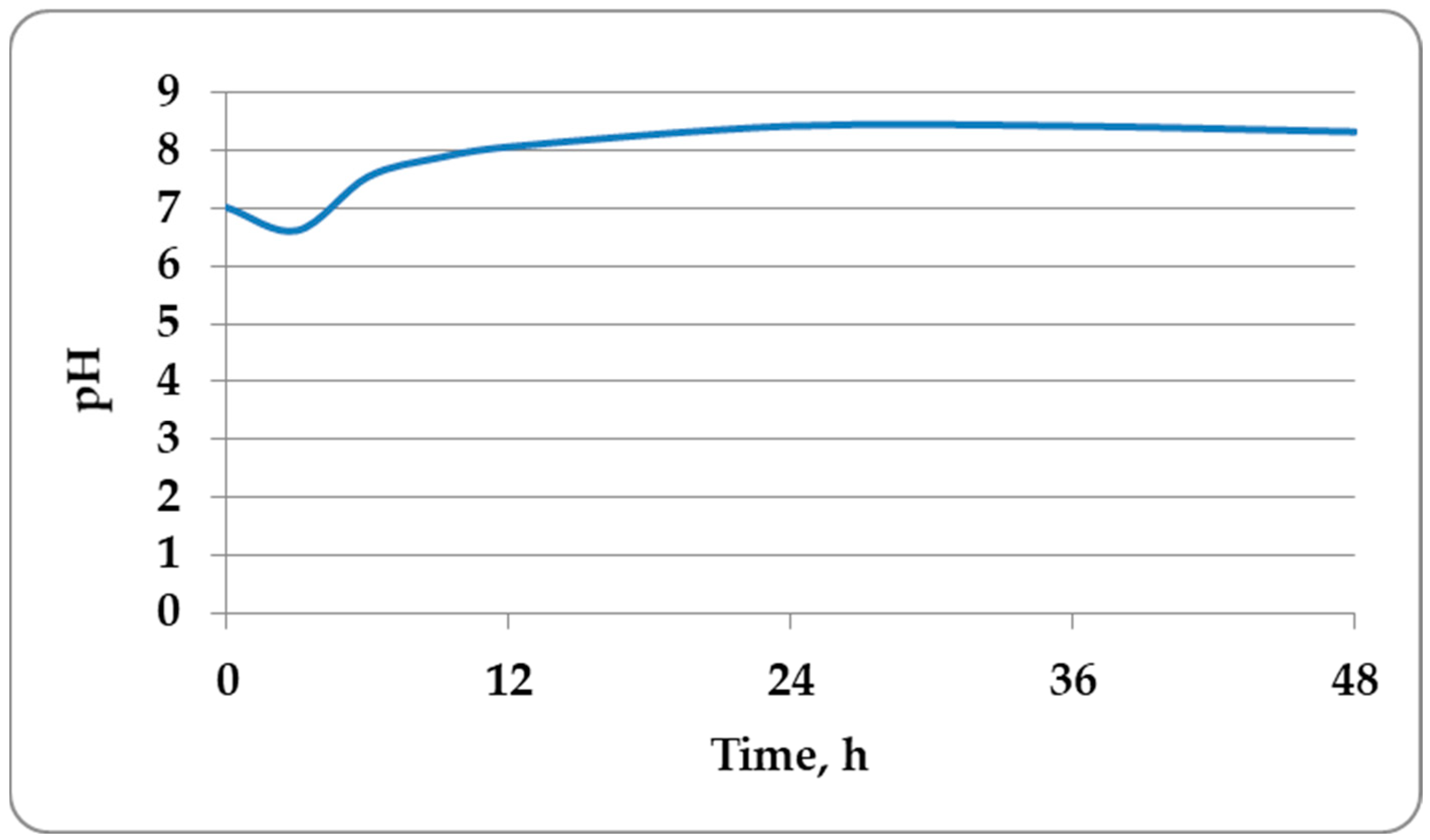
3.3. Cultivation of B. amyloliquefaciens M in a Laboratory Bioreactor and Determination of the Kinetic Parameters of the Process

To obtain antifungal preparations, the strains must allow cultivation with the formation of a significant amount of spores (above 109 cfu spores/cm3).
A mandatory condition for achieving a high concentration of spores is the research and optimization of the cultivation process. This goal cannot be successfully achieved if the maximum specific growth rate, the time to reach maximum vegetative cell productivity, and the sporulation rate are not known. The kinetic characteristics of the culture determine the optimal duration, management, and regulation of the process, resulting in products with a high concentration of spores. Therefore, a batch cultivation process of Bacillus amyloliquefaciens M was carried out in a laboratory bioreactor with mechanical stirring and a constant concentration of dissolved oxygen. The changes in the biomass, the concentration of spores, the redox potential, and the pH were monitored. Mathematical modeling of the kinetics of biomass accumulation and sporulation was performed (Figure 5, Figure 6, Figure 7 and Figure 8).
Mathematical models are valuable and help in the rational design of production plants, for scaling up, and management of the production process. Kinetic models make it possible to study the complex biochemical processes occurring in cells and their interrelationships [41].
The strain was characterized by a short lag phase of 1.5 h (Figure 5). The culture then entered the exponential growth phase, during which it grew with a very high maximum specific growth rate µm = 0.739 h−1 and a low intrapopulation competition coefficient of 0.057 logN/cm3·h. This indicates that the conditions in the bioreactor are suitable for the growth of the strain. This was due to the rapid entry of the strain into the stationary phase (as early as the 12th h), when a high concentration of vegetative cells (around 1013 cfu/cm3) was achieved. A study on the presence of spores at the 12th h was conducted, but no spores were found. As can be seen from Figure 1, the concentration of vegetative cells remains initially constant; therefore, the theoretical duration of the process was calculated, and the time at which the maximum concentration of vegetative cells would be reached was determined. Accurate determination of process duration is essential when scaling up in industrial settings, as it determines production costs. Due to its high maximum specific growth rate, the time to reach the maximum productivity of vegetative cells was short τex = 16.2 h.
Most likely, after this time, the sporulation process began, since at the 24th h, the amount of spores was of the magnitude order of 107 cfu/cm3. The monitoring of the total active cell concentration was therefore stopped and determination of the number of spores only began. An important parameter for biopreparation production is the sporulation rate because it determines the process duration. The sporulation rate for the studied strain was high µmS = 0.451 h−1, resulting in a high spore concentration at the end of the process—around 1012 spores/cm3. The percentage of sporulation was calculated according to a modified equation [42]:
%   o f   s p o r u l a t i o n = n u m b e r   o f   s p o r e s t o t a l   n u m b e r   o f   c e l l s × 100
A high sporulation rate of 86% was observed as early as the 48th h from the start of the cultivation process of Bacillus amyloliquefaciens M, confirming the high concentration of spores at the end of the cultivation. For practice, the time to reach this percentage of sporulation is of great interest, and it is calculated by the equation for τes (Equation (2)). An 86% sporulation percentage in this strain would be reached at the 24th h of cultivation, after initiation of sporulation.
A comparison of the experimental data with those obtained from the models was made (Figure 6). The mathematical models agreed well with the experimental data (Figure 6).
Both models describe the experimental data very well, as a result of which they can be used to control and predict the fermentation process carried out with Bacillus amyloliquefaciens M, which has also been found for other Bacillus amyloliquefaciens strains [41].
The results obtained for spore formation in Bacillus amyloliquefaciens M were better compared to those obtained by Tzeng, Y.-M., 2008 [42] for Bacillus amyloliquefaciens strain B 128 (5.93 × 10⁸ spores/cm3, after the 48th h) and Díaz-García A., 2015 [43], which achieved 88.7% sporulation at the 66th h. Figure 7 shows the changes in the redox potential during cultivation.
During intensive growth, the redox potential decreased until the culture entered into the stationary growth phase, when its value was retained (Figure 7). The redox potential started to increase when the sporulation of the strain began, which was explained by the lower intensity of respiration by the population. A similar trend was observed in the concentration of dissolved oxygen. During the exponential phase, the dissolved oxygen concentration dropped to 0, indicating a large rate of oxygen consumption and very high respiratory activity. At the start of sporulation, the concentration of dissolved oxygen increased and remained relatively constant—between 8% and 10% until the end of the process. This was due to the reduction in the number of vegetative cells, which reduced the respiratory activity of the population as a whole. The pH slightly decreased during the lag phase, after which it started to steadily increase (Figure 8). A relatively constant pH value between 8.43 and 8.33 was established when the sporulation process began. Thus, a concentrate (1012 spores/cm3) of Bacillus amyloliquefaciens M with high antibacterial and antifungal activity was obtained.

4. Conclusions

It was established that B. amyloliquefaciens M synthesizes antibiotic substances with Rf values of 0.47, 0.55, 0.67, and 0.75 with a peptide nature. The cultural medium and cultivation conditions for B. amyloliquefaciens M were optimized, thus obtaining a concentrate with a high concentration of spores (1012 spores/cm3) at the 48th h (86%). It has been proven that the sporulation process in Bacillus amyloliquefaciens M started after the 16th h from the start of the batch aerobic cultivation process. After additional research on the antiphytopathogenic activity of B. amyloliquefaciens M on plant species, the strain can be successfully applied as a biocontrol agent against phytopathogenic fungi, including Aspergillus flavus.

Author Contributions

Conceptualization, Z.D. and B.G.; methodology, Z.D., B.G., P.I., Y.G. and G.K.; software, G.K. and R.D.-K.; validation, Y.G., G.K. and R.D.-K.; formal analysis, Z.D., Y.G., P.I. and B.G.; investigation, Z.D., Y.G., B.G. and G.K.; resources, Z.D., B.G. and R.D.-K.; data curation, G.K., Y.G. and R.D.-K.; writing—original draft preparation, Z.D., R.D.-K., P.I. and B.G.; writing—review and editing, Z.D., G.K. and R.D.-K.; visualization, B.G., P.I., Y.G. and R.D.-K.; supervision, Z.D. and G.K. All authors have read and agreed to the published version of the manuscript.

Funding

This research received no external funding.

Institutional Review Board Statement

Not applicable.

Informed Consent Statement

Not applicable.

Data Availability Statement

The original contributions presented in the study are included in the article, further inquiries can be directed to the corresponding author.

Conflicts of Interest

The authors declare no conflicts of interest.

References

  1. Kheiri, F.; Faghani, M.; Landy, N. Evaluation of thyme and ajwain as antibiotic growth promoter substitutions on growth performance, carcass characteristics and serum biochemistry in Japanese quails (Coturnix japonica). Anim. Nutr. 2018, 4, 79–83. [Google Scholar] [CrossRef] [PubMed]
  2. Gheisari, A.; Shahrvand, S.; Landy, N. Effect of ethanolic extract of propolis as an alternative to antibiotics as a growth promoter on broiler performance, serum biochemistry and immune responses. Vet. World 2017, 10, 249–254. [Google Scholar] [CrossRef]
  3. Andremont, A. Consequences of antibiotic therapy to the intestinal ecosystem. Ann. Françaises D‘Anesthésie Réanimation 2000, 19, 395–402. [Google Scholar] [CrossRef]
  4. Henning, S.; Marianne, S. Resistence to antibiotics in the normal flora of animals. Vet. Res. 2001, 32, 227–241. [Google Scholar] [CrossRef] [PubMed]
  5. Denkova-Kostova, R.; Teneva, D.; Tomova, T.; Goranov, B.; Denkova, Z.; Shopska, V.; Slavchev, A.; Hristova-Ivanova, Y. Chemical composition, antioxidant and antimicrobial activity of essential oils from tangerine (Citrus reticulata L.), grapefruit (Citrus paradisi L.), lemon (Citrus lemon L.) and cinnamon (Cinnamomum zeylanicum Blume). Z. Naturforschung-Sect. C J. Biosci. 2021, 76, 175–185. [Google Scholar] [CrossRef] [PubMed]
  6. Goranov, B.; Teneva, D.; Denkova-Kostova, R.; Shopska, V.; Oulahal, N.; Denkova, Z.; Kostov, G.; Degraeve, P.; Pagan, R. Biopreservation of chocolate mousse with Lactobacillus helveticus 2/20: Microbial Challenge Test. Molecules 2022, 27, 5631. [Google Scholar] [CrossRef]
  7. FAO/WHO. Guidelines for the Evaluation of Probiotics in Food; Working Group Report; Food and Health Agricultural Organisation of the United Nations—World Health Organisation: London, ON, Canada, 2002. [Google Scholar]
  8. Sanders, M.E. Probiotics: Definition, sources, selection, and uses. Clin. Infect. Dis. 2008, 46, S58–S61. [Google Scholar] [CrossRef]
  9. Hill, C.; Guarner, F.; Reid, G.; Gibson, G.R.; Merenstein, D.J.; Pot, B.; Morelli, L.; Canani, R.B.; Flint, H.J.; Salminen, S.; et al. Expert consensus document. The International Scientific Association for Probiotics and Prebiotics consensus statement on the scope and appropriate use of the term probiotic. Nat. Rev. Gastroenterol. Hepatol. 2014, 11, 506–514. [Google Scholar] [CrossRef]
  10. Rodjan, P.; Soisuwan, K.; Thongprajukaew, K.; Theapparat, Y.; Khongthong, S.; Jeenkeawpieam, J.; Salaeharae, T. Effect of organic acids or probiotics alone or in combination on growth performance, nutrient digestibility, enzyme activities, intestinal morphology and gut microflora in broiler chickens. J. Anim. Physiol. Anim. Nutr. 2018, 102, e931–e940. [Google Scholar] [CrossRef]
  11. Abudabos, A.M.; Alhouri, H.A.A.; Alhidary, I.A.; Nassan, M.A.; Swelum, A.A. Ameliorative effect of Bacillus subtilis, Saccharomyces boulardii, oregano, and calcium montmorillonite on growth, intestinal histology, and blood metabolites on Salmonella-infected broiler chicken. Environ. Sci. Pollut. Res. 2019, 26, 16274–16278. [Google Scholar] [CrossRef]
  12. Guo, M.; Li, M.; Zhang, C.; Zhang, X.; Wu, Y. Dietary administration of the Bacillus subtilis enhances immune responses and disease resistance in chickens. Front. Microbiol. 2020, 11, 1768. [Google Scholar] [CrossRef] [PubMed]
  13. Montesinos, E. Antimicrobial Peptide and Plant Disease Control. FEMS Microbiol. Lett. 2007, 270, 1–11. [Google Scholar] [CrossRef] [PubMed]
  14. Bravo, A.; Pacheco, S.; Gómez, I.; Garcia-Gómez, B.; Onofre, J.; Soberón, M. Insecticidal Proteins from Bacillus thuringiensis and Their Mechanism of Action. In Bacillus thuringiensis and Lysinibacillus sphaericus; Springer International Publishing: Cham, Switzerland, 2017; pp. 53–66. [Google Scholar]
  15. Nayak, S.K. Multifaceted applications of probiotic Bacillus species in aquaculture with special reference to Bacillus subtilis. Rev. Fish. Sci. Aquac. 2020, 13, 862–906. [Google Scholar] [CrossRef]
  16. Jahan, R.; Siddique, S.S.; Jannat, R.; Hossain, M.M. Cosmos white rot: First characterization, physiology, host range, disease resistance, and chemical control. J. Basic Microbiol. 2022, 62, 911–929. [Google Scholar] [CrossRef] [PubMed]
  17. Islam, M.R.; Akanda, A.M.; Hossain, M.M.; Hossain, M.M. First characterization of a newly emerging phytopathogen, Sclerotinia sclerotiorum causing white mold in pea. J. Basic Microbiol. 2021, 61, 923–939. [Google Scholar] [CrossRef] [PubMed]
  18. Sumi, C.D.; Yang, B.W.; Yeo, I.-C.; Hahm, Y.T. Antimicrobial peptides of the genus Bacillus: A new era for antibiotics. Can. J. Microbiol. 2015, 61, 93–103. [Google Scholar] [CrossRef] [PubMed]
  19. Tran, C.; Cock, I.E.; Chen, X.; Feng, Y. Antimicrobial Bacillus: Metabolites and their mode of action. Antibiotics 2022, 11, 88. [Google Scholar] [CrossRef]
  20. Yoo, J.-S.; Zheng, C.-J.; Lee, S.; Kwak, J.-H.; Kim, W.-G. Macrolactin N, a new peptide deformylase inhibitor produced by Bacillus subtilis. Bioorg. Med. Chem. Lett. 2006, 16, 4889–4892. [Google Scholar] [CrossRef] [PubMed]
  21. Zhao, P.; Xue, Y.; Gao, W.; Li, J.; Zu, X.; Fu, D.; Bai, X.; Zuo, Y.; Hu, Z.; Zhang, F. Bacillaceae-derived peptide antibiotics since 2000. Peptides 2018, 101, 10–16. [Google Scholar] [CrossRef]
  22. Malviya, D.; Sahu, P.K.; Singh, U.B.; Paul, S.; Gupta, A.; Gupta, A.R.; Singh, S.; Kumar, M.; Paul, D.; Rai, J.P.; et al. Lesson from ecotoxity: Revisiting the microbial lipopeptides for the management of emerging diseases for crop protection. Int. J. Environ. Res. Public Health 2020, 17, 1434. [Google Scholar] [CrossRef]
  23. Stefanello, C.; Vieira, S.L.; Rios, H.V.; Simões, C.T.; Ferzola, P.H.; Sorbara, J.O.B.; Cowieson, A.J. Effects of energy, α-amylase, and β-xylanase on growth performance of broiler chickens. Anim. Feed Sci. Technol. 2017, 225, 205–212. [Google Scholar] [CrossRef]
  24. Sinnelä, M.T.; Park, Y.K.; Lee, J.H.; Jeong, K.C.C.; Kim, Y.-W.; Hwang, H.-J.; Mah, J.-H. Effects of Calcium and Manganese on Sporulation of Bacillus Species Involved in Food Poisoning and Spoilage. Foods 2019, 8, 119. [Google Scholar] [CrossRef]
  25. Baharudin, M.M.A.; Ngalimat, M.S.; Shariff, F.M.; Yusof, Z.N.B.; Karim, M.; Baharum, S.N.; Sabri, S. Antimicrobial activities of Bacillus velezensis strains isolated from stingless bee products against methicillin-resistant Staphylococcus aureus. PLoS ONE 2021, 16, e0251514. [Google Scholar] [CrossRef]
  26. Caulier, S.; Nannan, C.; Gillis, A.; Licciardi, F. Overview of the antimicrobial compounds produced by members of the Bacillus subtilis group. Front. Microbiol. 2019, 10, 435128. [Google Scholar] [CrossRef]
  27. Leifert, C.; Li, H.; Chidburee, S.; Hampson, S.; Workman, S.; Sigee, D.; Epton, H.A.S.; Harbour, A. Antibiotic production and biocontrol activity by Bacillus subtilis CL27 and Bacillus pumilus CL45. J. Appl. Bacteriol. 1995, 78, 97–108. [Google Scholar] [CrossRef]
  28. Arx, E.V.; Faupel, M.; Brugger, M. Das 4,4′-Tetramethyldiamino-diphenylmethan Reagens (TDM) Eine Modifikation der Chlor-o-Tolidin Farbereaktion für die Dünnschichtchromatographie. J. Chromatogr. 1976, 120, 224–228. [Google Scholar] [CrossRef]
  29. Laemmli, U.K. Cleavage of structural proteins during the assembly of the head of bacteriophage T4. Nature 1970, 227, 680–685. [Google Scholar] [CrossRef]
  30. Aitken, A.; Learmonth, P.M. The Protein Protocols Handbook, 2nd ed.; Walker, J.M., Ed.; Humana Press Inc.: Totowa, NJ, USA, 2002. [Google Scholar]
  31. Massawe, V.C.; Hanif, A.; Farzand, A.; Mburu, D.K.; Ochola, S.O.; Wu, L.; Samad Tahir, H.A.; Gu, Q.; Wu, H.; Gao, X. Volatile compounds of endophytic Bacillus spp. have biocontrol activity against Sclerotinia sclerotiorum. Phytopathology 2018, 108, 1373–1385. [Google Scholar] [CrossRef]
  32. Birnie, C.; Malamud, D.; Schnaare, R. Antimicrobial evaluation of N-Alkyl Betaines and N-Alkyl-N,N-Dimethylamine oxides with variations in chain length. Antimicrob. Agents Chemother. 2000, 44, 2514–2517. [Google Scholar] [CrossRef]
  33. Martins, C.; Correia, V.G.; Aguiar-Ricardo, A.; Cunha, Â.; Moutinho, M.G.M. Antimicrobial activity of new green-functionalized oxazoline-based oligomers against clinical isolates. Springer Plus 2015, 4, 382. [Google Scholar] [CrossRef]
  34. Soliman, S.A.; Khaleil, M.M.; Metwally, R.A. Evaluation of the Antifungal Activity of Bacillus amyloliquefaciens and B. velezensis and characterization of the bioactive secondary metabolites produced against plant pathogenic fungi. Biology 2022, 11, 1390. [Google Scholar] [CrossRef]
  35. Shobier, A.H.S.; Abdel Ghani, S.A.; Barakat, K.M. GC/MS spectroscopic approach and antifungal potential of bioactive extracts produced by marine macroalgae. Egypt. J. Aquat. Res. 2016, 42, 289–299. [Google Scholar] [CrossRef]
  36. Chandrasekaran, M.; Senthilkumar, A.; Venkatesalu, V. Antibacterial and antifungal efficacy of fatty acid methyl esters from the leaves of Sesuvium portulacastrum L. Eur. Rev. Med. Pharmacol. Sci. 2011, 15, 775–780. [Google Scholar]
  37. Mensah-Agyei, G.O.; Ayeni, K.I.; Ezeamagu, C.O. GC-MS analysis of bioactive compounds and evaluation of antimicrobial activity of the extracts of Daedalea elegans: A Nigerian mushroom. Afr. J. Microbiol. Res. 2020, 14, 204–210. [Google Scholar] [CrossRef]
  38. Shanga, B.M.; Lyon, G.D. Bacillus subtilis BS107as an antagonist in potato blackieg and soft rod bacteria. Can. J. Microbiol. 1998, 44, 777–783. [Google Scholar]
  39. McKeen, C.D.; Reilly, C.C.; Pusey, P.L. Production and Partial Characterization of Antifungal Substances Antagonistic to Monilinia fructicola from Bacillus subtilis. Phytopathology 1986, 76, 136–139. [Google Scholar] [CrossRef]
  40. Besson, F.; Michel, G. Influence of divalent ions on the solubility of iturin and bacillomycin L, antifungal peptidolipids of Bacillus subtilis. Microbios 1991, 65, 15–21. [Google Scholar]
  41. Zhao, R.; Wang, H.-H.; Gao, J.; Zhang, W.-J.; Li, X.; Zhou, J.-J.; Liang, P.; Gao, X.-Y.; Gu, S.-H. Plant volatile compound methyl benzoate is highly effective against Spodoptera frugiperda and safe to non-target organisms as an eco-friendly botanical-insecticide. Ecotoxicol. Environ. Saf. 2022, 245, 114101. [Google Scholar] [CrossRef]
  42. Tzeng, Y.-M.; Rao, Y.K.; Tsay, K.-J.; Wu, W.-S. Effect of cultivation conditions on spore production from Bacillus amyloliquefaciens B128 and its antagonism to Botrytis elliptica. J. Appl. Microbiol. 2008, 104, 1275–1282. [Google Scholar] [CrossRef]
  43. Díaz-García, A.; García-Riaño, J.; Zapata-Narvaez, J. Improvement of sporulation conditions of a new strain of Bacillus amyloliquefaciens in liquid fermentation. Adv. Biosci. Biotechnol. 2015, 6, 302–310. [Google Scholar] [CrossRef]
Figure 1. Elution profile in Sephadex G200 gel chromatography.
Applsci 14 03612 g001
Figure 2. SDS-PAGE of the obtained 4 peaks: 1—peak II; 2—peak III; 3—peak IV; 4—peak I; 5—markers, MW, kDa.
Applsci 14 03612 g002
Figure 3. Fractional profiles of the obtained 4 peaks (1—peak II; 2—peak III; 3—peak IV; 4—peak I; 5—markers). (1—250 kDa; 2—150 kDa; 3—100 kDa; 4—75 kDa; 5—50 kDa; 6—37 kDa; 7—25 kDa; 8—20 kDa; 9—15 kDa; 10—10 kDa).
Applsci 14 03612 g003
Figure 4. (a) Proportional contribution of individual protein fractions to the total protein profile (TPP) of peak II. (b) Proportional contribution of individual protein fractions to the total protein profile (TPP) of peak III. (c) Proportional contribution of individual protein fractions to the total protein profile (TPP) of peak IV. (d) Proportional contribution of individual protein fractions to the total protein profile (TPP) of peak I. The numbers on the individual bars of the histograms indicate the molecular weight of the respective fraction.
Applsci 14 03612 g004
Figure 5. Changes in the concentration of vegetative cells and spores during batch cultivation of Bacillus amyloliquefaciens M in a laboratory bioreactor with stirring.
Applsci 14 03612 g005
Figure 6. Comparison of experimental data with the logistic curve model and the exponential spore formation model (1).
Applsci 14 03612 g006
Figure 7. Dynamics of changes in the redox potential at dissolved oxygen concentration of 45%.
Applsci 14 03612 g007
Figure 8. Dynamics of pH change during batch cultivation of Bacillus amyloliquefaciens M in a laboratory bioreactor with stirring.
Applsci 14 03612 g008
Table 1. Antifungal activity of Bacillus amyloliquefaciens M. dwell = 6 mm. All values represent dinhibition zone.
Candida utilis ATCC 42402, 2 × 10⁷ cfu/cm3Saccharomyces cerevisiae ATCC 9763, 2.4 × 10⁷ cfu/cm3Aspergillus niger ATCC 1015, 1.2 × 10⁷ cfu/cm3Aspergillus flavus ATCC 9643, 2.8 × 10⁷ cfu/cm3Fusarium moniliforme ATCC 38932, 1.0 × 10⁶ cfu/cm3Penicillium chrysogenum ATCC 28089, 2 × 10⁷ cfu/cm3
Fermentation medium with molasses
B37.33 ± 0.4739.50 ± 0.4125.33 ± 0.4740.50 ± 0.4120.67 ± 0.4732 17 ± 0.24
ASN39.83 ± 0.2425.83 ± 0.2420.50 ± 0.4130.33 ± 0.4718.17 ± 0.2428.33 ± 0.47
Fermentation medium with malt
B30.50 ± 0.4125.50 ± 0.4130.50 ± 0.4130.67 ± 0.4718.17 ± 0.2420.50 ± 0.41
ASN26.33 ± 0.4727.67 ± 0.4720.83 ± 0.2432.33 ± 0.4720.33 ± 0.4718.67 ± 0.47
Fermentation medium MPB with salts
B37.33 ± 0.4730.17 ± 0.2422.33 ± 0.4737.83 ± 0.2420.17 ± 0.2415.33 ± 0.47
ASN32.50 ± 0.4124.17 ± 0.2422.50 ± 0.4125.33 ± 0.4712.67 ± 0.4712.83 ± 0.24
Table 2. Antifungal activity of Bacillus amyloliquefaciens M against Sclerotinia sclerotiorum var scleroria ATCC 18687.
Bacillus amyloliquefaciens Mdinhibition zone, mm
Sclerotinia sclerotium var scleroria ATCC 18687, 5.4 × 10⁷ cfu/cm3
24 h72 h
B-25.33 ± 0.47
ASN27.17 ± 0.2427.50 ± 0.41
CS30.33 ± 0.4730.67 ± 0.47
Table 3. Antibacterial activity of Bacillus amyloliquefaciens M against pathogenic microorganisms. dwell = 6 mm. All values represent dinhibition zone.
Escherichia coli ATCC 25922, 9 × 10¹⁰ cfu/cm3Salmonella enterica ssp. enterica serovar Enteritidis ATCC 25928, 2.5 × 10¹¹ cfu/cm3Salmonella abony NTCC 6017,
5.4 × 10¹¹ cfu/cm3
Staphylococcus aureus ATCC 6538P,
3 × 10¹¹ cfu/cm3
Pseudomonas aeruginosa NBIMCC 1390,
1.4 × 10¹¹ cfu/cm3
Listeria monocytogenes ATCC 19111,
2.0 × 10¹⁰ cfu/cm3
Bacillus cereus ATCC 14579,
5 × 10⁹ cfu/cm3
Fermentation medium with molasses
B18.33 ± 0.47-9.50 ± 0.4126.50 ± 0.4120.17 ± 0.2432.50 ± 0.4118.50 ± 0.41
ASN18.17 ± 0.24-9.33 ± 0.4723.17 ± 0.2418.17 ± 0.2428.17 ± 0.2420.50 ± 0.41
Fermentation medium with malt
B18.33 ± 0.47-9.17 ± 0.2415.50 ± 0.4120.17 ± 0.2433.33 ± 0.4713.50 ± 0.41
ASN20.67 ± 0.47-9.33 ± 0.4728.17 ± 0.2428.33 ± 0.4726.67 ± 0.4717.17 ± 0.24
Fermentation medium MPB with salts
B12.17 ± 0.24-9.17 ± 0.2425.33 ± 0.4720.50 ± 0.4120.33 ± 0.4713.17 ± 0.24
ASN10.33 ± 0.47--12.33 ± 0.479.17 ± 0.2418.67 ± 0.4717.33 ± 0.47
Table 4. Antiphytopathogenic activity of B. amyloliquefaciens M against phytopathogenic fungi in static cultivation at 30 °C.
Bacillus amyloliquefaciens M
Static Cultivation
30 °C
48 h
Test Microorganisms
Aspergillus niger ATCC 1015,
3.0 × 10⁵ cfu/cm3
Aspergillus awamori ATCC 22342,
1.2 × 10⁵ cfu/cm3
Aspergillus flavus ATCC 9643,
1.1 × 10⁵ cfu/cm3
LBG broth
pH = 4.5
ASN23.17 ± 0.2410.17 ± 0.2410.17 ± 0.24
CS23.33 ± 0.4712.33 ± 0.4710.33 ± 0.47
B26.33 ± 0.4710.17 ± 0.2410.33 ± 0.47
LBG broth
pH = 5
ASN25.67 ± 0.4740.67 ± 0.4725.17 ± 0.24
CS28.33 ± 0.4720.50 ± 0.4124.17 ± 0.24
B28.17 ± 0.2430.33 ± 0.4728.50 ± 0.41
LBG broth
pH = 6
ASN28.17 ± 0.2437.67 ± 0.4734.17 ± 0.24
CS30.33 ± 0.4723.33 ± 0.4732.17 ± 0.24
B31.67 ± 0.4732.50 ± 0.4135.17 ± 0.24
LBG broth
pH = 7
ASN37.67 ± 0.4735.33 ± 0.4734.67 ± 0.47
CS25.33 ± 0.4725.50 ± 0.4122.33 ± 0.47
B32.17 ± 0.2443.67 ± 0.4736.50 ± 0.41
LBG broth
pH = 8
ASN38.33 ± 0.4740.67 ± 0.4734.33 ± 0.47
CS31.17 ± 0.2430.50 ± 0.4125.67 ± 0.47
B32.33 ± 0.4742.17 ± 0.2435.17 ± 0.24
MPB broth
pH = 4.5
ASN12.17 ± 0.2412.50 ± 0.4120.33 ± 0.47
CS15.33 ± 0.4710.17 ± 0.2412.67 ± 0.47
B13.17 ± 0.2413.33 ± 0.4720.50 ± 0.41
MPB broth
pH = 5
ASN23.67 ± 0.4710.17 ± 0.2418.33 ± 0.47
CS13.33 ± 0.4710.17 ± 0.2422.17 ± 0.24
B27.50 ± 0.4115.33 ± 0.4722.50 ± 0.41
MPB broth
pH = 6
ASN28.67 ± 0.4725.50 ± 0.4123.17 ± 0.24
CS35.67 ± 0.4733.50 ± 0.4125.17 ± 0.24
B30.33 ± 0.4730.33 ± 0.4728.33 ± 0.47
MPB broth
pH = 7
ASN30.50 ± 0.4112.17 ± 0.2422.33 ± 0.47
CS28.17 ± 0.2410.17 ± 0.2412.17 ± 0.24
B32.33 ± 0.4714.50 ± 0.4126.67 ± 0.47
MPB broth
pH = 8
ASN20.17 ± 0.2412.17 ± 0.2415.17 ± 0.24
CS25.33 ± 0.4713.50 ± 0.4114.33 ± 0.47
B25.50 ± 0.4114.67 ± 0.4713.17 ± 0.24
Table 5. Antiphytopathogenic activity of B. amyloliquefaciens M after cultivation under dynamic conditions at 30 °C.
Bacillus amyloliquefaciens M
Dynamic Cultivation
30 °C
48 h
Test Microorganisms
Aspergillus niger ATCC 1015,
3.0 × 10⁵ cfu/cm3
Aspergillus awamori ATCC 22342,
1.2 × 10⁵ cfu/cm3
Aspergillus flavus ATCC 9643,
1.1 × 10⁵ cfu/cm3
LBG broth
pH = 4.5
ASN30.17 ± 0.2412.17 ± 0.2413.17 ± 0.24
CS25.50 ± 0.4112.33 ± 0.4710.33 ± 0.47
B27.33 ± 0.4712.33 ± 0.4715.33 ± 0.47
LBG broth
pH = 5
ASN28.33 ± 0.4733.33 ± 0.4734.50 ± 0.41
CS35.17 ± 0.2440.67 ± 0.4720.17 ± 0.24
B30.50 ± 0.4132.50 ± 0.4132.67 ± 0.47
LBG broth
pH = 6
ASN25.33 ± 0.4733.17 ± 0.2435.17 ± 0.24
CS32.17 ± 0.2428.67 ± 0.4735.17 ± 0.24
B34.50 ± 0.4133.33 ± 0.4735.33 ± 0.47
LBG broth
pH = 7
ASN42.67 ± 0.4732.33 ± 0.4730.17 ± 0.24
CS30.17 ± 0.2423.50 ± 0.4123.17 ± 0.24
B43.17 ± 0.2437.50 ± 0.4135.33 ± 0.47
LBG broth
pH = 8
ASN43.33 ± 0.4738.17 ± 0.2435.17 ± 0.24
CS34.50 ± 0.4137.17 ± 0.2430.50 ± 0.41
B43.50 ± 0.4143.67 ± 0.4737.50 ± 0.41
MPB broth
pH = 4.5
ASN22.33 ± 0.4722.67 ± 0.4720.17 ± 0.24
CS12.17 ± 0.2410.50 ± 0.4115.17 ± 0.24
B20.33 ± 0.4720.67 ± 0.4727.33 ± 0.47
MPB broth
pH = 5
ASN20.33 ± 0.4713.17 ± 0.2413.17 ± 0.24
CS15.17 ± 0.2414.33 ± 0.4718.33 ± 0.47
B20.50 ± 0.4115.67 ± 0.4720.50 ± 0.41
MPB broth
pH = 6
ASN25.33 ± 0.4724.50 ± 0.4128.17 ± 0.24
CS35.33 ± 0.4730.17 ± 0.2430.50 ± 0.41
B28.33 ± 0.4730.67 ± 0.4730.67 ± 0.47
MPB broth
pH = 7
ASN20.17 ± 0.2415.17 ± 0.2428.17 ± 0.24
CS22.33 ± 0.4715.50 ± 0.4125.33 ± 0.47
B28.67 ± 0.4718.33 ± 0.4728.50 ± 0.41
MPB broth
pH = 8
ASN18.17 ± 0.2415.33 ± 0.4725.17 ± 0.24
CS15.17 ± 0.2417.67 ± 0.4720.67 ± 0.47
B23.50 ± 0.4125.50 ± 0.4125.33 ± 0.47
Table 6. Antiphytopathogenic activity of B. amyloliquefaciens M after static cultivation at 37 °C.
Bacillus amyloliquefaciens M
Static Cultivation
37 °C
24 h
Test Microorganisms
Aspergillus niger ATCC 1015,
3 × 10⁵ cfu/cm3
Aspergillus awamori ATCC 22342,
1.2 × 10⁵ cfu/cm3
Aspergillus flavus ATCC 9643,
1.12 × 10⁵ cfu/cm3
LBG broth
pH = 4.5
ASN18.17 ± 0.2410.17 ± 0.2425.33 ± 0.47
CS20.33 ± 0.4712.17 ± 0.2425.67 ± 0.47
B15.50 ± 0.4115.50 ± 0.4130.17 ± 0.24
LBG broth
pH = 5
ASN30.50 ± 0.4130.50 ± 0.4137.17 ± 0.24
CS32.33 ± 0.4734.50 ± 0.4137.33 ± 0.47
B32.67 ± 0.4735.33 ± 0.4735.50 ± 0.41
LBG broth
pH = 6
ASN35.33 ± 0.4728.17 ± 0.2435.17 ± 0.24
CS40.67 ± 0.4732.50 ± 0.4140.33 ± 0.47
B40.33 ± 0.4733.33 ± 0.4743.33 ± 0.47
LBG broth
pH = 7
ASN30.50 ± 0.4130.17 ± 0.2437.67 ± 0.47
CS35.17 ± 0.2432.33 ± 0.4737.50 ± 0.41
B37.33 ± 0.4743.33 ± 0.4743.50 ± 0.41
LBG broth
pH = 8
ASN35.17 ± 0.2435.50 ± 0.4137.50 ± 0.41
CS37.67 ± 0.4740.17 ± 0.2435.33 ± 0.47
B34.33 ± 0.4737.17 ± 0.2440.67 ± 0.47
MPB broth
pH = 4.5
ASN30.17 ± 0.2434.33 ± 0.4735.33 ± 0.47
CS37.17 ± 0.2432.17 ± 0.2434.67 ± 0.47
B32.33 ± 0.4734.67 ± 0.4739.17 ± 0.24
MPB broth
pH = 5
ASN30.33 ± 0.4734.33 ± 0.4735.17 ± 0.24
CS37.17 ± 0.2440.17 ± 0.2434.67 ± 0.47
B37.50 ± 0.4135.50 ± 0.4137.33 ± 0.47
MPB broth
pH = 6
ASN32.17 ± 0.2432.33 ± 0.4740.50 ± 0.41
CS35.17 ± 0.2435.67 ± 0.4734.17 ± 0.24
B37.50 ± 0.4134.33 ± 0.4735.33 ± 0.47
MPB broth
pH = 7
ASN35.33 ± 0.4730.17 ± 0.2434.33 ± 0.47
CS33.67 ± 0.4739.50 ± 0.4125.67 ± 0.47
B35.17 ± 0.2439.50 ± 0.4132.67 ± 0.47
MPB broth
pH = 8
ASN30.17 ± 0.2430.67 ± 0.4742.33 ± 0.47
CS30.50 ± 0.4137.17 ± 0.2435.67 ± 0.47
B39.33 ± 0.4737.50 ± 0.4137.50 ± 0.41
Table 7. Antiphytopathogenic activity of B. amyloliquefaciens M after cultivation under dynamic conditions at 37 °C.
Bacillus amyloliquefaciens M
Dynamic Cultivation
37 °C
24 h
Test Microorganisms
Aspergillus niger ATCC 1015,
3 × 10⁵ cfu/cm3
Aspergillus awamori ATCC 22342,
1.2 × 10⁵ cfu/cm3
Aspergillus flavus ATCC 9643,
1.12 × 10⁵ cfu/cm3
LBG broth
pH = 4.5
ASN15.17 ± 0.2415.17 ± 0.2428.50 ± 0.41
CS25.33 ± 0.4720.50 ± 0.4137.17 ± 0.24
B22.33 ± 0.4717.50 ± 0.4130.33 ± 0.47
LBG broth
pH = 5
ASN43.67 ± 0.4735.17 ± 0.2442.67 ± 0.47
CS32.33 ± 0.4735.50 ± 0.4132.17 ± 0.24
B28.17 ± 0.2433.33 ± 0.4735.50 ± 0.41
LBG broth
pH = 6
ASN32.33 ± 0.4726.17 ± 0.2443.17 ± 0.24
CS45.33 ± 0.4743.33 ± 0.4752.17 ± 0.24
B43.67 ± 0.4735.50 ± 0.4140.33 ± 0.47
LBG broth
pH = 7
ASN27.17 ± 0.2433.67 ± 0.4740.33 ± 0.47
CS40.50 ± 0.4140.17 ± 0.2449.17 ± 0.24
B38.33 ± 0.4737.33 ± 0.4745.50 ± 0.41
LBG broth
pH = 8
ASN32.33 ± 0.4740.50 ± 0.4143.17 ± 0.24
CS43.17 ± 0.2442.33 ± 0.4745.33 ± 0.47
B35.17 ± 0.2439.17 ± 0.2445.33 ± 0.47
MPB broth
pH = 4.5
ASN28.17 ± 0.2430.17 ± 0.2435.50 ± 0.41
CS27.33 ± 0.4732.17 ± 0.2440.33 ± 0.47
B37.67 ± 0.4737.50 ± 0.4137.17 ± 0.24
MPB broth
pH = 5
ASN40.17 ± 0.2437.50 ± 0.4140.17 ± 0.24
CS32.33 ± 0.4742.33 ± 0.4735.17 ± 0.24
B43.17 ± 0.2430.50 ± 0.4140.17 ± 0.24
MPB broth
pH = 6
ASN35.33 ± 0.4734.50 ± 0.4137.33 ± 0.47
CS45.17 ± 0.2443.50 ± 0.4145.67 ± 0.47
B43.33 ± 0.4741.17 ± 0.2440.17 ± 0.24
MPB broth
pH = 7
ASN25.17 ± 0.2425.17 ± 0.2425.17 ± 0.24
CS35.33 ± 0.4732.67 ± 0.4732.33 ± 0.47
B35.33 ± 0.4730.50 ± 0.4135.50 ± 0.41
MPB broth
pH = 8
ASN30.17 ± 0.2428.17 ± 0.2435.17 ± 0.24
CS37.50 ± 0.4137.33 ± 0.4740.33 ± 0.47
B37.33 ± 0.4732.33 ± 0.4744.50 ± 0.41
Disclaimer/Publisher’s Note: The statements, opinions and data contained in all publications are solely those of the individual author(s) and contributor(s) and not of MDPI and/or the editor(s). MDPI and/or the editor(s) disclaim responsibility for any injury to people or property resulting from any ideas, methods, instructions or products referred to in the content.

