1. Introduction
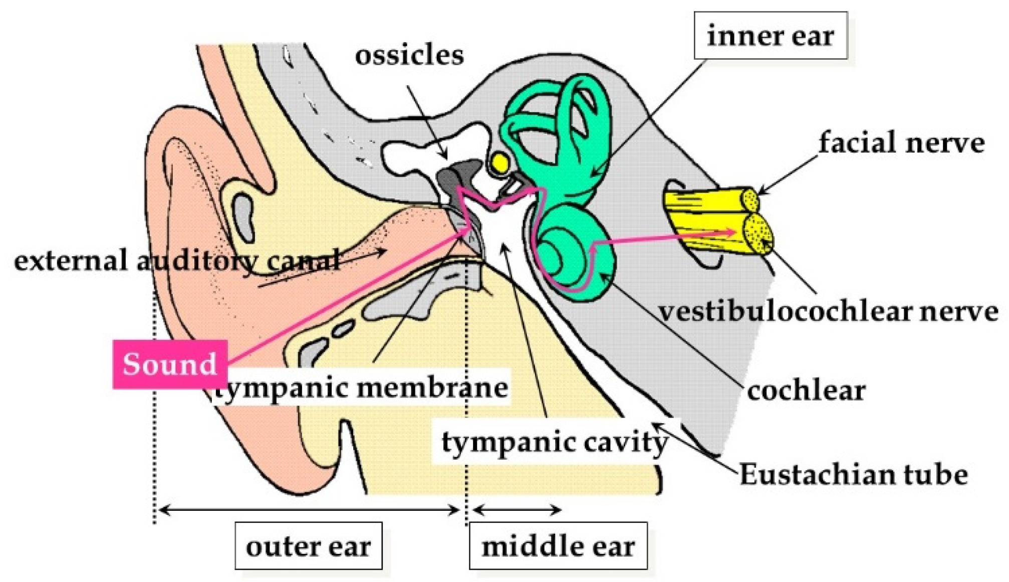
The internal anatomy of the ear is made up of extremely tiny, delicate, and interlocking anatomical structures that are surrounded by bone and muscle, with sound traveling through the external auditory canal as shown in
Figure 1. In particular, the mastoid portion of the temporal bone lies behind the ear and serves as a solid, normally impenetrable, barrier protecting the internal ear. This bony barrier has made it particularly challenging to access the anatomical structures within the internal ear, which has primarily been accomplished by drilling straight through and removing the mastoid bone in a procedure called a mastoidectomy. This procedure has been the mainstay of ear surgery up until the turn of the 20th century. While most ear surgery procedures are performed today with the objective of either preserving or improving hearing, the potential exists for noise and vibration generating surgical instruments required in a mastoidectomy to damage hearing. Of special concern has been the adverse impact on hearing of the use of powered surgical instruments, particularly drills, and the more recently introduced ultrasonic devices which are used to remove bone and expose the internal anatomy of the ear. This paper discusses issues available in the literature which have reported on the effects on hearing of these powered surgical instruments.
The majority of current ear surgery procedures can be broken down into two broad approaches: conventional microscopic ear surgery (MES) and the more recently developed transcanal endoscopic ear surgery (TEES). While powered surgical instruments are a standard part of MES, such instruments are only used in a subset of TEES procedures referred to as powered TEES. As was noted above, TEES has not totally eliminated the need for a mastoidectomy, because some surgeons are more comfortable with MES, and some procedures are not indicated for TEES, such as the mastoid air cells, inner ear, and skull base that are beyond its reach.
This paper presents an overview on types and causes of hearing loss in the context of ear surgery; a brief history of surgical instruments used to access the internal anatomy of the ear focusing on the powered surgical instruments, specifically electric drills and ultrasonic devices; an overview of the two main surgical procedures of MES and TEES used in ear surgery to access the internal anatomy of the ear; and a review of the literature on the potential for hearing loss caused by noise and vibrations produced by powered surgical instruments used in MES and TEES procedures, together with the presentation of previously unpublished data on the use of the ultrasonic aspirator, a powered surgical instrument, used in TEES.
2. Types and Causes of Hearing Loss
2.1. Types of Hearing Loss
Hearing loss falls into three broad categories: conductive hearing loss, sensorineural hearing loss and mixed hearing loss. Conductive hearing loss generally occurs when a physical impediment or barrier prevents the transmission of sound waves through the pathway from the outer ear through to the middle ear. Such impediments or barriers can range from a simple buildup of ear wax, accumulation of fluid within the ear due to an infection or an abnormal growth such as bony tissue or a tumor. The other type of hearing loss, sensorineural hearing loss, can be attributed to problems within the inner ear, primarily the cochlea and associated hair cells or the vestibulocochlear nerve (cranial nerve VIII). Sensorineural hearing loss can be caused by either intrinsic factors such as genetics resulting in congenital abnormalities, or extrinsic factors such as inner ear infections; ototoxic drugs such as aminoglycosides and cisplatin; or exposure to high noise levels both over an extended period of time such as in an industrial workplace, prolonged use of headphones or concert/entertainment venues or a single discrete event such as a blast of noise from equipment, gun shot, or bomb blast. The third type of hearing loss, mixed hearing loss, as the name implies, is a combination of the other two types of hearing loss [
1].
2.2. Hearing Loss and Powered Surgical Instruments
Hearing loss as related to powered surgical instruments has primarily been studied from two perspectives: noise levels (air-conducted) and vibrations (bone-conducted). The vast majority of studies have focused on noised-induced hearing loss, which is a clear subcategory of sensorineural hearing loss, while vibrations, or more precisely, skull vibrations, have garnered much less attention until recently and deserve further study and consideration. Such hearing loss is measured based on the degree to which the hearing threshold sensitivity has risen and is classified as either a permanent threshold shift or a temporary threshold shift [
2]. Most sensorineural hearing loss caused by powered surgical instruments fortunately falls into the temporary threshold shift category.
Separate from these two types of hearing loss caused by powered surgical instruments is the physical contact of an instrument with the ossicular chain. Such drill-to-bone contact results in vibrations which are transmitted via the ossicular chain to the cochlea. The subsequent damage to the cochlea generally results in permanent hearing loss. However, this hearing loss can be attributed to surgeon error rather than conventional use of the surgical instrument itself.
2.2.1. Noised-Induced Hearing Loss
Noised-induced hearing loss can, as alluded to above, be caused by either chronic, accumulative, and gradual exposure, or an acute, one-time event. The chronic, accumulative, and gradual exposure is generally defined in terms of daily exposure over years. Specifically, the National Institute for Occupational Safety and Health (NIOSH) has set down the recommended exposure limit (REL) as 85 decibels, A-weighted (dBA) for an 8-h time-weighted average (TWA) [
3] while the acute, one-time event is generally set at 140 dB or higher [
4]. However, any noise-induced hearing loss caused by powered surgical instruments falls into an undefined category between these two defined categories. While the noise levels tend to fall within chronic category, surgery time is only measured in minutes up to a couple of hours on a single day, rather than accumulated hours over multiple days. Moreover, while the noise levels generated by powered surgical instruments fall below noise levels defined as dangerous for acute one-time events, the fact that these instruments are used directly within the anatomy of the ear needs to be factored in.
The connection between possible noise-induced hearing loss and powered surgical instruments used during ear surgery has long been a source of concern and study in the case of surgical drills [
5,
6,
7,
8], as well as a recent target of research in the case of ultrasonic devices [
9].
2.2.2. Vibration-Induced Hearing Loss
The second potential way that powered surgical instruments can cause sensorineural hearing loss is by skull vibrations generated by these instruments. This cause has not been totally ignored, but has not received the same amount of attention over the years as noise levels. Moreover, occupational standards are not widely codified, particularly in regard to hearing, as opposed to damage to the circulatory system through the use of hand-held heavy equipment which can result in what is known as hand-arm vibration syndrome (HAVS), a type of Raynaud’s syndrome [
10]. Vibrations have been posited to cause damage to hearing through inner ear damage and Seki et al. [
11] and Miyasaka [
12] have both posited that morphological changes, specifically permeability, occur in stria vascularis capillaries, when the auditory ossicles or mastoid are subjected to vibrations. Again, as was the case with noised-induced hearing loss, vibration-induced hearing loss has been studied with surgical drills [
10,
13,
14], but less so with ultrasonic devices [
15].
3. History of Otological Surgical Instruments
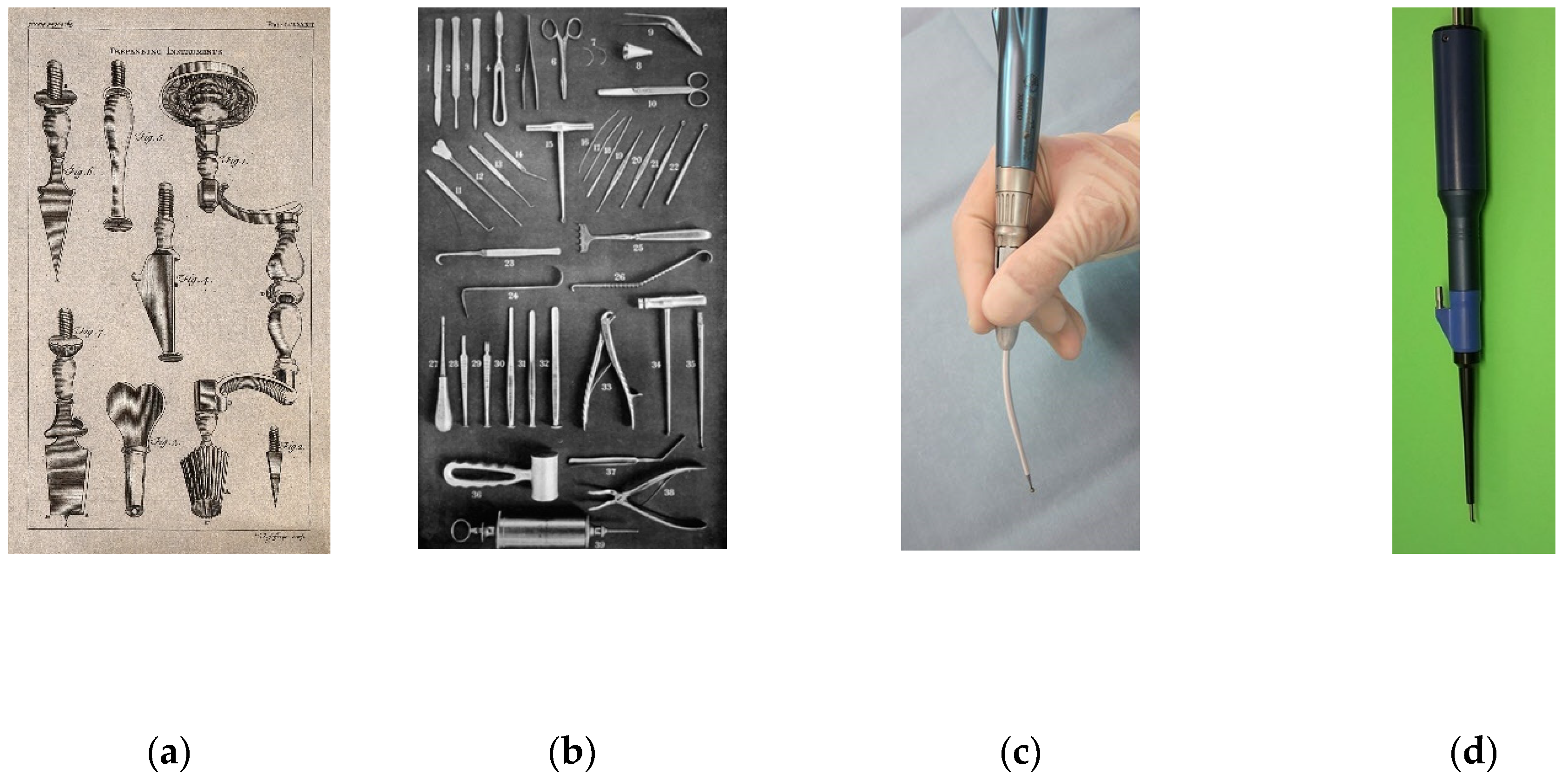
Progress in ear surgery has, like other surgical fields, been driven primarily by progress in the development of appropriate instruments. These instruments were often originally designed with another purpose in mind, often dentistry, but were eventually adapted by enterprising ear surgeons who co-opted them for their own purposes. Mudry has reported on the history of instruments in ear surgery and divided this history into three periods: trepans (
Figure 2a); chisels and gouges (
Figure 2b): and electrical drills (
Figure 2c [
16]. The introduction of powered TEES has added a fourth period of ultrasonic devices (
Figure 2d) [
17].
3.1. Pre-Powered Surgical Instruments
Though scattered references have been found throughout history that may be describing a mastoidectomy-like procedure such as by Galen of Pergamon (129 A.D−210 A.D.) [
19]. Surgical instruments had only developed to the point where a mastoidectomy could seriously be contemplated in the 18th century. These developments were attributed to advances made in metallurgy and machine tooling that fueled the Industrial Revolution and underscore the critical importance of technological advances outside medicine, leading to breakthroughs in medical procedures.
3.2. Powered Surgical Drills
The powered surgical drill, as opposed to a hand drill type instrument such as a trepan, was first used in the 1880s. However, its use did not catch on, and like many new approaches, was a bit ahead of its time. Instead, the electric drill was reintroduced into otological practice in the late 1920s by Julius Lempert, who is commonly recognized as the father of the use of the electric drill in ear surgery [
16]. The electrical drill has been the workhorse in ear surgery up until the present day.
3.3. Ultrasonic Devices
A new type of device technology began to make an appearance in the medical literature around the 1970s: ultrasonic devices. Broadly speaking, three types of ultrasonic devices used in surgery have appeared on the market: cavitation ultrasonic aspirators or CUSA devices; piezoelectric devices; and ultrasonic aspirators equipped with longitudinal and torsional oscillation.
A CUSA instrument selectively targets the fluid in soft tissue such as tumors, but leaves hard tissue such as bone untouched [
20]. CUSAs are quite frequently used in neurosurgery [
21] and renal surgery [
22] and have enabled surgeons in these fields to remove tumor tissue located in areas with little room for error with less possibility of damaging anatomical structures in comparison to electric drills.
Piezoelectric devices are another type of ultrasonic device that can cut both bone and soft tissue depending on the frequency setting [
23]. These devices are another example of borrowing a tool primarily used in dental surgery and expanding its reach into a wide variety of fields, including craniofacial surgery, to perform osteotomies [
24] and ear surgery to perform mastoidectomies [
25]. However, some surgeons have found that piezoelectric devices compare unfavorably to electric drills in terms of bone cutting power and speed [
9].
A third type of ultrasonic device offers the reverse of the CUSA by targeting hard tissue such as bone and leaving soft tissue relatively untouched. Both the CUSA and this new type of ultrasonic aspirator that removes only bone are often referred to in the literature as an ultrasonic bone curette (UBC) because they can both use the same handpiece to which a specialized tip is attached. These third types of ultrasonic aspirators remove bone using a longitudinal oscillation (L mode) or longitudinal and torsional oscillation (LT mode) to emulsify bone and these L mode/LT mode UBCs have been used in fields such as neurosurgery [
26], paranasal sinus surgery [
27], maxillofacial surgery [
28], and spinal surgery [
29].
Our original paper on powered TEES referred to the ultrasonic aspirator which we used as a Sonopet
® UBC [
17]. However, this ultrasonic aspirator that we used and still use today, emerged on the market in the early 2000s and has passed through a number of companies [
30]. This ultrasonic aspirator is today more properly called the Sonopet
® Omni and uses an H101 tip (model UST-2001 Ultrasonic Surgical Aspirator, Stryker Corporation, Kalamazoo, Michigan, USA) (
Figure 3).
It is the Sonopet
® Omni together with its H101 tip that has allowed us to perform powered TEES. The ability of this Sonopet
® Omni to target bone while generally avoiding soft tissue made it the perfect tool for working within the narrow confines of the external auditory canal and for removing bone from the ear canal. In addition, the Sonopet
® Omni not only removes bone, but also has irrigation and aspiration functions that were critical in opening the door to powered TEES, as will be described below. However, little information has been reported on the safety of the Sonopet
® Omni in terms of noise levels and vibrations [
15], which will also be discussed below.
4. History of Otological Surgical Procedures
The primary otological surgical procedure has been the mastoidectomy. A mastoidectomy involves the removal of the bone behind the ear in order to access the internal anatomy of the ear. However, the tools used in performing a mastoidectomy and related objectives and considerations in terms of safety have changed dramatically over the years. Moreover, since around the turn of the 20th century, a new surgical approach, TEES, has emerged that circumvents the need for a mastoidectomy. It should be noted that TEES has not totally eliminated the need for a mastoidectomy; some surgeons have yet to adopt the procedure and some procedures are not indicated for TEES. Moreover, TEES has raised its own safety issues that need to be addressed.
4.1. Pre-MES Procedures
Once tools were available for removing bone, humans opened holes in skulls for various purposes. The objective in prehistoric and early historic times could have been to release evil spirits, while other procedures may have been to relieve a buildup of pressure within the skull. This pressure buildup could be due to the presence of excess fluids caused by internal hemorrhaging or infection. Prior to the development of antibiotics and the appropriate tools, the main concern of otologists was the treatment of middle ear infections that resulted in pus accumulating within the ear, and having nowhere to go, were potentially life threatening. Such procedures often involved simple incisions of the tympanic membrane or abscesses located inside or outside of the ear. The French physician Ambroise Paré was the first to be credited in the 16th century with making a surgical incision to drain such an infection [
19].
The first confirmed mastoidectomy is credited to the French surgeon Jean Louis Petit in 1736 [
16] and thereafter the mastoidectomy became a standard, but not universally practiced part of ear surgery from around 1860 [
19]. A mastoidectomy was sometimes performed to allow draining of the pus and cleaning out of the infected area. However, removing the mastoid bone using the hand-drill-like trepan or by chipping away at the bone using chisels and gouges did not offer the level of control needed. Some patients were reported to have developed post-operative meningitis and subsequently died, an outcome which would have been due to removing too much bone and tissue. The inadequacy of the pre-powered age surgical instruments led to the mastoidectomy falling out of favor.
It should be noted that since these mastoidectomies were often performed as a life saving measure with little to no regard to the potential damage to internal ear anatomy and hearing caused by the surgical instruments. It is not a stretch, however, to assume that those patients who survived probably did have hearing loss. Eventually, Gustave Bondy developed improvements to the mastoidectomy with the aim of preserving the middle ear anatomical structures and better results were achieved. Bondy’s improvements and the introduction of the electric drill resulted in the mastoidectomy becoming a mainstay of ear surgery. This situation improved even further with the introduction of antibiotics in the 1940s, which dramatically cut down on the severity of middle ear infections [
19].
4.2. MES Procedures
While the introduction of an electric drill was a major breakthrough in ear surgery, an equally important breakthrough was the introduction of the microscope. This breakthrough is generally attributed to Dr. William House in the mid-1950s [
31]. The combination of the widespread use of antibiotics and the microscope led to the development of new surgical possibilities and techniques under the general category of microscopic ear surgery (MES). A majority of these procedures still involve the mastoidectomy as a first step to opening up the internal anatomy of the ear to the enhanced visualization afforded by the microscope. At the same time, higher standards emerged in terms of preserving hearing and preventing hearing loss. Concerns thus began to be raised, and are still raised today, about the potential for noised-induced hearing loss due to the use of these electric drills.
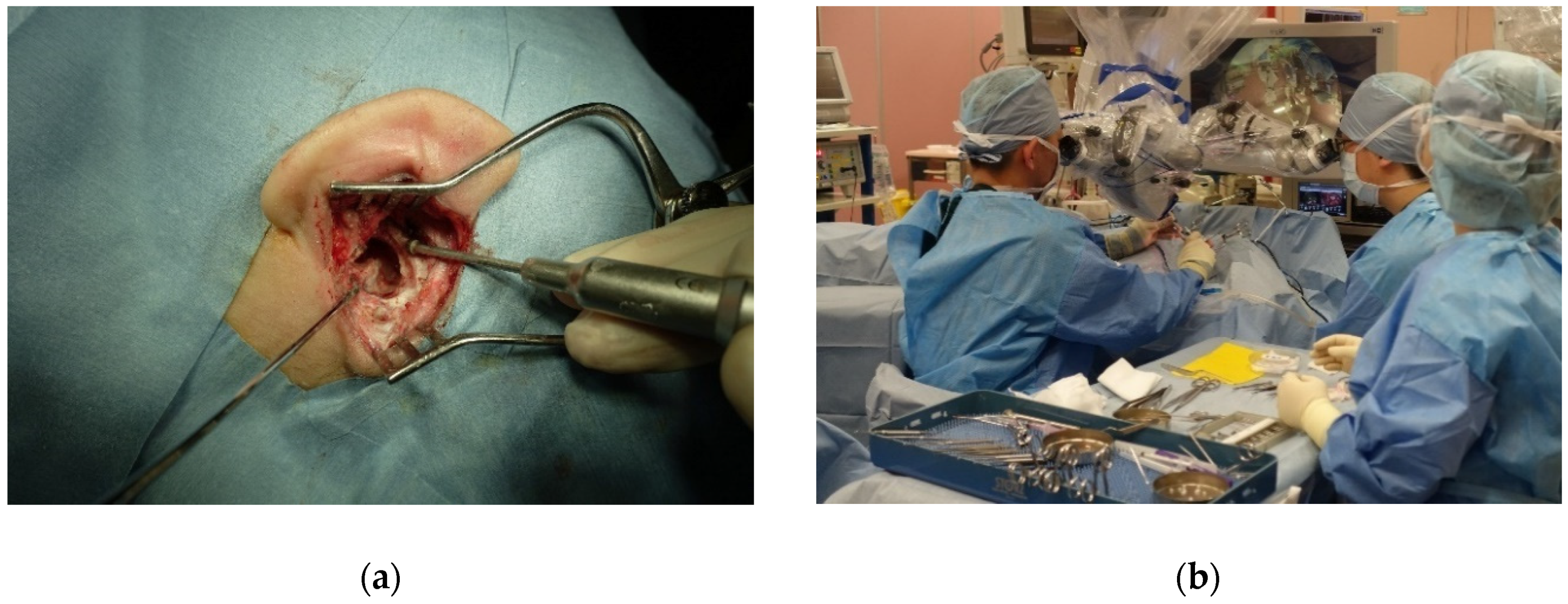
Figure 4a shows a close-up view of a mastoidectomy in progress while
Figure 4b shows the typical set up of the operating room and positioning of surgeons when performing an MES procedure.
4.3. TEES Procedures
While MES is still, by far, the common surgical approach employed in ear surgery, transcanal endoscopic ear surgery (TEES) has emerged as a viable alternative in the last twenty years. The microscope is not used in TEES, but instead an endoscope is employed to access and visualize the internal anatomy of the ear through the external auditory canal instead of the more invasive mastoidectomy approach. Even though the endoscope has long been a standard part of surgery in other surgical fields, ear surgeons have had to face a unique set of anatomical and technological challenges that delayed the use of the endoscope within the ear. Endoscopes equipped with cameras were first used together with the microscope when performing mastoidectomies in the 1990s as a way to get a better view of the internal anatomy of the ear [
32]. The first surgeries performed completely via the external auditory canal with the endoscope alone were reported on by Dr. Muaaz Tarabichi in 1997 [
33] and once again in 1999 [
34]. TEES really took off after 2008 with the further development of the 3-charged-coupled device (CCD) camera connected to high definition (HD) monitors, which resulted in high-resolution images during surgery of the tiny structures of the ear [
35,
36].
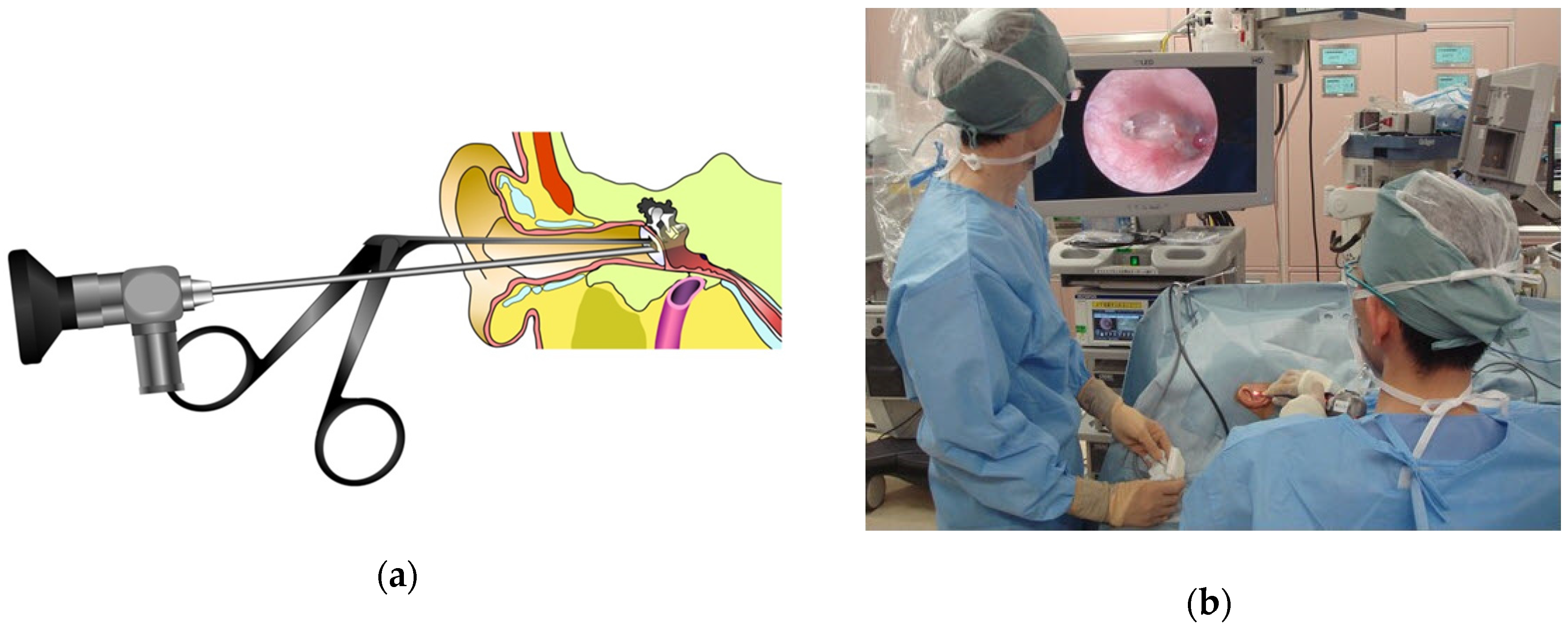
Figure 5a illustrates how the endoscope and forceps can be simultaneously inserted into the external auditory canal to perform TEES, and
Figure 5b shows the typical set up of the operating room and positioning of surgeons when performing a TEES procedure.
TEES offers a number of advantages over MES, including no need to perform an invasive mastoidectomy which requires bone loss, better visualization of the surgical field, the ability to see into deep recesses within the ear, no disfiguring retroauricular scarring, and a quicker recovery time. Moreover, most TEES procedures are performed entirely without powered surgical instruments, which eliminates the potential for sensorineural hearing loss resulting from noise levels or vibrations. However, the “conventional” or what we have unofficially dubbed “non-powered” TEES can be performed only so far into the middle ear, but a subset of TEES procedures use the Sonopet
® Omni to remove bone within the middle ear and enable access to the antrum which we call powered TEES.
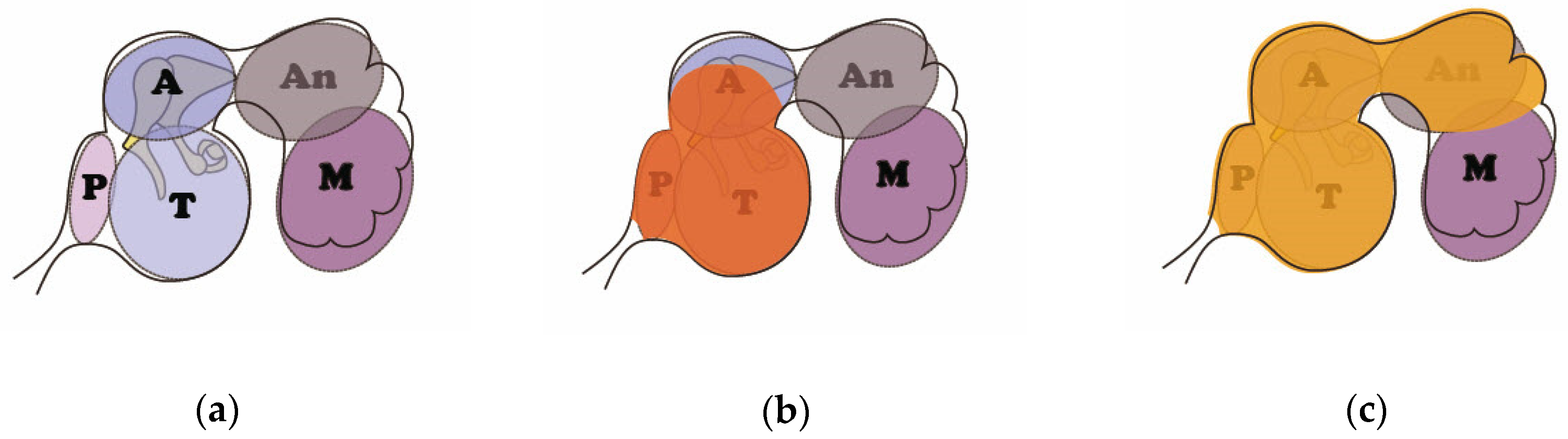
Figure 6 illustrates the internal anatomy of the middle ear (
Figure 6a); the scope of the indications for non-powered TEES which can only reach into the inferior portion of the attic (
Figure 6b); and the scope of the indications for powered TEES that can reach into the antrum (
Figure 6c). This figure underscores that the Sonopet
® Omni is reaching deep into the middle ear and closer to the ossicular chain and cochlea, which raises the specter of sensorineural hearing loss resulting from noise levels or vibrations, and is addressed below.
5. Generation of Noise and Vibrations by Otological Surgical Instruments and Hearing Loss
5.1. Pre-MES Procedures
One can reliably posit that the surgical instruments and procedures used in the pre-MES period were so crude that sensorineural hearing loss was common, but no data is available.
5.2. MES Procedures
5.2.1. Drill Generated Noise Levels
Many researchers have conducted basic research targeting the problem of measuring the noise levels generated by surgical drills in a non-clinical setting. Among the earliest studies of drill generated noise levels and most frequently cited is that of Kylén and Arlinger [
5], published in 1976. They measured vibrations generated by drills using isolated temporal bones and cadavers, whereafter the data was then converted into equivalent air-borne noise levels. They found that the isolated temporal bones produced lower noise levels than cadavers, and thus concluded that cadavers better simulated real surgical conditions. The maximum equivalent air-borne noise levels were found to be around 100 dB, which falls below the 130 to 140 dB threshold for causing permanent hearing loss from an acute one-time exposure. The same group reported additional results in 1977 [
6], looking at different variables focusing on the size of the burr; the type of the burr; (sizes: 2-mm, 4-mm and 6-mm burrs; types: diamond versus cutting burrs); drill rotation speed; and location of drilling. They found that the smaller the burr, the lower the noise levels, as well as that lower noise levels were obtained with diamond burrs, as opposed to cutting burrs with the highest noise level of 108 dB discovered with a 6-mm cutting burr. They further found that drill rotation speed and location of drilling had little effect on noise levels.
Several researchers subsequently collected intraoperative in vivo data with the objective of getting a better picture of drill generated noise during real-life conditions. Holmquist et al. [
38] reported higher in vivo noise levels in 1979 from the contralateral ear (the ear which was not being operated on) of six patients while undergoing a mastoidectomy on the ipsilateral ear (the ear which was being operated on). They reported noise levels in excess of the 108 dB recorded by the Kylén group at 116 dBA for 8-mm burrs; 109 dBA for 4-mm burrs; and surprisingly, a dangerously high 125 dBA for 2-mm burrs. Hickey and Fitzgerald O’Connor [
39] conducted an in vivo study in 1991 in which they attempted to directly measure the drill-generated noise levels by monitoring intraoperative
electrocochleographic (ECoG) noise levels and calculating these levels by using a masking technique. They were able to determine that drill-generated noise levels were present in excess of 90 dBHL at the level of the cochlea, but were unable to determine peak values.
Other researchers collected data on postoperative sensorineural hearing loss in the contralateral ear in order to attempt to eliminate surgical trauma as a possible cause of hearing loss from surgery, while at the same time acknowledging the added distance between the contralateral and ipsilateral ears. Man and Winerman [
40] conducted a study on 62 patients and reported in 1985 that they found a minimal difference between the peak noise levels which did not exceed 84 dB in the ipsilateral ear and 82 dB in the contralateral ear. Moreover, they found no hearing loss in the contralateral ear, but did find loss in 16 out of the 62 patients in the ipsilateral ear. They suggested that these results indicated that drill-generated noise levels did not cause sensorineural hearing loss. daCruz et al. [
41] reported on drill-induced hearing loss in the contralateral ear in 1997 based on determining outer hair cell (OHC) function using distortion-product otoacoustic emissions (DPOAE). They found a change in the amplitude of the intraoperative DPOAE in 2 out of 12 patients undergoing temporal bone surgery, indicating a transient OHC dysfunction that subsequently returned to normal. This transient but reversed dysfunction was attributed to drill-generated noise levels. Goyal et al. [
42] reported on the effect of mastoid bone drilling on the contralateral ear in 2013 based on otoacoustic emissions (OAE). Their study looked at the results for 30 patients and they stated that 15 of these patients exhibited a reduction in postoperative OAE levels out of which only 10 out of 15 completely recovered. However, data was only collected for up to 72 h, which makes a definite conclusion that the hearing loss was permanent a bit premature. In contrast, in two similar studies, Baradaranfar et al. in 2015 [
43] and Latheef et al. [
44] in 2018 reported transient hearing losses that all had disappeared by 72 h, while Badarudeen et al. [
45] in 2018 reported transient hearing losses that were still present on the 7th day after surgery.
The above findings thus paint a mixed picture on transient hearing loss in the contralateral ear caused by drilling.
5.2.2. Drill Generated Vibrations
Recent studies which have examined skull vibrations have indicated that this factor should not be discounted. Suits et al. reported on a guinea pig model in 1993 which was used to measure both noise and vibration levels based on the auditory brainstem response (ABR). They did find that a temporary threshold shift occurred, but that the shift had disappeared by three weeks [
13]. In 2001, Zou et al. also created a guinea pig model and compared the results for younger animals versus older animals when they were exposed to both noise and noise + vibrations. They reported that the older animals were more vulnerable to a threshold shift and that the sound-induced threshold shift was significantly less than the vibration + sound-induced threshold shift at three days after exposure [
14]. The same group of researchers reported in 2007 that temporal bone vibration in a guinea pig model showed that vibrations at high frequencies caused more severe hearing loss than at lower frequencies, but that the threshold shift was generally temporary [
10]. However, Hilmi et al. contended that high speed drills do not produce sufficient high levels of high frequency skull vibrations to result in damage to hearing [
8].
5.2.3. Hearing Loss after MES Procedures
A commonly accepted range for the incidence of sensorineural hearing loss in the ipsilateral ear after ear surgery is from 1.2% to 4.5% of patients. The higher figure of 4.5% is from a study of 1680 ear surgeries reported by Palva et al. [
46] in 1973, and the lower figure of 1.2% is from a study of 2,303 ear surgeries performed from 1965 to 1980, reported by Tos et al. [
47] in 1984. What is notable about these papers is that they are among the first large scale studies published in the literature and that they are from more than close to 35 years ago. Both authors attributed some of these hearing losses to the surgeon being too aggressive in the area of the ossicular chain, and Tos et al. [
47] in particular, noted that the incidence of sensorineural hearing loss was lower in patients treated from 1974 to 1980. They attributed this drop to better technique and better drills. In 1989, Doménech et al. [
48] reported what they characterize as an important sensorineural hearing loss after tympanoplasty in a larger percentage of patients at 16.7% than previously reported in literature, but the hearing loss was restricted to over 8000 Hz, which is typically not measured. Urquhart et al. [
7] reported in 1992 that they found no evidence of an even temporary threshold shift after ear surgery in a patient group of 40; however, they only tested up to 4000 Hz. A recent study by Kent et al. [
49] in 2017 looked at factors of the experience of the surgeon and the use of a powered drill in hearing outcomes for patients undergoing a tympanoplasty which required drilling in the ear canal, a less invasive procedure than a mastoidectomy, and they found that neither factor exhibited a correlation with high-frequency hearing loss. In contrast, Al Anazy et al. [
50] found in 2016 that the experience of the surgeon was a significant factor, but the use of a drill was not significant in the incidence of postoperative sensorineural hearing loss between 500 Hz to 4000 Hz after tympanoplasty which was 7% for residents, but only 1% for more experienced surgeons. However, they could not identify any obvious errors on the part of the residents.
Thus, the literature as it relates to the role of powered surgical instruments, specifically electric drills in postoperative sensorineural hearing loss, is inconclusive at best, and depends on the study design and definition of postoperative sensorineural hearing loss.
5.3. TEES Procedures
While a considerable amount of study has been done on noise-induced and vibration-induced hearing loss caused by surgical drills when performing MES, ultrasonic devices have not been studied in similar depth. CUSA devices are not used in ear surgery, and a search of the literature did not reveal any related studies from outside of ear surgery. Kramer et al. [
9] look at the potential for noise trauma caused by piezoelectric devices in craniofacial osteotomies and concluded that piezoelectric devices offer no advantage over regular drills in acoustic properties. They ultimately recommended using a drill because of the slower speed of the piezoelectric device. Research on TEES and the Sonopet
® Omni has, to our knowledge, only been conducted by our own group. The reason for this difference is that, as stated above, TEES procedures do not usually require the use of electric drills and only the small, but important subset of powered TEES, employ an ultrasonic aspirator together with a standard drill. The Sonopet
® Omni is inserted via the external auditory canal and used to remove part of the canal wall to expose the antrum while the surgical drill is used to polish a bony shelf which is designed to protect the facial nerve.
We took it upon ourselves to collect data on noise levels and vibration levels produced by the Sonopet
® Omni and compare it to data collected for standard surgical drills. The data on noise levels is presented herein for the first time while the data on skull vibrations was previously published in 2013 [
15].
5.3.1. Ultrasonic Aspirator Generated Noise Levels
Our study was designed to confirm that the noise levels generated by an ultrasonic aspirator during powered TEES fall within safe levels and should not induce sensorineural hearing loss. The study was conducted from September 2014 to February 2015 and data was collected during surgery from patients undergoing a powered TEES procedure to remove a cholesteatoma with a total of 14 patients (5 males/9 females) ranging in age from 15 to 76 and a median age of 50. All patients underwent a transcanal atticoantrotomy which was performed using a Sonopet
® Omni ultrasonic aspirator (model UST-2001 Ultrasonic Surgical Aspirator, Stryker Corporation, Kalamazoo, Michigan, USA) in the LT (longitudinal-torsion) mode at 25 kHz (
Figure 3a) and a high-speed drill with a curved burr at 80,000 rpm/1333 Hz (Visao
® High-Speed Otologic Drill, Medtronic, Shinagawa, Tokyo, Japan) (
Figure 7).
The noise levels were measured at 10 cm (
Figure 8a and 70 cm (
Figure 8b) from the devices from 0.5 kHz to 16 kHz. These distances were selected because 10 cm is the closest that it was physically possible to measure noise levels generated within the external auditory canal and 70 cm represents the distance to the surgeon’s ear. The noise level at 70 cm was measured in our original study because when new powered instruments are introduced, it is important to take into consideration the potential for hearing loss for anyone in the operating room due to long term noise exposure. Furthermore, we only measured up to 16 kHz despite the Sonopet
® LT mode generating frequencies of up to 25 kHz, because standard noise measurement equipment can only measure up to 16 kHz, which is also close to the maximum auditory threshold of a normal adult. The results for both the Sonopet
® Omni and the Visao
® drill at 10 cm were at 85 dB or below with the noise levels lower for the Sonopet
® Omni up to 2000 Hz and higher for the Sonopet
® Omni from 4000 Hz to 16,000 Hz. The results for both the Sonopet
® Omni and the Visao
® drill at 70 cm were below 70 dB with the noise levels for the Sonopet
® Omni lower or equal to the Visao
® drill up to 8000 Hz and higher for the Sonopet
® Omni from 8000 Hz to 16,000 Hz.
5.3.2. Ultrasonic Aspirator Generated Vibration Levels
Our group conducted a study designed to determine the vibration levels generated by an ultrasonic aspirator and compare the ultrasonic aspirator vibration levels to those of two surgical drills: an Osteon
® drill at 20,000 rpm/333 Hz (Zimmer Biomet, Warsaw, Indiana, USA) and a Visao
® High-speed Otologic Drill at 40,000 rpm/667 Hz and 80,000 rpm/1333 Hz (Medtronic, Shinagawa, Tokyo, Japan) [
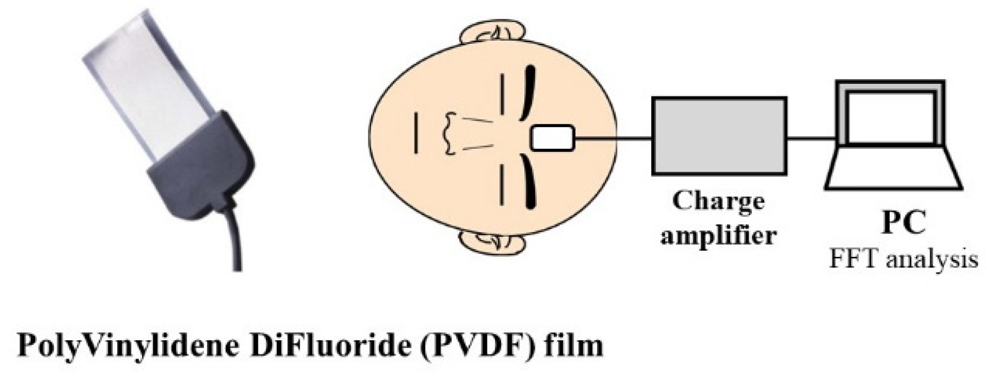
15]. All measurements were taken during an MES mastoidectomy and the skull bone vibrations were measured with a polyvinylidene difluoride (PVDF) film taped to the forehead as shown in
Figure 9. PVDF is a piezoelectric material and the charge builds up in the PVDF film in response to any applied mechanical stress.
Figure 10 shows the mean values of the measured skull vibrations and the background noise level at four frequency bands. In the frequency bands of 500–2000 Hz and 2000–8000 Hz, the mean values of the Sonopet
® Omni with an LT-vibration tip did not exceed the values for the Visao
® revolution speed of 40k rpm or 80k rpm as well as the Osteon
® drill. The peak values of skull vibrations by the Sonopet
® Omni with an LT-vibration tip were lower than the vibrations of Visao
® at 40 k rpm in the band of 500–2000 Hz; those of Visao
® at 80k rpm in the bands of 500–2000 Hz and 2000–8000 Hz; and those of Osteon
® in the bands of 500–2000 Hz and 2000–8000 Hz. No significant differences in the skull vibrations were observed among the three instruments below 500 Hz or above 8000 Hz.
Figure 11 shows the power spectrum produced by the Sonopet
® at 25 kHz in LT-mode versus background noise for the purpose of reference.
5.3.3. Hearing loss and TEES procedures
Our group has performed powered TEES since approximately 2011, and we have yet to record any postoperative sensorineural hearing loss in any patient up to a frequency of 8000 Hz. Thus, powered TEES can be performed, when indicated, without the worry of postoperative sensorineural hearing loss up to the highest measurable frequency.
6. Conclusions
The early days of ear surgery often focused on saving the lives of patients with little regard to postoperative hearing loss. However, the end of the 19th century and into the 20th century saw the introduction and steady improvement of surgical approaches and tools, particularly powered surgical instruments in the form of electric drills for MES and, more recently, ultrasonic aspirators for powered TEES. Concerns were raised about the impact that these powered surgical instruments could have on sensorineural hearing loss, and research has been done looking at both noise-induced hearing loss and vibration-induced hearing loss caused by powered surgical instruments when performing MES. The results of this research are long and often contradictory, as related to conventional MES and postoperative sensorineural hearing loss, particularly as related to high frequency hearing loss. Thus, effort and research should continue to be expended toward improving both MES techniques and electric drill noise level specifications, because MES, as stated above, will continue to be a standard and essential part of ear surgery, particularly in the areas of the mastoid air cells, inner ear, and skull base.
The introduction of TEES in the late 20th century eliminated the issue of sensorineural hearing loss for a circumscribed set of middle ear procedures because non-powered TEES does not require the use of powered surgical instruments. However, the emergence of powered TEES in the 21st century, once again, required that the research be conducted anew related to the use of powered surgical instruments, specifically the ultrasonic aspirator and curved burr, directly within the confines of the EAC and postoperative sensorineural hearing loss. We presented our research herein on whether powered TEES could be shown to cause either noise-induced hearing loss or vibration-induced hearing loss, and we found that powered TEES is as safe as, if not safer, in regard to the potential for postoperative sensorineural hearing loss than MES based on the data which we collected on noise levels, vibration levels, and the occurrence of postoperative sensorineural hearing loss for powered TEES. Thus, while MES will continue to be an essential part of ear surgery, surgeons can now rest assured that, when indicated, powered TEES can be performed safely and does not present any more of a risk of postoperative sensorineural hearing loss than MES.

 ,
, 


