Article

Proanthocyanidin Characterization and Bioactivity of Extracts from Different Parts of Uncaria tomentosa L. (Cat’s Claw)

by
Mirtha Navarro-Hoyos
1,
Rosa Lebrón-Aguilar
2,
Jesús E. Quintanilla-López
2,
Carolina Cueva
3,
David Hevia
4,
Silvia Quesada
5,
Gabriela Azofeifa
5,
M. Victoria Moreno-Arribas
3,
María Monagas
3 and
Begoña Bartolomé
3,*
1
Department of Chemistry, University of Costa Rica (UCR), Sede Rodrigo Facio, San Pedro de Montes de Oca, San José 2060, Costa Rica
2
Institute of Physical Chemistry “Rocasolano”, CSIC,C/ Serrano 119, Madrid 28006, Spain
3
Institute of Food Science Research (CIAL), CSIC-UAM, C/ Nicolás Cabrera 9, Madrid 28049, Spain
4
IUOPA-Redox Biology Group, Department of Cellular Morphology and Biology, Faculty of Medicine, University of Oviedo, C/ Julian Claveria 6, Oviedo 33006, Spain
5
Department of Biochemistry, School of Medicine, University of Costa Rica (UCR), Sede Rodrigo Facio, San Pedro de Montes de Oca, San José 2060, Costa Rica
*
Author to whom correspondence should be addressed.
Antioxidants 2017, 6(1), 12; https://doi.org/10.3390/antiox6010012
Submission received: 27 November 2016 / Revised: 11 January 2017 / Accepted: 26 January 2017 / Published: 4 February 2017
(This article belongs to the Special Issue Dietary Antioxidants and Health Promotion)

Abstract

:
Apart from alkaloids, bioactive properties of Uncaria tomentosa L. have been attributed to its phenolic constituents. Although there are some reports concerning low-molecular-weight polyphenols in U. tomentosa, its polymeric phenolic composition has been scarcely studied. In this study, phenolic-rich extracts from leaves, stems, bark and wood (n = 14) of Uncaria tomentosa plants from several regions of Costa Rica were obtained and analysed in respect to their proanthocyanidin profile determined by a quadrupole-time-of-flight analyser (ESI-QTOF MS). Main structural characteristics found for U. tomentosa proanthocyanidins were: (a) monomer composition, including pure procyanidins (only composed of (epi)catechin units) and propelargonidins (only composed of (epi)afzelechin units) as well as mixed proanthocyanidins; and (b) degree of polymerization, from 3 up to 11 units. In addition, U. tomentosa phenolic extracts were found to exhibit reasonable antioxidant capacity (ORAC (Oxygen Radical Absorbance Capacity) values between 1.5 and 18.8 mmol TE/g) and antimicrobial activity against potential respiratory pathogens (minimum IC50 of 133 µg/mL). There were also found to be particularly cytotoxic to gastric adenocarcinoma AGS and colon adenocarcinoma SW620 cell lines. The results state the particularities of U. tomentosa proanthocyanidins and suggest the potential value of these extracts with prospective use as functional ingredients.

1. Introduction

Proanthocyanidins are condensed flavan-3-ols with a high degree of structure variability, depending on their constituent monomers structure (propelargonidins, procyanidins, prodelphinidins, profisetinidins and prorobinetinidins), their interflavanic bond type (type A and type B proanthocyanidins), and its degree of polimerization (DP). Proanthocyanidins have become of high interest because of their biological properties, such as antioxidant, anti-inflammatory and anticancerigen properties with further investigation of interest due to proanthocyanidins potential use in cancer prevention [1].
Uncaria tomentosa L., also known as cat’s claw, is a plant used in traditional medicine, distributed in South America, mainly in Peru and Brazil as well as in Central America. There are numerous scientific reports on U. tomentosa bioactivity, comprising anti-inflammatory and antioxidant properties, and protective effects against cancer, as well as positive effects in the cardiovascular, central nervous and locomotor systems [2]. These bioactive properties of U. tomentosa have been attributed mainly to its alkaloid contents [3], however it has been reported that some properties such as its antioxidant effect could be related to its phenolic contents [4].
On the other hand, the antimicrobial activity of U. tomentosa has received little attention [5,6]. Phenolic compounds are recognized as antimicrobial agents on certain human bacteria. Some of them, such as hydroxybenzoic and hydroxycinnamic acids have shown antimicrobial effects against Salmonella [7] while monomeric flavonols are also capable to inhibit the growth of pathogen bacteria such as Clostridium perfringens, C. dificile, Bacteriodes spp., while probiotic bacteria strains from Lactobacillus and Bifidobacterium seems to be less sensitive [8]. In the case of proanthocyanidins, it has been described that a prolonged administration of these compounds produced a change in microbial population towards Gram-negative species (i.e., Enterobacteriaceae and Bacteriodes), indicating a greater susceptibility of Gram-positive bacteria to be inhibited by high molecular weight polyphenols [9].
U. tomentosa’s recent work on low-molecular weight polyphenols using liquid chromatography coupled to a triple quadrupole mass spectrometer indicated a high flavan-3-ol content, including procyanidin dimers and trimers, propelargonidin dimers, and cinchonain-type flavalignans (epicatechins substituted with phenylpropanoids), some of which were reported for the first time in U. tomentosa [10]. However, the study of greater molecular weight polyphenols requires the use of mass spectrometers with a broader m/z range, such as those based on Time of Flight (TOF) analysers. Thus, the objective of the present work was to perform a detailed characterization study of polymeric proanthocyanidins of U. tomentosa extracts using direct-injection (DI) mass spectrometry on an instrument equipped with electrospray ionization and a quadrupole-time of flight analyser (ESI-QTOF MS). In addition, extracts were assayed for different bioactivities, in particular, antioxidant activity, antimicrobial activity against respiratory pathogens and antitumoral effects in gastric and colon cancer cell lines. Finally, possible relationships between these activities and total proanthocyanidin content of the U. tomentosa extracts were explored.

2. Materials and Methods

2.1. Plant Material, Chemicals and Reagents

Uncaria tomentosa samples were collected from different places in Costa Rica: Asomat (northern part) and Aprolece-Palacios (Caribbean part), and others growing in the wild in Los Chiles (northern part) and Sarapiqui (Caribbean part). Vouchers for all plants are deposited in the Costa Rican National Herbarium, under series no. AQ2953, AQ3331, AQ3332 and AQ3510, respectively. The plant material was separated in its different parts: leaves (H), stems (T), bark (C) and wood (M), and then dried in a stove at 40 °C, being turned over every 24 h for a week until totally dry. The dried material was then ground and preserved in plastic recipients.
MTBE (methyl tert-butyl ether), chloroform, hexane, and methanol were purchased from Baker (Center Valley, PA, USA). Minimum essential Eagle’s medium (MEM) containing 10% fetal bovine serum (FBS), glutamine, penicillin-streptomycin, and amphotericin B were obtained from Life Technologies (Carlsbad, CA, USA). Trypsin-EDTA solution, DMSO (dimethyl sulfoxide) and MTT (3-(4,5-dimethylthiazol-2-yl)-2,5-diphenyltetrazolium bromide) were provided by Sigma-Aldrich (St. Louis, MO, USA).

2.2. Extraction of Phenolic Compounds from the Different Parts of U. tomentosa

Phenolic extracts from U. tomentosa leaves, stems, bark and wood were obtained as previously described [10]. Briefly, non-polar compounds were first extracted from the dried material in a mixture (0.05 mg/mL) of methyl ter-butyl ether (MTBE) and methanol (MeOH) 90:10 (v/v) at 25 °C during 30 min in ultrasound folllowed by letting the mixture standing for 24 h, and, after filtration, repeating the extraction process once. The solvent was evaporated to dryness and the residue washed with MeOH to extract residual polyphenols. After the MTBE-MeOH extraction, the polyphenolic-rich extract was obtained by extracting the residual plant material with MeOH at 25 °C during 30 min in ultrasound followed by letting the mixture standing for 24 h, and, after filtration, repeating the extraction process twice. These methanol extracts and the MeOH washings were combined and evaporated to dryness, under 40 °C, followed by succesive washings with hexane, MTBE and chloroform.

2.3. Total Proanthocyanidin Determination

Total proanthocyanidin content was determined by a modification of the Bate-Smith method, which is based on the acid-catalyzed oxidative cleavage of the C–C interflavanic bond of proanthocyanidins in butanol-HCl [11]. Briefly, 0.2 mL of each extract and 20 mL of butanol/HCl (50:50) (0.54 mM FeSO4) were incubated at 90 °C for 1 h. After cooling, the mixture volume was made up to 25 mL with butanol-HCl mixture, and the absorbance was measured at 550 nm against a blank prepared in the same way but without heating. Cyanidin chloride was used as standard to construct the calibration curve. Results were expresses as mg of cyanidin chloride equivalents/g of extract.

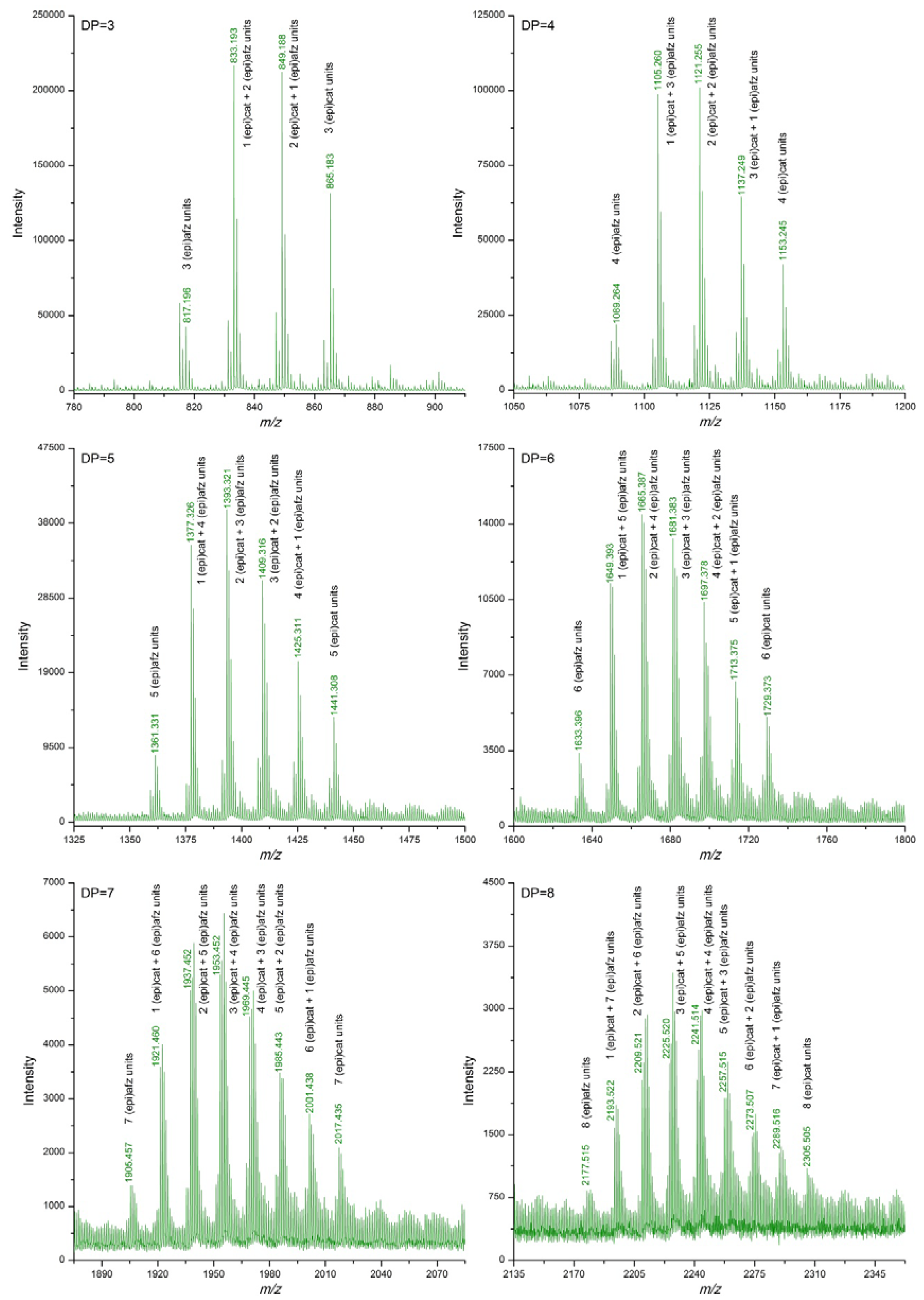
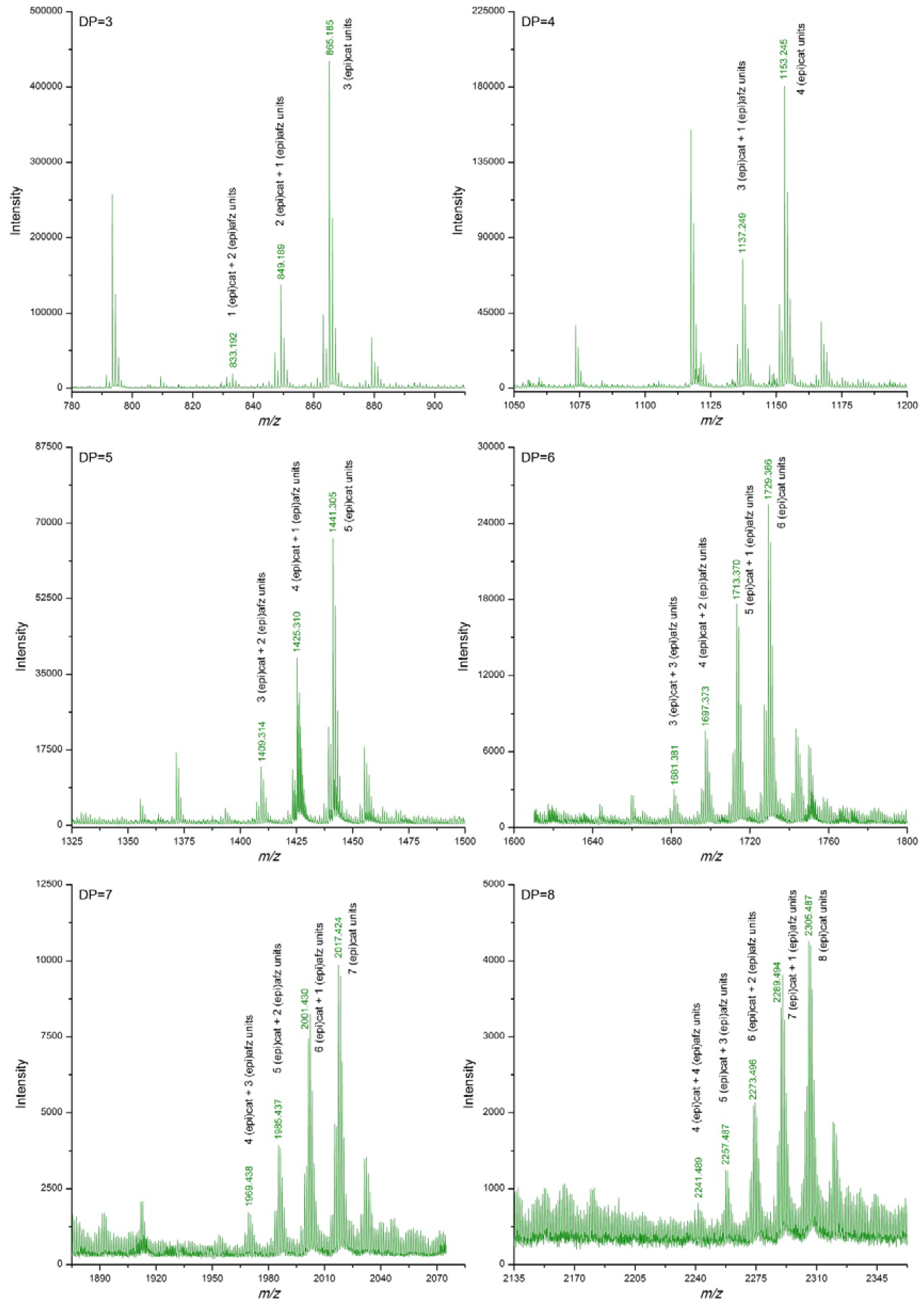
2.4. Characterization of the Proanthocyanidin Extracts by DI-ESI-QTOF MS

Mass Spectrometry experiments were carried out with an Agilent 1200 Series Liquid Chromatography system (equipped with a binary pump, an autosampler and an column oven) coupled to a 6520 quadrupole time-of-flight mass spectrometer using an electrospray interface (DI-ESI-QTOF MS instrument hereafter). All instruments were from Agilent Technologies (Santa Clara, CA, USA). 5 mg of the phenolic extracts were dissolved in 1 mL of methanol:water (1:1, v/v) and the solution filtered through 0.45 µm. 20 µL of the filtrate were injected using the autosampler into the LC system (without column) and carried through in acetonitrile:water (3:1, v/v) eluent at a flow rate of 100 µL/min [12]. The ESI source parameters were previously optimised, and adjusted as follows: spray voltage 4.5 kV; skimmer voltage, 300 V; fragmentor voltage, 500 V; drying gas temperature 300 °C; drying gas flow rate 6 L/min and nebulizer pressure 30 psi. Nitrogen (99.5% purity) was used as drying and nebulizer gas. Mass spectra were acquired in the negative mode, recording from m/z 100 to 5000. Data acquisition and processing were done using Agilent Mass Hunter Workstation Acquisition v. B.02.00 software (Agilent Technologies, Santa Clara, CA, USA). Assignation of DI ESI-QTOF MS signals (m/z) to a particular proanthocyanidin structure (i.e., propelargonidins, procyanidins, and prodelphinidins) was achieved by the calculation of the theoretical monoisotopic mass (as deprotonated ion, [M−H]), according to the Equation:
[M−H] (m/z) = 290.0790 × (EPI)CAT + 274.0841 × (EPI)AFZ − 2.0156 × (B) − 1.0078
where (EPI)CAT and (EPI)AFZ were, respectively, the numbers of (epi)catechin and (epi)afzelechin units contained in the proanthocyanidin molecule, and B were the numbers of B-type linkages between units.

2.5. In Vitro Antioxidant Activity

For the determination of the antioxidant activity, extracts (0.05 g) were treated with 10 mL of methanol/HCl (1000:1, v/v) by sonication for 5 min followed by an extra 15 min resting period. The mixture was then centrifuged (3024 g, 5 min, 5 °C) and filtered (0.45 µm). The radical scavenging activity of the extracts was determined by the ORAC (Oxygen Radical Absorbance Capacity) method using fluorescein as a fluorescence probe [13]. Briefly, the reaction was carried out at 37 °C in 75 mM phosphate buffer (pH 7.4) and the final assay mixture (200 µL) contained fluorescein (70 nM), 2,2'-azobis(2-methyl-propionamidine)-dihydrochloride (12 mM), and antioxidant (Trolox (1–8 µM) or phenolic extract (at different concentrations)). The plate was automatically shaken before the first reading and the fluorescence was recorded every minute for 98 min. A Polarstar Galaxy plate reader (BMG Labtechnologies GmbH, Offenburg, Germany) with 485-P excitation and 520-P emission filters was used. The equipment was controlled by the Fluostar Galaxy software version (v.4.11-0, BMG Labtechnologies GmbH, Offenburg, Germany) for fluorescence measurement. Black 96-well untreated microplates (Nunc, Denmark) were used. 2,2'-Azobis (2-methyl-propionamidine)-dihydrochloride and Trolox solutions were prepared daily and fluorescein was diluted from a stock solution (1.17 mM) in 75 mM phosphate buffer (pH 7.4). All reaction mixtures were prepared in duplicate and at least three independent runs were performed for each sample. Fluorescence measurements were normalized to the curve of the blank (no antioxidant). From the normalized curves, the area under the fluorescence decay curve (AUC) was calculated as:
AUC = 1 + i = 1 i = 98 i / 0
where 0 is the initial fluorescence reading at 0 min and i is the fluorescence reading at time i . The net AUC corresponding to a sample was calculated as follows:
Net AUC = AUCantioxidant − AUCblank
The regression equation between net AUC and antioxidant concentration was calculated. The ORAC value was estimated by dividing the slope of the latter equation by the slope of the Trolox line obtained for the same assay. Final ORAC values were expressed as mmol of Trolox equivalents (TE)/g of phenolic extract.

2.6. Cell Culture

The human gastric adenocarcinoma cell line AGS, human colorectal adenocarcinoma SW620 and monkey normal epithelial kidney cells Vero were obtained from American Type Culture Collection (ATCC, Rockville, MD, USA). They were grown in minimum essential Eagle’s medium (MEM) containing 10% fetal bovine serum (FBS) in the presence of 2 mmol/L glutamine, 100 IU/mL penicillin, 100 µg/L-streptomycin and 0.25 µg/mL amphotericin B. The cells were grown in a humidified atmosphere containing 5% CO2 at 37 °C and sub-cultured by detaching with trypsin–EDTA solution at about 70%–80% confluency. For the experiments, 100 µL of a cell suspension of 1.5 × 10⁵ cells/mL were seeded overnight into 96-well plates. The cells were further exposed for 48 h to various concentrations of U. tomentosa extracts (50 µL) in a humidified atmosphere containing 5% CO2 at 37 °C. For each experiment, the extract was dissolved in cell culture medium to a final concentration ranging between 15 and 500 µg/mL with a DMSO concentration below of 0.1 % (v/v). Control cultures were prepared with the addition of DMSO (vehicle control) to define the 100% of viability.

2.7. Assessment of Cytotoxicity by MTT Assay

After incubation for 48 h, MTT assays were performed to evaluate cytotoxicity. Briefly, the medium was eliminated, cells were washed once with 100 µL of PBS (Phosphate-Buffered Saline) and incubated with 100 µL of a MTT solution (3-(4,5-dimethylthiazolyl-2)-2,5-diphenyltetrazolium bromide; 0.5 mg/mL, final concentration) in PBS, for 2 h at 37 °C. Afterwards, MTT was removed, the formazan crystals formed were dissolved in 100 µL of ethanol 95%, and the absorbance was read at 570 nm in a microplate reader. For each extract concentration, the percentage of viable cells was calculated using the absorbance of the control (cells incubated with the vehicle solution (DMSO, 0.1 %)) as 100%. Dose-response curves were established for each extract and the concentration which is sufficient to reduce the cell number by 50% (IC50) was calculated. For each cell line, the extracts were tested in three independent experiments and in each experiment the different doses of extract were analysed in triplicate.

2.8. Antimicrobial Activity

Antimicrobial activity from phenolic extracts was evaluated on Staphylococcus aureus ATCC 25923, Enterococcus faecalis V583, and Pseudomonas aeruginosa PSP, which are bacteria characteristic of oral cavity and the respiratory system. S. aureus ATCC 25923 was obtained from the Spanish Type Culture Collection (CECT). E. faecalis V583 and P. aeruginosa PSP were clinical isolates obtained from human samples at the Ramón y Cajal Hospital (Madrid, Spain). Antibacterial assays were performed using a microdilution method in 96-well plates, according to Cueva et al. [14]. For each phenolic extract, a solution of 1mg/mL in sterilized water containing 10% DMSO (0.1 mg/mL final concentration on bacteria) was prepared. Eight serial dilutions were prepared so to get final concentrations of 100, 50, 25, 12.5, 6.25, 3.125, 1.562, 0.781 μg/mL. Inhibition percentages of the different extract concentrations were calculated by comparing the control growth with those obtained from cultures with phenolic extracts. Dose-response curves (% Inhibition versus extract concentration) were established for each extract and the concentration which was required to obtain 50% inhibition of growth (i.e., IC50 value) was calculated. For each bacteria strain, the extracts were tested in two independent experiments and in each experiment the different doses of extract were analysed in duplicate.

3. Results

3.1. Total Proanthocyanidin Content of U. tomentosa Extracts

As described in the methodology section, purification of U. tomentosa polar fractions with solvents of low and medium polarity allowed us to obtain phenolic enriched fractions of U. tomentosa. Total proanthocyanidin contents of the obtained phenolic extracts (n = 14) are summarized in Table 1. Findings indicated lower values for wood, ranging from 11.3 to 134.0 mg cyanidin chloride equivalents/g extract, and higher values ranging from 347.0 to 561.3 mg/g among all external parts (leaves, bark and stems), with values varying also according to plant origin. For instance, the highest values for the different plant parts (leaves, stems, bark and wood) clearly corresponding to extracts from the location of Sarapiqui whereas the lowest contents belonged to the Asomat location.

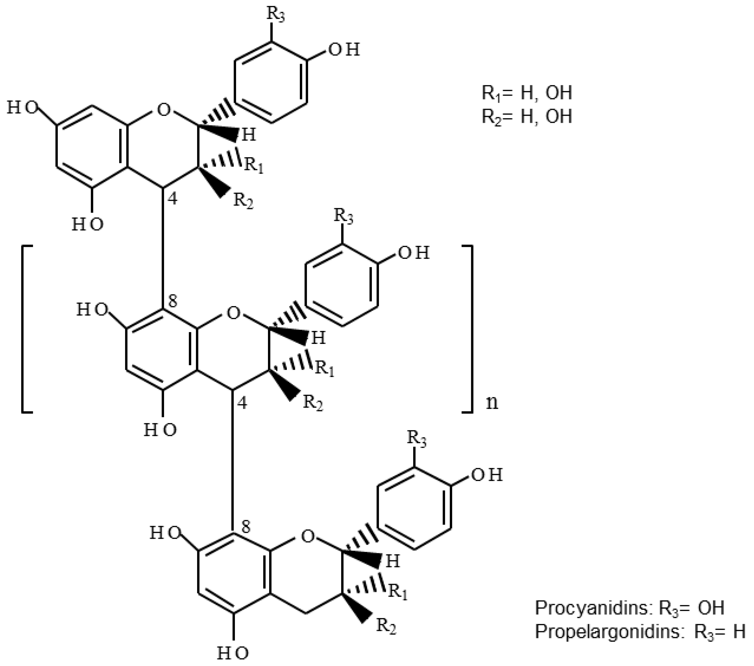
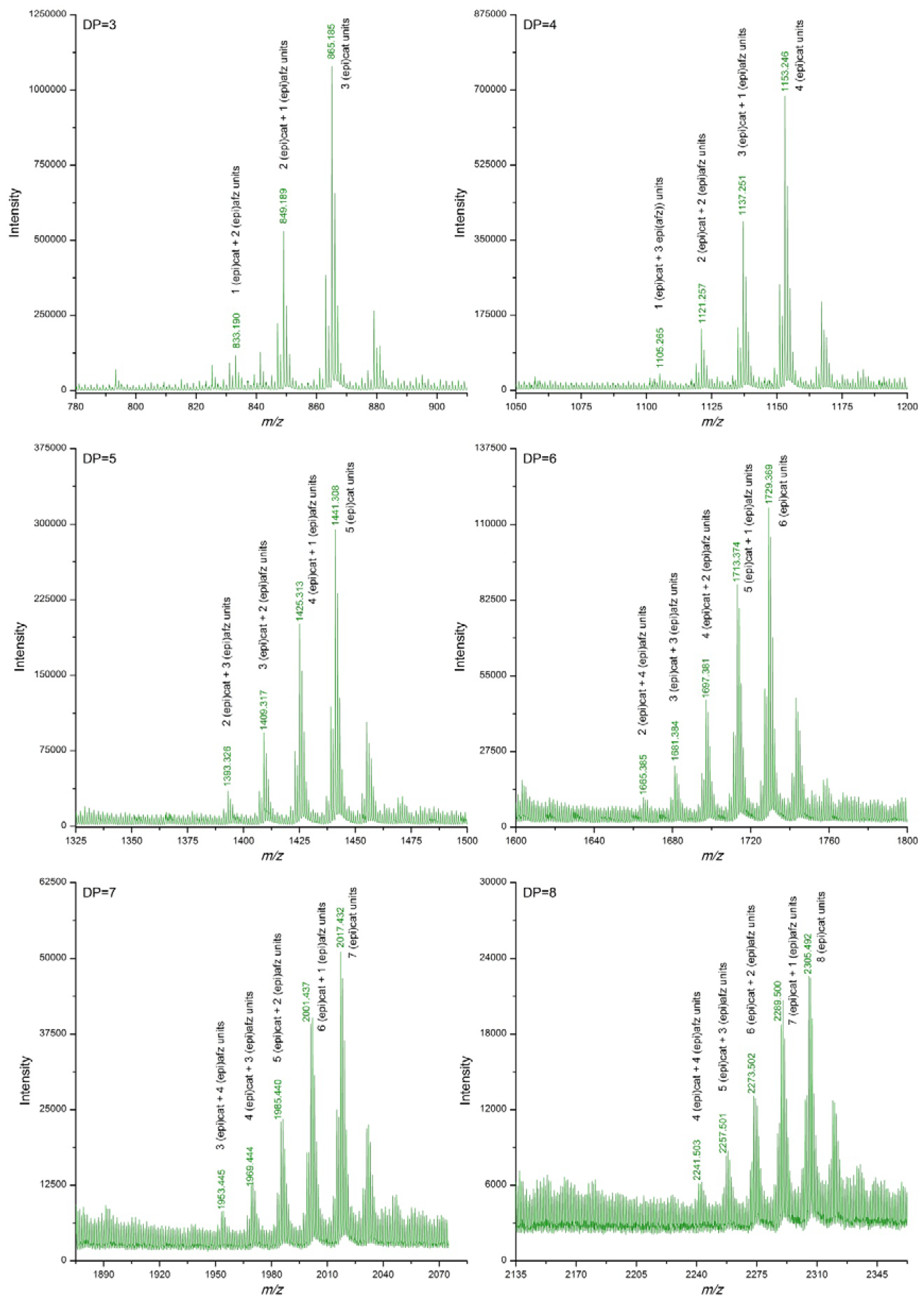
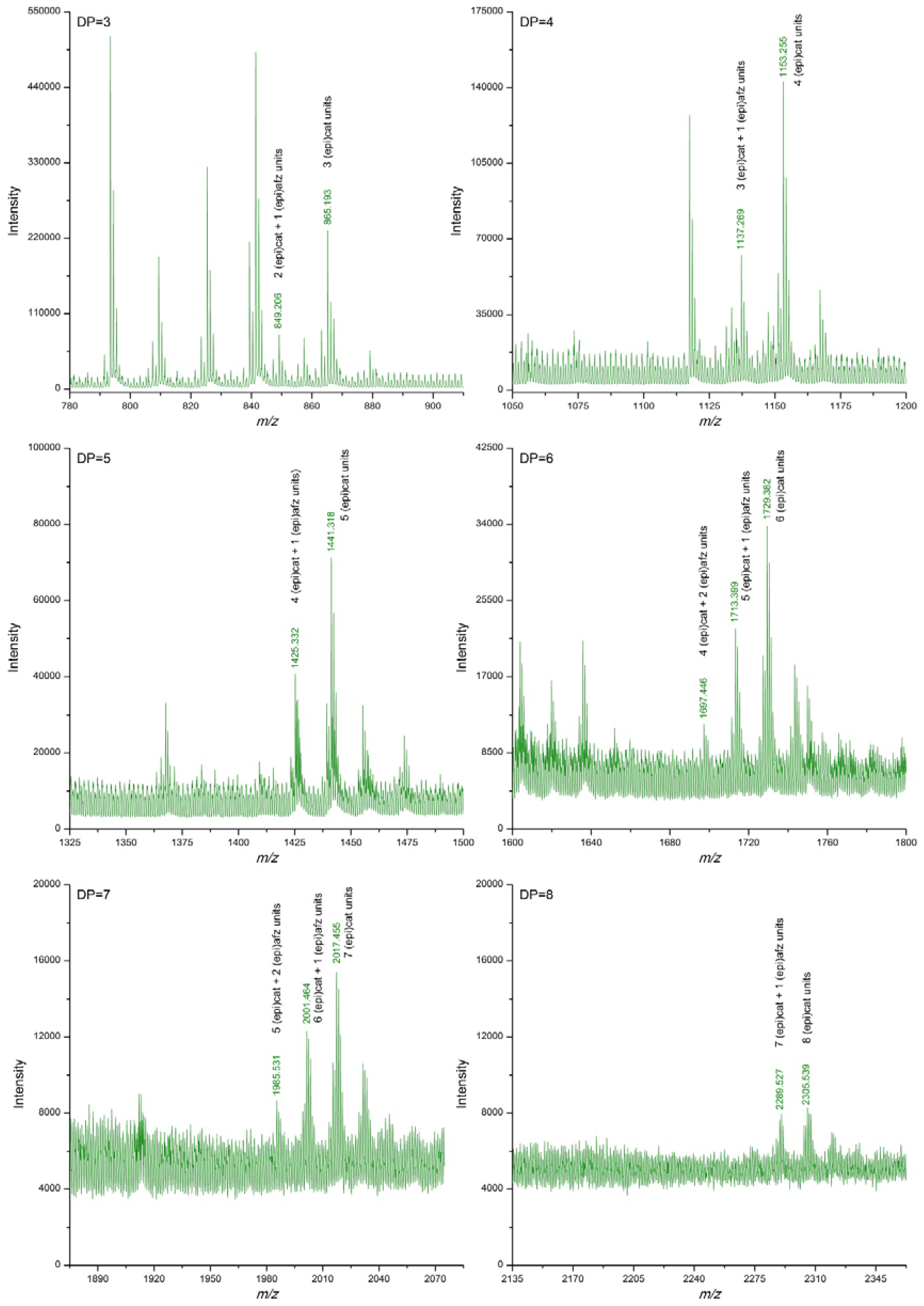
3.2. Proanthocyanidin Profile of U. tomentosa Extracts from Leaves, Stems, Bark and Wood

Table 2 summarizes the m/z signals corresponding to [M−H] ions of proanthocyanidins detected in the U. tomentosa extracts following the DI-ESI-QTOF MS analysis described in the methodology. Theoretical m/z data are also included in Table 2. In addition, Figure 1, Figure 2, Figure 3 and Figure 4 show amplifications of mass spectra corresponding to leaves, stems, bark and wood extracts, respectively. For the four types of U. tomentosa extracts, proanthocyanidins resulted to be composed by units of (epi)catechin and/or (epi)afzelechin linked through B-type linkages (Figure 1). However, some differences in molecular composition and degree of polymerization (DP) were found among extracts from the different parts of the plant. Proanthocyanidins in extracts from U. tomentosa leaves included pure procyanidins -only composed by (epi)catechin units- with a DP between 3 and 9 units-, pure propelargonidins -only composed by (epi)afzelechin units- with a DP between 3 and 8 units, and mixtures of both of them -composed by (epi)catechin and (epi)afzelechin units-, with a DP up to 10 units (decamers) (Table 2, Figure 2). For instance, in the case of pentamers (DP = 5) six signals were detected, corresponding to pure procyanidins (5 units of (epi)catechin) (m/z = 1441.308), pure propelargonidins (5 units of (epi)afzelechin units) (m/z =1361,331), and mixed proanthocyanidins containing 4, 3, 2, or 1 units of (epi)catechin and, respectively, 1, 2, 3 or 4 units of (epi)afzelechin (m/z = 1425.311, 1409.316, 1393.321, 1377.326, respectively) (Table 2, Figure 2).
Extracts from U. tomentosa stems contained proanthocyanidins up to decamers (DP = 10), either pure procyanidins or mixed proanthocyanidins with different (epi)catechin/(epi)afzelechin units ratio. In general, as the DP increased, signals corresponding to polymers with higher proportion of (epi)afzelechin units in relation to (epi)catechin units became non detected. No signals corresponding to pure propelargonidins were observed (Table 2, Figure 3). Similarly, extracts from U. tomentosa bark contained either pure procyanidins or mixed proanthocyanidins with different (epi)catechin/(epi)afzelechin units ratio, but both to at a DP of 11 units (Table 2, Figure 4). The proanthocyanidin profile of U. tomentosa wood extracts resulted simpler, being only detected signals corresponding to oligomers up to octamers, either consisted of pure procyanidins or mixed proanthocyanidins with 1 or 2 (epi)afzelechin units (Table 2, Figure 5). Whereas proanthocyanidin m/z signals followed a normal distribution for the different polymers detected in the leaves extracts (Figure 2), for stems, bark and wood extracts, higher signal intensities corresponded to pure procyanidins and proanthocyanidins with few units of (epi)afzelechin (Figure 3, Figure 4 and Figure 5).

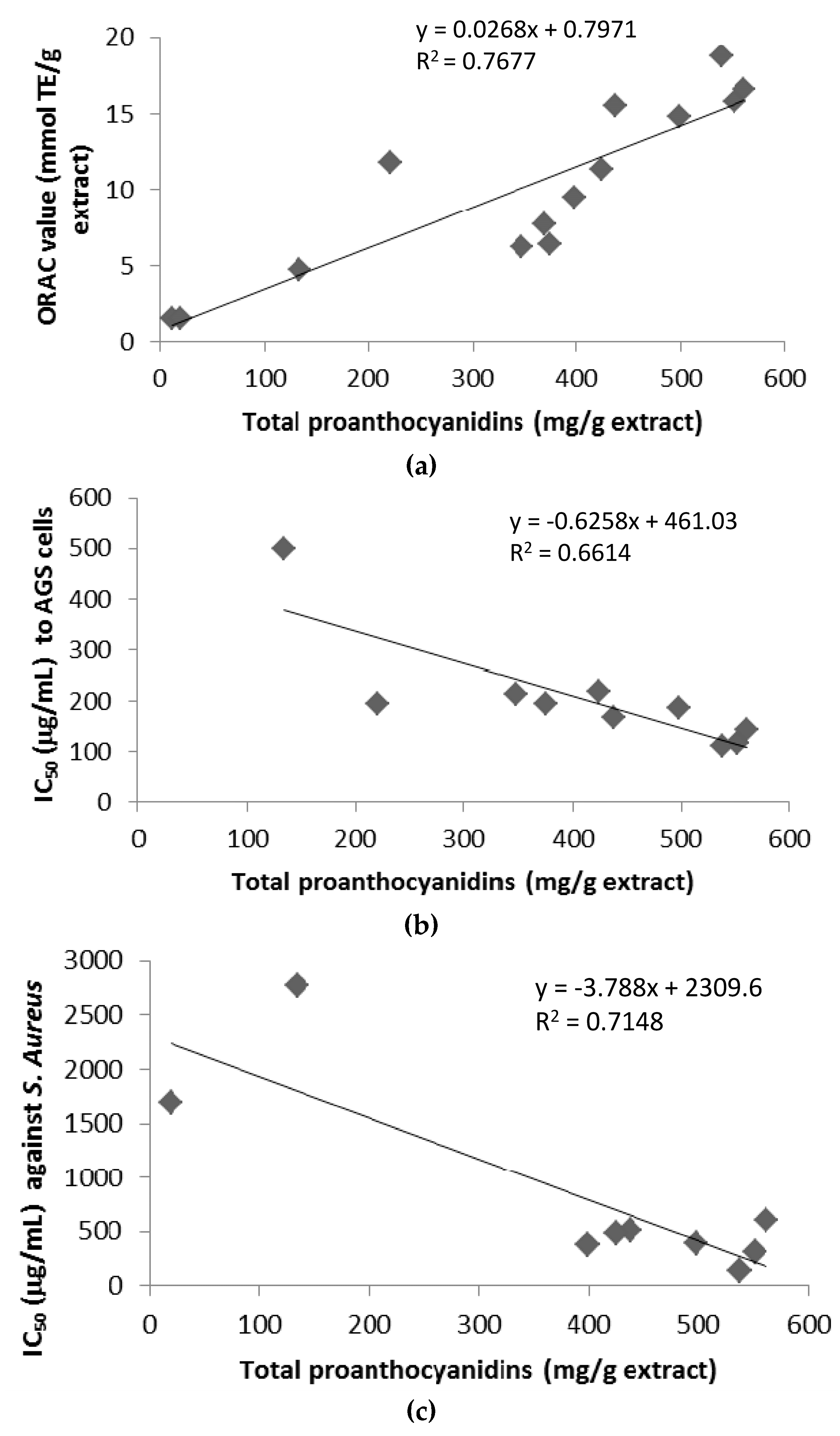
3.3. Antioxidant Activity of U. tomentosa Extracts

The antioxidant activity expressed as ORAC values of the U. tomentosa extracts varied in the order: leaves extracts (16.6–11.8 mmol Trolox equivalents/g) > stems (14.8–7.7 mol/mg) ~ bark (18.8–6.2 mmol/mg) > wood (4.7–1.5 mmol/mg) (Table 1). Among locations, Sarapiqui gave the highest values for the different type of extracts. In order to investigate if the proanthocyanidins contribute to the antioxidant activity of the extracts, a correlation analysis was carried out between the ORAC values and the total proanthocyanidin content (Table 1). A significant positive correlation was observed between them (R2 = 0.768) (Figure 6).

3.4. Citotoxicity of U. tomentosa Extracts

Table 3 reports the IC50 values for citotoxicity of the U. tomentosa extracts against the human gastric adenocarcinoma cell lines (AGS), human colon adenocarcinoma cell lines (SW620) and monkey normal epithelial kidney cells (Vero). IC50 values indicated that citotoxicity of U. tomentosa extracts resulted dependent on the cancer cell line used and also influenced by both the plant part and their collection site. Whereas growth of Vero cells were almost not affected by U. tomentosa extracts (IC50 > 500 µg/mL, with the only exception of leaves extracts from Los Chiles (IC50 = 458 µg/mL) and bark extracts from Sarapiquí (IC50 = 468 µg/mL)), growth of AGS and SW620 cells was strongly inhibited by all U. tomentosa extracts (IC50 < 220 µg/mL), except wood extracts that showed no cytotoxic effect towards any of the cell lines tested (Table 3). Among parts of the plants, IC50 variation intervals were quite similar for leaves and bark extracts: 116–195 and 111–220 µg/mL, respectively for leaves and bark extracts towards AGS cells, and 118–160 and 111–142 µg/mL respectively for leaves and bark extracts towards SW620 cells (Table 3). Of remarkable cytotoxicity for both adenocarcinoma cell lines were the extracts from leaves collected at Los Chiles (IC50 = 116 and 120 µg/mL respectively for AGS and SW620 cells) and from bark collected at Sarapiquí (IC50 = 111 and 142 µg/mL respectively for AGS and SW620 cells) (Table 3). As mentioned above, to investigate if proanthocyanidins contribute to the cytotoxicity of these extracts, a linear correlation analysis was carried out between the IC50 values and the total proanthocyanidin content (Table 1). A significant negative correlation was observed in the case of the AGS cells (R2 = 0.661) (Figure 6), but no significant correlation was found for the SW620 cells (data not shown).

3.5. Antimicrobial Activity of U. tomentosa Extracts

Antimicrobial activity of the U. tomentosa extracts was evaluated against three pathogenic strains known to be allocated in the oral cavity (Staphylococcus aureus ATCC 25923, Enterococcus faecalis V583 and Pseudomonas aeruginosa PSP) (Table 4). Most of the extracts were active against these different bacteria strains in the conditions used in this study. IC50 values varied between 133 and 6831 µg/mL and were influenced by both the part of the plants used and their location. Antimicrobial activity of extracts from Sarapiquí were in the order of bark > stems > leaves > wood for the three strains tested, whereas extracts from Palacios followed the order of stems > bark > leaves > wood for S. aureus ATCC 25923 and wood > leaves > stems > bark for E. faecalis V58 and P. aeruginosa PSP. In general, S. aureus ATCC 25923 and P. aeruginosa PSP were more sensitive (lower IC50 values) to the antimicrobial action of the U. tomentosa extracts than E. faecalis V583. As seen for the cytotoxic activity on AGS cells, significant negative correlations were observed between IC50 values and the total proanthocyanidin content for S. aureus ATCC 25923 (R2 = 0.715) (Figure 6). No significant correlations were found in the case of E. faecalis V583 either P. aeruginosa PSP (data not shown).

4. Discussion

Our previous studies on U. tomentosa [10,15] focused on the LC-ESI MS identification of low molecular weight polyphenols and demonstrated for the first time the occurrence of propelargonidin dimers in U. tomentosa extracts from different part plants. 13C-NMR studies also confirmed the presence of signals corresponding to epi(afzelechin) units in the different plant extracts. In the present study, we have carried out the characterization of oligomers and polymers of proanthocyanidins in U. tomentosa extracts from leaves, stem, bark and wood by accurate mass spectrometry (Q-TOF), and findings indicated that proanthocyanidins in U. tomentosa are composed of homopolymers of either (epi)catechin or (epi)afzlechins, as well as by heteropolymers constituted by both structural monomeric units. The occurrence of propelargonidins has been reported in food sources in lower quantities and dispersion than procyanidins, including A-type heteropolymers up to pentamers in strawberries [16] and up to undecamers with one (epi)afzelechin unit in cinnamon bark [17]; B-type propelargonidins in avocado fruit [18], kiwifruit pericarp [19] and raspberry [20], up to trimers in buckwheat grain [21] and boysenberry [22], up to tetramers in rhubarb [23], and up to heptamers with one (epi)afzelechin unit in almonds skins [24]. Some few reports exist in plants, including heteropolymers up to trimers in the leaves of Fagus silvatica [25] and Senna alata [26]. Studies in Rumex acetosa leaves showed predominance 5:1 of procyanidin over the propelargonidin units [27]; and while heteropolymers have been reported in Delonix regia leaves, only procyanidins existed in its bark [28]. In Acacia confuse and Casuarina equisetifolia bark, heteropolymers occurrence has been reported with predominance of procyanidins over propelargonidins [29,30]. Our results indicate a greater structural polydispersion in U. tomentosa proanthocyanidins, which was largely dependent on the part of plant, the complexity and DP range decreasing from the aerial parts of the plant to the inner wood. In our studies, the occurrence of homopolymers of properlagonidin up to DP 8 seems to be a very unique feature of U. tomentosa leaves as well as the presence of heteropolymers up to DP 10 with predominance 9:1 of (epi)afzelechin over the (epi)catechin units (Table 2). Besides the traditional use of U. tomentosa as an alkaloid source, our results demonstrated the potentiality of the plant as a source of proanthocyanidins, opening the possibilities of new applications in the nutraceutical industry [15], in particular for the leaves which has been neglected as source of bioactive components of U. tomentosa until now.
Previous studies indicated antioxidant activity of U. tomentosa aqueous and ethanol extracts with in-vitro DPPH, ABTS and TEAC tests [4,31,32] as well as of hydroethanol extracts under in vivo experiments [33]. These results appeared to be attributed to alkaloids, triterpenes and phenolic compounds, although these extracts were not fully characterized. Our study demonstrated for the first time the antioxidant activity of the proanthocyanidin enriched-fractions of U. tomentosa. ORAC values obtained for U. tomentosa leaves, stems and bark extracts (6.2–18.8 mmol Trolox/g extract) were similar to those of proanthocyanidin extracts from grape seeds [34], indicating once again the potential value of these extracts.
Considering that proanthocyanidins are known to reach the colon in intact form, anticancer cytotoxicity was evaluated in human gastric adenocarcinoma AGS and human colon adenocarcinoma SW620. In our case, results showed cell-dependence, since a negative correlation with PRO contents was observed for AGS gastric cells while no correlation was found for SW620 colon cells. U. tomentosa cytotoxic activity has been described mainly on alkaloids and triterpene extracts [2,35], however Pilarski et al. [36] studied triterpene-free and alkaloid-free U. tomentosa extracts on Lewis Lung carcinoma LL/2 cell lines, colon adenocarcinoma SW707, breast carcinoma MCF7, and cervical carcinoma KB, indicating that anticancer activity could be attributed to other secondary metabolites in U. tomentosa besides alkaloids. In addition, other reports indicate that fruits such as strawberry and raspberry containinig proanthocyanidins with (epi)catechin and (epi)afzelechin (propelargonidin) units have cytotoxicity activity on human colorectal adenocarcinoma HT-29 [37], for instance with antiproliferation EC50 values ranging from 110 to 120 mg/mL in the aforementioned strawberry and kiwifruit containing procyanidins and properlagonidins. Our results are in agreement demonstrating activity when using U. tomentosa extracts with propelargonidin and procyanidin oligomeric contents and showing high selectivity in both cancer cell lines in respect to normal vero cells. Regarding correlation of polyphenolic contents with cytotoxicity some studies argue in favour [38] while others [39] claim there is no correlation.
As stated earlier, U. tomentosa antimicrobial activity has received less attention and further, studies have reported divergent results. For instance, Ccahuana-Vasquez et al. [5] found that micropulverized U. tomentosa inhibited Staphylococus spp but failed to inhibit P. aeruginosa while another study [40] showed that U. tomentosa gel had an inhibitory effect on Staphylococus spp but was not active towards E. faecalis, suggesting that U. tomentosa type of extract may affect bacterial inhibition [2]. However, Herrera et al. [40] proved the antibacterial effect of U. tomentosa against E. faecalis in infected dentin. Our results align with these findings and evidence that antimicrobial activity varied according to U. tomentosa part, location and results suggested strain dependence. For instance, S. aureus was inhibited by all leaves, bark and stems extracts. Among them, Sarapiqui bark exhibited the strongest inhibition and was also the more effective extract towards E. facealis and P. aeruginosa strains. However, in overall, leaves -richer in proanthocyanidin contents with higher diversity- were the part that showed activity towards all three strains. These results align also with the ones of Cueva et al. [2] that showed proanthocyanidin oligomeric-rich extracts displaying stronger antimicrobial effects on both gram-positive and gram-negative strains. Therefore, these results suggest that proanthocyanidins would be, at least partly, responsible for these activities of the extracts, along with some other bioactive components such as low-molecular weight polyphenols, present in U. tomentosa extracts [10].

5. Conclusions

In conclusion, this paper constitutes the first detailed report of proanthocyanidin oligomers in U. tomentosa leaves, bark, stems and wood. Our findings indicate in general a rich and diverse procyanidin and propelargonidin oligomeric contents up to eleven monomeric units of (epi)catechin and (epi)afzelechin, in the extracts studied, as well as important antioxidant activity; cytotoxicity against gastric and colon cancer cell lines with selectivity in respect to vero normal cells; and antimicrobial effect against respiratory pathogens S. aureus, E. faecalis and P. aeruginosa. In addition, leaves demonstrated to be the part with widespread and better results in all activities studied, while individually leaves from Los Chiles and bark from Sarapiqui yield the best results. These findings suggest the potential value of U. tomentosa -especially leaves and bark- polyphenolic extracts with prospective use as functional ingredients.

Acknowledgments

This project was partially funded by grant from the Spanish International Development Cooperation Agency (AECID) (Ref. A/023397/09 and A/030037/10) and a joint grant from the Costa Rica-USA Foundation (CRUSA) and the Spanish Scientific Research Council (CSIC) (Ref. CR0024). Authors also thank financial support from the Comunidad de Madrid (Spain) and European funding from FEDER program (projects AVANSECAL-CM S2013/ABI-3028 and ALIBIRD-CM S2013/ABI-2728) and from the University of Costa Rica. Special thanks are due to Eng. Juan Jose Cordero from Costa Rican National Production Council (CNP) and Alonso Quesada from Costa Rican National Herbarium for their support in plant collection and with the vouchers.

Author Contributions

Mirtha Navarro-Hoyos and María Monagas participated in the conception and design of the study. Rosa Lebrón-Aguilar, Jesús E. Quintanilla-López, Carolina Cueva, David Hevia, Mirtha Navarro-Hoyos, Silvia Quesada, Gabriela Azofeifa, M. Victoria Moreno-Arribas and Begoña Bartolomé were involved in samples preparation, technical work and interpretation of data. Mirtha Navarro-Hoyos, María Monagas, M. Victoria Moreno-Arribas and Begoña Bartolomé drafted the manuscript that was revised and approved by all the authors.

Conflicts of Interest

The authors declare no conflicts of interest.

References

  1. Zhou, K.; Raffoul, J.J. Potential anticancer properties of grape antioxidants. J. Oncol. 2012, 803294. [Google Scholar] [CrossRef] [PubMed]
  2. Zhang, Q.; Zhao, J.J.; Xu, J.; Feng, F.; Qu, W. Medicinal uses, phytochemistry and pharmacology of the genus Uncaria. Review. J. Ethnopharmol. 2015, 173, 48–80. [Google Scholar] [CrossRef] [PubMed]
  3. Bertol, G.; Franco, L.; de Oliveira, B.H. HPLC analysis of oxindole alkaloids in Uncaria Tomentosa: Sample preparation and analysis optimisation by factorial design. Phytochem. Anal. 2012, 23, 143–151. [Google Scholar] [CrossRef] [PubMed]
  4. Sandoval, M.; Okuhama, N.N.; Zhang, X.J.; Condezo, L.A.; Lao, J.; Angeles, F.M.; Musah, R.A.; Bobrowski, P.; Miller, J.S. Anti-inflammatory and antioxidant activities of cat’s claw (Uncaria tomentosa and Uncaria guianensis) are independent of their alkaloid content. Phytomedicine 2002, 9, 325–337. [Google Scholar] [CrossRef] [PubMed]
  5. Ccahuana-Vasquez, R.A.; Ferreira dos Santos, S.S.; Koga-Ito, C.Y.; Cardoso, A.O. Antimicrobial activity of Uncaria tomentosa against oral human pathogens. Braz. Oral Res. 2007, 21, 46–50. [Google Scholar] [CrossRef] [PubMed]
  6. Herrera, D.; Tay, L.; Rezende, E.; Kozlowski, V.; dos Santos, E. In vitro antimicrobial activity of phytoterapic Uncaria tomentosa against endodontic pathogens. J. Oral Sci. 2010, 52, 473–476. [Google Scholar] [CrossRef] [PubMed]
  7. Alakomi, H.L.; Pimia, R.P.; Aura, A.M.; Helander, I.M.; Hohynek, L.; Osman-Caldente, K.M.; Saarela, M. Weakening of salmonella with selected microbial metabolites of berry-derived phenolic compounds and organic acids. J. Agric. Food Chem. 2007, 55, 3905–3912. [Google Scholar] [CrossRef] [PubMed]
  8. Lee, H.C.; Jenner, A.M.; Low, C.S.; Lee, Y.K. Effect of tea phenolics and their aromatic fecal bacterial metabolites on intestinal microbiota. Res. Microbiol. 2006, 157, 876–884. [Google Scholar] [CrossRef] [PubMed]
  9. Smith, A.H.; Mackie, R.I. Effect of condensed tannins on bacterial diversity and metabolic activity in the rat gastrointestinal tract. Appl. Environ. Microbiol. 2004, 70, 1104–1115. [Google Scholar] [CrossRef] [PubMed]
  10. Navarro-Hoyos, M.; Sanchez, F.; Murillo, R.; Martín, P.; Zamora, W.; Monagas, M.; Bartolomé, B. Phenolic assesment of Uncaria tomentosa L. (Cat’s Claw): Leaves, stem, bark and wood extracts. Molecules 2015, 20, 22703–22717. [Google Scholar] [CrossRef] [PubMed]
  11. Ribéreau-Gayon, P.; Stonestreet, E. Dósage des tannins du vin rouges et determination du leur structure. Chem. Anal. 1966, 48, 188–196. [Google Scholar]
  12. Sánchez-Patán, F.; Cueva, C.; Monagas, M.; Walton, G.E.; Gibson, G.R.; Quintanilla-López, J.E.; Lebrón-Aguilar, R.; Martín-Álvarez, P.J.; Moreno-Arribas, M.V.; Bartolomé, B. In vitro fermentation of a red wine extract by human gut microbiota: Changes in microbial groups and formation of phenolic metabolites. J. Agric. Food Chem. 2012, 60, 2136–2147. [Google Scholar] [CrossRef] [PubMed]
  13. Dávalos, A.; Gomez-Cordoves, C.; Bartolome, B. Extending applicability of the oxygen radical absorbance capacity (ORAC-Fluorescein) assay. J. Agric. Food Chem. 2004, 52, 48–54. [Google Scholar] [CrossRef] [PubMed]
  14. Cueva, C.; Mingo, S.; Munoz, I.; Bustos, I.; Requena, T.; del Campo, R.; Martın, P.; Bartolome, B.; Moreno, V. Antibacterial activity of wine phenolic compounds and oenological extracts against potential respiratory pathogens. Lett. Appl. Microb. 2012, 54, 557–563. [Google Scholar] [CrossRef] [PubMed]
  15. Monagas, M.; Sánchez, F.; Quintanilla, J.; Lebrón, R.; Bartolomé, B.; Navarro, M. Phenolic Extracts of Uncaria Tomentosa L. (Cat’s Claw) Containing Procyanidins, Propelargonidins and Flavanolignans. Method for the Production Thereof, and Applications of Same. Patent WO2014/096488 A1, 26 June 2014. [Google Scholar]
  16. Gu, L.; Kelm, M.; Hammestorne, J.; Beecher, G.; Holden, J.; Haytowitz, D.; Proir, R. Screening of foods containing proanthocyanidins and their structural characterization using LC-MS/MS and thiolytic degradation. J. Agric. Food Chem. 2003, 51, 7513–7521. [Google Scholar] [CrossRef] [PubMed]
  17. Mateos-Martín, M.L.; Fuguet, E.; Quero, C.; Pérez-Jiménez, J.; Torres, J.L. New identification of proanthocyanidins in cinnamon (Cinnamomum zeylanicum L.) using MALDI-TOF/TOF mass spectrometry. Anal. Bioanal. Chem. 2012, 402, 1327–1336. [Google Scholar] [CrossRef] [PubMed]
  18. Chai, M.K.; Wei, M.K.; Wang, R.; Deng, R.G.; Zou, Z.R.; Peng, Y.Y. Avocado proanthocyanidins as source of tyrosinase inhibitors: Structure characterization, inhibitory activity and mechanism. J. Agric. Food Chem. 2015, 63, 7381–7387. [Google Scholar] [CrossRef] [PubMed]
  19. Chai, W.M.; Shi, Y.; Feng, H.L.; Xu, L.; Xiang, Z.H.; Gao, Y.S.; Chen, Q.Z. Structure characterization and anti-tyrosinase mechanism of polymeric proanthocyanidins fractionated from kiwifruit pericarp. J. Agric. Food Chem. 2014, 62, 6382–6389. [Google Scholar] [CrossRef] [PubMed]
  20. Hellstrom, J.; Torronen, R.; Mattila, P. Proanthocyanidins in common food products of plant origin. J. Agric. Food Chem. 2009, 57, 7899–7906. [Google Scholar] [CrossRef] [PubMed]
  21. Kiprovski, B.; Petkovsek, M.; Slatnar, A.; Veberic, R.; Stampar, F.; Malencic, D.; Latkovic, D. Comparison of phenolic profiles and antioxidant properties of European Fagopyrum esculentum cultivars. Food Chem. 2015, 185, 41–47. [Google Scholar] [CrossRef] [PubMed]
  22. Furuuchi, R.; Yokoyama, T.; Watanabe, Y.; Hirayama, M. Identification and quantification of short oligomeric proanthocyanidins and other polyphenols in boysenberry seeds and juice. J. Agric. Food Chem. 2011, 59, 3738–3746. [Google Scholar] [CrossRef] [PubMed]
  23. Ge, Y.W.; Zhu, S.; Kazuma, K.; Wei, S.L.; Yoshimatsu, K.; Komatsu, K. Molecular ion index assisted comprehensive profiling of B-type oligomeric proanthocyanidins in rhubarb by high performance liquid chromatography-tandem mass spectrometry. Anal. Bioanal. Chem. 2016, 408, 3555–3570. [Google Scholar] [CrossRef] [PubMed]
  24. Bartolome, B.; Monagas, M.; Garrido, I.; Gómez, C.; Martín, P.; Lebrón, R.; Urpí-Sardà, M.; Llorach, R.; Andrés, C. Almond (Prunus dulcis (Mill.) D.A. Webb) polyphenols: From chemical characterization to targeted analysis of phenolic metabolites in humans. Arch. Biochem. Biophys. 2010, 501, 124–133. [Google Scholar] [CrossRef] [PubMed] [Green Version]
  25. Behrens, A.; Nagamitsu, M.; Knicker, H.; Kogel, I. MALDI-TOF mass spectrometry and PSD fragmentation as means for the analysis of condensed tannins in plant leaves and needles. Phytochemistry 2003, 62, 1159–1170. [Google Scholar] [CrossRef]
  26. Ramsay, A.; Mueller-Harvey, I. Senna alata leaves are a good source of propelargonidins. Nat. Prod. Res. 2015, 13, 1–4. [Google Scholar]
  27. Bicker, J.; Petereit, F.; Hensel, A. Proanthocyanidins and a phloroglucinol derivative from Rumex acetosa L. Fitoterapia 2009, 80, 483–495. [Google Scholar] [CrossRef] [PubMed]
  28. Chai, W.M.; Shi, Y.; Feng, H.L.; Qiu, L.; Zhou, H.C.; Deng, Z.W.; Yan, C.L.; Chen, Q.X. NMR, HPLC-ESI-MS, and MALDI-TOF MS analysis of condensed tannins from Delonix regia (Bojer ex Hook.) Raf. and their bioactivities. J. Agric. Food Chem. 2012, 60, 5013–5022. [Google Scholar] [CrossRef] [PubMed]
  29. Wei, S.D.; Zhou, H.C.; Lin, Y.M.; Liao, M.M.; Chai, W.M. MALDI-TOF MS analysis of condensed tannins with potent antioxidant activity from the leaf, stem bark and root bark of Acacia confuse. Molecules 2010, 15, 4369–4381. [Google Scholar] [CrossRef] [PubMed]
  30. Zhang, S.J.; Lin, Y.M.; Zhou, H.C.; Wei, S.D.; Lin, G.H.; Ye, G.F. Antioxidant tannins from stem bark and fine root of Casuarina equisetifolia. Molecules 2010, 15, 5658–5670. [Google Scholar] [CrossRef] [PubMed]
  31. Pilarski, R.; Zielinski, H.; Ciesiołka, D.; Gulewicz, K. Antioxidant activity of ethanolic and aqueous extracts of Uncaria tomentosa (Willd.) DC. J. Ethnopharm. 2006, 104, 18–23. [Google Scholar] [CrossRef] [PubMed]
  32. Amaral, S.; Mira, L.; Nogueira, J.M.; da Silva, A.P.; Florêncio, H.M. Plant extracts with anti-inflammatory properties. A new approach for characterization of their bioactive compounds and establishment of structure-antioxidant activity relationships. Biorg. Med. Chem. 2009, 17, 1876–1883. [Google Scholar] [CrossRef] [PubMed]
  33. Dreifuss, A.A.; Bastos-Pereira, A.L.; Avila, T.V.; Soley, B.S.; Rivero, A.J.; Aguilar, J.L.; Acco, A. Antitumoral and antioxidant effects of a hydroalcoholic extract of cat’s claw (Uncaria tomentosa) (Willd. Ex Roem. & Schult) in an in vivo carcinosarcoma model. J. Ethnopharm. 2010, 130, 127–133. [Google Scholar]
  34. Monagas, M.; Hernandez, B.; Garrido, I.; Martin-Álvarez, P.J.; Gomez, C.; Bartolomé, B. Quality assessment of commercial dietary antioxidant products from Vitis vinifera L. grape seeds. Nutr. Cancer 2005, 53, 244–254. [Google Scholar] [CrossRef] [PubMed]
  35. Heitzman, M.E.; Neto, C.C.; Winiarz, E.; Vaisberg, A.J.; Hammond, G.B. Ethnobotany, phytochemistry and pharmacology of Uncaria (Rubiaceae). Phytochemistry 2005, 66, 5–29. [Google Scholar] [CrossRef] [PubMed]
  36. Pilarski, R.; Filip, B.; Wietrzykb, J.; Kura´s, M.; Gulewicz, K. Anticancer activity of the Uncaria tomentosa (Willd.) DC. preparations with different oxindole alkaloid composition. Phytomedicine 2010, 17, 1133–1139. [Google Scholar] [CrossRef] [PubMed]
  37. Fan, Z.L.; Wang, Z.Y.; Liu, J.R. Cold-field fruit extracts exert different antioxidant and antiproliferative activities in vitro. Food Chem. 2011, 129, 402–407. [Google Scholar] [CrossRef]
  38. Loizzo, M.; Said, A.; Tundis, R.; Hawas, U.; Rashed, K.; Menichini, F.; Frega, N.; Menichini, F. Antioxidant and antiproliferative activity of Diospyros lotus L. extract and isolated compounds. Plant Foods Hum. Nutr. 2009, 64, 264–270. [Google Scholar] [CrossRef] [PubMed]
  39. Liu, R.H.; Sun, J. Antiproliferative activity of apples is not due to phenolic induced hydrogen peroxide formation. J. Agric. Food Chem. 2003, 51, 1718–1723. [Google Scholar] [CrossRef] [PubMed]
  40. Herrera, D.R.; Durand-Ramirez, J.E.; Falcao, A.; da Silva, E.J.L.N.; dos Santos, E.B.; de Gomes, B.P.F.A. Antimicrobial activity and substantivity of Uncaria tomentosa in infected root canal dentin. Braz. Oral Res. 2016, 30, e61. [Google Scholar] [CrossRef] [PubMed]
Figure 1. General chemical structure of B-type proanthocyanidins (composed by (epi)catechin units) and propelargonidins (composed by (epi)afzelechin units).
Antioxidants 06 00012 g001
Figure 2. Enlargements of DI-ESI-QTOF mass spectrum of proanthocyanidins with DP 3–8 from a sample of U. tomentosa leaves from Sarapiquí location.
Antioxidants 06 00012 g002
Figure 3. Enlargements of DI-ESI-QTOF mass spectrum of proanthocyanidins with DP 3–8 from a sample of U. tomentosa stems from Sarapiquí location.
Antioxidants 06 00012 g003
Figure 4. Enlargements of DI-ESI-QTOF mass spectrum of proanthocyanidins with DP 3–8 from a sample of U. tomentosa bark from Sarapiquí location.
Antioxidants 06 00012 g004
Figure 5. Enlargements of DI-ESI-QTOF mass spectrum of proanthocyanidins with DP 3–8 from a sample of U. tomentosa wood from Sarapiquí location.
Antioxidants 06 00012 g005
Figure 6. Linear correlation between total proanthocyanidin content and antioxidant capacity (ORAC value) (a), citotoxicity (IC50) to AGS cells (b), and antimicrobial activity (IC50) against S. aureus (c) of U. tomentosa extracts.
Antioxidants 06 00012 g006
Table 1. Total proanthocyanidins and antioxidant activity of leaves, stems, bark and wood extracts from U. tomentosa.
Sample LocationTotal Proanthocyanidins (mg/g Extract) 1ORAC Value (mmol TE/g Extract) 2
Leaves
Asomat220.9 ± 0.511.8 ± 0.2
Los Chiles551.5 ± 18.315.8 ± 0.2
Palacios437.2 ± 36.915.5 ± 0.6
Sarapiquí561.3 ± 13.516.6 ± 1.2
Stems
Asomat368.9 ± 3.37.7 ± 0.1
Palacios398.7 ± 13.39.5 ± 0.3
Sarapiquí497.8 ± 27.814.8 ± 0.1
Bark
Asomat347.0 ± 18.86.2 ± 0.1
Los Chiles374.3 ± 12.16.4 ± 0.2
Palacios424.4 ± 0.111.4 ± 1.1
Sarapiquí538.0 ± 29.218.8 ± 0.2
Wood
Asomat11.3 ± 4.31.5 ± 0.0
Palacios19.4 ± 2.21.5 ± 0.1
Sarapiquí134.0 ± 4.84.7 ± 0.3
1 mg cyanidin chloride equivalents/g extract. 2 mmol Trolox equivalents/g extract.
Table 2. Proanthocyanidin characterization of U. tomentosa extracts by DI-ESI-QTOF MS.
DP(epi)cat(epi)afzm/z Theoretical [M−H]m/z Experimental [M−H]
Bark (n = 4)Leaves (n = 4)Stems (n = 3)Wood (n = 3)
330865.198865.185865.183865.185865.193
21849.203849.189849.188849.189849.203
12833.208833.192833.193833.190
03817.213 817.196
4401153.2611153.2451153.2451153.2461153.255
311137.2661137.2491137.2491137.2511137.269
221121.272 1121.2551121.257
131105.277 1105.2601105.265
041089.282 1089.264
5501441.3251441.3051441.3081441.3081441.318
411425.3301425.3101425.3111425.3131425.332
321409.3351409.3141409.3161409.317
231393.340 1393.3211393.326
141377.345 1377.326
051361.350 1361.331
6601729.3881729.3661729.3731729.3691729.382
511713.3931713.3701713.3751713.3741713.399
421697.3981697.3731697.3781697.3811697.446
331681.4031681.3811681.3831681.384
241665.409 1665.3871665.385
151649.414 1649.393
061633.419 1633.396
7702017.4522017.4242017.4352017.4322017.455
612001.4572001.4302001.4382001.4372001.464
521985.4621985.4371985.4431985.4401985.513
431969.4671969.4381969.4451969.444
341953.472 1953.4521953.445
251937.477 1937.452
161921.482 1921.460
071905.487 1905.457
8802305.5152305.4872305.5052305.4922305.539
712289.5202289.4942289.5162289.5002289.527
622273.5252273.4962273.5072273.502
532257.5302257.4872257.5152257.501
442241.5352241.4892241.5142241.503
352225.540 2225.520
262209.545 2209.521
172193.551 2193.522
082177.556 2177.515
9902593.5782593.5472593.5962593.563
812577.5832577.5472577.5792577.571
722561.5892561.5532561.5782561.568
632545.5942545.5602545.5862545.563
542529.599 2529.5822530.574
452513.604 2513.587
362497.609 2497.585
272481.614 2481.588
182465.619 2465.579
101002881.6422881.618 2881.635
912865.6472865.606 2865.632
822849.6522849.642 2849.617
732833.6572833.6412833.6962833.649
642817.662 2817.626
552801.667 2801.656
462785.672 2785.670
372769.677 2769.657
282753.682 2753.680
192737.687 2737.666
111103169.7053169.693
1013153.7103153.685
923137.7153136.719
DP: Degree of polymerization; (epi)cat: (epi)catechin units; (epi)afz: (epi)afzelechin units; M: monoisotopic molecular mass.
Table 3. Cytotoxicity of U. tomentosa extracts to gastric (AGS) and colon (SW620) adenocarcinoma cells as well as to control Vero cells.
Sample LocationAGS Cells IC50 (μg/mL)SW620 Cells IC50 (μg/mL)Vero Cells IC50 (μg/mL)
Leaves
Asomat195 ± 14118 ± 12>500
Los Chiles116 ± 5120 ± 7458 ± 17
Palacios167 ± 17160 ± 14>500
Sarapiquí145 ± 14149 ± 29>500
Stems
Sarapiquí188 ± 20178 ± 13>500
Bark
Asomat215 ± 23111 ± 4>500
Los Chiles196 ± 17111 ± 8>500
Palacios220 ± 13132 ± 5>500
Sarapiquí111 ± 3142 ± 5468 ± 13
Wood
Sarapiquí>500>500>500
Table 4. Antimicrobial activity of U. tomentosa extracts against potential respiratory pathogens.
IC50 (μg/mL)
Sample LocationS. aureus ATCC 5923E. faecalis V583P. aeruginosa PSP
Leaves
Los Chiles31211641012
Palacios5161444521
Sarapiquí6032095475
Stems
Palacios3806831n.e
Sarapiquí3961383417
Bark
Palacios490n.e.n.e.
Sarapiquí133395152
Wood
Palacios16881112449
Sarapiquí2774n.e.811

